AniFOUND: analysing the associated proteome and genomic landscape of the repaired nascent non-replicative chromatin
←
→
Page content transcription
If your browser does not render page correctly, please read the page content below
Nucleic Acids Research, 2021 1
                                                                                                                                      doi: 10.1093/nar/gkab144

aniFOUND: analysing the associated proteome and
genomic landscape of the repaired nascent
non-replicative chromatin
Georgios C. Stefos1,† , Eszter Szantai1,† , Dimitris Konstantopoulos1 , Martina Samiotaki2 and
Maria Fousteri 1,*

                                                                                                                                                                  Downloaded from https://academic.oup.com/nar/advance-article/doi/10.1093/nar/gkab144/6163098 by guest on 25 March 2021
1
 Institute for Fundamental Biomedical Research, BSRC ‘Alexander Fleming’, Vari 16672, Greece and 2 Proteomics
Facility, BSRC ‘Alexander Fleming’, Vari 16672, Greece

Received August 03, 2020; Revised February 01, 2021; Editorial Decision February 16, 2021; Accepted February 22, 2021

ABSTRACT                                                                                 INTRODUCTION
Specific capture of chromatin fractions with distinct                                    During development but also throughout the whole life
and well-defined features has emerged as both chal-                                      of living organisms, chromatin is in a state of continuous
lenging and a key strategy towards a comprehensive                                       changes required to sustain vital biological cellular pro-
understanding of genome biology. In this context,                                        cesses. The complex nature of these events is reflected on
                                                                                         the sophisticated organization of chromatin structure and
we developed aniFOUND (accelerated native isola-
                                                                                         its spatial and functional features (1). Although in the re-
tion of factors on unscheduled nascent DNA), an
                                                                                         cent years a number of sophisticated proteomic based ap-
antibody-free method, which can label, capture, map                                      proaches (2) have been developed, novel technologically ad-
and characterise nascent chromatin fragments that                                        vanced strategies to isolate specific chromatin loci are still
are synthesized in response to specific cues outside                                     necessary. A small series of methods dedicated in identify-
S-phase. We used the ‘unscheduled’ DNA synthe-                                           ing proteins associated with certain chromatin states, such
sis (UDS) that takes place during the repair of UV-                                      as the newly replicated DNA (3–5) have greatly contributed
induced DNA lesions and coupled the captured chro-                                       to our understanding of the mechanisms of chromatin repli-
matin to high-throughput analytical technologies. By                                     cation (6,7). Nevertheless, DNA synthesis is not exclusive
mass-spectrometry we identified several factors with                                     to replication as it occurs also outside S-phase, for instance
no previously known role in UVC-DNA damage re-                                           during DNA repair. Given that DNA damage is a con-
                                                                                         stant threat to the integrity of the genome and it requires
sponse (DDR) as well as known DDR proteins. We ex-
                                                                                         a prompt and well-coordinated repair to take place in chro-
perimentally validated the repair-dependent recruit-
                                                                                         matin for its elimination, examining DNA synthesis cou-
ment of the chromatin remodeller RSF1 and the                                            pled to repair and its associated proteome/genomic land-
cohesin-loader NIPBL at sites of UVC-induced pho-                                        scape is crucial towards an in depth elucidation of the mech-
tolesions. Developing aniFOUND-seq, a protocol for                                       anisms of chromatin repair.
mapping UDS activity with high resolution, allowed                                          Considering that cancer is among the main causes of hu-
us to monitor the landscape of UVC repair-synthesis                                      man death, especially in countries with high life expectan-
events genome wide. We further resolved repair ef-                                       cies, scrutinizing every aspect of the factors and processes
ficacy of the rather unexplored repeated genome,                                         that lead to its development and uncovering new diag-
in particular rDNA and telomeres. In summary, an-                                        nostic, prognostic and treatment options is of pivotal im-
iFOUND delineates the proteome composition and                                           portance. To this end, nucleotide excision repair (NER)-
                                                                                         associated DNA damage responses (NER-DDR) have at-
genomic landscape of chromatin loci with specific
                                                                                         tracted prominent interest in cancer research. Exposure
features by integrating state-of-the-art ‘omics’ tech-                                   to environmental agents such as ultraviolet (UV) irradia-
nologies to promote a comprehensive view of their                                        tion, cigarette smoke and several chemotherapeutics cur-
function.                                                                                rently in clinical use (8), induce helix distorting DNA le-
                                                                                         sions that trigger a multi-layered cellular NER-DDR for
                                                                                         their repair (9). Increased numbers of mutations due to an

* To   whom correspondence should be addressed. Tel: +30 2109656235; Fax: +30 2109653934; Email: fousteri@fleming.gr
†
    The authors wish it to be known that, in their opinion, the first two authors should be regarded as Joint First Authors.

C The Author(s) 2021. Published by Oxford University Press on behalf of Nucleic Acids Research.
This is an Open Access article distributed under the terms of the Creative Commons Attribution-NonCommercial License
(http://creativecommons.org/licenses/by-nc/4.0/), which permits non-commercial re-use, distribution, and reproduction in any medium, provided the original work
is properly cited. For commercial re-use, please contact journals.permissions@oup.com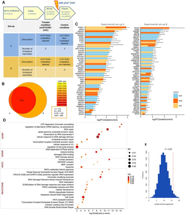
2 Nucleic Acids Research, 2021
overwhelmed or defective NER are causatively linked to Isolation of the UVC-UDS-associated chromatin
cutaneous melanoma carcinogenesis as well as basal and
For one pull-down experiment 120 million human fibrob-
squamous cell carcinoma and certain lung cancers (10–12).
lasts were used. Cells were plated in 150 mm dishes and
Moreover, research on NER-DDR may aid in deepening
grown until 100% confluency. Then they were maintained
our understanding and provide new strategies for treatment
for three more days in culture medium supplemented with
of Xeroderma Pigmentosum, Cockayne Syndrome, and Tri-
0.5% FBS. In all cell culture preparations that were sub-
chothiodystrophy, which are rare human disorders caused
jected to MS, 30 min-long pre-treatment with 10 mM HU
by NER defects (12).
was applied before irradiation. After washing with PBS the
The era of high throughput technologies has allowed new
cells were irradiated with 20 or 30 J/m2 UVC (254 nm,
insights into the NER field. Several strategies have been de-
Downloaded from https://academic.oup.com/nar/advance-article/doi/10.1093/nar/gkab144/6163098 by guest on 25 March 2021
TUV Lamp, Philips) as indicated (doses that induce suffi-
veloped for analysing the effects of NER-inducing geno-
cient amounts of DNA lesions for the subsequent analy-
toxic factors on the whole chromatin-associated proteome
ses), and incubated in FBS-free culture medium containing
(13,14) as well as the cell’s PTMome (post-translation mod-
10 M EdU (3,4) and 10 mM hydroxyurea (HU) for 4 h.
ifications) (15–17). In addition, a multiomic strategy entail-
Next, the cells were washed with PBS, scraped and pelleted
ing proteomic screens and a functional genomics screen has
in the presence of 1 mM PMSF. The following steps, from
been used to address transcription-related DNA damage re-
nuclei isolation till capturing, were adapted from Leung et
sponses (13). In parallel, novel methods have been devel-
al. (4) with minor changes. Briefly, the nuclei were isolated
oped for deep sequencing of damaged DNA (18–20) and the
by resuspending the cell pellet in nuclear extraction buffer
DNA that is excised from damaged sites (21). Despite the in-
(NEB) and rotating for 15 min at 4◦ C. The pelleted nu-
teresting findings emerging from these approaches, impor-
clei were washed with PBS and subjected to Click-reaction
tant questions remain unanswered, in particular in the con-
by resuspending in Click-reaction mix supplemented with
text of chromatin and its coupling to the repair.
1 mM THPTA and rotating for 1 h at room temperature
There are two sub-pathways of NER, the transcription
(RT). Next the nuclei were pelleted, washed with PBS, re-
coupled NER (TC-NER), which is triggered by an actively
suspended in 1ml B1 buffer supplemented with protease in-
transcribing RNA polymerase II (Pol II) and removes DNA
hibitors (PIs), incubated for 15 min and sonicated twice for
lesions from the transcribed strand of genes and gene reg-
10 s with 1 W and once for 120 s (with 10 s breaks after every
ulatory regions (21,22) and the global genomic NER (GG-
10 s of sonication) with 4 W in an VC 70 sonicator (Sonics
NER) that removes lesions from the entire genome. The two
& Materials). Between each sonication cycle the chromatin
sub-pathways mainly differ at the step of damage recogni-
was centrifuged, resuspended in a fresh B1 buffer and in-
tion. Following the removal by the NER core machinery of
cubated for 15 min on ice. This resulted in fragment sizes
DNA oligomers containing helix distorting DNA lesions,
between 300 and 600 bp (Supplementary Figure S1). After
unscheduled DNA synthesis (UDS) takes place and DNA
the last sonication cycle, the chromatin was pelleted, the su-
polymerases fill in the resulting single stranded DNA gap
pernatant was kept and an equal amount of buffer B2 (sup-
(of length of ∼30 nucleotides) using the non-damaged com-
plemented with PIs) and 40 l MyOne T1 Dynabeads were
plementary strand as a template (12). Here, we took advan-
added to it. The mixture was rotated overnight at 4◦ C and
tage of this property of NER and of previously reported
the next day the beads were washed three times with buffer
Click chemistry-based protocols for isolation of replication-
B2. The captured chromatin was eluted by boiling the beads
derived nascent chromatin (3,4) to develop aniFOUND (ac-
for 5 min in 0.1% SDS. Additional information is available
celerated native isolation of factors on unscheduled nascent
as a Supplementary Protocol.
DNA), using UVC as a specific NER-inducing geno-
toxic factor. aniFOUND is a novel unbiased (antibody-
Immunocytochemistry
free) method for the specific labelling, enrichment and
purification of repaired, newly-synthesized chromatin. By The following antibodies were used for immunocytochem-
coupling nascent UDS labelling to proteomic and Next istry: anti-CPD (CosmoBio, cat. no. NMDND001), anti-
Generation Sequencing (NGS) technologies, this method ␥ H2AX (Abcam, cat. no. ab2893), anti-XPG (Novus
provides an in-depth view of the repaired chromatin- Biologicals, cat. no. NB100-74611), anti-RSF1 (Abcam,
associated proteome composition and its genome-wide cat. no. ab109002), anti-NIPBL (SantaCruz, cat. no. SC-
distribution. 374625), anti-PCNA (Abcam, cat. no. ab15497). Cells that
were grown on coverslips were washed with PBS and were
locally irradiated with 30 or 100 J/m2 UVC through a 5 or
MATERIALS AND METHODS 8 m pore polycarbonate membrane filter (Merck, cat. no.
TMTP04700, TETP04700) or mock irradiated and left for
Cells and cell propagation
recovery in the presence or absence of EdU. After recovery
1BR.3 (23) and VH10 (24) normal and NER-deficient XPA the coverslips were washed with ice-cold PBS and incubated
(24) hTert immortalized human skin fibroblast cell lines in CSK buffer for 5 min on ice. Next, cells were fixed with
were used in this study. The cell lines were maintained 4% paraformaldehyde for 10 min and were washed three
under standard conditions in DMEM with high glucose times with PBS. If co-staining with anti-CPD was to be
and sodium pyruvate (Gibco) supplemented with 10% fetal done, an incubation step in 37◦ C in the presence of 0.1 N
bovine serum (FBS) (Gibco), 100 units/ml of penicillin and HCl for 10 min was included after the fixation followed by
100 g/ml of streptomycin (Gibco) at 37◦ C in a 5% CO2 two PBS washes. Then, cells were blocked in 10% FBS or
humidified incubator. 3% BSA (Sigma-Aldrich, cat. no. A9647), diluted in PBS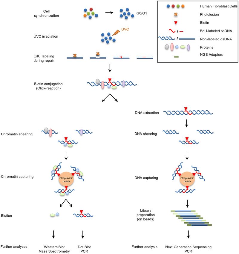
Nucleic Acids Research, 2021 3
for 20 min at RT. For EdU staining, the cells were subjected (Thermo-Fisher, cat. no. NP0321PK2). The proteins were
to Click-reaction by incubation in PBS containing 25 M transferred onto a PVDF membrane (Merck, cat. no.
Alexa Fluor Azide (Thermo Fisher, cat. no. A10270), 10 IPFL00010) by electrophoresis at 130 V for 1 h at 4◦ C. The
mM sodium L(+)-ascorbate (Applichem, cat. no. A5048) membrane was blocked with Odyssey Blocking Buffer (Li-
and 4 mM CuSO4 (Applichem, cat. no. A3327) for 1 h at Cor, cat. no. 927-40000) for 1 h at RT. Then the part of the
RT in dark. Cells were washed three times with PBS–Tween membrane corresponding to histones molecular weight was
(0.05%) and for co-staining with antibodies cells were incu- incubated overnight at 4◦ C with the appropriate antibodies:
bated with the appropriate antibody overnight at 4◦ C pro- H2A-ub (Millipore, cat. no. 05-678), H2B (Millipore, cat.
tected from light. Next day cells were washed three times no. 07-371), H4 (Abcam, cat no ab10158). Next day. the
with wash buffer, incubated for 1 h with Alexa Fluor sec- membrane was washed with PBS–Tween, incubated with
Downloaded from https://academic.oup.com/nar/advance-article/doi/10.1093/nar/gkab144/6163098 by guest on 25 March 2021
ondary antibody at RT, washed twice, stained with Dapi the relevant secondary antibodies (Li-Cor) for 1 h at RT,
for 5 min, washed twice again and mounted with Mowiol washed with PBS–Tween and scanned in an Odyssey scan-
(Fluka, 81381. polyvinyl alcohol 4–88). ner (Li-Cor).
Images were acquired with a LEICA DM2000 micro-
scope equipped with the DFC345 FX camera and pseudo-
Sample preparation for Mass Spectrometry
colour was applied using the LAS V4.12 software. Imaging
conditions, i.e. exposure time, brightness and contrast re- Samples were digested following filter-aided sample prepa-
mained identical between different conditions. ImageJ was ration (FASP) method published by Wisniewski et al. (25).
used for the quantification of the signal. The ImageJ images Samples were mixed with 200 l of 8 M urea in 0.1 M
were colour threshold adjusted for grey-scaled dapi stain- Tris/HCl (pH 8.5) and transferred onto a Vivacon 500 10
ing and then converted to binary. After filling holes, nuclear kDa MW cut-off filter and centrifuged at a constant 14 000
particles were analysed and added to ROI manager. Red × g. This step was repeated once more and then the flow-
channel was overlaid with regions of interest and Mean In- through solvent was discarded. Alkylation step was per-
tensity of each nucleus was measured on three photos for formed when 100 l of 1.5 mg/ml iodoacetamide was added
each condition. and incubated for 20 min in the dark at room temperature.
The filter was centrifuged for 10 min. Wash steps were per-
FACS sorting formed with 100 l of 8 M urea (three times) and 100 l of
0.05 M ammonium bicarbonate in H2 O (three times), each
Cells were trypsinized, collected and washed with ice-cold step the filter was centrifuged until dryness. Vials contain-
PBS. 1.5 million cells were resuspended in 500 l PBS con- ing the flow-through were exchanged with clean ones before
taining 0.1% glucose and were fixed with 5 ml 70% ethanol adding 80 l ammonium bicarbonate buffer containing 1
for 1 day at –20◦ C. After rehydration they were shaken for g trypsin/LysC (Promega) onto the filter. Trypsin diges-
40 min in the presence of 50 g/ml propidium iodide and tion was performed for 16 h at 37◦ C with shaking. Follow-
20 g/ml RNase A in the dark. Samples were measured in ing digestion, 40 l of water was added to the filter, and
a BD FACS CANTO II flow cytometer (Becton Dickinson) the peptides were eluted by centrifugation for 10 min at 14
and analysed with BD FACSDiva Software v6.0 (Becton 000 × g. The part containing the peptides was collected and
Dickinson). dried down by speed-vac-assisted solvent removal and re-
constituted in a solution of 2% (v/v) ACN and 0.1% (v/v)
Dot blot/slot blot formic acid. The peptide solution was incubated for 3 min
Labelled DNA for input was extracted either as described in a sonication water bath. Peptide concentration was deter-
in Note 8 of the supplementary protocol 1 or by treat- mined by Nanodrop absorbance measurement at 280 nm.
ment with Proteinase K and extraction with the QIAquick 2.5 g peptides were pre-concentrated with a flow of
PCR Purification Kit (Qiagen, cat. no. 28104). The captured 3 l/min for 10 min using a C18 trap column (Acclaim
and eluted DNA was extracted by the MinElute Reaction PepMap100, 100 m × 2 cm, Thermo Scientific) and then
Cleanup Kit (Qiagen, cat. no. 28204). DNA was quantified loaded onto a 50 cm long C18 column (75 m ID, parti-
with Nanodrop or Qubit and samples were boiled at 95◦ C cle size 2 m, 100 Å, Acclaim PepMap100 RSLC, Thermo
for 10 min to denature DNA. Samples were returned onto Scientific). The binary pumps of the HPLC (RSLCnano,
ice immediately and SSC buffer was added. Single stranded Thermo Scientific) consisted of Solution A (2% (v/v) ACN
DNA was loaded on a nitrocellulose membrane (Li-Cor, in 0.1% (v/v) formic acid) and Solution B (80% (v/v) ACN
cat. no. 926-31092) and the blots/slots were washed three in 0.1% (v/v) formic acid). The peptides were separated
times with a mixture of 30% SSC and 70% TE. The mem- using a linear gradient of 4% B up to 40% B in 210 min
brane was baked at 80◦ C for 1 h, blocked overnight with with a flow rate of 300 nl/min. The column was placed in
Odyssey Blocking Buffer (Li-Cor, cat. no. 927–40 000) and an oven operating at 35◦ C. The eluted peptides were ion-
incubated with Streptavidin Alexa Fluor (Thermo-Fisher, ized by a nanospray source and detected by an LTQ Or-
cat. no. S32357) for 1 h in the dark. The washed membrane bitrap XL mass spectrometer (Thermo Fisher Scientific,
was scanned in an Odyssey scanner (Li-Cor). Waltham, MA, USA) operating in a data dependent mode
(DDA). Full scan MS spectra were acquired in the orbitrap
(m/z 300–1600) in profile mode with the resolution set to
Western Blot
60,000 at m/z 400 and automatic gain control target at 106
Samples were boiled for 10 min in a Laemmli sample ions. The six most intense ions were sequentially isolated for
buffer and loaded in 4–12% gradient polyacrylamide gels collision-induced (CID) MS/MS fragmentation and detec-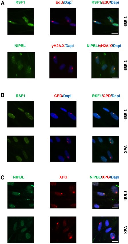
4 Nucleic Acids Research, 2021

tion in the linear ion trap. Dynamic exclusion was set to 1     NGS library construction
min and activated for 90 s. Ions with single charge states
                                                                Biotinylated chromatin was treated with Proteinase K
were excluded. Lockmass of m/z 445.120025 was used for
                                                                and DNA was extracted by phenol–chloroform. RNA
continuous internal calibration. XCalibur (Thermo Scien-
                                                                molecules were degraded by treatment with RNAse A.
tific) was used to control the system and acquire the raw
                                                                DNA was sheared in a Covaris S2 sonicator and selection
files.
                                                                of the fragments between 100 and 200 bp was done by run-
                                                                ning in agarose gel and DNA extraction with the QIAquick
Protein identification, quantification and data analysis        Gel Extraction Kit. The biotinylated DNA was isolated by
                                                                incubation with Dynabeads MyOne Streptavidin T1 mag-
The mass spectral files (.RAW files) were processed us-

                                                                                                                                Downloaded from https://academic.oup.com/nar/advance-article/doi/10.1093/nar/gkab144/6163098 by guest on 25 March 2021
                                                                netic beads for 20 min at RT. The next steps, up to PCR
ing MaxQuant software (1.5.8.3). Default parameters were
                                                                amplification, were done on beads. The ends of the DNA
used for protein identification and quantification. Trypsin
                                                                fragments were repaired by incubation with T4 DNA poly-
specificity with two missed cleavages was allowed and min-
                                                                merase, Klenow Fragment and T4 DNA Polynucleotide Ki-
imum peptide length was set to seven amino acids. Cys-
                                                                nase, then A-tailing was done by incubation with dATPs in
teine carbamidomethylation was set as fixed, and methio-
                                                                the presence of Klenow 3–5 exo and TruSeq adapters were
nine oxidation, deamidation of asparagine and glutamine
                                                                ligated with Quick Ligase. The DNA was amplified in an
and N-terminal acetylation were set as variable modifica-
                                                                end-point PCR instrument using the minimum number of
tions. A maximum of five modifications per peptide was
                                                                cycles for which PCR products could be seen. Finally, the
set. The false discovery rate both for peptide and protein
                                                                libraries were cleaned with AMPure XP beads. For the li-
were set to 5%. For the calculation of the protein abun-
                                                                braries of input DNA, the sheared and size-selected biotiny-
dances, label-free quantification (LFQ) was performed with
                                                                lated DNA was subjected to standard Illumina protocols as
both ‘second peptide’ and ‘match between run’ options en-
                                                                previously described (29).
abled. The complete human database was downloaded from
Uniprot 05/17. Statistical analysis was performed using
Perseus (version 1.5.3.2) (26). Proteins identified as ‘con-    NGS sequencing and bioinformatics analysis
taminants’, ‘reverse’ and ‘only identified by site’ were fil-
tered out. The LFQ intensities were transformed to loga-        Both aniFOUND-seq and input libraries were sequenced
rithmic and missing values were imputed––replaced by ran-       as single-end reads at Genecore-EMBL with an Illumina
dom numbers drawn from a normal distribution. The three         HiSeq 2000 instrument. To analyse the NGS datasets in an
replicas were grouped for each set of conditions (treated and   automated and reproducible manner, custom bioinformat-
control conditions for both experimental set A and B) and       ics pipelines were developed.
a two-sided Student’s t-test of the grouped proteins within
each experimental set was performed using FDR values for        Quality control and read alignment
truncation.
                                                                Quality control of raw FASTQ files was performed us-
                                                                ing fastQC version 0.11.5 (www.bioinformatics.babraham.
RNA sequencing                                                  ac.uk/projects/fastqc/). To avoid read length biases in the
Total RNA was isolated in duplicate from two 1BR.3 fi-          downstream analysis (see ‘Differential repair enrichment
broblasts cultures using Trizol (ThermoFisher Scientific).      analysis in the repeated genome’, and ‘Telomeric content
The Agilent RNA 6000 Nano kit with the bioanalyser from         enrichment analysis’ methodologies), the reads that were
Agilent were used for analyzing RNA samples in terms of         longer than 50 bases were initially trimmed at the 3 end,
quantity and quality in the BSRC ‘Alexander Fleming’ Ge-        until a constant read length of 50 bases was reached for all
nomics Facility. RNA samples with RNA integrity num-            samples. Remains of adapters were clipped and low-quality
ber (RIN) >7 were used for library construction using the       bases were trimmed from both ends of the sequenced reads
3 mRNA-Seq Library Prep Kit Protocol for Ion Torrent           (Phred score 20) using cutadapt version 2.4 version 0.7.12
(QuantSeq-LEXOGEN™) according to manufacturer’s in-             (30), allowing a minimum of 10 bases per read. High-
structions. The DNA High Sensitivity Kit was used along         quality reads were mapped against the UCSC hg19 refer-
with the bioanalyser for assessing the quantity and quality     ence genome using bwa-mem (31) with default settings, and
of the libraries. Next, libraries were pooled and templated     allowing 2 mismatches between the subject and the refer-
using the Ion PI™ HiQ OT2 200 Kit (ThermoFisher Scien-          ence sequences, to account for sequencing errors and SNPs
tific) on Ion One Touch System. Sequencing was performed        between the 1BR.3 cell line and the sequenced genome.
in BSRC ‘Alexander Fleming’ Genomics Facility using the
Ion PI™ HiQ Sequencing 200 Kit and Ion Proton PI™ V2
                                                                Read density analysis
chips (ThermoFisher Scientific) on an Ion Proton™ System,
according to the manufacturer’s instructions. Mapping of        To produce average read density profiles, read density
sequencing reads to hg19 human reference genome was per-        heatmaps, read density boxplots and UCSC Genome
formed using hisat2 (27) and gene expression quantification     Browser tracks (32), uniquely aligned and deduplicated
was performed by the Bioconductor package metaseqR2             reads were used. Specifically, to define a ‘uniquely’ aligned
(28), using the 3’ UTR pipeline with default settings. Genes    set, hits with mapping quality score less than 10 were fil-
with at least one read in both replicates were considered ac-   tered out using samtools version 1.9 (33), and chimeric
tively expressed.                                               and secondary alignments were filtered out using the ‘XA’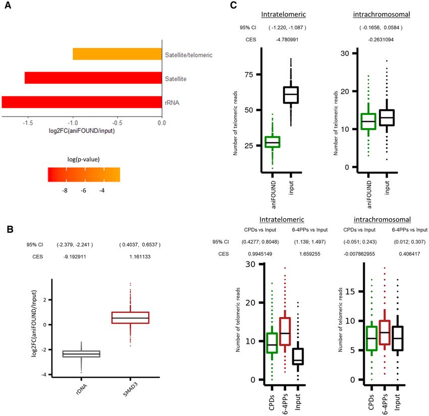
Nucleic Acids Research, 2021 5

and ‘SA’ tags. Duplicated alignments were eliminated us-        were used as an input to RepeatMasker software. To effi-
ing samtools markdup -r. Replicates were downsampled            ciently run the algorithm, FASTQ files were first converted
to the minimum sample read depth per biological condi-          to FASTA files and were split to 300,000 sequence chunks.
tion and merged in order to create a consensus dataset.         RepeatMasker was run with parameters: -e crossmatch -pa
Genomic annotations of hg19 RefSeq transcription start          30 -q -low -species human -a -inv -lcambig -html -source -
sites (TSSs) and enhancers were retrieved by UCSC ta-           gff -excln -u -nopost to produce pairwise alignment files of
ble browser (34) and FANTOM5 project (35), while active         repeat elements against the examined fasta sequences, using
and inactive characterization of TSSs, enhancers, and as-       RepBase (36) and Dfam (37) as repeat species reference. The
PROMPTs was applied using previously published RNA              output of the software was further processed using Process-
Pol II-ser2P, H3K27ac and H3K27me3 ChIP-seq data from           Repeats to produce repeat specific annotation files, contain-

                                                                                                                                  Downloaded from https://academic.oup.com/nar/advance-article/doi/10.1093/nar/gkab144/6163098 by guest on 25 March 2021
VH10 hTERT-immortalized human skin fibroblasts (22,29)          ing information about the alignment of every repeat species
and CAGE-seq data (35) from dermal and skin fibroblasts,        against each sequenced read. All annotation files of each
as described in Liakos et al. (22). All TSSs were extended to   library were summarized to produce a count-like matrix,
2 kb on each direction, subdivided to 20 bp genomic bins,       containing the number of total repeat species occurrences
and were used as a reference for read counting. Heatmaps        in each of the examined samples. A total of 1279 unique re-
were generated using seqMINER, while average profiles           peat species were identified in all datasets and Counts were
were generated using custom R scripts and ggplot2.              summarized to a total of 68 repeat families of origin.
   Boxplots of reads per million values (RPM) of each ex-           The final {repeat families × samples} count matrix was
tended TSS region were generated using custom R scripts,        further processed by DESeq2 (38) to perform differential
representing RPM distributions of actively transcribed, or      enrichment analysis. The input dataset was used as a refer-
inactive regions. To apply per sample comparisons between       ence sample, and size factors and dispersion were estimated
the active and inactive sets distributions, a permutation       using default settings. A test for significance of coefficients
strategy was applied. Briefly, for each distribution compar-    in a negative binomial Generalized Linear Model (GLM),
ison and for each active/inactive set, 10,000 samplings of      was applied using the abovementioned estimated size fac-
100 data points were randomly generated, and 95% confi-         tors. Only results with a P-adjusted value threshold lower
dence intervals of mean differences between active and in-      than 0.05 were reported.
active regions were calculated. Effect sizes of log2 counts         To further examine the degree of NER repair activity
between active and inactive sets were calculated using Co-      at rDNA regions, a differential enrichment analysis be-
hen’s method (CES).                                             tween aniFOUND-seq and input datasets at rDNA repeats
   Genome browser-compatible files (bigWig) were gener-         was conducted as follows. Initially, the hg19 human refer-
ated using deeptools bamCoverage with counts per million        ence genome FASTA file was extended by adding the 45S
normalization (cpm).                                            pre-ribosomal N5 (RNA45SN5) NCBI sequence (https://
                                                                www.ncbi.nlm.nih.gov/nuccore/NR 046235) as a new chro-
                                                                mosome, using the >NR 046235.3 identifier. High-quality
Genomic annotation of sequenced reads
                                                                aniFOUND-seq FASTQ reads were mapped against the ex-
To annotate the aniFOUND-seq, XR-seq and damage-seq             tended genome index using bwa mem (31) with quality fil-
samples in respect to their genomic origin, genomic annota-     ter -T 0. Low quality and duplicated alignment were not
tions from roadmap epigenomics project were used, and in        filtered out, and replicates were downscaled to a similar
particular the NHDF-Ad Adult Dermal Fibroblasts core            read depth (19,000,000 reads), merged to a single file and
15-state model, by excluding the 8th chromatin state, ‘ZNF      sampled 1000 times to produce 100,000 alignment chunks
genes & repeats’, since they were analysed in a separate        that were in turn summarized at the NR 046235.3 chromo-
analysis module (see below). For each biological condition,     some and SMAD3 gene to produce boxplots of log2 count
only uniquely aligned and deduplicated reads were used and      ratios between the aniFOUND-seq and input datasets. To
replicates were merged as described above, and each high-       apply a statistical comparison between the two count dis-
quality alignment was centered and assigned to a unique         tributions for each examined element, 1000 samplings of
chromatin state. All resulting region counts were aggregated    100 data points were randomly generated, and 95% confi-
per chromatin state and normalized using the total counts       dence intervals of mean log2 differences between PD and
of each dataset, as also the total genome coverage of each      INPUT regions were calculated as described in the ‘Read
chromatin state. These values were either visualized as ra-     density analysis‘ paragraph. Effect sizes of log2 counts be-
tios, normalized by their corresponding input dataset (Fig-     tween PD and INPUT sets were calculated using Cohen’s
ure 5A), or as percentages of the total annotations (Figure     method (CES).
5E).
                                                                Telomeric content enrichment analysis
Differential repair enrichment analysis in the repeated
                                                                In order to examine the extent of NER repair and the
genome
                                                                UVC-induced DNA damage burden on telomere sequences,
To determine potential differential patterns of repair preva-   an analysis pipeline was developed to compare the oc-
lence along the repeated genome, a differential enrich-         currence of TGAGGG repeats in both aniFOUND and
ment analysis between the aniFOUND-seq and the in-              damage-seq libraries and their input libraries. Initially,
put libraries was performed. In summary, quality filtered,      high-quality reads were mapped against the UCSC hg19
adapter-free, and trimmed to the same length FASTQ reads        reference genome using bwa-mem with quality threshold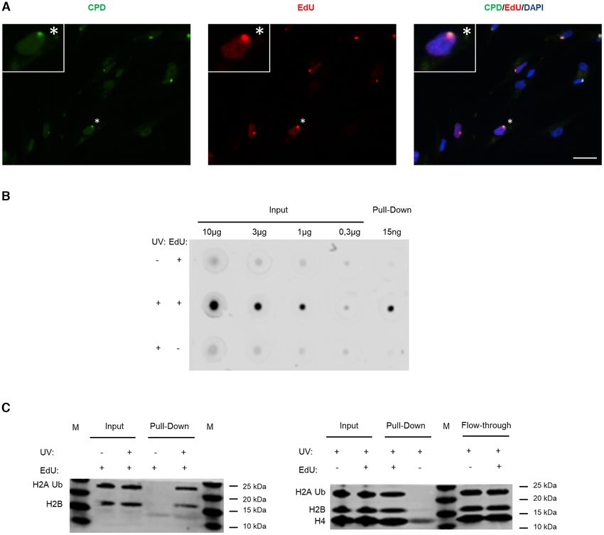
6 Nucleic Acids Research, 2021
-T 0, in order to minimize the fraction of the unmapped DNA synthesis during NER (EdU incorporation, Supple-
reads (31). Both mapped and unmapped reads might con- mentary S5) in line with earlier findings (41). Under these
tain TGAGGG repeats, but only the unmapped sequences conditions, UVC-induced UDS was the major source of de-
are considered as telomeric (39). Replicates were downsam- tectable DNA synthesis (Supplementary Figure S4). By us-
pled to the minimum sample read depth, merged per bio- ing established protocols (42), we confirmed that, under our
logical condition, and downsampled to a similar read depth conditions, only newly synthesized DNA undergoing NER
level of 16,000,000 reads. The resulting BAM files were ran- was labelled, as labelling occurred specifically at the sites
domly sampled for 1000 times, to generate BAM files of of locally-induced DNA damage. This is depicted in Fig-
100,000 reads that were further processed. Specifically, each ure 2A, illustrating the colocalization of cyclobutane pyrim-
subsample file was examined for TGAGGG enrichment, us- idine dimers (CPDs)––a type of UV-induced lesion––and
Downloaded from https://academic.oup.com/nar/advance-article/doi/10.1093/nar/gkab144/6163098 by guest on 25 March 2021
ing TelomereHunter (39) with default parameters, and with- EdU. We further verified that labelled nucleotide incorpora-
out using a control sample. Candidate telomeric reads were tion did not occur in the NER deficient XPA cells, as shown
classified into three categories: (a) Intrachromosomal reads, in Supplementary Figure S6.
which comprise of telomeric repeats that are mapped to the We then used the above cell synchronization protocol
chromosomal regions of the genome, except for the first and to specifically isolate UDS-associated chromatin under na-
last band. The particular regions were considered ‘pseudo’ tive conditions. The experimental pull down of interest,
telomeric and were used as a control set. (b) Subtelom- termed aniFOUND-UVC, was performed with cells that
eric reads, which consist of telomeric reads aligned to the had been UV-irradiated and left to recover in the pres-
first or last band of a chromosome. (c) Unmapped telom- ence of EdU (+UV/+EdU). We employed a short range
eric reads, which were categorized as intratelomeric and of UVC irradiation doses (20–30 J/m2 ) to induce suffi-
were considered as the ‘true’ telomeric content. The out- cient amounts of DNA damage for the subsequent analy-
puts of all the telomeric quantifications were summarized, ses while allowing the majority of cells to recover from the
to produce a telomeric content occurrence distribution for stress. The EdU concentration (10 m) that we used en-
each of the ‘true’ and ‘pseudo’ telomeric categories, for each abled efficient labelling of UDS-specific nucleotide incor-
dataset. To compare the intratelomeric and intrachromo- poration and nuclear fluorescent intensity (Figure 2, Sup-
somal distributions between aniFOUND or HS-damage- plementary Figures S4 and S6), in agreement with a pre-
seq datasets, as also their corresponding input libraries, a vious report (40) and it is considered the optimal EdU
similar approach to calculate confidence intervals and ef- concentration for Click chemistry-based enrichment pro-
fect sizes as described in the ‘Read density analysis‘ para- tocols (3,4). To distinguish any non-specific material, we
graph was applied. HS-Damage-seq samples used in this used two different negative controls. A non-irradiated but
analysis include: (i) damage-seq (CPDs): HS-Damage-seq labelled control (–UV/+EdU) to distinguish any newly syn-
(CPDs) (0 h after irradiation with 10 J/m2 UVC (20), (ii) thesized but non-UDS-derived chromatin and an irradiated
damage-seq (6-4PPs): HS-Damage-seq (6-4 PPs) (0 h after but non-labelled control (+UV/–EdU) to identify any non-
irradiation with 20 J/m2 UVC (20), (iii) damage-seq (input): specific binding on the beads. In both experimental sets, the
NHF1 input (20). isolated aniFOUND-UVC chromatin (+UV/+EdU) was
found to be highly enriched in EdU-labelled nucleotides
RESULTS AND DISCUSSION in comparison to the two negative controls –UV/+EdU
and +UV/–EdU (Figure 2B), validating the specificity of
aniFOUND isolates chromatin associated specifically with
the purification procedure for unscheduled, non-replicative,
UVC-induced, unscheduled DNA synthesis
newly synthesized DNA. The aniFOUND-UVC chromatin
To develop aniFOUND, we exploited UDS, which oc- was enriched for core histones (Figure 2C), including
curs during the repair of UVC-induced DNA lesions by the damage-associated ubiquitinated form of H2A (43).
NER and provides a direct measure of repair efficacy (40). Taken together, our findings show that the isolated ma-
We used 5-ethynyl-2 -deoxyuridine (EdU) and Click chem- terial is appropriate for both proteomic and genomic
istry to label and biotinylate the newly synthesized repaired analyses.
DNA in cells. EdU-labelled native chromatin was subse-
quently isolated by pull-down with streptavidin and sub-
aniFOUND coupled to proteomic analysis
jected to high-throughput analyses, such as mass spectrom-
etry and NGS as illustrated and described in detail in Figure We first employed mass spectrometry (aniFOUND-MS)
1. The complete elimination of active DNA synthesis apart for the isolation and identification of aniFOUND-UVC-
from NER-derived UDS is a key step in aniFOUND. As associated proteins. aniFOUND-MS is described in detail
replication-associated DNA synthesis is the major contrib- in Supplementary Protocol 1. We carried out two experi-
utor to nucleotide incorporation into DNA when cells are mental set-ups, set A and set B, with identical treatment
not synchronized outside of S phase, we arrested human condition (+UV/+EdU: 20 J/m2 of UVC irradiation and
skin fibroblasts in G0/G1 by both contact inhibition and 4 hour-long recovery in the presence of EdU, which we call
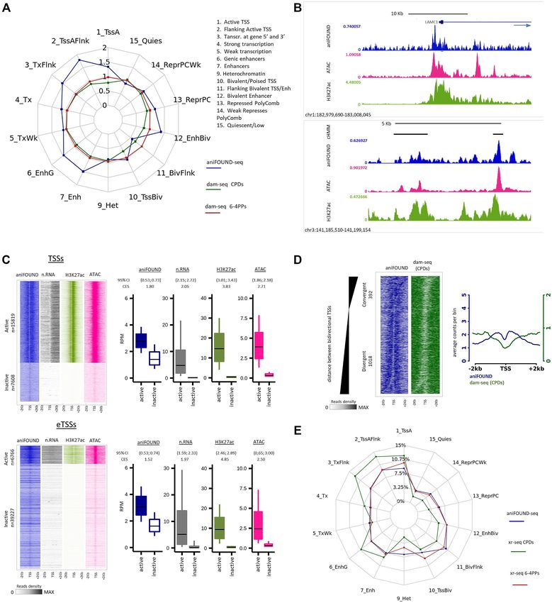
serum starvation (Supplementary Figure S2). Additionally, aniFOUND-UVC) (Figure 3A; Supplementary Figure S7)
to sufficiently diminish replication-derived DNA synthesis but different non-treated control conditions, as described
from the fraction of the cells that still escape to the S-phase, in the previous paragraph. For each of the two experimen-
cultures were treated with 10 mM hydroxyurea (HU) dur- tal set-ups, we conducted three biological replicates for the
ing the DNA labelling step (Supplementary Figure S3 and aniFOUND-UVC samples and the corresponding control
S4). We found no effect of HU at this concentration on samples. The isolated chromatin samples were analysed sep-Nucleic Acids Research, 2021 7
Downloaded from https://academic.oup.com/nar/advance-article/doi/10.1093/nar/gkab144/6163098 by guest on 25 March 2021
Figure 1. Schematic depiction of aniFOUND. An asynchronous population of fibroblasts is synchronized to G0/G1 phase by serum starvation and contact
inhibition. DNA photolesions are formed by UVC-irradiation and left to be repaired in the presence of EdU. HU is added in this step to eliminate the
replication of any escaper cells. Since no replication occurs, UDS is the only source of DNA synthesis. Biotin molecules are conjugated to EdU, the
chromatin or the extracted DNA is sheared and the biotin-labelled fragments are isolated with streptavidin beads. The captured material can be used for
several further analyses.
arately by protein-mass spectrometry following a label free negative control conditions (statistically significant or not in
quantification protocol. By carrying out two separate sta- A [–UV/+EdU] or B [+UV/-EdU]) were filtered out. This
tistical analyses (one for each set-up), we found two protein resulted in 182 and 308 proteins for set A and set B, respec-
lists significantly enriched in the aniFOUND-UVC samples tively. The union of the two lists gave 323 enriched proteins,
(+UV/+EdU) compared to the two negative controls. To constituting the aniFOUND-UVC protein list, while the in-
take full advantage of the outcome of both sets, the protein tersection gave a strict aniFOUND list consisted of 167 pro-
lists were merged and all proteins enriched in any of the two teins. Both lists contain a considerable fraction of known8 Nucleic Acids Research, 2021
Downloaded from https://academic.oup.com/nar/advance-article/doi/10.1093/nar/gkab144/6163098 by guest on 25 March 2021
Figure 2. Specific labelling and isolation of the UDS-associated chromatin. (A) 1BR.3 fibroblasts were locally irradiated with 100 J/m2 UVC using 8
m pore filters and incubated for 4 h in no serum medium containing EdU and HU. The coverslips were incubated with Alexa-azide for labelling the
incorporated EdU, and with anti-CPD. Scale bar: 25 m. (B) VH10 fibroblasts were synchronized for aniFOUND and were UVC- or mock-irradiated
with 30 J/m2 . They were kept in the presence of HU and in the presence/absence of EdU for 4 h. Next, cells were subjected to aniFOUND and the DNA
from the isolated material (as well as from input) was extracted, quantified and immobilized on a nitrocellulose membrane. The membrane was incubated
with Streptavidin-Alexa for detecting the incorporated EdU. C1 Dynabeads were used for this experiment. The blot was cropped to advance clarity. (C)
Fibroblasts were synchronized for aniFOUND and then were either UVC- or mock-irradiated with 30 J/m2 and kept for 4 h in the presence of EdU
and HU (1BR.3 fibroblasts; left panel) or were irradiated and kept for 4 h in the presence of HU and in the presence/absence of EdU (VH10 fibroblasts;
right panel). Next, cells were subjected to aniFOUND and the isolated material was used for western blot. Material from 1 million cells was loaded for
inputs while for the pull-down the whole amount was loaded. The membrane was labelled with the indicated antibodies using two discriminable secondary
antibodies with different wavelengths.
DDR factors (Figure 3B; Supplementary Table S1; Supple- The specificity of the aniFOUND-MS method was fur-
mentary Figure S8). ther validated by comparison to the results of two rele-
The aniFOUND-UVC protein list was significantly vant mass spectrometry studies. More specifically, 42% of
enriched in nucleus- (GO:0005634; P = 8.44E−66 ) the proteins in the aniFOUND-UVC list were present in
and chromatin-associated proteins (GO:0000785; P = the list of proteins identified by a multiomic analysis of
7.36E−20 ), as expected (Supplementary Table S2). In transcription-related DDR of Boeing et al. (13). From these
contrast, the proteins enriched in the two negative control 11.8% are annotated to the GO term ‘Nucleotide excision
conditions, were enriched for proteins such as cytoplasm- repair’ and 25.7% to DDR-related GO terms. Moreover,
(GO:0005737; P = 4.05E−11 ) and mitochondria-associated 28.2% of our proteins were enriched on DNA lesions in-
proteins (GO:0005739; P = 2.08E−06 ). duced by cisplatin (14), a chemotherapeutic that inducesNucleic Acids Research, 2021 9
Downloaded from https://academic.oup.com/nar/advance-article/doi/10.1093/nar/gkab144/6163098 by guest on 25 March 2021
Figure 3. Characterization of the proteins resulted by aniFOUND-MS. (A) Description of the two experimental sets that were used for the aniFOUND-
MS. 1BR.3 fibroblasts were treated as shown in the upper panel. The conducted experiments are described in the lower panel. HS: High serum-containing
medium (10%); LS: low serum-containing medium (0.5%); NS: no serum medium. (B) Venn diagram for the identified proteins of the two experimental
sets. The areas of the circles are proportional to the number of the identified proteins. The colour of each compartment represents the ratio of the identified
DDR-related genes (DDR ratio). (C) Proteins of both experimental sets that are significantly enriched in the treated conditions (two-sided Student’s t-test
of the grouped proteins was performed using FDR values for truncation; FDR < 0.1; s0 > 0.1) and fall under DDR-related GO terms. On the x-axis
the log2 -fold change and standard errors of the treated compared to control condition are shown (in each experimental set the treated condition has been
compared to its coupled control condition). With yellow are shown the proteins that are annotated to ‘nucleotide excision repair’ (GO:0006289), with red the
proteins that are annotated to ‘Double strand break repair’ (GO:0006302) and with blue the ones that are annotated to DDR-related GO terms. (D) Gene
Ontology and Reactome terms that are overrepresented in the aniFOUND-UVC protein list. As background universe all the expressed genes in the 1BR.3
cell line, as resulted by RNA-seq analysis (see Materials and Methods), were used. Gene Ontology: BP (Biological Process); CC (Cellular Component);
MF (Molecular Function). (E) Frequency histogram of randomization tests for the number of tumour suppressors (according to OncoKB (46)) that are
contained in random lists of equal size to the strict aniFOUND list and are annotated to GOCC ‘chromatin’ (GO:0005717). The randomization test was
conducted 10,000 times. The red line shows the number of tumour suppressors contained in the strict aniFOUND protein list (observed value) resulted
from the intersection of experimental sets A and B. P-values were calculated as the ratio of tests that resulted in values equal or greater than the observed
value divided by the total number of tests (*P < 0.05).10 Nucleic Acids Research, 2021
damage repaired mainly by NER (20.9% of the common genesis, but very little is known about their putative role
proteins between the two lists is annotated to DDR-related specifically in UV damage repair. In support of our findings,
GO terms). a recent study showed that Nup84 mutant strains, a compo-
nent of the yeast nuclear pore complex and homologue of
human Nup107 (present in aniFOUND-UVC list), display
aniFOUND-MS results in enrichment of DDR-related and
NER defects and increased UV sensitivity during S phase
other proteins and pathways
(44).
Gene ontology analysis of the aniFOUND-UVC protein Taking into account that UV irradiation and NER are
list revealed proteins that are annotated to GO terms re- relevant to mutagenesis that may lead to cancer (10,12,45),
lated to DNA damage responses (18.3%; 59 proteins) and we further explored whether aniFOUND-MS can be a prac-
Downloaded from https://academic.oup.com/nar/advance-article/doi/10.1093/nar/gkab144/6163098 by guest on 25 March 2021
to GO ‘nucleotide-excision repair’ (5.3%; 17 proteins). It tically useful tool in providing information related to can-
further identified proteins annotated to GO term ‘double- cer research. Indeed, the strict aniFOUND-UVC protein
strand break (DSB) repair’ (5.3%; 17 proteins) (Figure 3C) list comprise significantly more tumour suppressor pro-
in line with previously reported findings derived from dif- teins [as defined by oncoKB (46)] than random chromatin-
ferent omics strategies (13). Given that HU induces DSBs associated proteins lists (Figure 3E). By searching for highly
in replicating cells, the fact that we have minimized the S- interconnected clusters between aniFOUND proteins and
phase fraction and we have also treated the control condi- melanoma driver genes in PPI networks, we find that pro-
tions with HU, makes rather difficult the rare HU-derived teins such as TP53 as well as ARID2, which is part of
DSB events to give rise in statistically significant DDR fac- the BAF-complex, may also be of high importance in UV-
tors. It should be noted that these numbers of proteins cor- induced melanoma carcinogenesis (Supplementary Figure
respond strictly to the ones annotated under the respective S12).
GO terms and do not necessarily reflect all proteins that are
mentioned in the literature to be implicated in DDR, NER
Identification and validation of novel UVC-induced DDR
or DSBs.
players
All the significant ones as well as the most relevant to
DDR terms that resulted from gene ontology and pathway To experimentally validate the recruitment of aniFOUND-
overrepresentation analysis of the aniFOUND-UVC pro- MS proteins on repaired nascent chromatin or in its vicin-
tein list are depicted in Figure 3D and Supplementary Table ity, we induced local UV damage (LUDs) in cells and tested
S2. A parallel ontology analysis performed on the clusters the localization of selected candidates by immunofluores-
of highly interconnected proteins confirmed that the iden- cence (47). Here, we present evidence for the recruitment of
tified factors can be divided into four categories, with the two of the proteins that have not been reported to be im-
largest associated with chromatin organization and DNA plicated in NER, remodelling and spacing factor 1 (RSF1)
repair (Supplementary Figure S9; blue cluster). An out- and nipped-B-like protein (NIPBL) (Figure 4; Supplemen-
line of the epigenetic role of proteins associated with the tary Figure S13).
UDS-chromatin is further presented in Supplementary Fig- RSF1 is a chromatin-remodelling factor frequently over-
ure S10. expressed in a number of cancers; it is shown to partici-
The aniFOUND-UVC protein list was also enriched pate in DSB-DDR and to accumulate at DSB foci (48,49).
in several protein complexes according to the CORUM RSF1 is reported to form complex with SMARCA5 (also
database. For example, complexes related to ‘cellular re- present in the aniFOUND-UVC protein list), which is a
sponse to DNA damage stimulus’ (GO:0006974) and member of the SWI/SNF remodelling factors and is im-
‘DNA repair’ (GO:0006281) were identified, including com- plicated in NER-related DDR (50). NIPBL is one of the
plexes containing DDB1/2/Cullin4A, ERCC2/ERCC3, causal genes (mutated in 60% of cases) of Cornelia de Lange
RUVBL1/2 or XRCC5/6 (Supplementary Table S3). Of Syndrome (51), a rare developmental disorder with char-
note, the last two groups of complexes are principally re- acteristic facial features, stunted growth, and mental retar-
ported to be associated with DSBs, providing support to dation, as well as multiple other systemic abnormalities.
the notion that a number of protein complexes participate NIPBL is essential for cohesin loading to chromatin and
in both DSB- and UVC-related DDR. In addition to the has been detected at DSBs (52,53). Notably, the core mem-
core DNA damage and repair complexes, aniFOUND un- bers of cohesin (SMC1A, SMC3 and RAD21), which are
covered whole protein complexes that are involved in other also known to participate in DDR, and specifically in NER-
relevant biological processes, such as the reorganization of DDR in Caenorhabditis elegans (54), as well as certain reg-
chromatin and the post-translational modification of pro- ulatory proteins (STAG2 and PDS5B) were also present
teins. All these complexes are presented, along with cita- in the aniFOUND-UVC protein list (55). Recently, based
tions documenting their implication in DDR, in Supple- on transcription-associated analyses, NIPBL was suggested
mentary Table S3. as a Cockayne Syndrome marker (56). Nevertheless, to the
By searching for clusters of highly interconnected pro- best of our knowledge, no evidence exists for the recruit-
teins in the protein-protein interactions (PPI) networks ment of either RSF1 or NIPBL at sites of UV photolesions.
built on the aniFOUND-UVC protein list we identified pu- Figure 4A illustrates the recruitment of RSF1 and NIPBL
tative clusters consisting of nuclear pore complex (NPC) at LUDs (defined by EdU or ␥ H2A.X staining) in NER-
proteins (Supplementary Figure S11A) and others consist- proficient (wild type- WT) cells at 4 h post-UVC irradia-
ing of ribonucleoproteins (Supplementary Figure S11B). tion (30 J/m2 ). This recruitment is not dependent on HU
Both classes are implicated in DDR and linked to tumori- (Supplementary Figure S13). Next, we examined RSF1 andNucleic Acids Research, 2021 11
and left to recover for 1 h (Figure 4B and C). In these ex-
periments, LUDs were defined by anti-CPD staining or by
staining for XPG, a core NER structure-specific endonucle-
ase, which is assembled at NER complexes independently
of XPA. Figure 4B illustrates RSF1 and CPD colocaliza-
tion in 1BR.3 cells, but no recruitment of RSF1 at LUDs
in XPA-deficient cells. In line with these results, it was re-
ported that lack of RSF1 confers weak sensitivity to UVC
(48). Similar to the picture seen with RSF1, NIPBL colo-
calized with XPG in WT, but not in NER-deficient cells
Downloaded from https://academic.oup.com/nar/advance-article/doi/10.1093/nar/gkab144/6163098 by guest on 25 March 2021
(Figure 4C). Taken together, our data show that RSF1 and
NIPBL associate with UDS-enriched chromatin in response
to UV irradiation and are likely involved in NER-DDR
in human cells. We further provide important insights into
their spatio-temporal involvement in the cascade of NER
events, as their dependence on XPA suggests that they are
recruited at a later step of the process, probably after the in-
cision of the damage-containing oligonucleotide has taken
place.
aniFOUND is coupled to genomic analysis
In parallel to MS, we coupled aniFOUND to NGS to map
the landscape and dynamics of DNA repair/synthesis via
NER. We termed this protocol ‘aniFOUND-seq’ (Figure 1,
Supplementary Protocol 2). Serum starved human fibrob-
lasts were irradiated, left to recover for 4 h in the presence
of EdU and HU and the nascent DNA was biotinylated,
as described for aniFOUND-MS. Next, the DNA was ex-
tracted, fragmented, and the labelled fraction was isolated
by streptavidin pull-down, enabling high-stringency purifi-
cation. On-bead NGS library construction was performed
and NER-associated DNA was sequenced and mapped to
the genome. We also isolated DNA from control samples (–
UV/+EDU, as described above), however the amount was
negligible.
To obtain information on the genome wide distribution
of loci associated with UVC-induced UDS, we calculated
the overlap of aniFOUND-seq sequences, obtained from
two experiments (Supplementary Figure S14), with chro-
matin states defined by the 15-state ChromHMM annota-
tion (57) (Figure 5A). We excluded from this analysis repeat
Figure 4. RSF1 and NIPBL are recruited to sites of UV damage. (A) RSF1 sequences, as they were analysed separately (see below). In
and NIPBL are recruited at sites of UV damage in wild type cells upon contrast to the rather uniform formation of the UV-induced
30 J/m2 irradiation. Fibroblasts were locally irradiated using 5 m pore photolesions (CPDs and 6-4PPs) immediately after irradi-
filters and left for 4 h in no serum containing medium with HU (10 mM)
and with (upper panel) or without (lower panel) EdU. After fixation cells ation, as mapped by the high-sensitivity damage sequenc-
were labelled with Alexa-azide and anti-RSF1 (upper panel) or with anti- ing methodology (20) (HS-Damage-seq) (Figure 5A, HS-
␥ H2A.X and anti-NIPBL (lower panel). (B, C) RSF1 (B) and NIPBL (C) damage-seq), the aggregated UVC-UDS activity was un-
is recruited at the sites of DNA damage in wild type cells (upper panel) but equally distributed to the different chromatin states (Figure
not in XPA deficient cells (lower panel) upon 100 J/m2 UV irradiation.
Fibroblasts were locally irradiated using 5 m pore filters and left for 1
5A, aniFOUND-seq; Supplementary Figure S15A). Active
hour in no serum containing medium with HU (10 mM). After fixation transcription start sites (TSSs) and their flanking regions
cells were labelled with anti-CPDs and anti-RSF1 (B) or with anti-XPG (states 1, 2 and 3), as well as enhancer-associated regions
and anti-NIPBL (C). Scale bar: 12.5 m. (states 6, 7 and 12) showed elevated repair-synthesis. These
results suggest faster NER-gap filling activity during the 4-
h recovery period in actively transcribed genes and regula-
NIPBL accumulation at LUDs in both WT (1BR.3) and tory regions compared to repressed and quiescent regions.
Xeroderma pigmentosum A (XPA) cells, which carry mu- Our data are in agreement with and complete previous re-
tations in the XPA gene, an early core NER factor required ports showing how NER activity is implemented with al-
for NER pre-incision complex assembly. Cells were locally tered speeds in different genomic regions (20,21).
UVC irradiated (with 100 J/m2 , a dose high enough to en- We next analysed the repair patterns revealed by
sure the detection of DDR factors at the damage spots) aniFOUND-seq specifically around TSSs (see Materials12 Nucleic Acids Research, 2021
Downloaded from https://academic.oup.com/nar/advance-article/doi/10.1093/nar/gkab144/6163098 by guest on 25 March 2021
Figure 5. Genome-wide distribution of aniFOUND-seq signal. (A) Repair and damage ratios in different chromatin states. The chromatin states are
defined according to the 15-state ChromHMM annotation (see Materials and Methods). Repair ratios are calculated by aniFOUND-seq reads normalized
by their input reads for each state. Similarly, damage ratios have been resulted from normalized dam-seq (20) by their inputs (see Materials and Methods).
(B) UCSC Genome Browser snapshots of the signals of aniFOUND, H3K27 and ATAC-seq. Upper panel: depiction of a gene TSS and its flanking regions.
The direction of transcription is shown by arrow. Lower panel: enhancers located in an area free of genes. The top track (cHMM) shows the ChromHMM
states; black bars correspond to enhancers. (C) aniFOUND signal in active and inactive transcription start sites. Left panels: heat maps with the signal
of aniFOUND, nRNA, H3K27 and ATAC-seq 2 kb around the transcription start sites of active and inactive genes (TSSs) and enhancers (eTSSs). The
designation of human genes in active and inactive state was done as previously described (22). Right panels: Box plots with the signal distributions of the
gene sets shown in the corresponding heat maps of the left panel. Boxes show the 25th–75th percentiles and error bars show data range to the larger and
smaller values. For each active/inactive set, 10,000 samplings of 100 data points were randomly generated, and 95% confidence intervals of mean differencesNucleic Acids Research, 2021 13
and Methods). Inspection of the aniFOUND-seq signal Therefore, we next thought to relate NER-synthesis
and comparison with ATAC-seq, and H3K27ac ChIP-seq events (aniFOUND-UVC) with NER excision events as
data (22,29) highlighted enhanced levels of UDS at regions established by XR-seq (21,58) (Figure 5E; Supplementary
with increased chromatin accessibility (22), in particular, at Figure S15A). To do this, we took into consideration the
TSSs and the flanking regions of actively expressed genes particular features of the two methodologies, XR-seq and
(protein-coding and lnc-RNA genes, Materials and Meth- aniFOUND-seq: (a) aniFOUND maps the newly synthe-
ods) (Figure 5B; Supplementary Figure S15B, upper panel) sized DNA in chromatin after repair has taken place, while
and at highly accessible enhancer TSSs (eTSSs) (22) (Fig- XR-seq maps the damaged DNA fragments excised dur-
ure 5B; Supplementary Figure S15B, lower panel). Simi- ing the early steps of NER; (b) aniFOUND cumulatively
larly, as illustrated in Figure 5C and Supplementary Fig- captures repair-synthesis events, while XR-seq captures 10-
Downloaded from https://academic.oup.com/nar/advance-article/doi/10.1093/nar/gkab144/6163098 by guest on 25 March 2021
ure S15C, the aniFOUND signal was clearly prominent min-long excision activity; (c) aniFOUND captures total
around active TSSs and eTSSs compared to inactive ones UDS activity, which is associated with the repair of both
(subsets of active/inactive genes/enhancers defined in Ma- CPDs and 6-4 PPs, whereas XR-seq focuses on one type
terials and Methods and as described previously by Li- of lesion per experiment. In view of these differences and
akos et al. (22)). We note a characteristic pattern more to make aniFOUND-seq and XR-seq data as comparable
pronounced at TSSs (Figure 5C), matching previously ob- as possible, we merged the available XR-seq datasets cor-
served global increases of nascent-RNA reads in both the responding to different time-points (Figure 5E, from 5-min
sense (mRNA) and antisense (PROMPT - PROMoter uP- to 4-h recovery) of 6-4PP repair (58). Similarly, for CPDs,
stream transcripts) directions and confirming that NER oc- we merged the two available time-points (1 and 4 h). The
curs quickly and efficiently at open and actively transcrib- spider plot (Figure 5E) demonstrates that the distribution
ing regions (22). Nonetheless, a lower but detectable UDS of UDS signal (aniFOUND-seq) in different ChromHMM
signal was also detected at non-transcribed regions during states (57) is very similar to that of 6-4PP signal (21), in line
the first 4 h of recovery (Figure 5C, signal not detected in with the fact that the majority of 6-4PPs are repaired during
nascent RNA- and H3K27ac-seq; Supplementary Figure the first 4 hours after damage induction (Figure 5E). On the
S15C). other hand, the elevated levels of XR-seq CPD signal com-
Given that the underlying distribution of DNA dam- pared to aniFOUND-seq signal in states related to active
age would affect the localization of DNA repair-synthesis transcription (states 1, 2, 3, 4, 6 and 7) and the reduced lev-
events, we further plotted the UDS signal along with the els of CPD repair in repressed chromatin and heterochro-
damage signal (HS-Damage-seq data (20)) around active matin (states 9–14) are in agreement with CPDs being pref-
TSSs of bidirectional transcripts (Figure 5D; Supplemen- erentially repaired by TC-NER early in the post-UV recov-
tary Figure S15D). Active bidirectional promoters (TSSs ery process (21,58). Focusing on the repair activity around
of active mRNA-mRNA pairs transcribed in the oppo- TSSs and eTSSs, we find that during the first 4 h of repair
site direction) were sorted according to their InterCage dis- both methods detect increased signal in active regions, con-
tance between the sense and antisense TSSs (see Materi- trasting with lower repair in inactive loci (Supplementary
als and Methods; (22)), and further categorized as conver- Figure S16). We thus conclude that aniFOUND-seq, which
gent (when overlapping transcription occurred) or diver- has major differences and unique features in comparison to
gent (when overlapping transcription did not occur) (Fig- the established method, is a valid and fast method to accu-
ure 5D; Supplementary Figure S15D; left panels). Using rately map the cumulative pattern of DNA repair-synthesis
this set-up, we found that the observed UVC-UDS pat- events genome-wide.
tern around TSSs (Figure 5C) follows the shape of UV-
damage formation. The same is also seen in the aggregate
plot where the valley in the UDS curve coincides with the aniFOUND-seq estimation of NER efficacy on repetitive se-
absence of UV-lesions (Figure 5D; Supplementary Figure quences
S15D; right panels). Taken together these results confirm We next used the aniFOUND-seq data to study UDS ac-
that aniFOUND-seq can exclusively isolate and map in tivity at repetitive sequences, a considerable and under-
high resolution newly synthesized chromatin patches at sites explored part of the genome. We annotated the reads of
of UV lesions that have undergone NER. both aniFOUND-seq and input libraries to repeat elements
←−−−−−−−−−−−−−−−−−−−−−−−−−−−−−−−−−−−−−−−−−−−−−−−−−−−−−−−−−−−−−−−−−−−−−−−−−−−−−−−−−−−−−−−−−−−
between active and inactive regions were calculated. Effect sizes of log2 counts between active and inactive sets were calculated using Cohen’s method (CES).
(D) DNA damage and repair on bidirectional promoters. Left panel: heat maps of aniFOUND and dam-seq around the TSSs of bidirectional genes. The
sorting was done based on the distance between the TSSs of the two bidirectional genes (see Materials and Methods). Right panel: Aggregate plots of
aniFOUND and dam-seq around the TSSs for the gene sets shown in the left panel. (E) Distribution of aniFOUND and XR-seq signals among chromatin
states. For XR-seq all the available data sets up to 4 h after irradiation were merged (5 min, 20 min, 1 h, 2 h and 4 h for the 6-4PPs and 1 and 4 h for CPDs).
The states are defined according to the 15-state ChromHMM annotation. For each library the number of reads that correspond to a chromatin state has
been corrected for the length of the state. Y-axis shows the percentage of the corrected reads that fall in each state. For the hypothetical library in which
all states were equally represented, a polygon with all its sides positioned at around 7% (≈ 100%/14 states) would be resulted. aniFOUND: aniFOUND-
seq; dam-seq (6-4 PPs): HS-Damage-seq (6-4 PPs) (0 h after irradiation with 20 J/m2 UVC (20)); dam-seq (CPDs): HS-Damage-seq (CPDs) (0 h after
irradiation with 10 J/m2 UVC (20)); nRNA: nascent RNA-seq (2 h after irradiation with 20 J/m2 UVC (29)); ATAC: ATAC-seq (2 h after irradiation with
15 J/m2 UVC (22)); H3K27ac: ChIP-seq of H3K27ac (2 h after irradiation with 15 J/m2 UVC (22)); XR-seq (CPDs): NHF1 CPD XR-seq (merged data
sets of 1 and 4 h after irradiation with 10 J/m2 UVC (58)); XR-seq (6-4PPs): NHF1 (6-4)PP XR-seq (merged data sets of 5 min, 20 min, 1 h, 2 h and 4 h
after irradiation with 20 J/m2 UVC (58)).You can also read