The Glycobiology of Pulmonary Arterial Hypertension - MDPI
←
→
Page content transcription
If your browser does not render page correctly, please read the page content below
H

       OH
            OH
                 metabolites

Review
The Glycobiology of Pulmonary Arterial Hypertension
Shia Vang † , Phillip Cochran † , Julio Sebastian Domingo, Stefanie Krick                          and Jarrod Wesley Barnes *

                                          Division of Pulmonary, Allergy and Critical Care Medicine, Department of Medicine,
                                          University of Alabama at Birmingham, Birmingham, AL 35294, USA; svang35@uab.edu (S.V.);
                                          prcochran@yahoo.com (P.C.); julio10@uab.edu (J.S.D.); skrick@uabmc.edu (S.K.)
                                          * Correspondence: barnesj5@uab.edu; Tel.: +1-205-975-5300
                                          † These authors contributed equally to this work.

                                          Abstract: Pulmonary arterial hypertension (PAH) is a progressive pulmonary vascular disease of
                                          complex etiology. Cases of PAH that do not receive therapy after diagnosis have a low survival rate.
                                          Multiple reports have shown that idiopathic PAH, or IPAH, is associated with metabolic dysregu-
                                          lation including altered bioavailability of nitric oxide (NO) and dysregulated glucose metabolism.
                                          Multiple processes such as increased proliferation of pulmonary vascular cells, angiogenesis, apop-
                                          totic resistance, and vasoconstriction may be regulated by the metabolic changes demonstrated in
                                          PAH. Recent reports have underscored similarities between metabolic abnormalities in cancer and
                                          IPAH. In particular, increased glucose uptake and altered glucose utilization have been documented
                                          and have been linked to the aforementioned processes. We were the first to report a link between
                                          altered glucose metabolism and changes in glycosylation. Subsequent reports have highlighted
                                          similar findings, including a potential role for altered metabolism and aberrant glycosylation in IPAH
                                          pathogenesis. This review will detail research findings that demonstrate metabolic dysregulation in
                                          PAH with an emphasis on glycobiology. Furthermore, this report will illustrate the similarities in the

                                   pathobiology of PAH and cancer and highlight the novel findings that researchers have explored in
Citation: Vang, S.; Cochran, P.;          the field.
Sebastian Domingo, J.; Krick, S.;
Barnes, J.W. The Glycobiology of          Keywords: pulmonary hypertension; glycobiology; metabolism
Pulmonary Arterial Hypertension.
Metabolites 2022, 12, 316. https://
doi.org/10.3390/metabo12040316

Academic Editors: Monika Kubacka,
                                          1. Introduction
Magdalena Kotańska and                        Pulmonary vascular diseases (PVDs) are a group of pulmonary ailments resulting
Marek Bednarski                           from precapillary or postcapillary hypertension and include pulmonary embolism, chronic
                                          thromboembolic disease, pulmonary veno-occlusive disease, arteriovenous malformations,
Received: 1 March 2022
                                          pulmonary edema, and pulmonary hypertension (PH) [1,2]. Several clinical studies have
Accepted: 28 March 2022
                                          estimated that PVDs occur in 20–25 million persons worldwide; however, the occurrence
Published: 1 April 2022
                                          as well as the pathogenesis still remain unknown. Currently, PH is the most complex and
Publisher’s Note: MDPI stays neutral      widely studied PVD and is therefore the focus of this review.
with regard to jurisdictional claims in        In 2009, PH was defined by a mean pulmonary artery pressure (mPAP) of ≥25 mm
published maps and institutional affil-   Hg at rest that is measured during right heart catheterization (RHC) [3,4]. It was recently
iations.                                  recommended to lower the mPAP threshold to ≥20 mm Hg in 2019 based on scientific
                                          studies [5,6]. PH is often associated with other PVDs or diseases such as congenital
                                          heart disease, coronary artery disease, liver cirrhosis, and COPD, as well as personal and
                                          environmental factors such as tobacco use, drug use, and toxin stimuli. Therefore, PH has
Copyright: © 2022 by the authors.
                                          been classified by the World Health Organization (WHO) as a five-group system according
Licensee MDPI, Basel, Switzerland.
This article is an open access article
                                          to etiology: Group 1, PAH; Group 2, PH due to left heart disease; Group 3, PH due to
distributed under the terms and
                                          chronic lung disease or hypoxia; Group 4, Chronic Thromboembolic PH (CTEPH); and
conditions of the Creative Commons        Group 5, PH due to unclear multifactorial mechanisms [5].
Attribution (CC BY) license (https://          Pulmonary Arterial Hypertension (PAH), the Group 1 subcategory of PH, is charac-
creativecommons.org/licenses/by/          terized hemodynamically by the presence of pre-capillary PH with an end-expiratory pul-
4.0/).                                    monary artery wedge pressure (PAWP) ≤ 15 mm Hg and a pulmonary vascular resistance

Metabolites 2022, 12, 316. https://doi.org/10.3390/metabo12040316                                     https://www.mdpi.com/journal/metabolites
Metabolites 2022, 12, 316 2 of 17
>3 Wood units [7], which frequently leads to right ventricular (RV) hypertrophy/failure
and early mortality. RHC remains essential for a diagnosis of IPAH [8]. From the time
of diagnosis, PAH has an average survival of 2–3 years without treatment and affects
mostly women; however, men diagnosed with PAH have lower survival rates [9,10]. The
pathobiology of PAH is complex; features include vasoconstriction, inflammation, genet-
ics/epigenetics, and/or environmental cues [11–16] as well as increased cell proliferation,
vascular remodeling, and angiogenesis [17–22]. Furthermore, metabolic dysfunction that
includes deficits in nitric oxide (NO) production [23–27], insulin resistance [28–31], leptin
dysregulation [32–34], dyslipidemia, and increased lipid oxidation [35–42] have been es-
tablished in IPAH and may affect disease progression. Currently, the primary therapies
for PAH are those that target the mediators of vasoconstriction (e.g., NO, prostacyclin,
or endothelin) [23,43–45], while other pathomechanisms including cell proliferation, in-
creased angiogenesis, inflammation, and plexiform lesions (a hallmark of PAH) have no
known treatment options. This review will discuss metabolic dysregulation with an em-
phasis on novel findings in glycobiology observed in Group 1 PAH, where the most of the
investigations have been documented.
1.1. Metabolism Studies in PAH
Over the years, evidence for the role of metabolites in PAH has been documented and
determined to affect the many processes mentioned above. Other reports have shown that
metabolites including glucose [30,46–49], ammonia [50], arginine [51,52], glutamine [53–55],
and fatty acids are dysregulated [35,37,41,56]. Interestingly, altered glucose influx into cells
can lead to glucose intolerance in PAH, and studies have shown that glycated hemoglobin
levels correlate with idiopathic PAH (IPAH) diagnosis [30,57]. This suggests that chronic
hyperglycemia may perpetuate glucose influx and insulin resistance in IPAH patients.
Several reports have also emphasized the similarities between cancer and PAH [58–61].
For example, cancer cells, such as PAH, display increased cellular glucose uptake and al-
tered glucose metabolism [62–65]. Highly proliferative cancer cells require immense energy
and utilize excess metabolites, a characteristic that has become a primary research focus in
PAH. Highly proliferative cells have been postulated to alter their glycolytic metabolism
rates and to shift from glucose oxidation and CO2 production to aerobic glycolysis de-
spite normal oxygen levels (also known as the Warburg effect) [66,67]. Rapidly growing
cells benefit from aerobic glycolysis because it improves their survival rate when total cell
numbers increase, and the oxygen supply is drastically reduced. Highly proliferative cells,
which switch to aerobic glycolysis, yield less ATP than those that utilize mitochondrial
respiration (oxidative phosphorylation). In turn, these cells take up glucose more rapidly
when levels are abundantly available.
Similar findings of vascular cell proliferation and changes in glucose metabolism have
been determined in IPAH. In particular, (18F)-Fluoro-deoxyglucose-PET (FDG-PET) scans
have shown that the lungs of patients with IPAH have increased uptake of glucose and have
a higher glycolytic rate than healthy individuals [48,68]. In addition, IPAH endothelial cells
display three-fold more glycolysis than normal cells, which contributes to an increased rate
of growth and proliferation [68]. Other studies have shown that pulmonary vascular cells
from several PH animal models and IPAH human tissue have hyperpolarized mitochondria
and suppressed glucose oxidation and respiration. Glycolysis in PAH is enhanced, and
mitochondrial dysfunction with reduced oxidative phosphorylation has also been observed
in PAH (and cancer) [69–74]. Multiple reports have shown that a key enzyme, pyruvate
dehydrogenase (PDH), which regulates pyruvate influx (a product of glycolysis) into the
mitochondria, is inhibited and results in the suppression of glucose oxidation [72,75].
The role of PDH in PAH (and cancer) has been demonstrated through pharmacological
inhibition with dichloroacetate (DCA), a known activator of PDH, and knockout of fatty
acid metabolic enzymes [41,76–79]. Both of these interventions alter the ‘glycolytic shift’,
augment glucose oxidation, and reverse PAH phenotypes in several models of PAH. In
the PAH pulmonary vasculature, cells have been postulated to adapt to these metabolicMetabolites 2022, 12, 316 3 of 17
alterations (nutrient stress conditions) by reprograming their metabolism and protein
homeostasis, resulting in increased survival and proliferation.
1.2. Carbohydrate Metabolism and Glycosylation
Most of the proteins produced by the human body contain linked sugar chains or
glycans, and all multicellular organisms utilize these molecules as biosignals in normal
physiology [80–82]. Glycans are formed as secondary gene products by the concerted
action of glycosyltransferases, glycosidases, and high energy sugar nucleotides (e.g., UDP-
GlcNAc, UDP-GalNAc, and CMP-sialic acid). Therefore, the biosynthesis of glycans is
not controlled by an interventional template, and their structures are much less rigidly
defined than those of proteins and nucleic acids. In addition, carbohydrates and glycans are
dynamic and altered by the cellular microenvironment. This is exemplified by malignant
cells in which metabolic dysregulation has been shown to alter glycan structures [83–85].
An investigation of glycosylation changes driven by disease has already produced some
novel insights. In particular, changes in the molar proteoglycan (PG) ratios have been
demonstrated in patients with aging skin and with arthritis [86,87]. In addition, altered
glycan patterns of glycolipids have been linked to aging [88–90], and glycan changes on
glycoproteins have also been shown in patients with Alzheimer’s disease [91–97] as well as
cardiovascular disease [98–100], COPD [101–106], and other pulmonary diseases [107–115].
Studying glycans, carbohydrate precursors/pathways, and/or the glycan machinery (i.e.,
glycosyltransferases and glycosylhydrolases) may hold essential information that is critical
for uncovering mechanism(s) in these metabolic diseases, including PAH, where these
post-translational modifications have not been widely studied.
1.3. Hexosamine Biosynthetic Pathway
The hexosamine biosynthetic pathway (HBP) has been documented as a cellular sensor
for nutrient uptake [116–119]. Indeed, cells during proliferation use excess metabolites,
namely glucose, glutamine, acetyl-CoA, and uridine triphosphate (UTP) to fuel their
demanding energy requirements, all of which are also funneled into the HBP. This pathway
is critical for the synthesis of the highly ‘energy’ charged sugar nucleotide, UDP-GlcNAc,
which is one of the most fundamentally important building blocks for all glycosylation
events. UDP-GlcNAc is also a precursor to other sugar nucleotides such as UDP-GalNAc
and CMP-sialic acid that are essential for generating oligosaccharides, which make up all
glycoconjugates (e.g., glycoproteins and proteoglycans) and glycosaminoglycans (GAGs).
1.4. Intracellular Glycosylation (O-GlcNAc) in PAH
UDP-GlcNAc biosynthesis has been most studied for its role as the substrate/precursor
for the O-linked N-Acetylglucosamine (O-GlcNAc) post-translational protein modification.
The ‘GlcNAc’ moiety from UDP-GlcNAc is transferred and covalently attached to proteins
on serine/threonine residues [120,121] through catalytic activity of the O-GlcNAc trans-
ferase (OGT). Conversely, the removal of ‘GlcNAc’ is performed by O-GlcNAc hydrolase
(OGA) [122–124]. The O-GlcNAc modification is found throughout the cell including the
cytoplasm, nucleus, and mitochondria [125–127]. O-GlcNAc has been demonstrated to
have an antagonistic role on phosphorylation [128–130]. Since its discovery, more than
1900 articles have characterized protein O-GlcNAcylation with over 5000 human proteins
identified to bear O-GlcNAc [131].
The addition/removal of O-GlcNAc is a dynamic process under the influence of the
cellular environment (i.e., stress, hormones, and nutrient flux) [132–138]. For this reason,
the O-GlcNAc modification has been well documented as a nutrient sensor and flux media-
tor [123,125,133,137,139]. Alterations in glucose uptake, similar to those found in IPAH, can
regulate protein O-GlcNAc levels and are likely to influence protein activity/function. On
the other hand, an imbalance in the OGT, OGA, and O-GlcNAc has been documented to
impact glucose utilization in cancer metabolism [140–145]. The crux of O-GlcNAc modifi-
cation is regulated by the concentration of UDP-GlcNAc sugar pools, which is governed byMetabolites 2022, 12, 316 4 of 17
flux into the HBP, suggesting that the intrinsic mechanisms and regulators of the pathway
may be controlled by O-GlcNAc.
The O-GlcNAc modification of proteins in vascular disease has been described [100,135,146–149].
However, the exact role of the O-GlcNAc modification in vascular disease is still poorly
understood. In particular, the role of this modification in PAH is just beginning to be
recognized. We recently showed that alterations in glucose uptake and flux into the HBP
led to augmented OGT expression/activity, increased proliferation in IPAH pulmonary
artery smooth muscle cells (PASMCs), and faster time to clinical worsening in PAH (as
defined by hospitalization, lung transplantation, or death; n = 86 PAH patients) [139].
In addition, we later demonstrated that reductions in O-GlcNAc levels contributed to
impaired IPAH vascular sprouting and de novo vascularization [150] as well as alterations
in eNOS activity and NO production [151]. Other studies followed our initial reports
showing a connection between increased O-GlcNAc, RV glucose uptake, RV metabolic
derangements, and RV function in PAH [152,153]. These findings indicate a direct role for
the O-GlcNAc modification in PAH disease pathology.
1.5. The Role of Extracellular Matrix (ECM) Glycosaminoglycans (GAGs) in PAH
ECM is a dynamic scaffold with a fundamental role in regulating tissue and cellular
function. The ECM is made up of proteins and PGs that greatly influence ECM develop-
ment, stem cell differentiation, cellular morphology, and cell signaling [154]. In disease
pathology, dysregulated ECM remodeling has been shown to contribute to dysfunctional
cellular processes such as cell signaling, migration, proliferation, and adhesion. In par-
ticular, PGs are known to facilitate the aforementioned processes. In addition, PGs are
proteins glycosylated with anionic GAGs [155–161]. GAG chains are typically long and
unbranched with alternating disaccharide units of either D-glucuronic or L-iduronic acid
and either GlcNAc or GalNAc. Often, one or both sugars contain sulfate moieties with
the exception of hyaluronic acid (HA) that does not bear sulfation and is not covalently
linked to a core protein. PGs are categorized by their GAG chain and fall into groups
containing either heparan sulfate (HS), chondroitin sulfate (CS), dermatan sulfate (DS), or
keratan sulfate (KS) [162]. Based on the vascular abnormalities (e.g., remodeling, intimal
thickening, and plexiform lesion formation) observed in PAH, it is clear that altered PG
biosynthesis/turnover may contribute to the pathogenesis of the disease.
In PAH, remodeling of the pulmonary vasculature and neovascularization are known
consequences of disruptive changes to the ECM of vascular cells/tissue. Interestingly
enough, hypoxia has been shown to potentiate GAG synthesis in human primary lung
fibroblasts from normal lungs [163,164] and may offer similar regulation of GAG synthe-
sis/deposition in PH. In PH animal models, ECM remodeling has been suggested to occur
during early hypoxia exposure, which precedes increases in RV systolic pressure (RVSP)
and RV hypertrophy [165,166]. Furthermore, hypoxia, vascular dysfunction, and dysregu-
lated glucose metabolism, all of which are contributors to IPAH pathogenesis, have been
suggested to modulate PG synthesis/turnover [167–174]. Several groups have identified
specific PGs and their potential role in IPAH including HA, perlecan, versican, aggrecan,
and syndecan.
1.6. Hyaluronan (HA) in PAH
HA is a large GAG composed of an alternate repeat of two sugars, GlcNAc and glu-
curonic acid (GlcUA), and is found in the pericellular matrix and ECM [175]. HA also
exists in the synovial fluid, dermis, and vitreous body [176]. HA is formed at the cell
membrane by any of three HA synthase enzymes (HAS1, HAS2, or HAS3) by alternately
adding GlcUA and GlcNAc to the reducing end of the nascent polysaccharide, which is
extruded through the cell membrane into the extracellular space [177,178]. Conversely,
the turnover/degradation of HA differs from tissue to tissue and is performed by the
hyaluronidases (Hyal1 and Hyal2). Recent reports have shown that HA synthesis is con-
trolled through cytosolic UDP-GlcNAc levels generated from the HBP [179,180]. ThroughMetabolites 2022, 12, 316 5 of 17
the regulatory production of UDP-GlcNAc sugar pools, the HBP controls not only the
intracellular energy state of the cell but also the extracellular integrity surrounding the cell
through HA production.
Multiple studies over the past few decades have contributed to our understanding
of HA and its role in pulmonary health and disease [181–184]. Once thought to be only
a scaffolding molecule of the ECM, research has shown that HA is an important active
regulator of inflammation, airway hyperresponsiveness [185–187], lung injury [188–190],
and fibrosis in the lung [112,113,188,191]. The presence of HA in IPAH has also been
described. Elevated levels of HA in IPAH were first demonstrated by Aytekin et al. in
2008 [192]. They showed increased plasma HA levels in IPAH patients as well as abnormal
levels in the plexogenic lesions and PASMCs. In addition, Lauer, Aytekin, et al. showed
increased binding of inflammatory cells to a pathological form of HA (covalently modified
with heavy chains from inter-alpha-inhibitor) in IPAH, suggesting that there may be a role
for HA in regarding the processes of remodeling and inflammation in the disease [193].
Others showed similar findings, where HA content was elevated in IPAH compared to
control donor lungs [194], as well as a potential role for endothelin-1 in HA synthesis and
THP-1 monocyte adhesion [195].
Even though the role of HA has been recognized in PAH, its regulation and precise
functional role in the disease still remains unknown. Cell culture models may hold the
key to understanding the regulatory mechanisms associated with HA synthesis. In culture
models, HA production can be induced by inhibitors of protein synthesis as well as the
viral mimetic polyinosinic acid:polycytidylic (poly I:C) [196]. Similarly, the addition of
high glucose to cell cultures can stimulate HA synthesis in cultured cell models [197].
A possible mechanism for the increase in HA levels in PASMCs may be due to the vast
uptake of glucose [49,139], similar to the uptake described in PAECs [68]. HA, along with
other GAGs, in PAH may be functionally involved in the remodeling process as well as
the metabolic abnormalities that contribute to the overall disease pathology including
angiogenesis, proliferation, and inflammation.
1.7. Perlecan in PAH
Heparan sulfate proteoglycans (HSPGs) contain a core protein linked to HS GAG
chains [154]. Examples of HSPGs are perlecan, agrin, type XVIII collagen α1, and glypican.
HSPGs are located in the ECM and provide structural support as well as promote cell-to-cell
and cell-to-matrix interactions, and cell motility and migration [198]. In addition, the HSPGs
interact with cytokines, chemokines, morphogens, and growth factors while facilitating
spatial/temporal cell signaling and morphogen gradient formation between cells and
in tissues [155,157,199,200]. Perlecan, a major HSPG in the vasculature, is important in
vasculogenesis and maintaining vascular tone [201,202]. Interestingly, both perlecan and
agrin were found to be higher in PAH patients [203]. Most of the research that has been
carried out has been conducted on perlecan, which has been observed in high abundance
in junctions between PAH PAECs and PASMCs [204–206]. In addition, the role of perlecan
in PAH may be linked to its involvement in EC barrier function, cell-to-cell interactions
between PAECs and PASMCs, and inhibition of SMC proliferation [204], all of which are
known to be dysregulated in the disease pathogenesis.
1.8. Versican and Aggrecan in PAH
Versican and aggrecan both belong to a PG family called lecticans. These chondroitin
sulfate proteoglycans (CSPGs) also contain DS and KS, are found in the ECM, and are
the largest and most highly glycosylated PGs. Aggrecan is the best studied member of
this group due to its abundance in cartilage and brain and has the ability to withstand
compressive forces as well as changes in water composition (desorption/resorption) [154].
Versican is predominantly found in the pericellular and interstitial ECM of connective
tissues, blood vessels, brain, and leukocytes [154] and has been documented for its role in
cell adhesion, migration, and proliferation [207,208]. Interestingly enough, both versicanMetabolites 2022, 12, 316 6 of 17
and aggrecan can form super molecules with HA in the ECM. Recently, levels of CSPGs
were found to be high in PAH PASMCs. In particular, versican was shown to be increased
in medial thickened regions, the neointima, and in the plexiform lesions as well as overall
lung tissues of patients with IPAH [209]. Aggrecan was also found in the intima, media,
and adventitia of both small and large IPAH vessels [210]. These data suggest that CSPGs
may have a pathogenic role in PAH and may be involved in vascular remodeling and
cell proliferation.
1.9. Other PGs in PAH
Syndecan is a PG that contains both HS and CS GAGs and is made up of four members
(Syndecan 1–4). These PGs are membrane bound and are found in most nucleated cells [154].
Syndecan has been shown to be dysregulated in several cancers and facilitates cell adhesion,
migration, and actin cytoskeletal organization as well as to interact with multiple growth
factors and matrix components [211–213]. Syndecan can be cleaved, shedding an active
ectodomain containing GAGs, which retain potent biological activity [214–216]. It was
recently reported that while plasma levels of Syndecan-1 (along with other GAGs/PGs)
increased in monocrotaline (MCT)-induced PAH rats, they decreased in the pulmonary
arteries compared to wild-type controls [217], suggesting destruction of the pulmonary
vascular glycocalyx. Other reports have shown that prolargin, which interacts with PGs
(and does not contain GAGs), has some potential in differentiating PH subtypes based
on the level of pulmonary vascular remodeling [218], indirectly suggesting that PGs are
involved in the disease remodeling process. Overall, these data suggest that Syndecan, as
well as other PG interacting proteins, may regulate vascular remodeling and contribute to
overall PAH disease severity.
1.10. Galectins (Carbohydrate Lectins) in PAH
Galectins (Gal) are β-galactoside-binding lectins expressed in several cells types and
tissues [219]. They are involved in immune responses such as inflammation, metabolism,
wound healing, autophagy, signaling, and angiogenesis [220–224]. Currently, there are
15 different galectins that have been identified. They have been classified based on their
biochemical structure into three groups: (1) Gals-1, -2, -5, -7, -10, -11, -13, -14, and -15 exists
as monomers or dimers and display a single carbohydrate-recognition domain (CRD);
(2) Gals-4, -6, -8, -9, and -12 form tandem-repeats and have two CRDs with a linker
peptide; and (3) Gal-3 (chimera-type) CRD and an non-lectin domain with the ability to
oligomerize [225].
Gal-1 and -3 have been studied in cancer, fibrosis, infection, and auto-immune disease
and are known to interact with signaling molecules, transcriptional regulators, lysosomal
proteins, cell surface receptors, and ECM proteins through glycans on the surface of these
proteins [224–227]. They have also been shown to promote angiogenesis through interaction
with a specific glycoform of vascular endothelial growth factor receptor (VEGFR) [228,229]
and have been studied in cardiovascular disease [230,231].
In PAH, the majority of the investigations involve the functional consequences of
increased Gal-3 in the disease [232–235]. This may be due to the uniqueness of Gal-3 and
its single classification, limiting redundancy from other isoforms during the investigation.
Nevertheless, Gal-1 has also been studied in PAH [236] though there is no literature
exploring the other galectin isoforms in the disease. Interestingly, direct investigation of
carbohydrate-galectin interactions has not been studied in PAH. It is worth postulating
that these carbohydrate ligands may be affected by the same environmental cues (e.g.,
nutrient levels, ROS, etc.) that modulate metabolic pathways in PAH. In particular, the
alterations in glycan machinery (glycosyltransferases and glycosidases), levels of sugar
nucleotide substrates, and overall glycan composition could be similar to our findings with
OGT, UDP-GlcNAc, and O-GlcNAc in IPAH [150]. Therefore, strategies to determine the
changes in glycan as well as the glycosyltransferases/hydrolases involved are likely to be
of interest.Metabolites 2022, 12, 316 7 of 17
1.11. Sialylation in PAH
Not much is known about the role of sialylation in PAH. Recently, Morrow et al.
reported in a FASEB journal abstract that IgG sialylation/glycosylation was increased in
PAH patients, affecting IgG binding to endothelial cells [237]. Free sialic acid, which is a
signature found in cancer and cardiovascular disease, was also suggested to be increased in
the serum of PAH patients compared to controls in another abstract by Morrow et al. [238].
Other studies showed that sialylation of the von Willebrand factor, a known marker for
worse survival rates in patients with PAH [239], was reduced in PAH (or precapillary
PH) [240]. These data suggest that altered sialic acid levels in PAH may have a role in the
disease pathogenesis; specifically, terminal sialylation of glycoproteins can influence the
binding and function galectins as well as other carbohydrate lectins.
2. Conclusions
Pulmonary hypertension has key features of progressive thickening and remodeling
of the vasculature caused by endothelial dysfunction and aberrant pulmonary vascular cell
proliferation. Tremendous effort has been put into investigating the pathobiology of PAH
and key features that could reverse/slow the disease process. In particular, there is a need
to explore more therapeutic options other than vasodilation in PAH. Our knowledge of
metabolic dysfunction in PAH has advanced over the years; however, there are still impor-
tant molecular details regulated by metabolic changes that we do not fully understand, and
investigating glycans and their role in PAH may hold key details.
It is clear from past reports in cancer as well as recent reports in PAH that aberrant
glycosylation can cause or perpetuate disease processes (Figure 1). Intracellular glycosyla-
tion such as the O-GlcNAc modification is highly dynamic, and its tight regulation by the
cell is important in PAH. Targeting the O-GlcNAc transferase or the O-GlcNAc hydrolase
may help to slow or reverse the metabolic changes. Indeed, the O-GlcNAc modification
is important in the regulation of specific PAH cellular phenotypes and more studies are
Metabolites 2022, 12, x FOR PEER REVIEW 8 of 17
needed to determine specific proteins that may be under the control of the O-GlcNAc
modification in the disease.
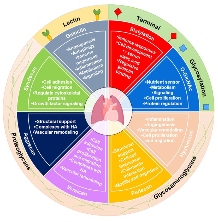
Figure
Figure 1. The
1. The glycobiology
glycobiology of PAH.
of PAH. Changes in Changes inhave
glycosylation glycosylation
been linked tohave been linked
dysregulated cel- to dysregulated
lular metabolism,
cellular a hallmark
metabolism, of both PAHof
a hallmark and cancer.
both PAHGalectins (Lectins); Galectins
and cancer. O-GlcNAc and sialylationO-GlcNAc and sialy-
(Lectins);
(Terminal Glycosylation); as well as hyaluronan, perlecan, versican, aggrecan, and syndecan (Pro-
lation (Terminal Glycosylation); as well as hyaluronan, perlecan, versican, aggrecan, and syndecan
teoglycans and Glycosaminoglycans) have been investigated in PAH.
(Proteoglycans and Glycosaminoglycans) have been investigated in PAH.
PGs/GAGs (HA) and galectins are increased in PAH. In addition, sialylation may be
altered. However, there specific roles and how they contribute to vascular remodeling and
regulation of vascular function as well as cell proliferation, cell signaling, and cell differ-
entiation/morphology have not been defined. Are the changes observed in these gly-
cans/glycoconjugates a consequence of the metabolic derangements observed in PAH? Or
do these glycoconjugates, when altered, have a direct role in the PAH pathogenesis? Stud-
ying the specific role of glycans and/or glycoconjugates is critical to fully understandingMetabolites 2022, 12, 316 8 of 17
PGs/GAGs (HA) and galectins are increased in PAH. In addition, sialylation may be
altered. However, there specific roles and how they contribute to vascular remodeling
and regulation of vascular function as well as cell proliferation, cell signaling, and cell
differentiation/morphology have not been defined. Are the changes observed in these
glycans/glycoconjugates a consequence of the metabolic derangements observed in PAH?
Or do these glycoconjugates, when altered, have a direct role in the PAH pathogenesis?
Studying the specific role of glycans and/or glycoconjugates is critical to fully understand-
ing the molecular mechanisms associated with dysregulated metabolism in PAH. These
efforts cannot move forward without some technical challenges, which are contingent on
the availability and accessibility of better tools for quantifying and precisely determin-
ing the roles of glycans and glycosylation events in cell biology. Targeted approaches to
regulating glycosyltransferases/hydrolases, sugar nucleotides, and/or specific glycoconju-
gates/glycoforms are needed to advance our knowledge of pulmonary hypertension as
well as other diseases defined by metabolic dysregulation.
Author Contributions: Conceptualization, J.W.B. and S.K.; writing—original draft preparation, S.V.,
P.C., S.K. and J.W.B.; writing—review and editing, S.V., P.C., J.S.D., S.K. and J.W.B. All authors have
read and agreed to the published version of the manuscript.
Funding: This work was supported by the National Institutes of Health (R01HL152246 to J.W.B. and
R01HL160911 to S.K.).
Conflicts of Interest: The authors declare no conflict of interest.
References
1. Mélot, C.; Naeije, R. Pulmonary vascular diseases. Compr. Physiol. 2011, 1, 593–619. [PubMed]
2. Barnes, J.W.; Tonelli, A.R.; Heresi, G.A.; Newman, J.E.; Mellor, N.E.; Grove, D.E.; Dweik, R.A. Novel Methods in Pulmonary
Hypertension Phenotyping in the Age of Precision Medicine (2015 Grover Conference Series). Pulm. Circ. 2016, 6, 439–447.
[CrossRef] [PubMed]
3. Badesch, D.B.; Champion, H.C.; Sanchez, M.A.G.; Hoeper, M.M.; Loyd, J.E.; Manes, A.; McGoon, M.; Naeije, R.; Olschewski,
H.; Oudiz, R.J.; et al. Diagnosis and Assessment of Pulmonary Arterial Hypertension. J. Am. Coll. Cardiol. 2009, 54, S55–S66.
[CrossRef] [PubMed]
4. Galiè, N.; Hoeper, M.M.; Humbert, M.; Torbicki, A.; Vachiery, J.L.; Barbera, J.A.; Beghetti, M.; Corris, P.; Gaine, S.; Gibbs, J.S.;
et al. Guidelines for the diagnosis and treatment of pulmonary hypertension: The Task Force for the Diagnosis and Treatment of
Pulmonary Hypertension of the European Society of Cardiology (ESC) and the European Respiratory Society (ERS), endorsed by
the International Society of Heart and Lung Transplantation (ISHLT). Eur. Heart J. 2009, 30, 2493–2537.
5. Simonneau, G.; Montani, D.; Celermajer, D.S.; Denton, C.P.; Gatzoulis, M.A.; Krowka, M.; Williams, P.G.; Souza, R. Haemodynamic
definitions and updated clinical classification of pulmonary hypertension. Eur. Respir. J. 2019, 53, 1801913. [CrossRef]
6. Simonneau, G.; Hoeper, M.M. The revised definition of pulmonary hypertension: Exploring the impact on patient management.
Eur. Heart J. 2019, 21, K4–K8. [CrossRef]
7. Hoeper, M.M.; Bogaard, H.J.; Condliffe, R.; Frantz, R.; Khanna, D.; Kurzyna, M.; Langleben, D.; Manes, A.; Satoh, T.; Torres, F.;
et al. Definitions and diagnosis of pulmonary hypertension. J. Am. Coll. Cardiol. 2013, 62, D42–D50. [CrossRef]
8. Galie, N.; Humbert, M.; Vachiery, J.L.; Gibbs, S.; Lang, I.; Torbicki, A.; Simonneau, G.; Peacock, A.; Vonk Noordegraaf, A.; Beghetti,
M.; et al. 2015 ESC/ERS Guidelines for the Diagnosis and Treatment of Pulmonary Hypertension. Rev. Esp. Cardiol. (Engl. Ed.)
2016, 69, 177.
9. Memon, H.A.; Park, M.H. Pulmonary Arterial Hypertension in Women. Methodist Debakey Cardiovasc. J. 2017, 13, 224–237.
[CrossRef]
10. D’Alonzo, G.E.; Barst, R.J.; Ayres, S.M.; Bergofsky, E.H.; Brundage, B.H.; Detre, K.M.; Fishman, A.P.; Goldring, R.M.; Groves, B.M.;
Kernis, J.T.; et al. Survival in patients with primary pulmonary hypertension. Results from a national prospective registry. Ann.
Intern. Med. 1991, 115, 343–349. [CrossRef]
11. Hassoun, P.M. Pulmonary Arterial Hypertension. N. Engl. J. Med. 2021, 385, 2361–2376. [CrossRef] [PubMed]
12. Yan, Y.; He, Y.Y.; Jiang, X.; Wang, Y.; Chen, J.W.; Zhao, J.H.; Ye, J.; Lian, T.Y.; Zhang, X.; Zhang, R.J.; et al. DNA methyltransferase
3B deficiency unveils a new pathological mechanism of pulmonary hypertension. Sci. Adv. 2020, 6, eaba2470. [CrossRef]
13. Wang, X.-J.; Lian, T.-Y.; Jiang, X.; Liu, S.-F.; Li, S.-Q.; Jiang, R.; Wu, W.-H.; Ye, J.; Cheng, C.-Y.; Du, Y.; et al. Germline BMP9 mutation
causes idiopathic pulmonary arterial hypertension. Eur. Respir. J. 2019, 53, 1801609. [CrossRef] [PubMed]
14. Evans, J.D.; Girerd, B.; Montani, D.; Wang, X.J.; Galie, N.; Austin, E.D.; Elliott, G.; Asano, K.; Grunig, E.; Yan, Y.; et al.
BMPR2 mutations and survival in pulmonary arterial hypertension: An individual participant data meta-analysis. Lancet Respir.
Med. 2016, 4, 129–137. [CrossRef]Metabolites 2022, 12, 316 9 of 17
15. Wang, X.-J.; Xu, X.-Q.; Sun, K.; Liu, K.-Q.; Li, S.-Q.; Jiang, X.; Zhao, Q.-H.; Wang, L.; Peng, F.-H.; Ye, J.; et al. Association of
Rare PTGIS Variants With Susceptibility and Pulmonary Vascular Response in Patients With Idiopathic Pulmonary Arterial
Hypertension. JAMA Cardiol. 2020, 5, 677–684. [CrossRef]
16. Klouda, T.; Yuan, K. Inflammation in Pulmonary Arterial Hypertension. Adv. Exp. Med. Biol. 2021, 1303, 351–372.
17. Voelkel, N.F.; Gomez-Arroyo, J.; Abbate, A.; Bogaard, H.J.; Nicolls, M.R. Pathobiology of pulmonary arterial hypertension and
right ventricular failure. Eur. Respir. J. 2012, 40, 1555–1565. [CrossRef]
18. Rabinovitch, M.; Guignabert, C.; Humbert, M.; Nicolls, M.R. Inflammation and immunity in the pathogenesis of pulmonary
arterial hypertension. Circ. Res. 2014, 115, 165–175. [CrossRef]
19. Meloche, J.; Renard, S.; Provencher, S.; Bonnet, S. Anti-inflammatory and immunosuppressive agents in PAH. Handb. Exp.
Pharmacol. 2013, 218, 437–476.
20. Humbert, M.; Guignabert, C.; Bonnet, S.; Dorfmuller, P.; Klinger, J.R.; Nicolls, M.R.; Olschewski, A.J.; Pullamsetti, S.S.; Schermuly,
R.T.; Stenmark, K.R.; et al. Pathology and pathobiology of pulmonary hypertension: State of the art and research perspectives.
Eur. Respir. J. 2019, 53, 1801887. [CrossRef]
21. Tuder, R.M.; Chacon, M.; Alger, L.; Wang, J.; Taraseviciene-Stewart, L.; Kasahara, Y.; Cool, C.D.; Bishop, A.E.; Geraci, M.; Semenza,
G.L.; et al. Expression of angiogenesis-related molecules in plexiform lesions in severe pulmonary hypertension: Evidence for a
process of disordered angiogenesis. J. Pathol. 2001, 195, 367–374. [CrossRef] [PubMed]
22. Jeffery, T.K.; Morrell, N.W. Molecular and cellular basis of pulmonary vascular remodeling in pulmonary hypertension. Prog.
Cardiovasc. Dis. 2002, 45, 173–202. [CrossRef]
23. Koress, C.; Swan, K.; Kadowitz, P. Soluble Guanylate Cyclase Stimulators and Activators: Novel Therapies for Pulmonary
Vascular Disease or a Different Method of Increasing cGMP? Curr. Hypertens. Rep. 2016, 18, 42. [CrossRef] [PubMed]
24. Xu, W.; Kaneko, F.T.; Zheng, S.; Comhair, S.A.; Janocha, A.J.; Goggans, T.; Thunnissen, F.B.; Farver, C.; Hazen, S.L.; Jennings, C.;
et al. Increased arginase II and decreased NO synthesis in endothelial cells of patients with pulmonary arterial hypertension.
FASEB J. 2004, 18, 1746–1748. [CrossRef] [PubMed]
25. Kaneko, F.T.; Arroliga, A.C.; Dweik, R.A.; Comhair, S.A.; Laskowski, D.; Oppedisano, R.; Thomassen, M.J.; Erzurum, S.C.
Biochemical reaction products of nitric oxide as quantitative markers of primary pulmonary hypertension. Am. J. Respir. Crit.
Care Med. 1998, 158, 917–923. [CrossRef]
26. Giaid, A.; Saleh, D. Reduced expression of endothelial nitric oxide synthase in the lungs of patients with pulmonary hypertension.
N. Engl. J. Med. 1995, 333, 214–221. [CrossRef]
27. Rubin, L.J. Primary pulmonary hypertension. N. Engl. J. Med. 1997, 336, 111–117. [CrossRef]
28. Brunner, N.W.; Skhiri, M.; Fortenko, O.; Hsi, A.; Haddad, F.; Khazeni, N.; Zamanian, R.T. Impact of insulin resistance on
ventricular function in pulmonary arterial hypertension. J. Heart Lung Transplant. 2014, 33, 721–726. [CrossRef]
29. Hansmann, G.; Wagner, R.A.; Schellong, S.; Perez, V.A.; Urashima, T.; Wang, L.; Sheikh, A.Y.; Suen, R.S.; Stewart, D.J.; Rabinovitch,
M. Pulmonary arterial hypertension is linked to insulin resistance and reversed by peroxisome proliferator-activated receptor-
gamma activation. Circulation 2007, 115, 1275–1284. [CrossRef]
30. Pugh, M.E.; Robbins, I.M.; Rice, T.W.; West, J.; Newman, J.H.; Hemnes, A.R. Unrecognized glucose intolerance is common in
pulmonary arterial hypertension. J. Heart Lung Transplant. 2011, 30, 904–911. [CrossRef]
31. Zamanian, R.T.; Hansmann, G.; Snook, S.; Lilienfeld, D.; Rappaport, K.M.; Reaven, G.M.; Rabinovitch, M.; Doyle, R.L. Insulin
resistance in pulmonary arterial hypertension. Eur. Respir. J. 2009, 33, 318–324. [CrossRef]
32. Aytekin, M.; Tonelli, A.R.; Farver, C.F.; Feldstein, A.E.; Dweik, R.A. Leptin deficiency recapitulates the histological features of
pulmonary arterial hypertension in mice. Int. J. Clin. Exp. Pathol. 2014, 7, 1935–1946.
33. Santos, M.; Reis, A.; Goncalves, F.; Ferreira-Pinto, M.J.; Cabral, S.; Torres, S.; Leite-Moreira, A.F.; Henriques-Coelho, T. Adiponectin
levels are elevated in patients with pulmonary arterial hypertension. Clin. Cardiol. 2014, 37, 21–25. [CrossRef]
34. Tonelli, A.R.; Aytekin, M.; Feldstein, A.E.; Dweik, R.A. Leptin Levels Predict Survival in Pulmonary Arterial Hypertension. Pulm.
Circ. 2012, 2, 214–219. [CrossRef]
35. Talati, M.; Hemnes, A. Fatty acid metabolism in pulmonary arterial hypertension: Role in right ventricular dysfunction and
hypertrophy. Pulm. Circ. 2015, 5, 269–278. [CrossRef]
36. Cracowski, J.L.; Cracowski, C.; Bessard, G.; Pepin, J.L.; Bessard, J.; Schwebel, C.; Stanke-Labesque, F.; Pison, C. Increased lipid
peroxidation in patients with pulmonary hypertension. Am. J. Respir. Crit. Care Med. 2001, 164, 1038–1042. [CrossRef]
37. Brittain, E.L.; Talati, M.; Fessel, J.P.; Zhu, H.; Penner, N.; Calcutt, M.W.; West, J.D.; Funke, M.; Lewis, G.D.; Gerszten, R.E.; et al.
Fatty Acid Metabolic Defects and Right Ventricular Lipotoxicity in Human Pulmonary Arterial Hypertension. Circulation 2016,
133, 1936–1944. [CrossRef]
38. Cathey, S.S. Breath analysis in pulmonary arterial hypertension. Eur. J. Hum. Genet. EJHG 2014, 145, 551–558.
39. Chen, J.; Tang, H.; Sysol, J.R.; Moreno-Vinasco, L.; Shioura, K.M.; Chen, T.; Gorshkova, I.; Wang, L.; Huang, L.S.; Usatyuk, P.V.;
et al. The sphingosine kinase 1/sphingosine-1-phosphate pathway in pulmonary arterial hypertension. Am. J. Respir. Crit. Care
Med. 2014, 190, 1032–1043. [CrossRef]
40. Ross, D.J.; Hough, G.; Hama, S.; Aboulhosn, J.; Belperio, J.A.; Saggar, R.; Van Lenten, B.J.; Ardehali, A.; Eghbali, M.; Reddy, S.;
et al. Proinflammatory high-density lipoprotein results from oxidized lipid mediators in the pathogenesis of both idiopathic and
associated types of pulmonary arterial hypertension. Pulm. Circ. 2015, 5, 640–648. [CrossRef]Metabolites 2022, 12, 316 10 of 17
41. Sutendra, G.; Bonnet, S.; Rochefort, G.; Haromy, A.; Folmes, K.D.; Lopaschuk, G.D.; Dyck, J.R.; Michelakis, E.D. Fatty acid
oxidation and malonyl-CoA decarboxylase in the vascular remodeling of pulmonary hypertension. Sci. Transl. Med. 2010, 2,
44ra58. [CrossRef]
42. Heresi, G.A.; Aytekin, M.; Newman, J.; DiDonato, J.; Dweik, R.A. Plasma levels of high-density lipoprotein cholesterol and
outcomes in pulmonary arterial hypertension. Am. J. Respir. Crit. Care Med. 2010, 182, 661–668. [CrossRef]
43. Hu, J.; Xu, Q.; McTiernan, C.; Lai, Y.C.; Osei-Hwedieh, D.; Gladwin, M. Novel Targets of Drug Treatment for Pulmonary
Hypertension. Am. J. Cardiovasc. Drugs 2015, 15, 225–234. [CrossRef]
44. Ozkan, M.; Dweik, R.A.; Laskowski, D.; Arroliga, A.C.; Erzurum, S.C. High levels of nitric oxide in individuals with pulmonary
hypertension receiving epoprostenol therapy. Lung 2001, 179, 233–243. [CrossRef]
45. Giaid, A.; Yanagisawa, M.; Langleben, D.; Michel, R.P.; Levy, R.; Shennib, H.; Kimura, S.; Masaki, T.; Duguid, W.P.; Stewart,
D.J. Expression of Endothelin-1 in the Lungs of Patients with Pulmonary Hypertension. N. Engl. J. Med. 1993, 328, 1732–1739.
[CrossRef]
46. Heresi, G.A.; Malin, S.K.; Barnes, J.W.; Tian, L.; Kirwan, J.P.; Dweik, R.A. Abnormal Glucose Metabolism and High-Energy
Expenditure in Idiopathic Pulmonary Arterial Hypertension. Ann. Am. Thorac. Soc. 2016, 14, 190–199.
47. Lundgrin, E.L.; Park, M.M.; Sharp, J.; Tang, W.H.; Thomas, J.D.; Asosingh, K.; Comhair, S.A.; DiFilippo, F.P.; Neumann, D.R.;
Davis, L.; et al. Fasting 2-deoxy-2-[18F]fluoro-D-glucose positron emission tomography to detect metabolic changes in pulmonary
arterial hypertension hearts over 1 year. Ann. Am. Thorac. Soc. 2013, 10, 1–9. [CrossRef]
48. Marsboom, G.; Wietholt, C.; Haney, C.R.; Toth, P.T.; Ryan, J.J.; Morrow, E.; Thenappan, T.; Bache-Wiig, P.; Piao, L.; Paul, J.;
et al. Lung (1)(8)F-fluorodeoxyglucose positron emission tomography for diagnosis and monitoring of pulmonary arterial
hypertension. Am. J. Respir. Crit. Care Med. 2012, 185, 670–679. [CrossRef]
49. Barnes, J.W.; Kucera, E.T.; Tian, L.; Mellor, N.E.; Dvorina, N.; Baldwin, W.W., III; Aldred, M.A.; Farver, C.F.; Comhair, S.A.;
Aytekin, M.; et al. BMPR2 Mutation-independent Mechanisms of Disrupted BMP Signaling in IPAH. Am. J. Respir. Cell Mol. Biol.
2016, 55, 564–575. [CrossRef]
50. Cikach, F.S., Jr.; Tonelli, A.R.; Barnes, J.; Paschke, K.; Newman, J.; Grove, D.; Dababneh, L.; Wang, S.; Dweik, R.A. Breath Analysis
in Pulmonary Arterial Hypertension. Chest 2013, 145, 551–558. [CrossRef]
51. Kao, C.C.; Wedes, S.H.; Hsu, J.W.; Bohren, K.M.; Comhair, S.A.A.; Jahoor, F.; Erzurum, S.C. Arginine Metabolic Endotypes in
Pulmonary Arterial Hypertension. Pulm. Circ. 2015, 5, 124–134. [CrossRef]
52. Demoncheaux, E.A.; Higenbottam, T.W.; Kiely, D.G.; Wong, J.M.; Wharton, S.; Varcoe, R.; Siddons, T.; Spivey, A.C.; Hall, K.; Gize,
A.P. Decreased whole body endogenous nitric oxide production in patients with primary pulmonary hypertension. J. Vasc. Res.
2005, 42, 133–136. [CrossRef]
53. Izquierdo-Garcia, J.L.; Arias, T.; Rojas, Y.; Garcia-Ruiz, V.; Santos, A.; Martin-Puig, S.; Ruiz-Cabello, J. Metabolic Reprogramming
in the Heart and Lung in a Murine Model of Pulmonary Arterial Hypertension. Front. Cardiovasc. Med 2018, 5, 110. [CrossRef]
54. Piao, L.; Fang, Y.H.; Parikh, K.; Ryan, J.J.; Toth, P.T.; Archer, S.L. Cardiac glutaminolysis: A maladaptive cancer metabolism
pathway in the right ventricle in pulmonary hypertension. J. Mol. Med. 2013, 91, 1185–1197. [CrossRef]
55. Egnatchik, R.A.; Brittain, E.L.; Shah, A.T.; Fares, W.H.; Ford, H.J.; Monahan, K.; Kang, C.J.; Kocurek, E.G.; Zhu, S.; Luong, T.; et al.
Dysfunctional BMPR2 signaling drives an abnormal endothelial requirement for glutamine in pulmonary arterial hypertension.
Pulm. Circ. 2017, 7, 186–199. [CrossRef]
56. Mey, J.T.; Hari, A.; Axelrod, C.L.; Fealy, C.E.; Erickson, M.L.; Kirwan, J.P.; Dweik, R.A.; Heresi, G.A. Lipids and ketones dominate
metabolism at the expense of glucose control in pulmonary arterial hypertension: A hyperglycaemic clamp and metabolomics
study. Eur. Respir. J. 2020, 55, 1901700. [CrossRef]
57. Belly, M.J.; Tiede, H.; Morty, R.E.; Schulz, R.; Voswinckel, R.; Tanislav, C.; Olschewski, H.; Ghofrani, H.A.; Seeger, W.; Reichen-
berger, F. HbA1c in pulmonary arterial hypertension: A marker of prognostic relevance? J. Heart Lung Transpl. 2012, 31, 1109–1114.
[CrossRef]
58. Paulin, R.; Michelakis, E.D. The metabolic theory of pulmonary arterial hypertension. Circ. Res. 2014, 115, 148–164. [CrossRef]
59. Rai, P.R.; Cool, C.D.; King, J.A.; Stevens, T.; Burns, N.; Winn, R.A.; Kasper, M.; Voelkel, N.F. The cancer paradigm of severe
pulmonary arterial hypertension. Am. J. Respir. Crit. Care Med. 2008, 178, 558–564. [CrossRef]
60. Guignabert, C.; Tu, L.; Le Hiress, M.; Ricard, N.; Sattler, C.; Seferian, A.; Huertas, A.; Humbert, M.; Montani, D. Pathogenesis of
pulmonary arterial hypertension: Lessons from cancer. Eur. Respir. Rev. Off. J. Eur. Respir. Soc. 2013, 22, 543–551. [CrossRef]
61. Cool, C.D.; Kuebler, W.M.; Bogaard, H.J.; Spiekerkoetter, E.; Nicolls, M.R.; Voelkel, N.F. The hallmarks of severe pulmonary
arterial hypertension: The cancer hypothesis—ten years later. Am. J. Physiol. Lung Cell. Mol. Physiol. 2020, 318, L1115–L1130.
[CrossRef]
62. Baggetto, L.G. Deviant energetic metabolism of glycolytic cancer cells. Biochimie 1992, 74, 959–974. [CrossRef]
63. Altenberg, B.; Greulich, K.O. Genes of glycolysis are ubiquitously overexpressed in 24 cancer classes. Genomics 2004, 84, 1014–1020.
[CrossRef]
64. Bos, R.; van der Hoeven, J.J.M.; van der Wall, E.; van der Groep, P.; van Diest, P.J.; ComansUrvi Joshi, E.F.I.; Semenza, G.L.;
Hoekstra, O.S.; Lammertsma, A.A.; Molthoff, C.F.M. Biologic Correlates of 18Fluorodeoxyglucose Uptake in Human Breast
Cancer Measured by Positron Emission Tomography. J. Clin. Oncol. 2002, 20, 379–387. [CrossRef]Metabolites 2022, 12, 316 11 of 17
65. Pedersen, P.L. Warburg, me and Hexokinase 2, Multiple discoveries of key molecular events underlying one of cancers’ most
common phenotypes, the “Warburg Effect”, i.e., elevated glycolysis in the presence of oxygen. J. Bioenerg. Biomembr. 2007, 39, 211.
[CrossRef]
66. Warburg, O. On the origin of cancer cells. Science 1956, 123, 309–314. [CrossRef]
67. Diaz-Ruiz, R.; Rigoulet, M.; Devin, A. The Warburg and Crabtree effects: On the origin of cancer cell energy metabolism and of
yeast glucose repression. Biochim. Biophys. Acta 2011, 1807, 568–576. [CrossRef]
68. Xu, W.; Koeck, T.; Lara, A.R.; Neumann, D.; DiFilippo, F.P.; Koo, M.; Janocha, A.J.; Masri, F.A.; Arroliga, A.C.; Jennings, C.;
et al. Alterations of cellular bioenergetics in pulmonary artery endothelial cells. Proc. Natl. Acad. Sci. USA 2007, 104, 1342–1347.
[CrossRef]
69. Diebold, I.; Hennigs, J.K.; Miyagawa, K.; Li, C.G.; Nickel, N.P.; Kaschwich, M.; Cao, A.; Wang, L.; Reddy, S.; Chen, P.-I.; et al.
BMPR2 Preserves Mitochondrial Function and DNA during Reoxygenation to Promote Endothelial Cell Survival and Reverse
Pulmonary Hypertension. Cell Metab. 2015, 21, 596–608. [CrossRef]
70. Rehman, J.; Archer, S.L. A proposed mitochondrial-metabolic mechanism for initiation and maintenance of pulmonary arterial
hypertension in fawn-hooded rats: The Warburg model of pulmonary arterial hypertension. Adv. Exp. Med. Biol. 2010, 661,
171–185.
71. Rabinovitch, M. Molecular pathogenesis of pulmonary arterial hypertension. J. Clin. Investig. 2012, 122, 4306–4313. [CrossRef]
72. Bonnet, S.; Michelakis, E.D.; Porter, C.J.; Andrade-Navarro, M.A.; Thébaud, B.; Bonnet, S.; Haromy, A.; Harry, G.; Moudgil, R.;
McMurtry, M.S.; et al. An abnormal mitochondrial-hypoxia inducible factor-1alpha-Kv channel pathway disrupts oxygen sensing
and triggers pulmonary arterial hypertension in fawn hooded rats: Similarities to human pulmonary arterial hypertension.
Circulation 2006, 113, 2630–2641. [CrossRef]
73. Gomez-Arroyo, J.; Mizuno, S.; Szczepanek, K.; Tassell, B.V.; Natarajan, R.; dos Remedios, C.G.; Drake, J.I.; Farkas, L.; Kraskauskas,
D.; Wijesinghe, D.S.; et al. Metabolic Gene Remodeling and Mitochondrial Dysfunction in Failing Right Ventricular Hypertrophy
Secondary to Pulmonary Arterial Hypertension. Circ. Heart Fail. 2013, 6, 136–144. [CrossRef]
74. Nagendran, J.; Gurtu, V.; Fu, D.Z.; Dyck, J.R.; Haromy, A.; Ross, D.B.; Rebeyka, I.M.; Michelakis, E.D. A dynamic and chamber-
specific mitochondrial remodeling in right ventricular hypertrophy can be therapeutically targeted. J. Thorac. Cardiovasc. Surg.
2008, 136, 168–178.e163. [CrossRef]
75. Michelakis, E.D.; Gurtu, V.; Webster, L.; Barnes, G.; Watson, G.; Howard, L.; Cupitt, J.; Paterson, I.; Thompson, R.B.; Chow, K.;
et al. Inhibition of pyruvate dehydrogenase kinase improves pulmonary arterial hypertension in genetically susceptible patients.
Sci. Transl. Med. 2017, 9, eaao4583. [CrossRef]
76. Bonnet, S.; Archer, S.L.; Allalunis-Turner, J.; Haromy, A.; Beaulieu, C.; Thompson, R.; Lee, C.T.; Lopaschuk, G.D.; Puttagunta,
L.; Bonnet, S.; et al. A Mitochondria-K+ Channel Axis Is Suppressed in Cancer and Its Normalization Promotes Apoptosis and
Inhibits Cancer Growth. Cancer Cell 2007, 11, 37–51. [CrossRef]
77. Chu, Q.S.; Sangha, R.; Spratlin, J.; Vos, L.J.; Mackey, J.R.; McEwan, A.J.; Venner, P.; Michelakis, E.D. A phase I open-labeled,
single-arm, dose-escalation, study of dichloroacetate (DCA) in patients with advanced solid tumors. Investig. New Drugs 2015, 33,
603–610. [CrossRef]
78. McMurtry, M.S.; Bonnet, S.; Wu, X.; Dyck, J.R.; Haromy, A.; Hashimoto, K.; Michelakis, E.D. Dichloroacetate prevents and reverses
pulmonary hypertension by inducing pulmonary artery smooth muscle cell apoptosis. Circ. Res. 2004, 95, 830–840. [CrossRef]
79. Dyck, J.R.; Hopkins, T.A.; Bonnet, S.; Michelakis, E.D.; Young, M.E.; Watanabe, M.; Kawase, Y.; Jishage, K.; Lopaschuk, G.D.
Absence of malonyl coenzyme A decarboxylase in mice increases cardiac glucose oxidation and protects the heart from ischemic
injury. Circulation 2006, 114, 1721–1728. [CrossRef]
80. Reily, C.; Stewart, T.J.; Renfrow, M.B.; Novak, J. Glycosylation in health and disease. Nat. Rev. Nephrol. 2019, 15, 346–366.
[CrossRef]
81. Varki, A. Biological roles of oligosaccharides: All of the theories are correct. Glycobiology 1993, 3, 97–130. [CrossRef]
82. Varki, A. Biological roles of glycans. Glycobiology 2017, 27, 3–49. [CrossRef]
83. Häuselmann, I.; Borsig, L. Altered Tumor-Cell Glycosylation Promotes Metastasis. Front. Oncol. 2014, 4, 28. [CrossRef]
84. Kamigaito, T.; Okaneya, T.; Kawakubo, M.; Shimojo, H.; Nishizawa, O.; Nakayama, J. Overexpression of O-GlcNAc by prostate
cancer cells is significantly associated with poor prognosis of patients. Prostate Cancer Prostatic Dis. 2014, 17, 18–22. [CrossRef]
85. Kizuka, Y.; Taniguchi, N. Enzymes for N-Glycan Branching and Their Genetic and Nongenetic Regulation in Cancer. Biomolecules
2016, 6, 25. [CrossRef]
86. Mathews, M.B.; Glagov, S. Acid mucopolysaccharide patterns in aging human cartilage. J. Clin. Investig. 1966, 45, 1103–1111.
[CrossRef]
87. Pal-Ghosh, S.; Tadvalkar, G.; Stepp, M.A. Alterations in Corneal Sensory Nerves During Homeostasis, Aging, and after Injury in
Mice Lacking the Heparan Sulfate Proteoglycan Syndecan-1. Investig. Opthalmol. Vis. Sci. 2017, 58, 4959–4975. [CrossRef]
88. Kobata, A. Glycobiology in the field of aging research—Introduction to glycogerontology. Biochimie 2003, 85, 13–24. [CrossRef]
89. Beeley, J.G.; Blackie, R.; Baxter, A. Glycoprotein and glycolipid changes in aged erythrocytes [proceedings]. Biochem. Soc. Trans.
1977, 5, 1725–1726. [CrossRef]
90. Ovsepian, L.M.; Kazarian, G.S.; Akopdzhanian, A.A.; L’Vov, M.V. Age-dependent changes in phospholipid content and neutral
lipid contents in aging. Adv. Gerontol. 2012, 25, 250–254. [CrossRef]Metabolites 2022, 12, 316 12 of 17
91. Wang, W.; Gopal, S.; Pocock, R.; Xiao, Z. Glycan Mimetics from Natural Products: New Therapeutic Opportunities for Neurode-
generative Disease. Molecules 2019, 24, 4604. [CrossRef]
92. Kizuka, Y.; Kitazume, S.; Fujinawa, R.; Saito, T.; Iwata, N.; Saido, T.C.; Nakano, M.; Yamaguchi, Y.; Hashimoto, Y.; Staufenbiel, M.;
et al. An aberrant sugar modification of BACE1 blocks its lysosomal targeting in Alzheimer’s disease. EMBO Mol. Med. 2015, 7,
175–189. [CrossRef]
93. Maguire, T.M.; Gillian, A.M.; O’Mahony, D.; Coughlan, C.M.; Breen, K.C. A decrease in serum sialyltransferase levels in
Alzheimer’s disease. Neurobiol. Aging 1994, 15, 99–102. [CrossRef]
94. Fodero, L.R.; Sáez-Valero, J.; Barquero, M.S.; Marcos, A.; McLean, C.A.; Small, D.H. Wheat germ agglutinin-binding glycoproteins
are decreased in Alzheimer’s disease cerebrospinal fluid. J. Neurochem. 2001, 79, 1022–1026. [CrossRef]
95. Saito, F.; Yanagisawa, K.; Miyatake, T. Soluble derivatives of β/A4 amyloid protein precursor in human cerebrospinal fluid are
both N- and O-glycosylated. Mol. Brain Res. 1993, 19, 171–174. [CrossRef]
96. Griffith, L.S.; Mathes, M.; Schmitz, B. Beta-amyloid precursor protein is modified with O-linked N-acetylglucosamine. J. Neurosci.
Res. 1995, 41, 270–278. [CrossRef]
97. Zhu, Y.; Shan, X.; Yuzwa, S.A.; Vocadlo, D.J. The emerging link between O-GlcNAc and Alzheimer disease. J. Biol. Chem. 2014,
289, 34472–34481. [CrossRef]
98. Dashti, H.; Pabon Porras, M.A.; Mora, S. Glycosylation and Cardiovascular Diseases. In The Role of Glycosylation in Health and
Disease; Lauc, G., Trbojević-Akmačić, I., Eds.; Springer International Publishing: Cham, Switzerland, 2021; pp. 307–319.
99. 99Gudelj, I.; Lauc, G. Protein N-Glycosylation in Cardiovascular Diseases and Related Risk Factors. Curr. Cardiovasc. Risk Rep.
2018, 12, 16. [CrossRef]
100. Wright, J.N.; Collins, H.E.; Wende, A.R.; Chatham, J.C. O-GlcNAcylation and cardiovascular disease. Biochem. Soc. Trans. 2017, 45,
545–553. [CrossRef]
101. Komaromy, A.; Reider, B.; Jarvas, G.; Guttman, A. Glycoprotein biomarkers and analysis in chronic obstructive pulmonary
disease and lung cancer with special focus on serum immunoglobulin G. Clin. Chim. Acta 2020, 506, 204–213. [CrossRef]
102. Pavić, T.; Dilber, D.; Kifer, D.; Selak, N.; Keser, T.; Ljubičić, Ð.; Vukić Dugac, A.; Lauc, G.; Rumora, L.; Gornik, O. N-glycosylation
patterns of plasma proteins and immunoglobulin G in chronic obstructive pulmonary disease. J. Transl. Med. 2018, 16, 323.
[CrossRef] [PubMed]
103. Wang, X.; Inoue, S.; Gu, J.; Miyoshi, E.; Noda, K.; Li, W.; Mizuno-Horikawa, Y.; Nakano, M.; Asahi, M.; Takahashi, M.; et al.
Dysregulation of TGF-β1 receptor activation leads to abnormal lung development and emphysema-like phenotype in core
fucose-deficient mice. Proc. Natl. Acad. Sci. USA 2005, 102, 15791–15796. [CrossRef] [PubMed]
104. Yamada, M.; Ishii, T.; Ikeda, S.; Naka-Mieno, M.; Tanaka, N.; Arai, T.; Kumasaka, T.; Gemma, A.; Kida, K.; Muramatsu, M.; et al.
Association of fucosyltransferase 8 (FUT8) polymorphism Thr267Lys with pulmonary emphysema. J. Hum. Genet. 2011, 56,
857–860. [CrossRef] [PubMed]
105. Mészáros, B.; Járvás, G.; Farkas, A.; Szigeti, M.; Kovács, Z.; Kun, R.; Szabó, M.; Csánky, E.; Guttman, A. Comparative analysis of
the human serum N-glycome in lung cancer, COPD and their comorbidity using capillary electrophoresis. J. Chromatogr. B 2019,
1137, 121913. [CrossRef] [PubMed]
106. Krick, S.; Helton, E.S.; Easter, M.; Bollenbecker, S.; Denson, R.; Zaharias, R.; Cochran, P.; Vang, S.; Harris, E.; Wells, J.M.; et al.
ST6GAL1 and α2-6 Sialylation Regulates IL-6 Expression and Secretion in Chronic Obstructive Pulmonary Disease. Front.
Immunol. 2021, 12, 693149. [CrossRef]
107. Schulz, B.; Sloane, A.J.; Robinson, L.J.; Prasad, S.S.; Lindner, R.A.; Robinson, M.; Bye, P.T.; Nielson, D.W.; Harry, J.L.; Packer, N.H.;
et al. Glycosylation of sputum mucins is altered in cystic fibrosis patients. Glycobiology 2007, 17, 698–712. [CrossRef]
108. Arnold, J.N.; Saldova, R.; Galligan, M.C.; Murphy, T.B.; Mimura-Kimura, Y.; Telford, J.E.; Godwin, A.K.; Rudd, P.M. Novel Glycan
Biomarkers for the Detection of Lung Cancer. J. Proteome Res. 2011, 10, 1755–1764. [CrossRef]
109. Xia, B.; Royall, J.A.; Damera, G.; Sachdev, G.P.; Cummings, R.D. Altered O-glycosylation and sulfation of airway mucins
associated with cystic fibrosis. Glycobiology 2005, 15, 747–775. [CrossRef]
110. Davril, M.; Groux-Degroote, S.; Humbert, P.; Galabert, C.; Dumur, V.; Lafitte, J.-J.; Lamblin, G.; Roussel, P. The sialylation of
bronchial mucins secreted by patients suffering from cystic fibrosis or from chronic bronchitis is related to the severity of airway
infection. Glycobiology 1999, 9, 311–321. [CrossRef]
111. Collum, S.D.; Chen, N.; Hernandez, A.M.; Hanmandlu, A.; Sweeney, H.; Mertens, T.C.J.; Weng, T.; Luo, F.; Molina, J.G.; Davies, J.;
et al. Inhibition of hyaluronan synthesis attenuates pulmonary hypertension associated with lung fibrosis. J. Cereb. Blood Flow
Metab. 2017, 174, 3284–3301. [CrossRef]
112. Bjermer, L.; Lundgren, R.; Hallgren, R. Hyaluronan and type III procollagen peptide concentrations in bronchoalveolar lavage
fluid in idiopathic pulmonary fibrosis. Thorax 1989, 44, 126–131. [CrossRef] [PubMed]
113. Li, Y.; Jiang, D.; Liang, J.; Meltzer, E.B.; Gray, A.; Miura, R.; Wogensen, L.; Yamaguchi, Y.; Noble, P.W. Severe lung fibrosis requires
an invasive fibroblast phenotype regulated by hyaluronan and CD44. J. Exp. Med. 2011, 208, 1459–1471. [CrossRef] [PubMed]
114. Dittner-Moormann, S.; Lourenco, C.M.; Reunert, J.; Nishinakamura, R.; Tanaka, S.S.; Werner, C.; Debus, V.; Zimmer, K.-P.; Wetzel,
G.; Naim, H.Y.; et al. TRAPγ-CDG shows asymmetric glycosylation and an effect on processing of proteins required in higher
organisms. J. Med. Genet. 2021, 58, 213–216. [CrossRef]You can also read