The Role of HECT-Type E3 Ligase in the Development of Cardiac Disease - MDPI
←
→
Page content transcription
If your browser does not render page correctly, please read the page content below
International Journal of
               Molecular Sciences

Review
The Role of HECT-Type E3 Ligase in the Development of
Cardiac Disease
Jun Goto, Yoichiro Otaki *, Tetsu Watanabe                      and Masafumi Watanabe

                                          Department of Cardiology, Pulmonology and Nephrology, Yamagata University School of Medicine,
                                          2-2-2 Iida-Nishi, Yamagata 990-9585, Japan; j-goto007008@med.id.yamagata-u.ac.jp (J.G.);
                                          tewatana@med.id.yamagata-u.ac.jp (T.W.); m-watanabe@med.id.yamagata-u.ac.jp (M.W.)
                                          * Correspondence: y-otaki@med.id.yamagata-u.ac.jp; Tel.: +81-23-628-5302; Fax: +81-23-628-5305

                                          Abstract: Despite advances in medicine, cardiac disease remains an increasing health problem
                                          associated with a high mortality rate. Maladaptive cardiac remodeling, such as cardiac hypertrophy
                                          and fibrosis, is a risk factor for heart failure; therefore, it is critical to identify new therapeutic targets.
                                          Failing heart is reported to be associated with hyper-ubiquitylation and impairment of the ubiquitin–
                                          proteasome system, indicating an importance of ubiquitylation in the development of cardiac disease.
                                          Ubiquitylation is a post-translational modification that plays a pivotal role in protein function and
                                          degradation. In 1995, homologous to E6AP C-terminus (HECT) type E3 ligases were discovered. E3
                                          ligases are key enzymes in ubiquitylation and are classified into three families: really interesting
                                          new genes (RING), HECT, and RING-between-RINGs (RBRs). Moreover, 28 HECT-type E3 ligases
                                          have been identified in human beings. It is well conserved in evolution and is characterized by the
                                          direct attachment of ubiquitin to substrates. HECT-type E3 ligase is reported to be involved in a
                                          wide range of human diseases and health. The role of HECT-type E3 ligases in the development
                                of cardiac diseases has been uncovered in the last decade. There are only a few review articles
                                   summarizing recent advancements regarding HECT-type E3 ligase in the field of cardiac disease.
Citation: Goto, J.; Otaki, Y.;            This study focused on cardiac remodeling and described the role of HECT-type E3 ligases in the
Watanabe, T.; Watanabe, M. The Role       development of cardiac disease. Moreover, this study revealed that the current knowledge could be
of HECT-Type E3 Ligase in the             exploited for the development of new clinical therapies.
Development of Cardiac Disease. Int.
J. Mol. Sci. 2021, 22, 6065. https://     Keywords: ubiquitylation; HECT-type E3 ligase; cardiac disease
doi.org/10.3390/ijms22116065

Academic Editor: Tomoaki Ishigami

                                          1. Introduction
Received: 14 May 2021
Accepted: 1 June 2021
                                                Despite advances in medicine, cardiovascular disease remains a significant public
Published: 4 June 2021
                                          health problem associated with high mortality [1,2]. Heart failure (HF) is a major cause of
                                          cardiovascular deaths. Maladaptive cardiac remodeling caused by hypertension, ischemic
Publisher’s Note: MDPI stays neutral
                                          heart disease, and other cardiac diseases is accompanied by complex mechanisms that lead
with regard to jurisdictional claims in
                                          to the development of HF [3,4]. Further studies are needed to prevent maladaptive cardiac
published maps and institutional affil-   remodeling and subsequent heart failure.
iations.                                        The modification of eukaryotic proteins with ubiquitin, named ubiquitylation, con-
                                          trols their lifetimes, abundance, localization, interactions, and activities, thereby regulating
                                          protein function at all levels. Thus, ubiquitylation plays a pivotal role in a wide range
                                          of cellular processes, such as signal transduction, transcriptional regulation, and main-
Copyright: © 2021 by the authors.
                                          tenance of homeostasis. Failing hearts from patients with dilated cardiomyopathy and
Licensee MDPI, Basel, Switzerland.
                                          those with ischemic heart disease show hyper-ubiquitylation compared to donor hearts [5].
This article is an open access article
                                          An increase in key components of ubiquitylation (ubiquitin, ubiquitin-activating enzyme
distributed under the terms and           (E1), ubiquitin-conjugating enzyme (E2), and some ubiquitin ligase (E3)) and a decrease
conditions of the Creative Commons        in deubiquitinating enzymes have been observed [5–7]. Furthermore, several cardiac
Attribution (CC BY) license (https://     diseases, such as cardiac amyloidosis, hypertrophic cardiomyopathy, and hereditary car-
creativecommons.org/licenses/by/          diomyopathy, impair the ubiquitin–proteasome system [8,9]. The overall observed change
4.0/).                                    in the ubiquitylation cascade in failing hearts is considered an adaptive response to an

Int. J. Mol. Sci. 2021, 22, 6065. https://doi.org/10.3390/ijms22116065                                            https://www.mdpi.com/journal/ijms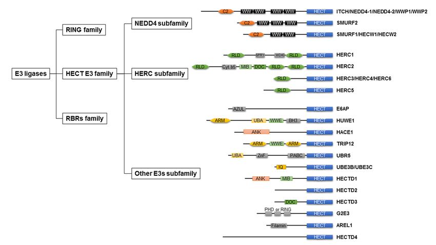
Int. J. Mol. Sci. 2021, 22, 6065                                                                                          2 of 26

                                   increased protein burden derived from increased protein synthesis that accompanies the
                                   hypertrophic response or an excess of damaged or modified proteins to be targeted for
                                   proteasomal degradation.
                                        Ubiquitin E3 ligase is pivotal in conferring specificity to ubiquitylation and provides
                                   particularly interesting targets for therapeutic interventions. In the 2000s, many stud-
                                   ies focused on the cardiac ubiquitin E3 ligases to clarify the role of ubiquitylation in
                                   the development of cardiac diseases, such as the carboxyl terminus of Hsp70 interact-
                                   ing protein (CHIP), atrogen-1, muscle ring finger (MuRF) family, mouse double mutant
                                   2 homolog (MDM2), cellular inhibitor of apoptosis, casitas b-lineage lymphoma, and E6-
                                   associated protein (E6AP) [10–17]. Cardiac ubiquitin E3 ligase plays several roles in protein
                                   turnover, energy metabolism, receptor internalization, hypertrophic response, apoptosis,
                                   and tolerance to ischemia/reperfusion (I/R) in cardiomyocytes [18,19]. Although elevated
                                   expression levels of E6AP were observed in mice after pressure overload [16], the functional
                                   role of E6AP has never been examined. Thus, our knowledge of the molecular mechanism
                                   of HECT-type E3 ligase in the development of cardiac disease is still lacking.
                                        HECT-type E3 ligase is reported to be involved in a wide range of human diseases and
                                   health including neurodegenerative diseases, neurological syndromes, and cancers [20–22].
                                   Notably, it is considered as an intriguing target in drug discovery in the context of cancer
                                   biology [23]. HECT-type E3 ligases are highly conserved between cells and tissues; as a
                                   result, it is tempting to speculate that they also contribute to human cardiac health and
                                   disease. The role of HECT-type E3 ligases in the development of cardiac diseases has
                                   been examined and uncovered in the last decade. There are only a few review articles
                                   summarizing recent advancements regarding HECT-type E3 ligase in the field of cardiac
                                   disease [24]. This study focused on cardiac remodeling and described the role of HECT-
                                   type E3 ligases in the development of cardiac disease. Moreover, this study revealed that
                                   the current knowledge can be exploited for the development of new clinical therapies.

                                   2. Ubiquitylation
                                   2.1. Ubiquitylation
                                        Ubiquitylation is a post-translational modification that covalently conjugates the
                                   ubiquitin molecule through the C-terminus to a lysine residue on a substrate protein.
                                   Ubiquitylation results in the turnover of the ubiquitylated substrate protein by either the
                                   proteasome or lysosome, a change in subcellular localization of the substrate protein, or
                                   alteration of substrate protein function [25]. Ubiquitylation is mediated by three enzymes
                                   and scaffolding proteins: E1, E2, and E3. There are only few E1s and several E2s; however,
                                   E3 ligases constitute a large class of proteins with the human genome encoding more
                                   than 600 putative E3 ligases and E3 ligase complexes [26–29]. Therefore, the specificity of
                                   ubiquitylation is determined by the numerous E3 ligases that recognize a specific substrate
                                   protein [30]. E3 ligases are also modulators of the rate-limiting step in this enzymatic
                                   cascade, participating in substrate protein recognition and catalytic transfer of ubiquitin.
                                        As shown in Figure 1, E3 ligases are classified into three groups: really interesting
                                   new genes (RING), homologous to E6AP C-terminus (HECT), and RING-between-RINGs
                                   (RBRs). The domain architecture and mechanism of ubiquitylation depend on the class of
                                   E3 ligases [31].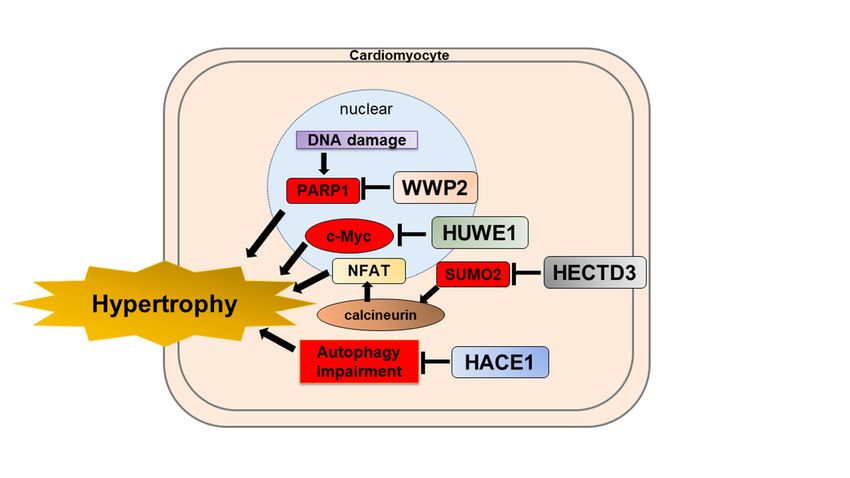
Int.
Int. J.J. Mol.
Mol. Sci.
Sci. 2021,
2021, 22,
22, x6065
FOR PEER REVIEW 3 3of
of 26
26
Figure 1.
Figure Classification of
1. Classification of ubiquitin
ubiquitinE3
E3ligases
ligasesand
andthethedomain
domainarchitecture
architectureofof HECT-type
HECT-type E3E3
ligases. E3 E3
ligases. ligases are are
ligases classified
clas-
into three
sified groups:
into three RING
groups: family,
RING HECT
family, HECTE3 family, RBRs
E3 family, RBRsfamily. HECT
family. HECT E3E3family
familyare
aregrouped
groupedintointothree
three subfamilies:
subfami-
lies:
NEDD4 NEDD4 subfamily,
subfamily, HERC HERC subfamily,
subfamily, otherother E3 subfamily.
E3 subfamily. RING,RING,
reallyreally interesting
interesting new genes;
new genes; HECT,HECT, homologous
homologous to E6AP
to E6AP C-terminus;
C-terminus; RBRs, RING-between-RINGs;
RBRs, RING-between-RINGs; NEDD4,NEDD4, neural precursor
neural precursor cell expressed
cell expressed developmentally
developmentally downreg-4;
downregulated
ulated
HERC,4; HERC,
HECT andHECT
RLD and
domainRLDcontaining
domain containing E3 protein
E3 ubiquitin ubiquitin protein
ligase; WWP,ligase;
WWWWP,domainWW domain containing
containing E3 ubiquitinE3protein
ubiq-
uitin protein ligase; SMURF, SMAD ubiquitin regulatory factor; HECW, HECT, C2, and WW domain
ligase; SMURF, SMAD ubiquitin regulatory factor; HECW, HECT, C2, and WW domain containing E3 protein ligase; E6AP, containing E3 pro-
tein ligase; E6AP, E6-associated protein; HUWE1, HECT, UBA, and WWE domain containing E3 ubiquitin protein ligase
E6-associated protein; HUWE1, HECT, UBA, and WWE domain containing E3 ubiquitin protein ligase 1; HACE1, HECT
1; HACE1, HECT domain and ankyrin repeat containing E3 ubiquitin protein ligase 1; TRIP12, thyroid hormone receptor
domain and ankyrin repeat containing E3 ubiquitin protein ligase 1; TRIP12, thyroid hormone receptor interactor 12;
interactor 12; UBR5, ubiquitin protein ligase E3 component N-recognin 5; UBE3B, ubiquitin–protein ligase E3B; UBE3C,
UBR5, ubiquitin
ubiquitin proteinprotein ligaseHECTD,
ligase E3C; E3 component
HECT N-recognin 5; UBE3B,protein
domain E3 ubiquitin ubiquitin–protein ligase
ligase; G2E3, G2/ME3B; UBE3C, ubiquitin
phase-specific protein
E3 ubiqui-
ligase E3C; HECTD, HECT domain E3 ubiquitin protein ligase; G2E3, G2/M phase-specific E3 ubiquitin
tin protein ligase; AREL1, apoptosis-resistance E3 ubiquitin protein ligase 1; C2, C2 domain; WW, WW domain; RLD, protein ligase;
AREL1,
RCC-likeapoptosis-resistance E3 ubiquitin
domain; ARM, armadillo repeat;protein
UBA, ligase 1; C2, C2WWE,
UBA domain; domain;WWEWW,domain;
WW domain;
ANK, RLD, RCC-like
ankyrin repeat.domain; ARM,
armadillo repeat; UBA, UBA domain; WWE, WWE domain; ANK, ankyrin repeat.
Substrate proteins are modified by a single ubiquitin moiety on one or multiple sites,
givingSubstrate proteins
rise to mono- andaremulti-mono-ubiquitylated
modified by a single ubiquitin moiety
proteins, on one or multiple
respectively. sites,
In addition, a
wide variety of polyubiquitin chains can be formed on substrate proteins, in which thea
giving rise to mono- and multi-mono-ubiquitylated proteins, respectively. In addition,
wide variety
ubiquitin of polyubiquitin
moieties can be linkedchains can either
through be formed onthe
one of substrate proteins,
seven internal in which
lysine the
residues
ubiquitin moieties can be linked through either one of the seven internal lysine
(Lys6, Lys11, Lys27, Lys29, Lys33, Lys48, and Lys63) in ubiquitin or through its N-termi-residues
(Lys6,
nal Lys11,
amino Lys27, Lys29, Lys33, Lys48, and Lys63) in ubiquitin or through its N-terminal
group.
amino group.
Polyubiquitylation through the Lsy48-linked ubiquitin chain is generally used for the
Polyubiquitylationdegradation
ubiquitin–proteasomal through the pathway.
Lsy48-linked ubiquitin
Substrate chain isthat
proteins generally
receiveused for
Lys48-
the ubiquitin–proteasomal degradation pathway. Substrate proteins that receive
linked polyubiquitin chains migrate to and are degraded by the 26S proteasome. The ubiq- Lys48-
linked polyubiquitin
uitin–proteasome chains
system is amigrate
protein to and are
quality anddegraded by the 26S
quantity control proteasome.
system The
that mediates
ubiquitin–proteasome system is a protein quality and quantity control system that medi-
approximately 80–90% of intracellular protein degradation under optimal nutritional con-
ates approximately 80–90% of intracellular protein degradation under optimal nutritional
ditions [32–36]. Furthermore, mono-ubiquitylation of lysine residues or polyubiquityla-
conditions [32–36]. Furthermore, mono-ubiquitylation of lysine residues or polyubiq-
tion through Lys63-linked ubiquitin chains are used for nonproteolytic pathways such as
uitylation through Lys63-linked ubiquitin chains are used for nonproteolytic pathways
DNA repair, relocalization, modifying activity (signal transcriptional activity), or endocy-
such as DNA repair, relocalization, modifying activity (signal transcriptional activity),
tosis [37–39]. The consequences for the modified substrate are determined by the type of
or endocytosis [37–39]. The consequences for the modified substrate are determined by the
ubiquitin modification it receives [40].
type of ubiquitin modification it receives [40].
RING E3 ligases catalyze the direct transfer of ubiquitin from the E2 conjugating en-
zyme to the substrate, suggesting that the linkage type of the ubiquitin chain is deter-
mined by the E2 conjugating enzyme. In contrast to RING-type E3 ligases, HECT-type E3Int. J. Mol. Sci. 2021, 22, 6065 4 of 26
RING E3 ligases catalyze the direct transfer of ubiquitin from the E2 conjugating en-
zyme to the substrate, suggesting that the linkage type of the ubiquitin chain is determined
by the E2 conjugating enzyme. In contrast to RING-type E3 ligases, HECT-type E3 ligases
include an active-site cysteine in the HECT domain, which forms an intermediate thioester
bond with ubiquitin before it is conjugated to the substrate protein [41,42]. HECT-type E3
ligase has enzymatic activity and directly catalyzes the covalent attachment of ubiquitin
to substrate proteins; therefore, it could determine the linkage type of ubiquitin chain
preferred [43].
2.2. HECT-Type E3 Ligase
In 1995, HECT-type E3 ligases were found in all eukaryotic organisms and ubiq-
uitin [41]. E6AP transcribed from the ubiquitin–protein ligase E3A gene was the first
identified HECT-type E3 ligase, leading to the discovery of the HECT-type E3 ligase fam-
ily [41,44,45]. There are 28 types of HECT-type E3 ligases in humans [46], which are
commonly grouped into three groups based on the presence of distinct amino acid se-
quence motifs or domains within the N-terminal: NEDD4 subfamily, HERC subfamily,
and other HECT-type E3 ligases [47] (Figure 1). The HECT domain is an approximately
40 kDa domain positioned at the C-terminus of the E3 ligases, consisting of the N-lobe and
C-lobe. The N-lobe represents the E2 binding domain, whereas the C-lobe contains an ac-
tive site cysteine to receive ubiquitin. In the HECT family, 16–92% amino acid identity was
found for this domain [48]. The domain architecture of the HECT-type E3 ligases is shown
in Figure 1.
2.2.1. NEDD4 Subfamily
The NEDD4 subfamily member includes nine types of HECT-type E3 ligases and
accounts for approximately 30% of HECT-type E3 ligases [22]. The NEDD4 family is
characterized by the presence of C2 and 2–4 WW domains. The N-terminal C2 domain
is defined as a Ca+ phospholipid binder [49]. The WW domains are responsible for
recognizing substrates and have also been found to form intramolecular interactions with
the HECT domain of the E3 ligases [50,51]. Some NEDD4 subfamily members are often
expressed as alternative splice isoforms [47].
2.2.2. HERC Subfamily
The HERC subfamily is characterized by a HECT domain and one or more regulators
of chromosome condensation-like domains (RLDs), an effector protein domain that was
first identified as a regulator of chromosome condensation 1 [52]. In humans, the HERC
subfamily comprises six members, which can be further organized into two large and
four small HERCs. Large HERCs (HERC1 and 2) have two or three RLDs; however, small
HERCs (HERCs 3, 4, 5, and 6) have one RLD. RLD has dual functions: one side of the
domain acts as a guanine nucleotide exchange factor for the small GTPase Ran, whereas
the opposite side interacts with chromatin through histones H2A and H2B [53,54].
2.2.3. Other HECT E3s
Each member of another HECT E3 ligase lacks WW or RLD domains and has a distinct
variety of N-terminal domains. There are several N-terminal domains of other HECT
E3 ligases, such as WWE and armadillo repeats (HUWE1 and TRIP12), UBA domain
(HUWE1 and UBR5), ankyrin repeats (HACE1 and HECTD1), and IQ motifs (UBE3B and
UBE3C) [55]. In addition, the MIB domain in HECTD1 and the DOC domain in HECTD3
were common in the N-terminus of HERC2.Int. J. Mol. Sci. 2021, 22, 6065 5 of 26
3. Importance of Ubiquitylation in Cardiac Disease
The adult heart endures a wide range of physiological and pathophysiological stresses
during life. Accumulating evidence indicates that ubiquitylation is involved in devel-
oping cardiac diseases [17,56–60]. Cardiac proteins are in a dynamic state of continual
degradation and resynthesis and are thought to replace all in 30 days under normal circum-
stances. Protein turnover is critical to cardiomyocytes as post-mitotic cells with minimal
regenerative capacity because protein aggravation is cytotoxic [61,62]. Eukaryotic cells
have developed multilayered protein quality control mechanisms primarily carried out by
chaperones, lysosomal autophagy, and the ubiquitin–proteasome system [63]. More than
70% of the protein turnover is regulated by the ubiquitin–proteasome system [64–66]. An
experimental study demonstrated that the balance of protein turnover could lead to protein
accumulation and aggravation during cardiac remodeling [67]. The discovery that cardiac
ubiquitin E3 ligases, such as muscle-specific ubiquitin ligase atrogin-1 and MuRF family,
yields cardiac growth, and remodeling through sarcomeric protein turnover, indicated that
the ubiquitylation cascade is fundamental to the maintenance of normal cardiac function
through protein quality control [17,68,69].
As previously mentioned, ubiquitylation is involved in most aspects of eukaryotic
cell biology, such as intracellular signaling, transcriptional control, and regulation of cell
death. Research regarding the role of cardiac ubiquitin E3 ligases has developed from
protein turnover to cellular processes such as signal transduction, transcriptional regulation,
maintenance of homeostasis, mitochondrial dynamics, receptor turnover, and energy
metabolism [17,19]. In the following paragraph, we will limit ourselves to a discussion of
HECT-type E3 ligases that have been associated with cardiac diseases.
4. Cardiac Hypertrophy and HECT-Type E3 Ligase
Pathological cardiac hypertrophy caused by hypertension, aortic stenosis, and other
disease-related stresses is an early milestone during the clinical course of HF [70]. A
study detected an accumulation of ubiquitylated proteins and abnormal aggregation in
cardiomyocytes collected from patients with decompensated cardiac hypertrophy [71].
Pressure overload-induced cardiac hypertrophy is associated with a marked increase in
protein synthesis at a rate exceeding degradation with an increase in ubiquitin, E2, and
several E3 ligases [16,72–74], indicating hyper-ubiquitylation in rat and mouse models
of hypertrophy.
At the cellular level, cardiomyocyte hypertrophy is characterized by an increase in
cell size, enhanced protein synthesis, multiplication of sarcomeres, a switch of proteins and
enzymes to fetal isoforms, changes in intracellular Ca2+ handling, metabolic alterations,
and increased rates of apoptosis [75,76]. Numerous key regulatory pathways that pro-
mote cardiac hypertrophy are either targets or components of the ubiquitylation cascade.
Several signaling pathways contribute to cardiac hypertrophy, such as Wnt/β catenin,
calcineurin/nuclear factor of activated T-cells (NFAT), the Janus kinase (JAK)/signal trans-
duction and the activator of transcription (STAT) pathway [77–79].
This study shows the current understanding of HECT-type E3 ligases in the develop-
ment of cardiac hypertrophy. An overview of HECT-type E3 ligases in cardiac hypertrophy
is summarized in Table 1.Int. J. Mol. Sci. 2021, 22, 6065 6 of 26
Table 1. HECT-type E3 ligase and cardiac remodeling.
HECT-Type E3 Ligase Substrate/Target Main Findings Reference
Cardiac hypertrophy
Cardiac-specific ITCH
transgenic mice inhibited
ITCH Dishevelled maladaptive hypertrophy [80]
via Wnt/β catenin
signal inhibition.
Cardiac hypertrophy was
observed in NEDD4-2 null
NEDD4-2 ENaC in kidney [81,82]
mice on chronic
high-salt diet.
Circular RNA WWP1 was
Circular RNA WWP1 ANF and miR-23a dysregulated in the heart [83]
treated with isoproterenol.
WWP2 conditional
knockout mice
(MycCre+;WWP2Fl/Fl )
WWP2 PARP1 [84]
exacerbated
isoproterenol-induced
cardiac hypertrophy.
Increased myocardium
E6AP E6AP expression after [16]
pressure overload.
HUWE1 conditional
knockout mice
HUWE1 c-myc [85]
spontaneously developed
cardiac hypertrophy.
HACE1 conditional
knockout mice
HACE1 Unknown [86]
spontaneously developed
cardiac hypertrophy.
AAV9-medited
overexpression of HECTD3
HECTD3 SUMO2/STAT1 [87]
inhibited pathological
hypertrophy in mice.
Cardiac fibrosis
WWP2mut/mut mice
attenuated cardiac fibrosis
WWP2 SMAD2 [88]
after angiotensin II infusion
and myocardial infarction.
SMURF1 was involved in
BMP-2 antagonization for
TGF-β1 signal.
SMURF1 SMURF1 was a target of [89,90]
miR-10b-5p, which inhibits
cardiac
fibroblast activation.
Mediator of TGF-β signal.
SMURF2 mediated SMAD7
SMURF2 SMAD7 [91,92]
degradation was inhibited
by SMAD3 inhibitor.
HFpEF
Cardiac-specific
overexpression of WWP1
WWP1 Not described developed cardiac [93]
hypertrophy with
diastolic dysfunction.
ANF, atrial natriuretic factor; BMP, bone morphogenic protein; ENaC, epithelial Na+ channel; HFpEF, heart failure
with preserved ejection fraction; NEDD4-2, neural precursor cell expressed developmentally downregulated
4-2; PARP1, poly(ADP-ribose)polymerase-1; SMAD, small mother against decapentaplegic; SMURF, SMAD
ubiquitin regulatory factor; STAT, signal transduction and activator of transcription; SUMO2, small ubiquitin-like
modifier 2.Int. J. Mol. Sci. 2021, 22, 6065 7 of 26
4.1. ITCH
The ubiquitin E3 ligase ITCH is a 903 amino acid residue protein that contains an
N-terminal C2 domain, four WW domains, and an HECT domain [55]. ITCH was orig-
inally identified after genetic analysis of a mutant mouse with aberrant immunological
phenotypes and constant skin scratching [94]. The WW domain recognizes the proline-rich
PPXY consensus sequence in substrate proteins, and the HECT domain attaches ubiquitin
molecules to substrates [95]. Thioredoxin-interacting protein, disheveled (Dvl), c-jun, Jun-B,
Notch, Gli, p73, p63, cellular FLICE-inhibitory protein, B cell lymphoma/leukemia 10,
Forkhead box O1, transforming growth factor-β-activated kinase 1, and ITCH are known
substrates of ITCH for degradative ubiquitylation. Frizzled receptor 4, phospholipase C
gamma 1, protein kinase C-θ, Deltex, ErbB4, CXC chemokine receptor 4, and transient
receptor potential vanilloid 4 are known substrates of ITCH for degradative ubiquitylation
by the non-ubiquitin–proteasome system. Furthermore, small mothers against decapenta-
plegic (SMAD2) are known substrates of ITCH for non-degradative ubiquitylation [96,97].
Accumulating evidence demonstrates that Wnt/β-catenin signaling is activated after
transverse aortic constriction (TAC) and induces pathological cardiac hypertrophy [79,98–100].
Dvl is an intracellular principle component of signaling [101]. Dvl has approximately
700 amino acids, harboring conserved DIX, basic and serine threonine-rich region, PDZ,
proline-rich PPXY consensus sequence region, and DEP domains. We previously reported
Int. J. Mol. Sci. 2021, 22, x FOR PEER REVIEW 7 of 26
that key molecules of the Wnt/β-catenin signaling pathway after TAC are inhibited in
cardiac-specific overexpression of ITCH transgenic mice. In this study, ITCH targets Dvl
proteins for ubiquitin–proteasome degradation in cardiomyocytes and attenuates cardiac
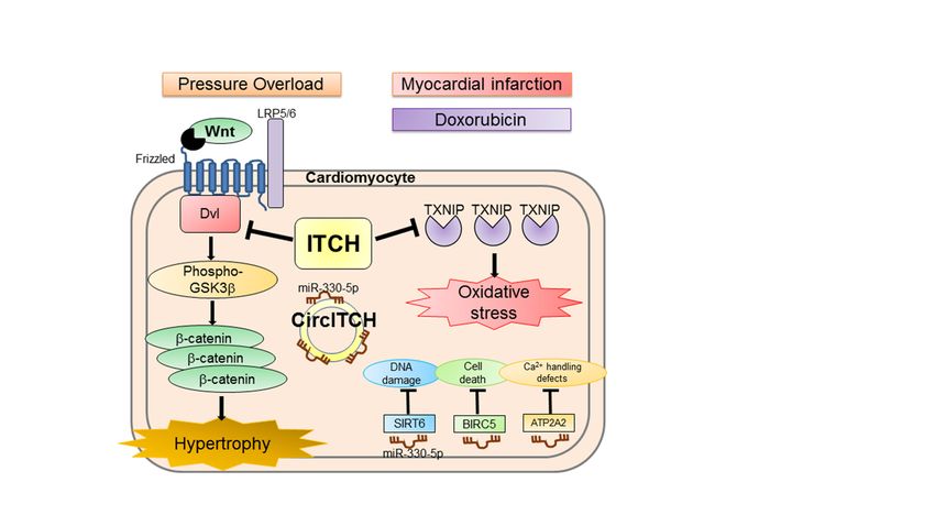
hypertrophy by suppressing the Wnt/β-catenin signaling pathway (Figure 2) [80].
Figure 2. A simplified model depicting the function of ITCH in the development of cardiac hyper-
trophy, myocardial infarction, and doxorubicin-induced cardiomyopathy. ITCH targets disheveled
Figure 2. A simplified model depicting the function of ITCH in the development of cardiac hypertrophy, myocardial
protein for ubiquitin-proteasome
infarction, and doxorubicin-induced degradation,
cardiomyopathy. resulting
ITCH targets in inhibition
disheveled protein of
forthe Wnt/β-catenin signal-
ubiquitin-proteasome degra-
ing pathway;
dation, resulting ITCH
in inhibition attenuates
of the cardiac
Wnt/β-catenin hypertrophy
signaling pathway;induced by pressure
ITCH attenuates overload.
cardiac ITCH
hypertrophy targets
induced by
pressure overload. ITCH targets thioredoxin-interacting
thioredoxin-interacting protein for ubiquitin–proteasome
protein for ubiquitin–proteasome degradation anddegradation
amelioratesand ameliorates
reactive oxy-
reactive oxygen species-induced cardiotoxicity
gen species-induced cardiotoxicity through the thioredoxin
through systemsystem
the thioredoxin in myocardial infarction and
in myocardial doxorubicin-
infarction and
induced cardiomyopathy models. Circ ITCH absorbs miR-330-5p, resulting in the activation of miR-330-5p targets (SIRT6,
doxorubicin-induced cardiomyopathy models. Circ ITCH absorbs miR-330-5p, resulting in the
BIRC5, and ATP2A2), thereby ameliorating doxorubicin-induced cardiomyopathy by reducing DNA damage, cell death,
and calciumactivation
handlingofdefects.
miR-330-5p
LRP5/6,targets (SIRT6,
low-density BIRC5, and
lipoprotein ATP2A2), thereby
receptor-related protein ameliorating
5/6; Circ ITCH,doxorubicin-
circular RNA
ITCH; miR, induced
microRNA; cardiomyopathy
SIRT6, sirtuin 6;by reducing
BIRC5, DNAIAP
baculoviral damage, cell death,5; and
repeat containing calcium
ATP2A2, ATPasehandling defects.
sarcoplasmic/en-
doplasmicLRP5/6, Ca2+ transporting
reticulumlow-density 2; Ca2+, calcium.
lipoprotein receptor-related protein 5/6; Circ ITCH, circular RNA ITCH; miR,
microRNA; SIRT6, sirtuin 6; BIRC5, baculoviral IAP repeat containing 5; ATP2A2, ATPase sarcoplas-
ITCH potentially mediates the degradation of non-Dvl substrates during cardiac hy-
mic/endoplasmic reticulum Ca2+ transporting 2; Ca2+ , calcium.
pertrophy [102,103]; therefore, further studies are needed to clarify the effect of ITCH on
other substrates during cardiac hypertrophy. Overall, ITCH may be a therapeutic target
for cardiac hypertrophy.
4.2. NEDD4-2
NEDD4-2, the most ancient member of the NEDD4 subfamily, was originally identi-Int. J. Mol. Sci. 2021, 22, 6065 8 of 26
ITCH potentially mediates the degradation of non-Dvl substrates during cardiac
hypertrophy [102,103]; therefore, further studies are needed to clarify the effect of ITCH on
other substrates during cardiac hypertrophy. Overall, ITCH may be a therapeutic target for
cardiac hypertrophy.
4.2. NEDD4-2
NEDD4-2, the most ancient member of the NEDD4 subfamily, was originally iden-
tified in a screening of genes downregulated during development of the central nervous
system [104]. NEDD4-2 consists of a C2 domain, four WW domains, and an HECT do-
main. NEDD4-2 is a multifunctional protein whose mutations are associated with develop-
ment disorders, hypertension, epilepsy, and end-stage renal disease [105,106]. NEDD4-2
has been suggested to function as an E3 ligase for several PY motif-containing proteins
such as SMADs, Dvl2, epithelial Na+ channel (ENaC), voltage-dependent cardiac Na+
channel (Nav1.5), KCNQ1 potassium channel [107], and human ether-a-go-go-related
gene [108,109].
It was reported that high salt diet-induced cardiac hypertrophy and systolic function
were exacerbated in NEDD4-2 deficient mice with higher expression levels of ENaC in the
kidney [82]. Furthermore, Galiana-Simal et al. demonstrated that NEDD4-2 is phosphory-
lated; therefore, NEDD4-2 functions to ubiquitylate ENaC in hypertrophied myocardium
of aldosterone-treated rats [81]. These findings suggest an association between NEDD4-2
and cardiac hypertrophy.
4.3. WWP2
The WW domain-containing E3 ubiquitin protein ligase 2 (WWP2), also known as
atrophin-1-interacting protein 2, was originally identified in screening for WW domain-
containing proteins [110]. WWP2 is ubiquitously expressed in the heart, placenta, lung,
liver, muscle, kidney, pancreas, and brain [111]. There are three isoforms in WWP2:
a full-length WWP2, an N-terminal isoform presumably generated by failure to splice-out
intron 9-10, and a C-terminal isoform possibly generated from a second promoter within
introns 10-11. WWP2 interacts with multiple substrates, such as phosphatase and tensin
homolog deleted from chromosome 10 (PTEN), SMADs, Oct4, and ENaC [112,113]. WWP2
is a multifunctional E3 ubiquitin ligase, which is involved in palatogenesis, craniofacial
development, innate immune response, tumorigenesis, and cell death [114–119]. WWP2
was also reported to regulate PTEN/ phosphatidylinositol 3-kinase (PI3K)/Akt signaling
and transforming growth factor-beta (TGF-β)/SMAD signaling [119].
Isoproterenol is a chronic infusion of β-stimulant, which induces cardiac hypertrophy
and fibrosis, leading to heart failure. Poly(ADP-ribose) polymerase-1 (PARP1) is a critical
injury factor in cardiac remodeling. Increases in PARP1 and poly(ADP-ribosyl)ation (PARy-
latin; PARP1 activity) have been observed in cardiac remodeling, leading to extreme energy
consumption by myocardial cells [120,121]. PARP1 is activated to generate PARylation via
recognition of damaged DNA fragments and induces cardiomyocyte damage, leading to
apoptosis and necrosis [122]. Zhang N et al. reported that full-length WWP2 knockout
mice exacerbated cardiac systolic function, hypertrophy, and fibrosis compared to WT
mice after isoproterenol infusion. In addition, the protein expression of WWP2 decreased;
however, PARP1 increased in response to isoproterenol in vitro and in vivo. WWP2 inter-
acts with PARP1 and degrades it through the ubiquitin–proteasome system by regulating
ubiquitylation of PARP1 K418 and K249 sites in the BRCT domain, leading to the inhibition
of isoproterenol-induced cardiac remodeling (Figure 3) [84]. WWP2 is a protective factor
against isoproterenol-induced cardiac remodeling.vivo. WWP2 interacts with PARP1 and degrades it through the ubiquitin–pro
tem by regulating ubiquitylation of PARP1 K418 and K249 sites in the BR
leading to the inhibition of isoproterenol-induced cardiac remodeling (F
WWP2 is a protective factor against isoproterenol-induced cardiac remodeli
Int. J. Mol. Sci. 2021, 22, 6065 9 of 26
Figure 3. Schematics depicting the protective role of HECT-type E3 ligases for cardiac hypertrophy. WWP2 targets PARP1
for ubiquitin-proteasome degradation. HUWE1 targets c-Myc for ubiquitin–proteasome degradation. HECTD3 targets
SUMO2 for ubiquitin–proteasome degradation. HACE1 ameliorates autophagy impairment. These HECT-type E3 ligases
are a protective role for cardiac hypertrophy. WWP2, WW domain-containing E3 ubiquitin protein ligase 2; PARP1, Poly
(ADP-ribose) polymerase-1; HUWE1, HECT, UBA, and WWE domain containing E3 ubiquitin protein ligase 1; HECTD3,
HECT domain E3 ubiquitin protein ligase 3; SUMO2, small ubiquitin-like modifier 2; NFAT, nuclear factor of activated
T-cells; HACE1, HECT domain and ankyrin repeat containing E3 ubiquitin protein ligase 1.
4.4. HUWE1
The ubiquitin E3 ligase HECT, UBA, and WWE domain containing E3 ubiquitin
protein ligase 1 (HUWE1), also termed UREB1, HECTH9, ARF-BP1, MULE, E3 Histone,
and LASU1, is a 4347 amino acid residue protein that contains an N-terminal armadillo
repeat domain, a UBA domain, a WWE domain, a BH3 domain, and a HECT domain [55].
c-Myc, p53, and myeloid cell leukemia-1 are known substrates of HUWE1 for degrada-
tive ubiquitylation [123]. In contrast, Dvl and herpesvirus-associated ubiquitin-specific
proteases are known substrates of HUWE1 for non-degradative ubiquitylation [124,125].
HUWE1 is associated with proliferation, differentiation, apoptosis, DNA repair, and stress
response [124]. HUWE1 has a general preference for attaching the K48 polyubiquitin chain
to substrate proteins [126,127].
Recently, Dadson et al. reported that HUWE1 expression was reduced in the left
ventricle of patients with end-stage heart failure. Furthermore, conditional cardiac-specific
HUWE1 knockout mice develop spontaneous cardiac hypertrophy, left ventricular dys-
function, and premature death with an increase in c-Myc in the heart (Figure 3) [85]. In
addition, conditional cardiac-specific HUWE1 knockout mice showed impaired mitochon-
drial energy metabolism and reactive oxygen species defense, accompanied by reduced
protein expression levels of key regulators such as Pgc-1α and Pink1 [128–130]. Transcrip-
tomic analysis of HUWE1 knockout mice revealed that elevated c-Myc directly inhibits
the transcription of Pgc-1α and Pink1. Cardiac hypertrophy and left ventricular dysfunc-
tion were diminished in conditional cardiac-specific HUWE1 and c-Myc double knockout
mice. Therefore, HUWE1 in the heart could be a potential therapeutic target for cardiac
hypertrophy through its interaction with c-Myc.Int. J. Mol. Sci. 2021, 22, 6065 10 of 26
4.5. HACE1
The ubiquitin E3 ligase HECT domain and ankyrin repeat containing E3 ubiquitin
protein ligase 1 (HACE1) is a 909 amino acid residue protein that contains an N-terminal
ankyrin repeat domain and an HECT domain [55]. HACE1 was discovered as a chromo-
some 6q21 tumor-suppressor gene in Wilms’ tumors [131]. Subsequently, HACE1 is noted
as a tumor suppressor in multiple cancers [132]. As a mechanism of tumor suppression, it
has been shown to catalyze the ubiquitylation of Ras-related C3 botulinum toxin substrate
1, a potent oncogene [133].
Zhang et al. demonstrated that HACE1 gene expression is upregulated in dilated car-
diomyopathy compared with non-failing hearts in silico. They demonstrated that HACE1
deficiency in mice exacerbated heart failure and increased mortality after severe TAC.
HACE1 knockout mice show abnormal cardiac hypertrophy, left ventricular dysfunction,
accumulation of LC3, p62, and ubiquitylated proteins enriched for cytoskeletal species
(Figure 3) [86]. This study reveals that HACE1 is required to fuse autophagosomes with
lysosomes, independent of its E3 ligase activity. Hence, HACE1 may play an important
role in suppressing protein accumulation and aggravation by regulating autophagy during
cardiac remodeling.
4.6. HECTD3
The ubiquitin E3 ligase HECT domain E3 ubiquitin protein ligase 3 (HECTD3) is an
861 amino acid residue protein that contains a DOC domain and an HECT domain. Trio-
associated repeats on actin and RAF proto-oncogene serine/threonine-protein kinase are
known substrates of HECTD3 for degradative ubiquitylation by the ubiquitin–proteasome
system. Furthermore, mucosa-associated lymphoid tissue proteins 1, caspase-8, caspase-9,
and STAT3 are substrates of HECTD3 for non-degradative ubiquitylation [134,135].
Small ubiquitin-like modifier 2 (SUMO2) is the most efficient activator of calcineurin/
NFAT signaling to induce cardiac hypertrophy [136]. Furthermore, the inflammatory re-
sponse is associated with myocardial fibrosis and hypertrophy [78,137]. In cardiac and
other systems, the JAK/STAT signaling pathway is associated with the inflammatory
response [138]. Recently, Rangrez et al. demonstrated that HECTD3 targets SUMO2 for
ubiquitin–proteasome degradation and suppresses calcineurin/NFAT signaling (Figure 3).
In addition, they showed that HECTD3 reduced the activation of STAT1 by attenuat-
ing its phosphorylation through the induction of its polyubiquitylation. In this study,
AAV9-mediated overexpression of HECTD3 in mice reduced cardiac SUMO2/STAT1
levels, pathological hypertrophy, largely abolished macrophage infiltration, and fibrosis
induced by pressure overload [87]. Thus, HECTD3 may be a potential therapeutic target for
cardiac hypertrophy.
5. Cardiac Fibrosis and HECT-Type E3 Ligase
Pathological cardiac fibrosis is a process characterized by excessive deposition of
extracellular matrix (ECM), leading to the development of cardiac dysfunction, arrhythmia,
and HF [139–141]. Several pathophysiological conditions induce cardiac fibrosis, such as
pressure overload, volume overload, myocardial infarction, dilated and hypertrophied
cardiomyopathy, various toxic insults, metabolic disturbances, and aging [142–145].
Cardiac fibroblasts are key effector cells in cardiac fibrosis and are responsible for
ECM homeostasis in the heart [139]. After cardiac fibroblasts are activated by regulators of
tissue fibrosis, such as angiotensin II, connective tissue growth factor, bone morphogenetic
protein (BMP), Wnt ligands, cytokines, and TGF-β superfamily, they differentiate into
myofibroblasts with an increase in ECM protein [146]. TGF-β1 contributes to cardiac
fibrosis development through SMAD-dependent and SMAD-independent pathways. TGF-
β1 generally exerts its biological effects by activating downstream mediators, including
SMAD2 and SMAD3, while negatively regulated by SMAD7 expression [147–149]. SMADs
have been reported to play a pivotal role in the transcription of ECM proteins [150].Int. J. Mol. Sci. 2021, 22, 6065 11 of 26
Although the pathophysiological conditions leading to cardiac fibrosis are different
from those of cardiac diseases, it is valuable to explore the common mechanisms involved
in cardiac fibrosis. This study shows the current understanding of the role of HECT-type
E3 ligases in cardiac fibrosis. An overview of HECT-type E3 ligases in cardiac fibrosis is
summarized in Table 1.
5.1. WWP2
An outline of the domain architecture, function, and substrates of WWP2 is described
in the previous paragraph. Chen et al. identified the WWP2 N-terminal isoform as a
positive regulator of the pro-fibrotic gene network associated with cardiac fibrosis using
systems genetics in human and murine dilated cardiomyopathy and repaired tetralogy
of Fallot. The WWP2 N-terminal isoform consists of a C2 domain and a WW domain,
indicating the absence of the HECT domain. The left ventricular single-cell RNA sequence
indicated that WWP2 is mainly expressed in fibroblasts, immune cells, and endothelial
cells. WWP2mut/mut mice lacking the N-terminal isoform and full-length WWP2 attenuated
cardiac fibrosis and preserved cardiac function after angiotensin II infusion or myocardial
infarction. The N-terminal region of WWP2 interacts with SMADs potentially through
its mono-ubiquitylation and mediates the TGF-β1-induced nucleocytoplasmic shuttling
and transcriptional activity of SMAD2 [88,151]. Thus, WWP2 is an important regulator of
pro-fibrotic and ECM genes. These findings provide new understanding into the role of
HECT-type E3 ligases independently of the HECT domain.
5.2. SMURF1
SMAD ubiquitin regulatory factor (SMURF) was initially identified as a regulator of
SMAD1 stability [152]. SMURFs are composed of two members, SMURF1 and SMURF2.
SMURFs have been implicated in determining the competence of cells in response to
the TGF-β/BMP signaling pathway [153]. SMURF1 consists of a C2 domain, two WW
domains, and an HECT domain. SMURF is a multifunctional protein that is involved
in cell cycle progression, cell proliferation, differentiation, DNA damage response, and
maintenance of genomic stability. SMURF1 targets Dvl2, SMADs, RhoA, and Runx2,
3 [154]. SMURF1 modulates several signal transduction pathways, such as the TGF-
β/BMP signaling pathway, Wnt signaling, mitogen-activated protein kinase signaling, and
RhoA/Rho-associated kinase signaling [155]. SMURF1 plays an important role in heart
development, including outflow tract septation and cell-type specification, by controlling
cilium-associated BMP signaling [156,157].
BMP-2, as a novel fibrosis-antagonizing cytokine, have a potential beneficial effect in
attenuating pressure overload-induced cardiac fibrosis. Wang S et al. demonstrated that
SMURF1 interacted with SMAD6 and that this SMURF1/SMAD6 complex was involved
in BMP2 antagonization of TGF-β1 mediated protein kinase C-δ and SMAD3 signaling
in cardiomyocytes [89]. This finding suggests that SMURF1 may contribute to cardiac
fibrosis development.
Endothelial colony-forming cells have been reported to reduce cardiac fibrosis in
myocardial infarction due to their proliferation and secretion of exosomes, which transfer
microRNAs. Cardiac fibroblast activation is ameliorated by exosomes from endothelial
colony-forming cells treated with normoxia compared to those treated with hypoxia. Liu
et al. found that miR-10b-5p was enriched in exosomes from normoxia and targeted
SMURF1 and histone deacetylase 4 using next-generation RNA sequencing. Thus, inhibi-
tion of mRNA expression of SMURF1 by miR-10b-5p was suggested to participate in the
antifibrotic effects of exosomes derived from endothelial colony-forming cells treated with
normoxia [90].
5.3. SMURF2
SMURF2 consists of a C2 domain, three WW domains, and an HECT domain. SMURF2
targets SMADs, heat shock proteins 27, and p53 [154]. SMURF2 was reported to beInt. J. Mol. Sci. 2021, 22, 6065 12 of 26
downregulated in DCM [158]. SMURF functions as a mediator of TGF-β signaling via
interaction with SMAD7 containing PY motif during cardiac fibrosis [91].
Meng et al. examined the effect of a SMAD3 inhibitor on angiotensin II-induced
cardiac fibrosis. The protein expression level of SMURF2 in the mouse heart increased,
while that of SMAD7 decreased after angiotensin II administration. However, this effect
was reversed by the SMAD3 inhibitor, suggesting that the SMAD3 inhibitor protected
cardiac SMAD7 from SMURF2-mediated ubiquitin–proteasome degradation. Since SMAD7
functions as an inhibitor of both TGF-β/SMAD and NF-κB signaling, an increase in
cardiac SMAD7 could be another mechanism through which SMAD3 inhibitor blocked
SMAD3-mediated cardiac fibrosis and NF-κB-driven cardiac inflammation [92]. This
finding suggests that SMRUF2 may contribute to the development of cardiac fibrosis.
6. HECT-Type E3 Ligase and HF with Preserved Ejection Fraction
HF is classified into three groups: HF with reduced ejection fraction, HF with mid-
range ejection fraction, and HF with preserved ejection fraction (HFpEF) [159]. The preva-
lence rate of HFpEF has been estimated to range from one-third to one-half of all HF
patients and is projected to increase [160,161]. Aging is associated with progressive fibrosis,
leading to the development of HFpEF [162]. HFpEF in older persons is typified by a broad
range of cardiac and non-cardiac abnormalities and reduced reserve capacity in multiple
organ systems [163]. To date, there are no approved therapies available for reducing mor-
tality or hospitalization for these patients due to the heterogeneity of HFpEF [164]. This
study demonstrates the role of HECT-type E3 ligases in HFpEF development.
WWP1
The WW domain containing E3 ubiquitin protein ligase 1 (WWP1) is a multifunctional
protein containing an N-terminal C2 domain, four tandem WW domains for substrate bind-
ing, and a C-terminal catalytic HECT domain for ubiquitin transfer. WWP1 was reported
to be associated with cancer, aging, neurological disorders, and bone homeostasis [165,166].
WWP1 has been suggested to function as an E3 ligase for several PY motif-containing
proteins such as connexin 43, large tumor suppressor 1 and 2, SMAD2, Krüppel-like
transcription factor 5, p63, ErbB4/HER4, Runx2, JunB, atrophin-1, and several non-PY
motif-containing proteins such as TGF-β receptor 1, SMAD4, Krüppel-like transcription
factor 2, and p53 [167]. WWP1 modulates several signal transduction pathways, such as
TGF-β signaling, epidermal growth factor signaling, and apoptosis signaling [166].
WWP1 is reported to be highly expressed in the heart [111,168,169] and increases with
aging [166]. It was reported that the WWP1 overexpressed mice showed left ventricular
hypertrophy, extracellular matrix remodeling, diastolic dysfunction, except systolic dys-
function, indicating the HFpEF phenotype [93]. The precise mechanism by which HFpEF
develops has not yet been fully elucidated. Gene analysis data from RNA sequencing
using right ventricular endomyocardial biopsies indicated enrichment in mitochondrial
adenosine triphosphate synthesis/electron transport and a decrease in endoplasmic retic-
ulum stress, autophagy, and angiogenesis [170]. The NEDD4 family is involved in the
development of HFpEF, suggesting the importance of post-translational modification by
ubiquitylation in HFpEF as well as HF with reduced ejection fraction. This knowledge
provides new insights into HFpEF physiology. WWP1 may have potential therapeutic
relevance in the context of HFpEF.
7. Ischemia/Reperfusion Injury and HECT-Type E3 Ligase
Early reperfusion of the ischemic myocardium plays a vital role in minimizing my-
ocardial infarction. However, the effects of reperfusion are complex and include harmful
effects, collectively referred to as reperfusion injury [171,172]. The underlying mechanisms
of I/R injury are associated with reactive oxygen species generation, intracellular Ca2+
disturbance, rapid pH restoration, and inflammation [173]. Therefore, I/R injury is accom-
panied by detrimental manifestations, including myocardial necrosis and apoptosis [174].Int. J. Mol. Sci. 2021, 22, 6065 13 of 26
Cell death contributes to an increase in infarct size, and regulation of this mechanism
contributes to improved cardiac function [175]. The ubiquitin–proteasome system plays
a pivotal role in I/R injury protection in the heart, organ transplantation, and cerebral
ischemia [176–178]. CHIP has been reported to be required for cardioprotection after my-
ocardial infarction in mice [179]. Furthermore, MDM2, which targets p53 for degradative
ubiquitylation, demonstrated a protective role against hypoxia/reoxygenation-induced
cell death [15]. Thus, the role of cardiac E3 ligases in the injured myocardium is important.
Recently, some HECT-type E3 ligases have been reported to be involved in I/R injury. This
study shows the current understanding of the role of HECT-type E3 ligases in I/R injury.
An overview of HECT-type E3 ligases in I/R injury is summarized in Table 2.
Table 2. HECT-type E3 ligase and ischemia reperfusion injury, doxorubicin cardiotoxicity, and arrhythmia.
HECT-Type E3 Ligase Substrate/Target Main Findings Reference
I/R injury
Overexpression of NEDD4-1
ameliorated myocardial apoptosis
NEDD4-1 p-Akt, PTEN [180]
after I/R injury in rat injected with
NEDD4-1 lentivirus vector.
miR-322/503 ameliorated I/R injury
SMURF2 EZH2 [181]
via inhibition of SMURF2 translation.
Doxorubicin cardiotoxicity
Cardiac specific ITCH transgenic
mice attenuated doxorubicin
TXNIP cardiotoxicity and myocardial
ITCH [182,183]
Unknown infarction.
miR-34b/c inhibited myocardial
injury through ITCH.
Inhibition of apoptosis caused by
miR-17-5p H2 O2 . [184]
Circular RNA ITCH
miR-330-5p Inhibition of doxorubicin [185]
cardiotoxicity.
Arrhythmia
Contribution to Nav1.5
NEDD4-2 Nav1.5 [186]
downregulation in HF.
Cardiac-specific overexpression of
WWP1 Connexin 43 WWP1 die due to ventricular [187]
arrhythmia.
H2 O2 , hydrogen peroxide; I/R, ischemia reperfusion injury; Nav1.5, voltage-dependent Na+ channel; NEDD, neural precursor cell
expressed developmentally downregulated; SMAD, small mother against decapentaplegic; SMURF, SMAD ubiquitin regulatory factor;
TXNIP, thioredoxin-interacting protein.
7.1. NEDD4-1
Neural precursor cell expressed developmentally downregulated protein 4-1 (NEDD4-1),
also termed NEDD4 and RPF1, is a 1319 amino acid residue protein that contains an N-
terminal C2 domain, four WW domains, and an HECT domain [116]. In 1992, NEDD4-1
was discovered in mouse neural precursor cells, whose mRNA levels were downregulated
during mouse brain development [104]. To date, the role of NEDD4-1 has been identi-
fied as an oncogene, tumor suppressor gene, autophagy regulation, and anti-Parkinson’s
disease effect [188–191]. PTEN, RNA polymerase II, and N-Myc are known substrates
of NEDD4-1 for degradative ubiquitylation. In contrast, Akt, MDM2, and α-Synuclein
are known substrates of NEDD4-1 for non-degradative ubiquitylation [192]. NEDD4-1
is required for heart development [193]. Activation of the PI3K/Akt pathway regulating
cellular processes involved in the cell cycle has been shown to protect the heart from
I/R injury [194,195]. PTEN is a tumor suppressor that inhibits PI3K/Akt signaling [196].
NEDD4-1 negatively regulates PTEN stability by catalyzing PTEN polyubiquitylation [188].
Furthermore, NEDD4-1 has been shown to positively regulate the nuclear trafficking of
phospho-Akt by K63-linked polyubiquitylation [197]. NEDD4-1 was reported to be down-Int. J. Mol. Sci. 2021, 22, 6065 14 of 26
regulated in rat hearts after I/R injury [198]. Recently, it was reported that overexpression of
NEDD4-1 ameliorated myocardial apoptosis after I/R injury in rats injected with NEDD4-1
lentivirus vector via activation of PI3K/Akt signaling, suggesting the cardioprotective
role of NEDD4-1 [180]. However, NEDD4-1 has numerous other substrates that need to
be taken into account when interpreting experimental results because it does not directly
demonstrate substrates of NEDD4-1 in the heart.
7.2. SMURF2
An outline of the domain architecture, function, and substrates of SMURF2 is described
in the previous paragraph. Enhancer of zeste homolog 2 (EZH2) is reported to be one
of the SMURF2 substrates. EZH2 is a polycomb group protein associated with pivotal
functions, including cell division, embryonic development, and cancer development [199].
EZH2 binds to PTEN, negatively regulates its expression, and upregulates PI3K/Akt
signaling [200]. Recently, Dong et al. demonstrated that miR-322/503 plays a beneficial
role in myocardial I/R injury. Inhibition of SMURF2 translation induces EZH2 expression
and activates PI3K/Akt signaling via miR-322/503, thereby protecting cells from I/R
injury [181]. Therefore, SMURF2 might be a potential target for improving the prognosis of
myocardial I/R injury.
8. Other Cardiac Disease and HECT-Type E3 Ligase
An overview of HECT-type E3 ligases in other cardiac diseases such as doxorubicin
cardiotoxicity and arrhythmia is briefly summarized in Table 2.
8.1. ITCH and Doxorubicin Cardiotoxicity
The outline of the domain architecture, function, and substrates of ITCH is described
in the previous paragraph. Doxorubicin is one of the most widely used anticancer therapies
for malignant lymphoma (CHOP therapy), breast cancer (AC therapy), and uterine cancer
(AP therapy) [201–203]. Although doxorubicin has a significant effect on reducing mortality
of these cancer patients, its cumulative and dose-dependent toxicity harms the heart [204].
The mechanisms of doxorubicin cardiotoxicity are involved in inflammation, oxidative
stress, apoptosis, mitochondrial impairment, and dysregulation of autophagy [205]. We
previously reported that ITCH targets the thioredoxin-interacting protein for ubiquitin–
proteasome degradation in cardiomyocytes and ameliorates cardiotoxicity induced by
reactive oxygen species, including doxorubicin cardiotoxicity and myocardial infarction
(Figure 2). After doxorubicin injection or myocardial infarction, the survival rates were
significantly higher in cardiac-specific overexpression of ITCH transgenic mice than in WT
littermates [182]. In addition, Zhang et al. demonstrated that miR-34b/c targeted ITCH
and suppressed its expression, promoting NFκB and subsequent inflammatory cytokine
expression in doxorubicin-treated HL-1 cells [183].
Circular RNAs (circRNAs) are important modulators of cardiac development and
disease. Recently, it was reported that circular RNA ITCH derived from 7–14 exons in
ITCH acts as a sponge of miR-330-5p, thereby upregulating sirtuin 6, baculoviral IAP
repeat containing 5, and ATPase sarcoplasmic/endoplasmic reticulum Ca2+ transporting
2 to alleviate doxorubicin cardiotoxicity [185] (Figure 2). Therefore, ITCH and circular
RNA ITCH have cardioprotective roles against doxorubicin and could be novel therapeutic
targets in doxorubicin cardiotoxicity.
8.2. NEDD4-2 and Arrhythmia
An outline of the domain architecture, function, and substrates of NEDD4-2 is de-
scribed in the previous paragraph. The activity of NEDD4-2 is regulated by Ca2+ concentra-
tion in cells, and an increase in Ca2+ concentration disrupts the autoinhibitory conformation
of NEDD4-2, leading to its activation [206].
Nav1.5, which is responsible for the action potential upstroke, is essential for main-
taining an adequate conduction velocity of the electrical impulse [207]. It has been reportedInt. J. Mol. Sci. 2021, 22, 6065 15 of 26
that protein expression of Nav1.5 is reduced in HF without any change in its mRNA
level [208]. Luo et al. uncovered the mechanism of downregulating Nav1.5 in HF. The
protein expression of Nav1.5 decreased, while that of NEDD4-2 increased after ionomycin-
induced intracellular Ca2+ increase. This was reversed by the calcium chelator BAPTA-AM,
suggesting an inverse relationship between Nav1.5 and NEDD4-2 expression in neonatal
rat cardiomyocytes. Nav1.5 was ubiquitylated and downregulated in NEDD4-2 transfected
neonatal rat cardiomyocytes. Reduced expression of Nav1.5 and augmented expression of
NEDD4-2 were observed in a volume overload rat heart failure model and cardiomyocytes
treated with isoproterenol and angiotensin II. Therefore, calcium-mediated increase in
NEDD4-2 induced Nav1.5 ubiquitylation with a resultant reduction in Nav1.5 cardiomy-
ocyte membrane density in HF [186]. Mutations in the gene encoding Nav1.5, SCN5A, have
been associated with various arrhythmic disorders such as long QT syndrome, Brugada
syndrome, and sick sinus syndrome, indicating the association of NEDD4-2 with cardiac
arrhythmia [209,210].
8.3. WWP1 and Arrhythmia
An outline of the domain architecture, function, and substrates of WWP1 is described
in the previous paragraph. Sudden cardiac death is a tragedy and the most common cause
of death worldwide. The cause of sudden cardiac death in most instances is ventricular
arrhythmias such as ventricular fibrillation and ventricular tachycardia [211,212]. Con-
nexin 43 is a main component of gap junctions in the ventricular myocardium and has
a PY motif in its C-terminus. Reduced expression or altered subcellular localization of
connexin 43 has been reported to be associated with contractile dysfunction, aging, and
arrhythmia [213,214]. Basheer et al. reported that WWP1 targets connexin 43 for ubiquitin–
proteasome degradation. Both global- and cardiomyocyte-specific overexpression of WWP1
mice die around 8 weeks of age due to lethal ventricular arrhythmia accompanied by a dra-
matic reduction in connexin 43. Therefore, these findings suggest an association between
WWP1 and ventricular arrhythmia [187].
9. Drug Discovery
The most successful clinically applied drug in this field is a proteasome inhibitor
for multiple myeloma [215]. However, proteasome inhibitors attenuate cardiac remod-
eling after pressure overload [73,216], it showed unexpected cardiac complications, in-
cluding HF [217]. Therefore, the clinical application of proteasome inhibitors for cardiac
diseases appears to be limited. Since E3 ligases are often focal points of cellular regula-
tion [218–220], targeting E3 ligases is thought to yield higher specificity and less toxicity
than other ubiquitylation cascades [221,222]. HECT-type E3 ligases have recently been
described as druggable by peptide and small molecule inhibitors, and several compounds
targeting NEDD4-1, NEDD subfamily, ITCH, SMURF1, E6AP, and HUWE1 have been
identified [223].
Rossi et al. identified clomipramine, a commonly used drug in treating depression, as
an inhibitor of ITCH, by using high-throughput screening of ITCH auto-ubiquitylation [224].
Clomipramine binds to the SH residue irreversibly in the C-lobe of the HECT domain
required for ubiquitin translocation. The therapeutic potential of clomipramine in cancer
has also been examined [225].
The E3 ligase activity of ITCH is modulated by several factors such as intra- and
intermolecular interactions, post-translational modification, adaptor proteins, and ITCH
expression itself [80,182,226,227]. Lithium is widely used as a drug for acute mania in bipo-
lar disorder [228]. Recently, Wang X et al. demonstrated that lithium induces upregulation
of ITCH and promotes ubiquitylation and degradation of Gli1, a regulator of the Hedgehog
signaling pathway, in PANC-1 cells [229]. A recent report demonstrated that low-dose
lithium feeding increases sarcoplasmic/endoplasmic reticulum Ca2+ ATPase 2a (SERCA2a)
to phospholamban ratio and improved SERCA function in the murine left ventricle [230],
suggesting a protective role in the heart. Although the precise mechanism by which lithiumYou can also read