Thinking Outside the Box: Utilizing Nontraditional Animal Models for COVID-19 Research
←
→
Page content transcription
If your browser does not render page correctly, please read the page content below
Review
Thinking Outside the Box: Utilizing Nontraditional Animal
Models for COVID-19 Research
Sachithra Gunasekara, Miruthula Tamil Selvan , Craig A. Miller and Jennifer M. Rudd *
Department of Veterinary Pathobiology, College of Veterinary Medicine, Oklahoma State University, Stillwater,
OK 74078, USA; sachithra.gunasekara@okstate.edu (S.G.); mtamils@ostatemail.okstate.edu (M.T.S.);
craig.miller@okstate.edu (C.A.M.)
* Correspondence: jennifer.rudd@okstate.edu
Abstract: The ongoing COVID-19 pandemic continues to affect the lives, wellbeing, and stability of
communities worldwide. The race to save human lives is critical, and the development of useful
translational animal models to elucidate disease pathogenesis and prevention, and to test therapeutic
interventions, is essential to this response. However, significant limitations exist with the currently
employed animal models that slow our ability to respond to the pandemic. Non-human primates
serve as an excellent animal model for SARS-CoV-2 disease and interventions, but the availability of
these animals is scarce, and few facilities are able to house and utilize this model. Adapted murine
models are accessible and improving but lack natural hACE-2 receptors and are only moderate
representatives of human COVID-19 disease, transmission, and immune responses. On the other hand,
there are several animal species that are both naturally and experimentally infected, such as domestic
cats, hamsters, ferrets, and mink. Several of these have proven animal-to-animal transmission
and evidence of significant clinical and histopathologic disease that mimics acute COVID-19 in
humans. Mobilizing these nontraditional animal models could have a crucial role in SARS-CoV-
2 research efficiency and impact. This review focuses on what is known about these nontraditional
animal models, including their immune responses to SARS-CoV-2 infection, evidence of clinical
Citation: Gunasekara, S.; Tamil and histopathologic disease, transmission potential, and the practicality of each model in a research
Selvan, M.; Miller, C.A.; Rudd, J.M. setting. Comparative insight into these animal models for COVID-19 can strengthen the efforts to
Thinking Outside the Box: Utilizing mitigate this pandemic.
Nontraditional Animal Models for
COVID-19 Research. Int. J. Transl. Keywords: COVID-19; SARS-CoV-2; animal models; domestic cats; hamsters; mink; ferrets
Med. 2022, 2, 113–133. https://
doi.org/10.3390/ijtm2010010
Academic Editor: Pier Paolo Claudio
1. Introduction
Received: 1 February 2022
Accepted: 2 March 2022
The ideal animal model for COVID-19 should be susceptible to infection and capable
Published: 9 March 2022
of replicating the varied clinical and histologic disease seen in humans [1]. In order to meet
these criteria, infected animals should have comparable receptors with efficient binding of
Publisher’s Note: MDPI stays neutral
SARS-CoV-2, and ideally, be capable of natural infection as well as transmission to other
with regard to jurisdictional claims in
animals. While there is definite value in optimized in vitro studies, these cannot precisely
published maps and institutional affil-
mimic human pathophysiology. Furthermore, human immune components are exceedingly
iations.
complex, and also cannot be completely translated in vitro. For these reasons, animal
models are the critical bridge to the development and evaluation of prophylactic treatments,
specifically drugs, vaccines, and therapeutic agents, and are required to elucidate key
Copyright: © 2022 by the authors.
mechanisms underlying pathology in vivo. Traditional lab animal species, including mice
Licensee MDPI, Basel, Switzerland. and nonhuman primates (NHPs), have been used extensively in SARS-CoV-2 research,
This article is an open access article and their involvement and utility has been thoroughly cataloged [2–13]. This review,
distributed under the terms and however, focuses on the contribution of nontraditional animal models to understanding
conditions of the Creative Commons the pathophysiology and transmission kinetics of SARS-CoV-2 infection, as well as their
Attribution (CC BY) license (https:// practicality in a research setting.
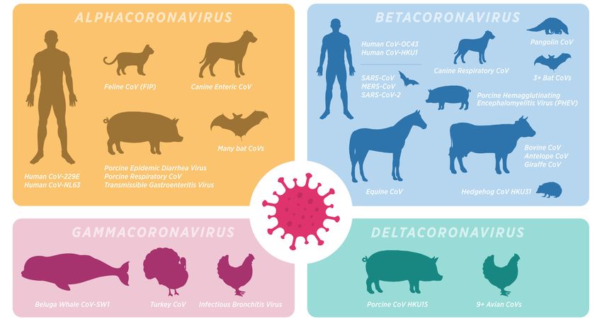
creativecommons.org/licenses/by/ There are many well-studied coronaviruses (CoVs) in mammals that offered insight
4.0/). into this virus species even prior to the emergence of SARS-CoV-2 (Figure 1). CoVs are
Int. J. Transl. Med. 2022, 2, 113–133. https://doi.org/10.3390/ijtm2010010 https://www.mdpi.com/journal/ijtm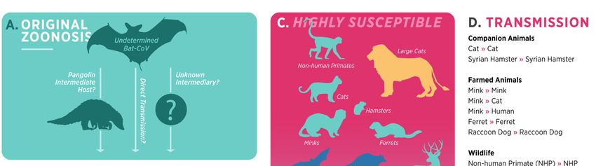
Int. J. Transl. Med. 2022, 12, FOR PEER REVIEW 2
Int. J. Transl. Med. 2022, 2 114
There are many well-studied coronaviruses (CoVs) in mammals that offered insight
into this virus species even prior to the emergence of SARS-CoV-2 (Figure 1). CoVs are
positive-stranded, encapsulated
positive-stranded, encapsulated RNA RNAviruses
viruseswithwithgenomes
genomesranging
ranging inin
size
sizefrom
from26 26to 32
to
kb [14]. In humans, typical alphacoronaviruses (229E and NL63)
32 kb [14]. In humans, typical alphacoronaviruses (229E and NL63) and betacoronaviruses and betacoronaviruses
(OC43 and
(OC43 and HKU1)
HKU1) produceproduce mild mild and
and self-limiting
self-limiting respiratory
respiratory tract
tract infections
infections [15].[15]. In
In
contrast, SARS-CoV-2, the betacoronavirus responsible for the
contrast, SARS-CoV-2, the betacoronavirus responsible for the coronavirus disease 2019 coronavirus disease 2019
(COVID-19) pandemic,
(COVID-19) pandemic, causescauses marked
marked inflammation
inflammation of of the
the airways
airways andand lungs
lungs andand results
results
in substantial
in substantial respiratory
respiratory disease
disease inin many
many infected
infected patients.
patients. SARS-CoV-2
SARS-CoV-2 attaches
attaches to to and
and
infects cells through the angiotensin-converting enzyme-II (ACE2)
infects cells through the angiotensin-converting enzyme-II (ACE2) receptor, resulting in receptor, resulting in
subsequent internalization and proliferation of the virus. A robust
subsequent internalization and proliferation of the virus. A robust immune response must immune response must
be activated
be activated for for viral
viral clearance,
clearance, butbut inflammation
inflammation from from activation
activation of of the
the host
host immune
immune
responses may also result in injury to host tissue [16]. Clinical symptoms
responses may also result in injury to host tissue [16]. Clinical symptoms of COVID-19 are of COVID-19 are
characterized byby
characterized fever,
fever, lethargy,
lethargy, cough, shortness
cough, shortness of breath, and of occasional
breath, and occasional
gastrointestinal
gastrointestinal
signs signs such
such as diarrhea. Mild asdisease
diarrhea.mayMild disease
progress tomay
severeprogress to severe SARS-CoV-
SARS-CoV-2-induced acute
2-induced
lung injuryacute
(ALI)lung andinjury
acute (ALI) and acute
respiratory respiratory
distress syndrome distress syndrome
(ARDS), which(ARDS), which
is the leading
is the of
cause leading
mortalitycause in of mortalitypatients.
COVID-19 in COVID-19 patients. Hyperinflammatory
Hyperinflammatory responses (e.g.,responses
cytokine
(e.g., cytokine storm syndrome), lymphopenia, and
storm syndrome), lymphopenia, and microthrombosis may all contribute microthrombosis maytoallwidespread
contribute
to widespread
alveolar alveolar
destruction, destruction,
significant significant
lung injury, lung injury,
and increased and increased
morbidity morbidity
and mortality and
[17–20].
mortality
Many [17–20]. Many
hospitalized hospitalized
COVID-19 patientsCOVID-19
also havepatients also have
comorbidities comorbidities
such as diabetes, such as
cardio-
diabetes,disease,
vascular cardiovascular disease,
renal disease, renal disease,[21],
or hypertension or hypertension
and age-related [21], and age-related
comorbidities have
had a tremendous
comorbidities haveimpact
had a on the progression
tremendous impactofon thethedisease [22]. The
progression of specific
the disease mechanisms
[22]. The
by whichmechanisms
specific these comorbidities
by which worsen SARS-CoV-2 patient
these comorbidities worsendisease remain patient
SARS-CoV-2 largely elusive,
disease
highlighting the critical need for animal models that can mimic
remain largely elusive, highlighting the critical need for animal models that can mimicboth COVID-19 and these
comorbid
both COVID-19conditions to help
and these mitigateconditions
comorbid human disease.to help mitigate human disease.
Figure 1.1.Mammalian
Mammaliancoronaviruses.
coronaviruses. The Coronaviridae
The family
Coronaviridae frequently
family infectsinfects
frequently and causes
and disease
causes
disease
in a wideinvariety
a wideofvariety
mammals.of mammals. Disease presentations
Disease presentations vary basedvary based tropism
on tissue on tissuefrom
tropism
mild,from
self-
mild, self-limiting
limiting upper respiratory
upper respiratory infections
infections (such (such
as with theas with the alphacoronaviruses
alphacoronaviruses CoV-229E andCoV-229E and
CoV-NL63)
CoV-NL63) to more severe respiratory or systemic diseases such as the SARS-like viruses
to more severe respiratory or systemic diseases such as the SARS-like viruses and enteric diseases and
enteric diseases of critical veterinary importance, especially those in pigs, cats, dogs, and cattle.
of critical veterinary importance, especially those in pigs, cats, dogs, and cattle. Coronaviruses
Coronaviruses undergo frequent host-shifts between mammals, creating a need to better delineate
undergo frequent host-shifts between mammals, creating a need to better delineate both origins for
both origins for non-human mammal coronaviruses that infect humans and mammals that have
non-human mammal
potential to act coronaviruses
as natural thathuman
reservoirs for infect humans and mammals
and veterinary diseases.that have potential to act as
natural reservoirs for human and veterinary diseases.
Int. J. Transl. Med. 2022, 2                                                                                               115

                                     Since SARS-CoV-2 is best theorized to have zoonotic origins, the human–animal–
                               environment interaction of the COVID-19 pandemic is of enormous scientific, public health,
                               and animal welfare importance. The recognition of animal species that are naturally
                               vulnerable to SARS-CoV-2 infection may aid in the investigation of the virus’s possible
                               origin, the identification of potential reservoirs and intermediate hosts, as well as the
                               clarification of mechanisms underlying cross-species transmission back to humans and
                               other species. Such animals that are naturally susceptible for these studies and do not
                               require genetic modification to improve susceptibility or replicate human disease in an
                               experimental setting [23] could provide additional information and assistance in preventing
                               future reverse zoonosis, which might result in the creation of new animal hosts.

                               2. Nontraditional Animal Models for COVID-19
                                     Murine (Mus musculus) and non-human primate (NHP) models for COVID-19 are
                               widely utilized and offer some clear benefits in their use. Rodent models have widely
                               available reagents and can be handled and housed with ease and at less cost than larger an-
                               imals, allowing greater animal numbers and improved statistical significance in results [1].
                               Despite these obvious benefits, undeniable limitations to murine models exist. There are
                               inherent biological differences between humans and rodents that specifically affect their
                               susceptibility to SARS-CoV-2 infection, resulting in the need for genetically modified mice
                               or viruses to better mimic human disease. The absence of suitable ACE2 receptors in
                               most mice species is one such key limitation [24]. Other limitations include the lack of
                               infectivity of SARS-CoV-2 clinical isolates in murine species, as well as the lack of persistent
                               infection, immunopathology, severe acute respiratory distress syndrome, and systemic
                               complications that characterize COVID-19 clinically [25,26]. The short lifespan of these
                               animals also makes it difficult to track the disease’s long-term effects [9,23,27]. Recently,
                               aged murine models have been utilized to better mimic SARS-CoV-2 in older patients with
                               comorbidities [28,29], but many studies exclusively utilize young and immunologically
                               naïve laboratory animals, posing a construct validity issue [30].
                                     Alternatively, nonhuman primates (NHP) are excellent models for human disease
                               due to the close link in physiological, immunological, and genetic aspects that replicate
                               human disease pathophysiology [31,32]. However, the high cost of these animals and
                               limitations to housing and care are obstacles that are difficult to overcome in many research
                               settings. These limited resources and availability of NHP for COVID-19 studies have been
                               a challenge for many researchers even before the emergence of SARS-CoV-2. Furthermore,
                               most NHPs are outbred animals with a broad range of genetic backgrounds, making it
                               challenging to evaluate study results owing to heterogeneity in outcomes across individual
                               animals [32].
                                     It is more crucial now than ever to explore the use of nontraditional animal models
                               that can fill these gaps. Much has been learned about natural infection, transmission,
                               and disease outcomes in mammals subsequently infected with SARS-CoV-2, offering a
                               great opportunity in their use for COVID-19 studies (Figure 2). While each model has
                               its own set of constraints, having a wider variety of optimal animal models will allow
                               for the investigation of a wider variety of important research problems. Importantly,
                               continuous identification and development of the nontraditional innovative animal models
                               will aid in researching disease pathophysiology, testing therapies, and aiding with vaccine
                               development, providing a very consistent framework. This review aims to summarize
                               what we know about several of these less traditional animal models and encourage further
                               studies to optimize and implement their usage.
Int. J.J. Transl.
Int. Transl.Med.
Med.2022,
2022,212, FOR PEER REVIEW 1164
Figure 2. Current
Figure Currentsummary
summaryofof SARS-CoV-2
SARS-CoV-2 origin,
origin,animal susceptibility,
animal susceptibility, andand
transmission.
transmission.(A)
(A) The original zoonosis for the SARS-CoV-2 is yet to be confirmed. It is suspected to be evolveda
The original zoonosis for the SARS-CoV-2 is yet to be confirmed. It is suspected to be evolved in
selected
in species
a selected of bats.
species BatsBats
of bats. maymayhave shed
have shedCoVs andand
CoVs infected
infected humans
humans (solid
(solidarrows)
arrows)viaviaa
suspected intermediate host (pangolin), direct transmission, or other intermediate species that
a suspected intermediate host (pangolin), direct transmission, or other intermediate species that
remain to be discovered (see references [33,34]). (B) SARS-CoV-2 transmission from human to
remain to be discovered (see references [33,34]). (B) SARS-CoV-2 transmission from human to animal
animal (reverse zoonosis events) have been documented in several instances to date (see references
(reverse zoonosis events) have been documented in several instances to date (see references [35–40]).
[35–40]). (C) The susceptibility of animal species for SARS-CoV-2 infection is shown in a
(C) The susceptibility of animal species for SARS-CoV-2 infection is shown in a descending order,
descending order, with NHPs and felids being most susceptible to infection while livestock species
with NHPs
(cattle, pigs,and
andfelids beinggenerally
poultry) most susceptible
being the to least
infection while livestock
susceptible. (D) Animalsspecies (cattle,
are color pigs,
codedandto
poultry) generally being the least susceptible. (D) Animals are color coded to represent
represent the mode of infection (natural vs. experimental) and a summary of animal–animal the mode of
infection (natural
transmission vs. experimental)
confirmed and a summary
by relevant methods (viral RNAof animal–animal
and sequencing) transmission
is shown atconfirmed
the right. by
relevant methods (viral RNA and sequencing) is shown at the right.
2.1. Domestic Cats
2.1. Domestic Cats
Domestic cats (Felis catus) offer an intriguing option as a translational model for
Domestic cats (Felis catus) offer an intriguing option as a translational model for
several reasons. Cats have been infected with SARS-CoV-2 both experimentally [26,41–44]
several reasons. Cats have been infected with SARS-CoV-2 both experimentally [26,41–44]
and naturally [45,46], and human-to-cat as well as cat-to-cat transmission is now well
and naturally [45,46], and human-to-cat as well as cat-to-cat transmission is now well
established [35,36,47]. This comes with little surprise as SARS-CoV was identified in wild
established [35,36,47]. This comes with little surprise as SARS-CoV was identified in wild
cats in
cats in the
the early
early 2000s
2000s [48],
[48], and
and felids
felids have
have the
the natural
natural expression
expression of of ACE2
ACE2 receptors
receptors that
that
act as an efficient binding site for this virus [36,49–51]. Experimentally,
act as an efficient binding site for this virus [36,49–51]. Experimentally, intranasal SARS- intranasal SARS-
CoV-2 inoculation
CoV-2 inoculation results
results in
in efficient
efficient viral
viral replication
replication in in the
the upper
upper and
and lower
lower respiratory
respiratory
tracts, peaking at 3 days post-inoculation (dpi) until decreasing below
tracts, peaking at 3 days post-inoculation (dpi) until decreasing below detectable limits detectable limits
around 14 dpi [42,44,52,53]. The virus is detectable in the nasal turbinates,
around 14 dpi [42,44,52,53]. The virus is detectable in the nasal turbinates, soft palate, soft palate,
tonsils, trachea,
tonsils, trachea, lungs,
lungs, and
and small
small intestines,
intestines, with
with the
the live
live virus
virus in
in all
all tissues
tissues except
except the
the
intestines or feces, suggesting low viral shedding via that pathway.
intestines or feces, suggesting low viral shedding via that pathway. The spleen, lymph The spleen, lymph
node, liver,
node, liver, heart,
heart, and
andolfactory
olfactory bulb
bulbalso
alsoexhibit
exhibitviral
viralreplication.
replication. While
While viral
viral detection
detection
and replication were evident, early studies indicated limited to
and replication were evident, early studies indicated limited to no clinical disease wasno clinical disease was
associated with intranasal experimental inoculation routes [26,42–44],
associated with intranasal experimental inoculation routes [26,42–44], even though mild even though mild
clinical disease
clinical disease was
was apparent
apparentin innatural
naturalfelid
felidinfections
infections[51].
[51].
That barrier
That barrier was
was later
later overcome
overcome by by using
using an an intratracheal
intratracheal inoculation
inoculation routeroute and
and
higher infective
higher infective dose
dose of
of SARS-CoV-2
SARS-CoV-2 than than had
had been
been previously
previously described,
described, resulting
resulting inin
clinical and histopathologic disease in cats consistent with that seen
clinical and histopathologic disease in cats consistent with that seen in acutely infected in acutely infected
hospitalized patients
hospitalized patients with
with COVID-19
COVID-19 [41]. [41]. Clinical
Clinical signs
signs noted
noted in
in infected
infected cats
cats included
included
fever, cough, lethargy, and increased respiratory effort [41,42,44,53,54]. These signs mimic
the most common clinical symptoms observed in humans with COVID-19 including fever,Int. J. Transl. Med. 2022, 2 117
fever, cough, lethargy, and increased respiratory effort [41,42,44,53,54]. These signs mimic
the most common clinical symptoms observed in humans with COVID-19 including fever,
shortness of breath, dry cough, and lethargy [55]. Similarities between COVID-19 and
respiratory disease in SARS-CoV-2-infected felids are also evident histologically. Cats
exhibit inflammation in the nasal turbinates and trachea as well as interstitial pneumonia
accompanied by marked pulmonary edema, widespread alveolar damage with hyaline
membrane formation, and vasculitis [41,53], mirroring the pulmonary and perivascular
changes seen in people with acute COVID-19 pneumonia [18,56–60].
Contrary to prior studies that used intranasal delivery of SARS-CoV-2, intratracheal in-
oculation resulted in lower viral RNA levels in the lungs [41]. However, SARS-CoV-2 virus
was readily detectable in the nasal turbinates at 4 and 8 dpi, indicating the mobility of
the virus via the mucociliary escalator to establish the upper respiratory infection despite
the absence of intranasal inoculation [41]. After the emergence of COVID-19 in Wuhan,
China, domestic cats in the region had 14.7% seroprevalence [40]. Viral RNA detection
in lung tissue is prolonged in juvenile cats, and while juvenile cats shed more infectious
virus, sub-adult cats exhibit greater antibody titers [51]. In cats, the adaptive immune
response is initiated early on, with neutralizing antibodies being produced as early as 7 dpi
and increasing beyond 28 days, whereas reinfected cats had an even stronger and quicker
humoral immune response to the viral infection [43,44].
Concerning the transmission potential of SARS-CoV-2 to domestic cats, several studies
support natural transmission from infected humans to domestic cats [54,61–64]. After
contact with an infected person, viral RNA was found in oropharyngeal swabs taken from
cats, and infections were verified serologically [48]. Several other investigations have
confirmed household transmission to cats from infected owners [65], with the infected
animals remaining asymptomatic in the majority of instances [62]. In addition to human-to-
cat transmission, airborne transmission between cats in adjacent cages was demonstrated
under experimental conditions, although infection was limited to one out of every three
sentinel animals [26]. Direct contact transmission between cats is even more evident,
with infection occurring in all sentinel animals [42,44,47]. Serial transmission of the virus
between cats was shown to reduce the effectiveness of transmission from infected to
naive cats [53,66], although further transmission from reinfected cats has not yet been
established [42]. Transmissibility for the most recent variants of concern (delta and omicron),
however, has not been established [67], and as variants continue to emerge, an established
feline model is needed to best understand the potential for cat-to-human transmission of
SARS-CoV-2.
Hypertension, diabetes, renal disease, and obesity, all of which increase COVID-19 ill-
ness, are naturally occurring comorbidities in domestic cats and are easily adapted to feline
models [68–73], offering another potential advantage of this model. Limitations do exist,
such as required experience with handling cats (in particular within an ABSL-3 environ-
ment), and the moderate cost involved compared to mouse studies. In addition, public
opinion regarding the use of companion animals in translational research must be taken
into account. Additional research on transmission efficiency is required, particularly with
regard to cats as intermediary hosts between SARS-CoV-2 and people, as well as on in-
flammation and how it mimics human illness [74]. Despite these limitations, felids remain
a promising option for modeling natural SARS-CoV-2 infection and COVID-19 disease
progression for future studies.
2.2. Ferrets
Ferrets (Mustela putorius furo) originated from the domestication of the European pole-
cat, and have been a valued model for many viral respiratory diseases including influenza,
respiratory syncytial virus (RSV), adenovirus, and SARS-CoV [75–77]. This is in part due to
the fact that they have an anatomically comparable respiratory tract to humans, with similar
features of glandular density in the bronchial wall, upper and lower respiratory tract propor-
tions, and terminal bronchioles as well as receptor distribution, resulting in similar clinicalInt. J. Transl. Med. 2022, 2 118
courses of disease [76,78–80]. Ferret and human ACE2 receptors vary by only two amino
acids and contain the crucial residues required for binding by the SARS-CoV-2 receptor-
binding domain [79,81]. The ferret model for COVID-19 has been utilized since early in
the COVID-19 pandemic. A sentinel study conducted by Shi et al. revealed that infection
with early variants of SARS-CoV-2 (SARS-CoV-2/F13/environment/2020/Wuhan and
SARS-CoV-2/CTan/human/2020/Wuhan) resulted in high viral susceptibility in nasal
turbinates, soft palates, and tonsils, although low quantities of the virus were identified
in the lungs [26]. Clinical signs included elevated body temperature and reduced activity,
but fever and appetite loss were observed in just 1 out of 3 ferrets, with the dose and
heterogeneity of the isolate affecting the presence of clinical disease [26]. In general, weight
reduction has not been a consistent finding in the ferret model [75,82], but clinical disease
in CoV-2-infected ferrets was noted in another study in which increased body temperature,
lethargy, and coughing were evident at 2 dpi, with all animals recovering by day 8 [75]. In
this study, viral loads peaked at 4 dpi in the lungs and nasal turbinates. Viral RNA was
detectable in the intestine, saliva, urine, rectal swabs, and feces through 8 dpi, and viral
loads were highest in the upper respiratory tract with overall mild disease. Additional
studies have reported similar results, with viral loads highest in the nasal turbinates and
throat, mild clinical disease, and viral shedding beyond 10 dpi [75,82–86].
Ryan et al. went a step further to try and optimize inoculum dosages for the fer-
ret COVID-19 model [87]. Ferrets were challenged intranasally with 1 mL of Victo-
ria/1/2020 SARS-CoV-2 at three different titers: high (5 × 10⁶ PFU/mL), medium
(5 × 10⁴ PFU/mL), and low dose (5 × 10² PFU/mL). While the high and medium doses
successfully infected the upper respiratory tract of all the animals, the lower inoculum dose
was only successful in 1 of 6. Some in the high dose group developed bronchopneumonia,
and viral shedding continued into days 14 to 21 [80]. Age also affects disease pathogenesis
and outcome in the ferret model, with infections in older ferrets (>3 years of age) resulting
in increased viral loads, prolonged viral replication and shedding, and more severe disease
than their younger counterparts [75,88,89].
Ferrets have also been utilized for longitudinal studies of immune responses to
SARS-CoV-2 infection through intranasal inoculation with SARS-CoV-2 and repeated as-
sessments of upper respiratory tract gene transcripts from nasal washes. Ferrets infected
with SARS-CoV-2 demonstrate unique inflammatory transcriptional responses, as evi-
denced by low levels of type I and III interferons in contrast to up-regulated IL-6, CCL2,
CCL8, and CXCL9 [90]. In depth studies into specific gene signatures and transcripts in
infected ferrets have shown that SARS-CoV-2 gene signatures induce more respiratory
immune responses than influenza A and SARS-CoV-2 gene signatures are identified in
both short-term and longer-term (21 days) infections in ferrets [91]. In both short- and
long-term scenarios, metabolic, glucocorticoid, and reactive oxygen species genes were
enriched. Activated T cells, macrophages, and type I IFN signaling are all involved in the
ferret’s immunological responses [92–94], yet all animals survive experimental infection,
with a strong adaptive response and neutralizing antibody generation as early as 8 days
post-infection [75,90,95,96]. When ferrets are rechallenged with SARS-CoV-2, the generated
immune response protects them from harmful disease, decreases viral replication, and no
remarkable lesions are noted [26,83,97].
Ferret-to-ferret transmission is also readily achieved. Direct contact transmission
appears to be the most efficient method of transmission as all naïve ferrets cohoused with
intranasally inoculated ferrets exhibited clinical symptoms (elevated body temperature
and reduced activity) 2–6 days after exposure [75,85,97]. Transmission rates in ferrets
vary amongst spike glycoprotein mutations seen with different variants, with higher
transmissibility of S(614G) over S(614D) glycoprotein structures [98]. Airborne transmission
of SARS-CoV-2 was also shown [3,85,96]; however, this transmission appeared less efficient
and diminished at distances greater than 1 m [3,99]. Despite multiple studies supporting
direct and indirect transmission between ferrets, differences in disease pathogenesis and
outcomes are noted between studies. When comparing direct recipient animals to donorInt. J. Transl. Med. 2022, 2 119
animals, most studies found that infection via either route resulted in comparable durations
and levels of virus shedding in direct recipients [85,96]. Kim et al., however, found lower
levels of SARS-CoV-2 RNA in nasal washes of indirect recipient ferrets, as well as shorter
viral shedding with no infectious virus isolated [75]. Viral RNA was detected in ferret
nasal washes 48 h after direct contact with intranasally infected animals, demonstrating
that transmission is rapid and can occur before peak disease at 4 dpi. Overall, there is a
resemblance between the clinical characteristics of infected naïve ferrets and those of injected
ferrets in terms of viral replication, pathology, and immune response [75,82,85,97,100].
The ferret model for COVID-19 is also well utilized in evaluating antiviral and
other treatment effects. The use of repurposed medications (hydroxychloroquine sul-
fate, lopinavir-ritonavir, and emtricitabine-tenofovir) in the treatment of infected ferrets
was shown to provide no substantial advantage over standard therapy [95]. In fact, emtric-
itabine, tenofovir, lopinavir-ritonavir, and hydroxychloroquine sulfate markedly enhanced
clinical symptoms in ferrets infected with SARS-CoV-2 and did not significantly lower
viral titers [95]. Those who received 18-azathioprine had a longer duration of clinical
illness, higher viral titers in the nasal turbinates, delayed virus clearance, and lower serum-
neutralizing antibody titers compared to those who did not receive the drug [95]. In
contrast, MK-4482/EIDD-2801, a ribonucleoside analog inhibitor developed for influenza
virus therapy, demonstrated promise as a SARS-CoV-2 therapy as evidenced by reduced
viral load and prevention of transmission to untreated contact ferrets [101]. Using this
model, newly developed lipoprotein fusion inhibitors were shown to successfully sup-
press S protein conformational changes and prevent virus–host cell membrane fusion
by integrating into host cell membranes, also decreasing SARS-CoV-2 transmission be-
tween ferrets [98,102]. Recent studies also demonstrate that intranasal treatment with
the TLR2/6 agonist INNA-051 can effectively reduce viral levels in ferrets infected with
SARS-CoV-2 [103].
Although the application of ferrets in vaccine evaluation is limited due to dose-
dependent responses to SARS-CoV-2 infection [87], several studies have utilized this animal
model in the process of vaccine development. Immunization of ferrets with Ad5-nCoV,
an adenovirus-vectored vaccine, suppressed viral multiplication in the upper respiratory
tract (URT) through both mucosal and intramuscular routes, with mucosal vaccination
completely protecting from infection [104]. Furthermore, according to Dong et al., intranasal
immunization with CVXGA1 inhibited viral infection in ferrets and prevented contact
transmission, additionally suppressing SARS-CoV-2 replication in the URT and limiting
disease progression to the lower respiratory tract [105].
Given their susceptibility to SARS-CoV-2, histopathological changes, detectable viral
loads, and shedding from the respiratory tract during infection, ferrets continue to be a
highly regarded model for asymptomatic or mild COVID-19 [3,79]. Unfortunately, the
lack of pulmonary viral replication and edema or the development of ARDS in ferrets
suggest a major limitation for the study of lung pathology with COVID-19 [106]. Ferrets
are larger than mice or hamsters and, thus, add some housing and handling limitations;
furthermore, as with most nontraditional models, they have costlier and less available
optimized reagents. However, ferrets live a long time, and have the potential for researching
aging and long-term effects of SARS-CoV-2, only adding to their value as a useful model to
research SARS-CoV-2 pathogenesis and therapeutic targets [26].
2.3. Mink
Similar to ferrets, mink (Neovison vison) belong to the Mustelidae family and are
naturally prone to SARS-CoV-2 infection. In fact, mink represents an exceptional an-
imal model to study SARS-CoV-2 since they show efficient interspecies transmission
and clinical features of SARS-CoV-2 infection comparable to humans [37,107]. Recent
SARS CoV-2 outbreaks on mink farms in the Netherlands and Spain resulted in the culling
of thousands of animals. In these cases, though the initial infection was likely to be trans-
mitted from infected farm laborers, studies suggest human reinfection can occur fromInt. J. Transl. Med. 2022, 2 120
infected minks [108,109]. Inter-species transmission is supported through an analysis of
mutations in the spike protein of SARS-CoV-2 virus, with specific mutations (L452M, Y453F,
F486L, and N501T) identified in both mink and workers closely in contact with animals
on those farms [110]. Two of these mutations (Y435F and N501T) adapt the spike protein
of SARS CoV-2 to improve transmission to both mink and ferrets. These mutations arose
independently in mink and seem to have minimal effect on human airway epithelial cells
in vitro. However, studies suggest that these mutations carry minimal effects on human
disease and are tolerated by the available vaccines against SARS-CoV-2 [111].
In mink, SARS-CoV-2 infection produces vigorous virus replication in the upper and
lower respiratory tracts, including the nasal turbinates, tonsils, soft palate, trachea, and lung
tissue, peaking at 2 dpi and continuing until 8 dpi before falling below measurable limits at
14 dpi [112]. SARS-CoV-2 also replicates in the gastrointestinal tract of mink, limiting feed
intake and exhibiting mild to severe symptoms including up to 20% body-weight loss [112].
Neutrophil infiltration, epithelial degeneration, and necrosis are found in the inflamed
nasal turbinates of SARS-CoV-2-infected minks [112–114]. Mink intranasally infected with
106 PFU of WA1 isolate resulted in a productive viral replication in the upper and lower res-
piratory tract, and viral replication was detected in the left cranial lung and nasal turbinates,
but not from nasal washes of 3–5 dpi [115]. Infection results in severe, diffuse interstitial
pneumonia with swelling and degeneration of bronchial epithelial cells, thickening of
alveolar septa, collagen deposition, hemorrhage, and marked pulmonary edema [112–114].
Mink also exhibit pneumocyte proliferation and pulmonary consolidation with diffuse
alveolar damage during SARS-CoV-2 infection, as well as infiltration of macrophages,
monocytes, and neutrophils throughout the lungs in addition to perivascular lymphocyte
accumulations [112–114]. The virus can cause lethal disease in mink; additionally, naïve
mink infected experimentally exhibit enhanced clinical features [112,113].
Mink not only offer potential as an animal model for human COVID-19, but should be
evaluated as a possible farmed animal reservoir for SARS-CoV-2 and for their role, through
mutants, on viral fitness, contagiousness, re-infection, immunotherapy, and vaccine efficacy.
Limitations related to the use of minks as an animal model include animal behavior, the
scarcity of reagents and resources, and difficulty in housing and handling mink under
laboratory conditions [108,109,113,114].
2.4. Hamsters
Syrian hamsters (Mesocricetus auratus) are widely used in viral respiratory disease
research, being valuable models for influenza virus, adenovirus, and most recently, SARS-
CoV-2 [116]. With these viral infections, hamsters show productive viral replication, patho-
logical signs and successful disease pathogenesis in the lung [117]. Hamsters are naturally
infected with SARS-CoV-2 virus, in part due to a high degree of homology with the
human ACE2 receptor domain and successful interaction with SARS-CoV-2 spike glyco-
protein [118]. Experimentally infected hamsters show significant weight loss [119–121],
respiratory distress, lethargy, ruffled fur, and hunched posture with recovery around
7 dpi [121]. In a study conducted to determine if age differences appear to influence disease
outcomes, it was found that weight loss was faster and more consistent in older hamsters
developing severe SARS-CoV-2 symptoms, while juvenile hamsters resisted severe disease
with a stronger and earlier immune cell influx than adult hamsters. Only young hamsters
showed fast lung recovery on day 14 following infection [119].
In a comparative study including several variants, namely 1.617.2, B.1.617.3, and
B.1.351, a similar degree of viral shedding was shown regardless of the variant, although
the delta variant (1.617.2) resulted in the least body-weight gain and the most severe lung
disease, affecting 40% of infected animals [122]. Infected hamsters produced neutralizing
antibodies, but the neutralizing response was lower with B.1.351 variant infection. This
study supports that 1.617.2 infection (delta variant) induces a stronger neutralizing antibody
response and that B.1.351 infection in hamsters is more immune evasive than the other
variants studied [122].Int. J. Transl. Med. 2022, 2 121
Histopathological features of SARS-CoV-2 infection in Syrian hamsters include necro-
suppurative bronchitis, intra-alveolar and intrabronchial infiltration of macrophages and neu-
trophils, pulmonary edema, and severe alveolar hemorrhage in infected lungs [119,120,123,124].
SARS-CoV-2 viral replication was observed in both the upper and lower respiratory tracts,
with peak viral titer at 3 dpi progressing to viral clearance at 7 to 10 dpi [118]. The innate
immune response exhibited by hamsters involves rapid augmentation of local antiviral
responses [125] facilitated by the production of pro-inflammatory cytokines, namely IFN
and IL-6, which peak at 2 to 4 dpi and resolve by 14 dpi [118]. The immune response also
incorporates significant production of chemokines and the recruitment of a potent type
1 T cell response [125]. The macrophage response to intracellular viral RNA is driven by
CCL2 and CXCL10, and the expression profile of pro-inflammatory cytokines in mono-
cytic macrophages favored effector T cell recruitment chemokines targeting CXCR3 and
CCR5 [125]. Emergence of IgM-neutralizing antibodies was shown to precede viral clear-
ance at day 5 post infection [125]. Hamsters also show significant potential in studies
involving intraspecies viral transmission. Sia et. al. [121] showed that SARS-CoV-2 is
transmitted between hamsters through both close contact and non-contact routes such
as aerosols and fomites. Naive hamsters were successfully infected within one day of
direct contact with intranasally infected hamsters, highlighting the potential to evaluate
the transmission risk of continually evolving variants and to study the mutation rates of
SARS-CoV-2 [121].
Apart from transmission studies, Syrian hamsters have been utilized to study pro-
phylactic modalities against SARS-CoV-2 infection. According to Rosenke et al., oral
administration of the nucleoside analog MK-4482 suppressed SARS-CoV-2 replication, and
led to decreased lung pathology [124]. The aforementioned outcomes were also observed in
a study that was conducted using ranitidine bismuth citrate [126]. Notably, viral shedding
was reduced by the administration of therapeutic neutralizing antibodies that targeted the
receptor-binding domain (RBD) of the virus [5,102,120,127,128].
Syrian hamsters have also been widely utilized as an animal model for preliminary
vaccine studies. According to Tostanoski et al., Ad26.COV2.S vaccination reduces humoral
immune responses in hamsters and protects against severe disease [129]. In addition,
pre-clinical trials conducted using the Newcastle disease virus (NDV-S) vaccine showed
substantial immunogenicity, generating spike-protein-specific neutralizing antibodies and
lower lung viral titers with less body-weight loss [130]. Other pre-clinical vaccine trials,
such as that of the PTX-COVID19-B mRNA vaccine, exhibited significant humoral and
cellular immune responses and protected the upper respiratory tract from SARS-CoV-2 in-
fection in hamsters [131]. A single dosage of another approved adeno virus-based vaccine,
ChAdOx1 nCoV-19 (AZD1222), protected hamsters against SARS-CoV-2 illness and pneu-
monia [132]. In this study, vaccinated hamster sera for B.1.351 had a 9.5-fold lower viral
neutralizing antibody titer than B.1.1.7, and the lungs of vaccinated animals exhibited no
gross lesions in comparison to control animals [132]. Hamsters intranasally inoculated
with another vaccine candidate (COVI-VAC) showed lower tissue virus loads, milder lung
pathology, and less weight loss compared to wild type hamsters, and this was followed
by the development of spike IgG antibody levels and plaque-reduction-neutralization
titers [133]. Moreover, inoculation of a number of live attenuated vaccine candidates gen-
erated by the recoding of the SARS-CoV-2 genome resulted in two-fold immunogenicity,
while two of the candidates elicited substantial protective immunity [134]. The attenuated
viruses caused minor pulmonary histopathology in the upper—but not the lower—airways,
and after challenge, the hamsters developed no signs of disease and virus could not be
recovered from the lungs of the infected animals [134]. Drawbacks to the Syrian hamster
model for COVID-19 include limited disease severity and fatality, which may be explained
by the presence of asparagine at position 82 in the sequence of the ACE2 receptor instead
of lysine in human ACE2, thus limiting their potential as a model for moderate-to-severe
COVID-19 [118]. Additionally, reagents and bioinformatic resources are more limited for
hamsters when compared to several other models [118,121].Int. J. Transl. Med. 2022, 2 122
The susceptibility of Chinese hamsters to SARS-CoV-2 infection was recently demon-
strated by the presence of viral replication in the upper and lower respiratory tract, accom-
panied by the development of pneumonia and bronchitis [74]. In this model, intranasal
infection results in transient but significant body-weight loss and a slight reduction in body
temperature. Lung lesions are less severe but are associated with diffuse alveolar damage
and the persistence of viral RNA in tissues may still be found up to 14 dpi [74]. In fact, this
animal model may prove advantageous to the Syrian hamster model due to prolonged and
more prominent clinical symptoms, smaller size, a well-characterized genome (supported
by transcriptome and translatome data), and the availability of molecular tools specific
to the species [74]. However, while most commercial breeders of laboratory animals only
provide a single line of Syrian hamsters, Chinese hamsters that meet laboratory criteria
such as uniform genetics and specific-pathogen-free (SPF) status are far less available.
SARS-CoV-2 infection in Roborovski dwarf hamsters leads to the rapid and robust
development of severe clinical illness due to early destructive and lethal lung pathology, as
well as signs of systemic SARS-CoV-2 infection, recapitulating human COVID-19 symptoms
such as coughing, snuffling, dyspnea, labored breathing, and gradual weight loss [135,136].
Roborovski dwarf hamsters may represent an important addition to current animal models
given the advantage of a small animal model and faster and more consistent development
of clinical symptoms following infection [135,136]. It is also less prone to aggressive
behavior than other hamster species, making it easier to handle and house in greater
groups [135,136]. Roborovski dwarf hamsters are commercially accessible and farmed in
large quantities for the pet industry, and the species reaches sexual maturity at 4 weeks of
age and reproduces easily in a laboratory setting. However, unrecognized co-infections
with other pathogens, different individual infection histories, and changeable, uncontrolled
microbiomes may substantially influence their responses to experimental infections with
specific pathogens [137].
In addition to Chinese hamsters and Roborovski dwarf hamsters, a recent study
indicated that cardiomyopathic J2N-k hamsters display characteristics comparable to those
associated with severe COVID-19 complications. Male J2N-k hamsters of 7 weeks of age
were infected with 106 PFU SARS-CoV-2 and demonstrated high viral titers in the lung
with a weight loss of 25.7% at 3 dpi. Mortality was reported in all infected J2N-k hamsters
at 9 dpi and pathological characteristics were consistent with SARS-CoV-2-associated lung
damage [138].
Overall, the beneficial impacts of the hamster model for SARS-CoV-2 infection are
vast and include easy handling for ABSL-3 and well-documented natural susceptibility to
SARS-CoV-2 virus with disease pathogenesis and severity comparable to mild COVID-19
in humans. Recently, significant progress has been achieved in spatial proteomics and
transcriptomics in these species, allowing the determination of the individual expression
levels of almost all proteins and mRNA molecules with single-cell-type precision [137].
Although the use of hamsters in biomedical research is somewhat limited by the absence
of suitable inbred strains and species bred under standardized, controlled circumstances,
these species offer an intriguing alternative to current SARS-CoV-2 animal models.
2.5. Other Species
Current research indicates that the origins of SARS-like coronaviruses stem from
bats, with these coronaviruses having the capability of docking and entrance via dif-
ferent orthologues of human ACE2 according to research prior to the emergence of
SARS-CoV-2 [139,140]. As bats are natural reservoirs for several coronaviruses, including
SARS-CoV and SARS-CoV-2, recent studies highlight the need for continued coronavirus
surveillance in bats [141,142]. While ongoing research into coronavirus origins and main-
tenance in bats is clearly indicated, questions remain as to the usefulness of the bat as an
animal model for human COVID-19. Studies have shown that intranasal inoculation of
SARS-CoV-2 in fruit bats (Rousettus aegyptiacus) results in viral replication in the upper
respiratory tract and seroconversion in 7/9 animals, and infection occurred in only 1 out ofInt. J. Transl. Med. 2022, 2 123
every 3 animals during direct contact [96]. However, clinical signs were lacking apart from
mild rhinitis [96]. In contrast, another study demonstrated that a coronavirus related to
SARS did not grow in fruit bats after an experimental infection [143,144]. While Rousettus
bats are not the major reservoir species for SARS-CoV-2, these findings suggest further
studies are needed to evaluate if experimental infection in fruit bats might help simulate
the physiopathology of the virus in its host [145–149].
Another emerging reservoir of interest for SARS-CoV-2 are white-tailed deer
(Odocoileus virginianus). In a recent study, rRT-PCR detected SARS-CoV-2 in 129 out of
360 (35.8%) free-ranging white-tailed deer from northeast Ohio (USA) sampled between
January and March 2021 [150]. At least three lineages of SARS-CoV-2 were identified in six
different locations (B.1.2, B.1.596, and B.1.582), and deer of the B.1.2 lineage, which were
prevalent in Ohio at the time, likely spread to deer populations in several different locales
over different timepoints [150]. It was further concluded that deer-to-deer transmission
likely occurred in at least three different sites [150]. Based on this study and others, white-
tailed deer may serve as a natural reservoir for SARS-CoV-2, which could lead to distinct
evolutionary trajectories and potential spillover to people, confounding long-term COVID-
19 control attempts [150,151]. Another study of 624 pre- and post-pandemic blood samples
collected from wild deer in four different states in the United States identified antibodies
to SARS-CoV-2 in 152 of 200 (40%) via surrogate virus neutralization testing [152]. These
findings and others underscore the need for further evaluation of white-tailed deer as a
natural reservoir and potential animal mode for COVID-19 [153].
Deer mice and bushy-tailed woodrats are also susceptible to SARS-CoV-2 infec-
tion [154–156]. In a recent study, busy-tailed woodrats were observed to shed virus orally
forInt. J. Transl. Med. 2022, 2 124
Table 1. Summary of current SARS-CoV-2 animal models.
Hamsters Mink Ferrets Cats Mice NHP
Naturally susceptible? Yes Yes Yes Yes Not all variants Yes
Experimental inoculation 103 –105.3
PFU; 105 PFU (I.N.); 4× 10⁵ –105 PFU
Not reported 105 –105.5 PFU (I.N.) (2.4–4.75) × 106 TCID50
dose 105 –107 TCID50 (I.N.) 1.2 × 10⁶ TCID50 (I.T.) 3 × 10⁴ –105 TCID50
Alveolar flooding
Interstitial pneumonia and
Inflammatory cell infiltration, Diffuse alveolar damage Diffuse alveolar damage
lymphocyte infiltration in Pulmonary edema
pulmonary edema, and alveolar and interstitial pneumonia with hyaline membrane
Bronchopneumonia transgenic mice; some Alveolar flooding
hemorrhage with hyaline membrane formation
Infiltration of inflammatory severe disease with alveolar Hyaline membranes
Lung pathology Apoptosis of the cells in upper formation Vasculitis
cells to the lung necrosis Histopathological lesions
and lower respiratory tract Histopathological lesions Pulmonary edema
Thickening of alveolar septa Diffuse alveolar damage, exhibit ARDS and mimic
Increased IL-6 and IL-10 in exhibit ARDS and mimic Histopathological lesions
exudation, and hemorrhage human COVID-19
lungs human COVID-19 exhibit ARDS and mimic
in mouse-adapted virus
human COVID-19
Neutropenia, anemia
Weight loss, respiratory
Weight loss (CBC)
distress, lethargy, ruffled fur Labored breathing and
Lymphopenia Increased respiratory effort Nasal congestion and Inflammatory cytokine
and hunched posture watery to mucoid nasal
Clinical signs and/or Pyrexia (Fever) Cough dyspnea boost (IL-1Ra, IL-1β,
Younger hamsters more exudates, anorexia
Systemic effects Mild clinical signs (fever, Pyrexia (Fever) Increase in IL-6, IL-1β, TNF- IFNγ, TNF-α, IL-6, IL-2,
resistant to severe disease Natural infection can
lethargy, coughing) Lethargy α, MCP-1, G-CSF and IL-4, IL-5, RANTES,
than more aged hamsters progress to death
GM-SCF in BALB/c mice G-CSF, GM-CSF, CCL-2)
Increase in IL-6, IL-1β, TNF-α
Increased respiratory rate
Active transmission via Naturally infected
Small size and rapid breeding
direct or indirect contact Mimic human lung
High susceptibility to
Similar distribution of pathology (ARDS) and Useful for pathogenesis Useful for pathogenesis,
SARS-CoV-2 Naturally infected
ACE2 receptors in clinical features of studies and testing of vaccine, and therapeutic
Useful for immunological Mimic human lung
respiratory tract; high COVID-19 antiviral therapeutics studies
studies for vaccine pathology (ARDS) and
Advantages similarity to hACE2 Can be used for Small size Mimic human lung
Lung pathology similar to other clinical features
Suitable for longitudinal transmission studies Rapid breeding pathology (ARDS) and
COVID-19 Can produce severe disease
studies of immune COVID-19 comorbidities Widespread availability of clinical features of
Active transmission via direct as in COVID-19
responses and treatment (hypertension, diabetes, research reagents COVID-19
contact and aerosol
efficacy during obesity, renal disease) occur
development
SARS-CoV-2 infection naturally in catsInt. J. Transl. Med. 2022, 2 125
Table 1. Cont.
Hamsters Mink Ferrets Cats Mice NHP
Do not mimic all clinical Short supply and high cost Requires species-specific
signs of COVID-19 of hACE2 transgenic mice training to handle animals
Do not mimic all clinical
Mortality is not observed Difficultly in handling Requires species-specific Mild inflammatory Not all macaques can be
signs of COVID-19
Viral clearance is rapid; Limited available resources training to handle animals responses and lung damage infected
Mortality is not observed.
cannot be used for and reagents Moderate cost involved Cannot be used directly; Clinical signs are
Disadvantages Low viral titer in the lungs
long-term pathogenesis Specific pathogen free Intratracheal inoculation needs a transgenic animal mild/moderate
Do not fully represent the
Fail to develop diffuse animals not yet available for needed to induce severe or mouse-adapted virus to Small sample size and
severe cases of human
alveolar disease and acute longitudinal studies respiratory disease cause infection availability
infection
respiratory distress found in Fatal encephalitis in High costs for housing
COVID-19 transgenic mice facilities and animal care
References [118,121,160,161] [109,112–114] [75,76,78–81,83,90] [18,26,41,47,62] [24,25,158,160,162–165] [31,32,166–170]
SARS-CoV-2, severe acute respiratory syndrome coronavirus 2; IN, intranasal; IT, intratracheal; TCID50, 50% Median Tissue Culture Infectious Dose; PFU, plaque-forming units; dpi,
days post infection.Int. J. Transl. Med. 2022, 2 126
Many questions concerning the SARS-CoV-2 transmission route remain
unresolved [157,171,172]. The dynamic differences in outcomes following infection en-
courage the identification of different animals for different disease outcomes in order to
better elucidate the pathophysiology and underlying circumstances that may trigger in-
creased susceptibility to disease, as well as host genetics that may contribute to these varied
outcomes [173,174]. There are numerous unanswered questions regarding SARS-CoV-2 co-
morbidities, coinfections, and their long-term effects, many of which can be successfully
mirrored in naturally infected animals—such as diabetes, obesity, and hypertension in the
cat [175–178]. Varied models also offer unique opportunities to test the effectiveness of
repurposed therapies for COVID-19 [179–181].
Due to fundamental variations in the biology and physiology of different animal
species, no single animal model will be able to address all human translational concerns.
The emergence of new variants may also lead to increased or decreased potential utiliza-
tion for specific animal models of COVID-19. While some offer more value for studying
transmission, other may have greater benefit for studying severe disease, new therapies,
vaccines, or other preventions. By thinking outside of the box with our choices in trans-
lational animal models, we add significant potential to select experimental designs that
will best address specific research questions. As available animal resources and limitations
continue to evolve, it is important to have a strong knowledge base supporting a more
varied approach to animal models for diseases such as COVID-19.
Author Contributions: Conceptualization, S.G. and J.M.R.; writing—original draft preparation, S.G.;
writing—review and editing, J.M.R., C.A.M. and M.T.S. All authors have read and agreed to the
published version of the manuscript.
Funding: This research received no external funding.
Acknowledgments: The authors would like to thank Benton Rudd for his efforts in the creation of
the figures used in this review.
Conflicts of Interest: The authors declare no conflict of interest.
References
1. Sutton, T.C.; Subbarao, K. Development of animal models against emerging coronaviruses: From SARS to MERS coronavirus.
Virology 2015, 479–480, 247–258. [CrossRef] [PubMed]
2. Singh, A.; Singh, R.S.; Sarma, P.; Batra, G.; Joshi, R.; Kaur, H.; Sharma, A.R.; Prakash, A.; Medhi, B. A Comprehensive Review of
Animal Models for Coronaviruses: SARS-CoV-2, SARS-CoV, and MERS-CoV. Virol. Sin. 2020, 35, 290–304. [CrossRef] [PubMed]
3. De Vries, R.D.; Rockx, B.; Haagmans, B.L.; Herfst, S.; Koopmans, M.P.G.; de Swart, R.L. Animal models of SARS-CoV-2 transmis-
sion. Curr. Opin. Virol. 2021, 50, 8–16. [CrossRef] [PubMed]
4. Yuan, L.; Tang, Q.; Cheng, T.; Xia, N. Animal models for emerging coronavirus: Progress and new insights. Emerg. Microbes. Infect.
2020, 9, 949–961. [CrossRef] [PubMed]
5. Li, W.; Chen, C.; Drelich, A.; Martinez, D.R.; Gralinski, L.E.; Sun, Z.; Schäfer, A.; Kulkarni, S.S.; Liu, X.; Leist, S.R.; et al. Rapid iden-
tification of a human antibody with high prophylactic and therapeutic efficacy in three animal models of SARS-CoV-2 infection.
Proc. Natl. Acad. Sci. USA 2020, 117, 29832. [CrossRef]
6. Lee, C.-Y.; Lowen, A.C. Animal models for SARS-CoV-2. Curr. Opin. Virol. 2021, 48, 73–81. [CrossRef]
7. Khoury, D.S.; Wheatley, A.K.; Ramuta, M.D.; Reynaldi, A.; Cromer, D.; Subbarao, K.; O’Connor, D.H.; Kent, S.J.; Davenport, M.P.
Measuring immunity to SARS-CoV-2 infection: Comparing assays and animal models. Nat. Rev. Immunol. 2020, 20, 727–738.
[CrossRef]
8. Brooke, G.N.; Prischi, F. Structural and functional modelling of SARS-CoV-2 entry in animal models. Sci. Rep. 2020, 10, 15917.
[CrossRef]
9. Gretebeck, L.M.; Subbarao, K. Animal models for SARS and MERS coronaviruses. Curr. Opin. Virol. 2015, 13, 123–129. [CrossRef]
10. Mullick, J.B.; Simmons, C.S.; Gaire, J. Animal Models to Study Emerging Technologies Against SARS-CoV-2. Cell. Mol. Bioeng.
2020, 13, 293–303. [CrossRef]
11. Neerukonda, S.N.; Katneni, U. A Review on SARS-CoV-2 Virology, Pathophysiology, Animal Models, and Anti-Viral Interventions.
Pathogens 2020, 9, 426. [CrossRef] [PubMed]
12. Subbarao, K.; Roberts, A. Is there an ideal animal model for SARS? Trends Microbiol. 2006, 14, 299–303. [CrossRef] [PubMed]
13. Renn, M.; Bartok, E.; Zillinger, T.; Hartmann, G.; Behrendt, R. Animal models of SARS-CoV-2 and COVID-19 for the development
of prophylactic and therapeutic interventions. Pharmacol. Ther. 2021, 228, 107931. [CrossRef] [PubMed]You can also read