The cathelicidins LL-37 and rCRAMP are associated with pathogenic events of arthritis in humans and rats - OPUS 4
←
→
Page content transcription
If your browser does not render page correctly, please read the page content below
Downloaded from http://ard.bmj.com/ on October 26, 2016 - Published by group.bmj.com

                                                                                                             Basic and translational research

                                     EXTENDED REPORT

                                     The cathelicidins LL-37 and rCRAMP are associated
                                     with pathogenic events of arthritis in humans and
                                     rats
                                     Markus H Hoffmann,1 Heiko Bruns,2 Liselotte Bäckdahl,1 Petra Neregård,3
                                     Birgit Niederreiter,4 Martin Herrmann,5 Anca Irinel Catrina,3 Birgitta Agerberth,6
                                     Rikard Holmdahl1

▸ Additional data are                ABSTRACT                                                       genes involved in cell adhesion, communication
published online only. To view       Background In rheumatoid arthritis (RA), neutrophil            and motility.7 In psoriasis, LL-37 produced by kera-
these files please visit the
journal online (http://dx.doi.org/
                                     granulocytes fuel inflammation and damage tissue in the         tinocytes and neutrophils appears to contribute to
10.1136/annrheumdis-2012-            joint by releasing cytotoxic agents, antimicrobial peptides,   the pathogenesis by inducing production of type I
202218)                              proteases and other inflammatory mediators. The human           interferons (IFNs) by altering membrane fluidity8
1
 Division of Medical                 cathelicidin LL-37 has recently been implicated in the         or via the formation of complexes with locally
Inflammation Research,                development of systemic lupus erythematosus and                released self-DNA and self-RNA, thereby leading to
Department of Medical                psoriasis.                                                     extended activation of Toll-like receptor (TLR)-7
Biochemistry and Biophysics,         Objective To elucidate if antimicrobial peptides (AMPs)        and TLR-9.1–4 In the serum samples of patients
Karolinska Institutet,
Stockholm, Sweden
                                     contribute to the pathogenesis of arthritis.                   with systemic lupus erythematosus (SLE), immune
2
 Department of Internal              Methods Expression of LL-37 was determined in                  complexes of AMPs and self-DNA produced by
Medicine 5, University of            synovial membranes from patients with arthritis and            activated neutrophils in the form of neutrophil
Erlangen-Nuremberg, Erlangen,        control subjects. Expression of the rat cathelicidin           extracellular traps trigger activation of TLR9.
Germany
3                                    rCRAMP and defensins was characterised in joints, blood        Furthermore, patients with SLE were found to
 Rheumatology Unit,
Department of Medicine at the        and secondary lymphoid organs during pristane-induced          develop autoantibodies to both self-DNA and
Karolinska University Hospital       arthritis (PIA) in rats and in a transfer model of PIA         AMPs.2 3
Solna, Sweden
4
                                     induced by CD4 T cells. Serum samples of rats with                Patients with SLE9 10 and a subgroup of patients
 Division of Rheumatology,           arthritis were tested for IgG and IgM autoantibodies           with rheumatoid arthritis (RA)11 display a type I
Department of Internal
Medicine 3, Medical University       against rCRAMP by immunoblot and for interferon (IFNα)         IFN signature of their peripheral blood mono-
of Vienna, Vienna, Austria           by ELISA.                                                      nuclear cell (PBMC) gene expression. In view of
5
 Department of Internal              Results Cathelicidins are strongly upregulated in RA           the importance of AMPs for the development of
Medicine 3, University of            synovial membranes and in joints from rats with arthritis      SLE and psoriasis they may possibly also stimulate
Erlangen-Nuremberg, Erlangen,
                                     as compared with healthy joints. Expression was most           TLR pathways involved in other autoimmune dis-
Germany
6
 Division of Chemistry II,           prominent in neutrophil granulocytes and macrophages/          eases characterised by reactivity to nucleic acids,
Department of Medical                osteoclasts. Cathelicidin expression is also upregulated in    such as arthritis.
Biochemistry and Biophysics,         the blood and spleen of pristane-injected rats, with              Under inflammatory conditions, such as in psor-
Karolinska Institutet,               strongest expression detected in activated CD62L− cells        iasis or SLE12 13 and in the joints of patients with
Stockholm, Sweden
                                     coexpressing granulocyte and monocyte markers. Pristane        RA,14 AMPs are induced at the transcriptional
Correspondence to                    injection caused accumulation of low-density                   level. Interestingly, also, expression of proteinase 3,
Dr Markus H Hoffmann,                granulocytes in the blood. After pristane injection, the       which mediates LL-37 processing and release from
Department of Internal               increased expression of rCRAMP coincided with higher           neutrophil granulocytes, is increased in patients
Medicine 3, University of
                                     levels of cell death, raised levels of interferon (IFN)α and   with RA.15 Therefore, we aimed to investigate the
Erlangen-Nuremberg,
Glückstrasse 4a, 91054               development of autoantibodies.                                 expression of AMPs during the course of RA and
Erlangen, Germany; markus.           Conclusions Our results show strong upregulation of            experimental arthritis.
hoffmann@uk-erlangen.de              cathelicidins and β-defensins coinciding with pathological        Arthritis in susceptible rat strains can be provoked
                                     events of arthritis. Higher expression and release of          by intradermal or subcutaneous injection of the
BA and RH contributed
equally.                             AMPs might contribute to development and/or                    hydrocarbon oil pristane (2,6,10,14-tetramethylpen-
                                     maintenance of disease by systemic or local                    tadecane).16 Pristane-induced arthritis (PIA) shares
Accepted 21 October 2012             mechanisms.                                                    many pathological features with RA, such as sym-
Published Online First                                                                              metrical involvement of peripheral joints, a
21 November 2012
                                                                                                    chronic-relapsing disease course and the presence of
                                     INTRODUCTION                                                   rheumatoid factor. PIA also shares several features
                                     Anti-microbial peptides (AMPs) such as the cathe-              with SLE. Thus, nucleic acid-restimulated macro-
                                     licidin LL-37 and defensins have recently been                 phages render T cells arthritogenic in the transfer
                                     implicated in the pathogenesis of autoimmune dis-              model of PIA. Furthermore, reactivity against the
                                     eases.1–4 In addition to their antimicrobial activ-            nucleic acid-associated autoantigen hnRNP-A2 has
                                     ities, AMPs possess potent immunomodulatory                    been detected in PIA.17 PIA is therefore a well-suited
                                     properties.5 LL-37 attracts neutrophils, monocytes,            model for analysing the role of AMPs in the develop-
                                     T cells and mast cells6 and induces a wide range of            ment of arthritis.

Ann Rheum Dis 2013;72:1239–1248. doi:10.1136/annrheumdis-2012-202218                                                                                  1239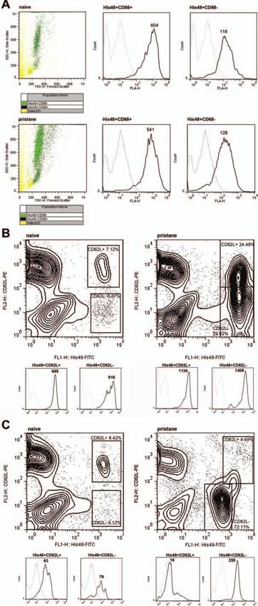
Downloaded from http://ard.bmj.com/ on October 26, 2016 - Published by group.bmj.com

 Basic and translational research

MATERIALS AND METHODS                                                 West Grove, USA) or anti-rat IgM, and bands were visualised by
Patients                                                              enhanced chemiluminescence (GE Healthcare).
Synovial samples were obtained by arthroscopy or open surgery
from an area adjacent to the cartilage–pannus junction from 49
patients with early and longstanding RA and 10 healthy volun-         Measurement of IFNα in serum
teers at the Karolinska University Hospital. Samples were snap-       Serum/plasma levels of IFNα were detected in naïve rats and in
frozen in liquid nitrogen and maintained at −70°C until sec-          rats 6, 12, 19 and 21 days after pristane injection or after cell
tioned. All experiments were approved by the local ethical            transfer, respectively, according to the instructions of the ELISA
committee.                                                            kit (Uscn Life Science, Wuhan, China).

Experimental arthritis
Dark Agouti (DA) rats originating from Harlan were bred and           Flow cytometry
maintained under specific pathogen-free conditions (Felasa II)         Anti-rat granulocytes (His48)-fluorescein isothiocyanate (FITC),
at the Division of Medical Inflammation Research, Karolinska           CD62L, anti-CD45RA (OX33)-phycoerythrin (PE), anti-CD45R
Institute. Experiments were performed on rats frequency-              (His24)-PE, anti-CD62L (HRL1)-biotin, anti-CD161a-biotin,
matched for age and sex and evaluated blindly. PIA was induced        streptavidin-FITC, streptavidin-PE were from BD Biosciences
by a subcutaneous injection of 150 ml pristane (Sigma-Aldrich,        (Franklin Lakes, USA); anti-CD11b/c, anti-CD11c (8A2)-FITC,
St Louis, USA) in the tail.                                           anti-CD68 (ED1)-Alexa Fluor 647, anti-CD68 (ED1)-FITC from
                                                                      Serotec (Oxford, UK); anti-γδ T cell receptor (TCR) (V65)-PE
                                                                      from Biolegend (San Diego, USA). Antibodies to rCRAMP
Peptide/protein extraction
                                                                      (Innovagen), rat β-defensin 1 (Santa Cruz Biotechnology, Santa
Tissues ( paws, spleens, lymph nodes, livers) were homogenised
                                                                      Cruz, USA) and rat α-defensin 4 (Santa Cruz) were labelled with
in 60% acetonitrile/1% trifluoroacetic acid. Extraction from
                                                                      Alexa Fluor 647 using the APEX labelling kit (Invitrogen).
blood was performed in pelleted whole blood (including ery-
                                                                         Antibodies were applied to total blood, to isolated PBMCs/
throcytes) or from PBMCs/neutrophils isolated as previously
                                                                      neutrophils or to synovial fluid (SF) collected from rat ankle
described.18 Debris was removed by centrifugation and the
                                                                      joints by lavage using cooled PBS and a 27G needle (BD
supernatants were lyophilised and dissolved in 0.1% trifluoroa-
                                                                      Microlance). To obtain a sufficient number of cells, SF of three
cetic acid. Proteins and peptides were then enriched by reverse
                                                                      naïve rats had to be pooled in some of the experiments. For
phase chromatography using Oasis cartridges (Waters, Milford,
                                                                      intracellular staining (rCRAMP, defensins, CD68), cells were
USA).
                                                                      permeabilised and fixed with Cytofix/Cytoperm (BD
                                                                      Biosciences) before staining. In all experiments, control anti-
Western blotting                                                      bodies of the respective IgG isotypes were included. Flow cyto-
Denaturing sodium dodecyl sulphate-polyacrylamide gel elec-           metry was performed on a FACS Calibur machine (BD
trophoresis was performed using 2–14% gradient gels                   Biosciences) and was analysed using FloJo software.
(NuPAGE, Invitrogen). The peptide/protein material was then
blotted to polyvinylidene difluoride membranes (Invitrogen).
Membranes were blocked for 1 h with phosphate-buffered                Immunohistochemistry and immunofluorescence microscopy
saline (PBS)/5% milk powder and incubated for 1 h with a              For LL-37 expression analysis in humans, cryostat sections of
polyclonal affinity-purified IgG anti-rCRAMP antibody raised            synovial membranes were fixed for 20 min with 2% formalde-
in rabbit (Innovagen, Lund, Sweden). After washing, the mem-          hyde and dissolved in PBS at 4°C. Synovial LL-37, CD3, CD31,
branes were incubated for 1 h with peroxidase-conjugated anti-        CD66b and CD68 were detected using an affinity-purified rabbit
rabbit secondary antibody (GE Healthcare, Little Chalfont,            polyclonal IgG anti-LL-37 (Innovagen), a mouse IgG1 anti-
UK). Finally, bands were visualised by enhanced chemilumines-         human CD66b (Beckman Coulter, Marseille, France), mouse
cence (GE Healthcare). Densitometric analysis was performed           IgG1 anti-human CD68, CD31 and CD3 (DakoCytomation,
using ImageJ software (Rasband, WS, ImageJ, NIH, Bethesda,            Glostrup, Denmark), respectively. Isotype controls using rabbit
USA). Full-length synthetic rCRAMP (Innovagen) with the               Ig and mouse IgG1 (both from DakoCytomation) were included.
sequence                                                              Visualisation was performed using diaminobenzidine, goat anti-
RFKKISRLAGLLRKGGEKFGEKLRKIGQKIKDFFQKLAPEIEQ-                          rabbit IgG-Alexa Fluor 546 (Invitrogen) and goat anti-mouse
COOH19 was used as positive control.                                  IgG1-Alexa Fluor 488 (Invitrogen).
                                                                         To assess the expression of rCRAMP by immunohistochemis-
Analysis of autoantibodies                                            try, serial paraffin sections of hind paws from naïve and arth-
For detection of autoantibodies to rCRAMP, serum samples              ritic DA rats were deparaffinised and rehydrated. After blocking
were taken from rats at days 0, 6, 14 and 25 after pristane injec-    endogenous peroxidase using hydrogen peroxidase, sections
tion or during the chronic phase of PIA (day 100+) or at day 6        were incubated for 60 min with rabbit anti-rCRAMP and for
and 18 after cell transfer. Synthetic rCRAMP peptide was trans-       30 min with a biotinylated horse anti-rabbit IgG antibody
ferred to polyvinylidene difluoride membranes by western blot.         (Ab), followed by incubation with the Vectastain Elite reagent
Membranes were cut into strips, which were incubated with             (Vector Laboratories), leading to a brown staining of
individual or pooled rat serum samples (diluted 1:50 in PBS/1%        rCRAMP-expressing cells. Finally, slides were counterstained
milk powder) over night at 4°C. As a control for specificity,          with haematoxylin (Merck). As a negative control, rabbit IgG
some of the samples were preincubated with rCRAMP or                  from pooled rabbit serum was used instead of rabbit
β-defensin 1 (Innovagen) and some of the strips were preincu-         anti-rCRAMP.
bated with rabbit anti-rCRAMP or rabbit IgG from pooled rabbit           Apoptosis was analysed in serial paraffin-embedded sections
serum (BioCare Medical). Detection was performed with a               of draining inguinal lymph nodes of DA rats by staining with
peroxidase-conjugated goat anti-rat IgG ( Jackson Immunoresearch,     an antibody to cleaved caspase 3 (Cell Signaling, Beverly, USA).

1240                                                                        Ann Rheum Dis 2013;72:1239–1248. doi:10.1136/annrheumdis-2012-202218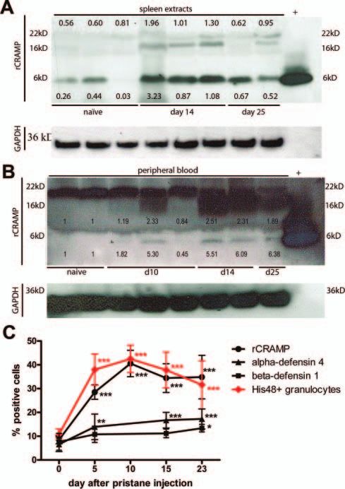
Downloaded from http://ard.bmj.com/ on October 26, 2016 - Published by group.bmj.com
Basic and translational research
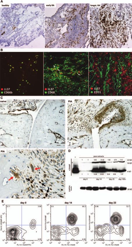
Figure 1 Continued.
Ann Rheum Dis 2013;72:1239–1248. doi:10.1136/annrheumdis-2012-202218 1241Downloaded from http://ard.bmj.com/ on October 26, 2016 - Published by group.bmj.com Basic and translational research Statistical analyses Flow cytometry analysis of SF samples obtained at different Comparisons of positive cells in flow cytometry and histology stages of PIA revealed that rCRAMP is expressed almost exclu- were made by analysis of variance tests. Where significant effects sively in cells recognised by the antibody His48 (denoted His48+ were found, subsequent comparisons were performed using cells) (figure 1E) showing mostly granulocyte but partly also Dunnett’s test or Bonferroni corrections. Comparisons with pre- monocyte morphology in forward-scatter/side-scatter (FSC/SSC) morbid values within groups (percentage positive cells in plots. His48 is commonly used as a phenotypic marker for rat fluorescence-activated cell sorting from peripheral blood, PBMCs granulocytes. Interestingly, cells expressing His48medium cells and neutrophil counts, serum IFNα levels) were tested using stained most strongly positive for rCRAMP, whereas His48high repeated-measures analysis of variance with Dunnett’s test or cells, constituting monocytes and granulocytes (see online supple- Bonferroni correction. Levels of significance are labelled through- mentary Figure S1b), were rCRAMP−. Apart from expression in out the figures as follows: *p
Downloaded from http://ard.bmj.com/ on October 26, 2016 - Published by group.bmj.com

                                                                                                    Basic and translational research

                                                                             of rCRAMP in liver protein extracts during PIA (see online sup-
                                                                             plementary Figure S2c).

                                                                             Phenotypic characterisation of cathelicidin-expressing cells in
                                                                             peripheral blood and SF
                                                                             The vast majority of rCRAMP-positive cells in whole blood
                                                                             was bound by the His48 antibody and exhibited granulocyte
                                                                             morphology on FSC/SSC plots. On His48− cells, rCRAMP was
                                                                             only expressed on very small fractions of B and αβ T cells, but
                                                                             was expressed on higher numbers of γδ T cells after pristane
                                                                             injection (see online supplementary Table SI).
                                                                                The unknown antigen recognised by His48 is believed to be
                                                                             exclusively expressed on rat granulocytes and cells of the eryth-
                                                                             roid lineage during their developmental stage in the bone marrow.
                                                                             Interestingly, rCRAMP cells bound by His48 also expressed low
                                                                             to intermediate levels of CD68. These His48+CD68+ cells exhib-
                                                                             ited both granulocyte and monocyte morphology on FSC/SSC
                                                                             and increased strongly in frequency after pristane priming (see
                                                                             online supplementary Table SI). His48+CD68+ cells showed sub-
                                                                             stantially stronger expression of rCRAMP than His48+CD68−
                                                                             cells in both naïve and pristane-primed animals (figure 3A). CD68
                                                                             is a glycoprotein that binds to low-density lipoprotein and has
                                                                             been used as a marker for monocytes and macrophages.20 A popu-
                                                                             lation of granulocyte-like cells expressing macrophage markers
                                                                             has been described in Toxoplasma-challenged mice21 and in trau-
                                                                             matised rat brain.22 Similarly, expression of these markers appears
                                                                             to be also less exclusive in the peripheral blood of DA rats.
                                                                             However, analysis by fluorescence-activated cell sorting and
                                                                             immunohistochemistry showed that rCRAMP can be expressed
                                                                             in cells with both granulocyte and monocyte morphology (see
                                                                             online supplementary Figure S3).
Figure 2 Expression of rCRAMP in spleen and peripheral blood during             Importantly, rCRAMP expression was higher in activated
pristane-induced arthritis (PIA). (A) Western blot analysis of rCRAMP        than in resting His48+ cells in blood and SF, as determined by
expression in spleens of naïve and pristane-injected Dark Agouti rats.       expression of CD62L (figure 3B,C). CD62L (L-selectin) is a cell
Proteins (extracts of two rats pooled per lane) were separated by            adhesion molecule found on leukocytes that mediates the
sodium dodecyl sulphate-polyacrylamide gel electrophoresis
                                                                             initial rolling on the endothelium.23 On neutrophils, CD62L is
(SDS-PAGE) and probed with polyclonal anti-rCRAMP. Synthetic
rCRAMP was run in parallel as positive control (+),                          downregulated after activation by shedding. Although the
glyceraldehyde-3-phosphate dehydrogenase (GAPDH) served as loading           number of CD62L+ cells was decreased after pristane injection
control. rCRAMP protein bands were analysed by densitometry, and             in both blood and SF, a substantial number of His48+ cells in
expression levels compared with GAPDH bands are depicted. The figure          blood were still CD62L+ and therefore in a resting state,
shows a representative result of three experiments. (B) Western blot         whereas the vast majority of cells at the site of inflammation,
analysis of rCRAMP expression in whole peripheral blood, including           in the SF, were activated (CD62L−).
erythrocytes. Proteins were separated by SDS-PAGE and probed with               Cathelicidins are highly expressed in neutrophil granules, but cell
anti-rCRAMP ( proteins from three rats pooled per lane). The intracellular   surface LL-37 has been described on activated neutrophils of
proform of rCRAMP is expressed in all samples tested, whereas the            patients with SLE.3 We tested flow cytometry staining of rCRAMP
mature rCRAMP peptide was only detected in pristane-injected
                                                                             also on non-permeabilised cells isolated from peripheral blood of
animals. For densitometric analysis ratios between rCRAMP ( proform
or mature peptide, relatively) and GAPDH bands were calculated.              naϊve and pristane-primed rats but could not detect rCRAMP
Upregulation during PIA was compared with bands from naïve rats. The         expression on the surface (see online supplementary Figure S4).
figure shows one of two independent experiments. (C) Time course of
percentages of His48+ cells compared with number of antimicrobial            Low-density neutrophils accumulate in the blood after pristane
peptide-expressing cells in peripheral blood of rats at various time         priming
points after pristane injection. Curves show mean±SD of 6. *p
Downloaded from http://ard.bmj.com/ on October 26, 2016 - Published by group.bmj.com Basic and translational research Figure 3 Continued. 1244 Ann Rheum Dis 2013;72:1239–1248. doi:10.1136/annrheumdis-2012-202218
Downloaded from http://ard.bmj.com/ on October 26, 2016 - Published by group.bmj.com

                                                                                                   Basic and translational research

Figure 4 Pristane triggers accumulation of low-density blood neutrophils and expression of antimicrobial peptides (AMPs). (A) Western blot
analysis of rCRAMP expression in peripheral blood mononuclear cells (PBMCs) isolated by conventional density gradient centrifugation. Lanes show
pools of proteins extracted from four animals each, separated by sodium dodecyl sulphate-polyacrylamide gel electrophoresis and stained with rabbit
anti-rCRAMP. Glyceraldehyde-3-phosphate dehydrogenase (GAPDH) was used as loading control. Densitometric analysis shows slight upregulation of
rCRAMP expression in PBMCs isolated from rats 15 days after pristane injection (B). Percentage of His48+ cells in the PBMC fraction after density
gradient centrifugation performed on blood of rats before and at various time points after pristane injection. Each bar shows mean±SD from five
animals. *p
Downloaded from http://ard.bmj.com/ on October 26, 2016 - Published by group.bmj.com Basic and translational research Figure 5 Autoantibodies to rCRAMP are found together with increased apoptosis and raised serum levels of interferon (IFNα) in pristane-injected rats. Western blot analysis of anti-rCRAMP IgG (A) and IgM (B) autoantibodies in serum samples of rats at various time points after injection of pristane, using blotted rCRAMP peptide. Some lanes were preincubated with a polyclonal rabbit anti-rCRAMP antibody to control for specificity of the observed bands. Each lane represents a strip incubated with a serum sample of one individual rat, except for IgM day 0, where serum samples of five animals have been pooled. (C) Quantification of apoptotic cells in lymph nodes from naive and pristane-injected rats. Bars show mean±SEM from pooled lymph nodes of seven individual animals for each time point. (D) Determination of IFNα serum levels in rats at various time points after injection of pristane or transfer of arthritogenic lymph node cells, respectively. ***p
Downloaded from http://ard.bmj.com/ on October 26, 2016 - Published by group.bmj.com

                                                                                                                      Basic and translational research

with PBMCs during gradient centrifugation has been described.                          REFERENCES
Their presence has been attributed to premature release from                            1.   Ganguly D, Chamilos G, Lande R, et al. Self-RNA-antimicrobial peptide complexes
                                                                                             activate human dendritic cells through TLR7 and TLR8. J Exp Med
the bone marrow owing to recruitment of mature neutrophils
                                                                                             2009;206:1983–94.
to sites of inflammation, or owing to accelerated mature neu-                            2.   Garcia-Romo GS, Caielli S, Vega B, et al. Netting neutrophils are major inducers of type I
trophil turnover.47 48 We detected low buoyant-density neutro-                               IFN production in pediatric systemic lupus erythematosus. Sci Transl Med 2011;3:73ra20.
phils also in pristane-injected rats, suggesting that similar                           3.   Lande R, Ganguly D, Facchinetti V, et al. Neutrophils activate plasmacytoid dendritic
mechanisms might be at work during inflammatory arthritis.                                    cells by releasing self-DNA-peptide complexes in systemic lupus erythematosus. Sci
                                                                                             Transl Med 2011;3:73ra19.
   The direct effects of pristane can be distinguished well from                        4.   Lande R, Gregorio J, Facchinetti V, et al. Plasmacytoid dendritic cells sense
its secondary effects, which are driven by articular inflamma-                                self-DNA coupled with antimicrobial peptide. Nature 2007;449:564–9.
tion, by comparing disease parameters in PIA with those in T                            5.   Nijnik A, Hancock R. Host defence peptides: antimicrobial and immunomodulatory
cell-transferred PIA. Although the transfer is strictly dependent                            activity and potential applications for tackling antibiotic-resistant infections. Emerg
                                                                                             Health Threats J 2009;2:e1.
on pristane priming of the donor cells, the acceptor rats
                                                                                        6.   De Y, Chen Q, Schmidt AP, et al. LL-37, the neutrophil granule- and epithelial
develop an arthritis that is very similar to PIA without having                              cell-derived cathelicidin, utilizes formyl peptide receptor-like 1 (FPRL1) as a receptor
been in direct contact with the oil. We found upregulation of                                to chemoattract human peripheral blood neutrophils, monocytes and T cells. J Exp
rCRAMP expression and accumulation of neutrophils in both                                    Med 2000;192:1069–74.
PIA and the PIA transfer model, but with different kinetics (see                        7.   Mookherjee N, Brown KL, Bowdish DM, et al. Modulation of the TLR-mediated
                                                                                             inflammatory response by the endogenous human host defense peptide LL-37.
online supplementary Figure S7). In contrast, induction of                                   J Immunol 2006;176:2455–64.
serum IFNα and development of autoantibodies are restricted                             8.   Morizane S, Yamasaki K, Muhleisen B, et al. Cathelicidin antimicrobial peptide
to PIA, indicating that they are caused by direct effects of pris-                           LL-37 in psoriasis enables keratinocyte reactivity against TLR9 ligands. J Invest
tane operating independently of T cell activation—for example,                               Dermatol 2012;132:135–43.
                                                                                        9.   Baechler EC, Batliwalla FM, Karypis G, et al. Interferon-inducible gene expression
related to a B cell response against stressed and dying cells.
                                                                                             signature in peripheral blood cells of patients with severe lupus. Proc Natl Acad Sci
   In summary, we performed a comprehensive expression analysis                              U S A 2003;100:2610–15.
in joints, lymphoid organs and blood and detected strong overex-                       10.   Bennett L, Palucka AK, Arce E, et al. Interferon and granulopoiesis signatures in
pression of cathelicidins during arthritis. In the joint, cathelicidins                      systemic lupus erythematosus blood. J Exp Med 2003;197:711–23.
were mainly expressed in cells of the inflammatory pannus, mostly                       11.   van der Pouw Kraan TC, Wijbrandts CA, van Baarsen LG, et al. Rheumatoid
                                                                                             arthritis subtypes identified by genomic profiling of peripheral blood cells:
those of monocytic, fibroblastoid and granulocytic morphology. A                              assignment of a type I interferon signature in a subpopulation of patients. Ann
phenotypic characterisation of rCRAMP-expressing cells in periph-                            Rheum Dis 2007;66:1008–14.
eral blood and SF showed that rCRAMP expression is highest in                          12.   Frohm M, Agerberth B, Ahangari G, et al. The expression of the gene coding for
activated CD62L− cells coexpressing granulocyte and monocyte                                 the antibacterial peptide LL-37 is induced in human keratinocytes during
                                                                                             inflammatory disorders. J Biol Chem 1997;272:15258–63.
markers and exhibiting granulocyte or monocyte morphology.
                                                                                       13.   Sun CL, Zhang FZ, Li P, et al. LL-37 expression in the skin in systemic lupus
Furthermore, IgG and IgM autoantibodies to rCRAMP were trig-                                 erythematosus. Lupus 2011;20:904–11.
gered by pristane and coincided with increased levels of cell death                    14.   Paulsen F, Pufe T, Conradi L, et al. Antimicrobial peptides are expressed and
and increased serum levels of IFNα.                                                          produced in healthy and inflamed human synovial membranes. J Pathol
   The possibility that global and local overexpression of AMPs                              2002;198:369–77.
                                                                                       15.   Matsumoto T, Kaneko T, Seto M, et al. The membrane proteinase 3 expression on
directly influences development and/or maintenance of arthritis                               neutrophils was downregulated after treatment with infliximab in patients with
will be examined in future studies. Local effects might be eli-                              rheumatoid arthritis. Clin Appl Thromb Hemost 2008;14:186–92.
cited by excessive release of mature AMPs that exert immune-                           16.   Vingsbo C, Sahlstrand P, Brun JG, et al. Pristane-induced arthritis in rats: a new
modulating and chemotactic effects. Formation of complexes                                   model for rheumatoid arthritis with a chronic disease course influenced by both
                                                                                             major histocompatibility complex and non-major histocompatibility complex genes.
between cationic AMPs and locally released nucleic acids may
                                                                                             Am J Pathol 1996;149:1675–83.
alert the immune system by the activation of TLR7 and/or                               17.   Hoffmann MH, Tuncel J, Skriner K, et al. The rheumatoid arthritis-associated
TLR9 expressed in the arthritic joints. Systemic effects might                               autoantigen hnRNP-A2 (RA33) is a major stimulator of autoimmunity in rats with
be fuelled by the development of autoantibodies and immune                                   pristane-induced arthritis. J Immunol 2007;179:7568–76.
complexes in a way that has been described in SLE,2 3 or by                            18.   Russo-Carbolante EMS, Azzolini AECS, Plizello ACM, et al. Comparative study of
                                                                                             four isolation procedures to obtain rat neutrophils. Comp Clin Pathol
influencing T cell polarisation.                                                              2002;11:71–6.
                                                                                       19.   Termen S, Tollin M, Olsson B, et al. Phylogeny, processing and expression of the rat
Acknowledgements The authors thank Monica Lindh for technical help in
                                                                                             cathelicidin rCRAMP: a model for innate antimicrobial peptides. Cell Mol Life Sci
establishing the western blot analysis and peptide extraction protocols, Carl-Walter         2003;60:536–49.
Steiner for help with fluorescence activated cell sorting of rCRAMP-positive cells,     20.   Holness CL, Simmons DL. Molecular cloning of CD68, a human macrophage
Marianne Engström for technical help with immunohistochemistry of human samples
                                                                                             marker related to lysosomal glycoproteins. Blood 1993;81:1607–13.
and Anita Fischer, Ulrika Norin and Jonatan Tuncel for providing rat samples.
                                                                                       21.   Mordue DG, Sibley LD. A novel population of Gr-1+-activated macrophages
Contributors MHH, MH, AIC, BA and RH were involved in conception of the study.               induced during acute toxoplasmosis. J Leukoc Biol 2003;74:1015–25.
MHH, LB, PN, HB and BN were involved in performing experiments. All authors            22.   Matsumoto H, Kumon Y, Watanabe H, et al. Antibodies to CD11b, CD68 and lectin
approved the final version to be published. BA and RH contributed equally to this             label neutrophils rather than microglia in traumatic and ischemic brain lesions.
work.                                                                                        J Neurosci Res 2007;85:994–1009.
                                                                                       23.   Gearing AJ, Newman W. Circulating adhesion molecules in disease. Immunol Today
Funding The research was funded by the Austrian Science Fund FWF (roject
                                                                                             1993;14:506–12.
J3102-B13), the Swedish Research Council, the Swedish Strategic Foundation (SSF),
                                                                                       24.   Calvani N, Caricchio R, Tucci M, et al. Induction of apoptosis by the hydrocarbon
the Lars Hiertas minne foundation, the EU project Masterswitch
                                                                                             oil pristane: implications for pristane-induced lupus. J Immunol 2005;175:4777–82.
(HEALTH-F2-2008-223404) and by the IMI program BTCure.
                                                                                       25.   Herman S, Kny A, Schorn C, et al. Cell death and cytokine production induced by
Competing interests None.                                                                    autoimmunogenic hydrocarbon oils. Autoimmunity 2012; Epub ahead of print.
Ethics approval Local ethical committee, Sweden.                                       26.   Kleinau S, Dencker L, Klareskog L. Oil-induced arthritis in DA rats: tissue
                                                                                             distribution of arthritogenic 14C-labelled hexadecane. Int J Immunopharmacol
Provenance and peer review Not commissioned; externally peer reviewed.                       1995;17:393–401.
Correction notice This article has been corrected since it was published Online        27.   Fuchs TA, Abed U, Goosmann C, et al. Novel cell death program leads to
First. Correspondence and author affiliations have been corrected. Occurrences of             neutrophil extracellular traps. J Cell Biol 2007;176:231–41.
“His48 cells”, “CD62 cells” and “pristine” have been changed to “His48+ cells”,        28.   Cederlund A, Gudmundsson GH, Agerberth B. Antimicrobial peptides important in
“CD62+ cells” and “pristane” respectively.                                                   innate immunity. FEBS J 2011;278:3942–51.

Ann Rheum Dis 2013;72:1239–1248. doi:10.1136/annrheumdis-2012-202218                                                                                                             1247
Downloaded from http://ard.bmj.com/ on October 26, 2016 - Published by group.bmj.com
Basic and translational research
29. Yount NY, Wang MS, Yuan J, et al. Rat neutrophil defensins. Precursor structures serum levels of anti-type II collagen auto-antibodies. Scand J Immunol
and expression during neutrophilic myelopoiesis. J Immunol 1995;155:4476–84. 1990;31:147–57.
30. Jia HP, Mills JN, Barahmand-Pour F, et al. Molecular cloning and characterization of rat 40. Holmdahl R, Jonsson R, Larsson P, et al. Early appearance of activated CD4+ T
genes encoding homologues of human beta-defensins. Infect Immun 1999;67:4827–33. lymphocytes and class II antigen-expressing cells in joints of DBA/1 mice
31. Eisenhauer PB, Harwig SS, Szklarek D, et al. Purification and antimicrobial immunized with type II collagen. Lab Invest 1988;58:53–60.
properties of three defensins from rat neutrophils. Infect Immun 1989;57:2021–7. 41. Wipke BT, Allen PM. Essential role of neutrophils in the initiation and progression of
32. Sorensen OE, Follin P, Johnsen AH, et al. Human cathelicidin, hCAP-18, is a murine model of rheumatoid arthritis. J Immunol 2001;167:1601–8.
processed to the antimicrobial peptide LL-37 by extracellular cleavage with 42. Satoh M, Reeves WH. Induction of lupus-associated autoantibodies in BALB/c mice
proteinase 3. Blood 2001;97:3951–9. by intraperitoneal injection of pristane. J Exp Med 1994;180:2341–6.
33. Zanetti M, Gennaro R, Romeo D. Cathelicidins: a novel protein family with a common 43. Wooley PH, Seibold JR, Whalen JD, et al. Pristane-induced arthritis. The
proregion and a variable C-terminal antimicrobial domain. FEBS Lett 1995;374:1–5. immunologic and genetic features of an experimental murine model of autoimmune
34. Turner J, Cho Y, Dinh NN, et al. Activities of LL-37, a cathelin-associated disease. Arthritis Rheum 1989;32:1022–30.
antimicrobial peptide of human neutrophils. Antimicrob Agents Chemother 44. Dahlgren J, Takhar H, Anderson-Mahoney P. Cluster of systemic lupus
1998;42:2206–14. erythematosus (SLE) associated with an oil field waste site: a cross sectional study.
35. Johansson J, Gudmundsson GH, Rottenberg ME, et al. Conformation-dependent Environ Health 2007;6:8.
antibacterial activity of the naturally occurring human peptide LL-37. J Biol Chem 45. Sverdrup B, Kallberg H, Bengtsson C, et al. Association between occupational
1998;273:3718–24. exposure to mineral oil and rheumatoid arthritis: results from the Swedish EIRA
36. Mohr W, Kohler G, Wessinghage D. Polymorphonuclear granulocytes in rheumatic case-control study. Arthritis Res Ther 2005;7:R1296–303.
tissue destruction. II. Demonstration of PMNs in rheumatoid nodules by electron 46. Holmberg J, Tuncel J, Yamada H, et al. Pristane, a non-antigenic adjuvant, induces
microscopy. Rheumatol Int 1981;1:21–8. MHC class II-restricted, arthritogenic T cells in the rat. J Immunol
37. Wright HL, Moots RJ, Bucknall RC, et al. Neutrophil function in inflammation and 2006;176:1172–9.
inflammatory diseases. Rheumatology (Oxford) 2010;49:1618–31. 47. Doreau A, Belot A, Bastid J, et al. Interleukin 17 acts in synergy with B cell–
38. Chou RC, Kim ND, Sadik CD, et al. Lipid-cytokine-chemokine cascade drives activating factor to influence B cell biology and the pathophysiology of systemic
neutrophil recruitment in a murine model of inflammatory arthritis. Immunity lupus erythematosus. Nat Immunol 2009;10:778–85.
2010;33:266–78. 48. Lit LC, Wong CK, Tam LS, et al. Raised plasma concentration and ex vivo
39. Holmdahl R, Jansson L, Larsson A, et al. Arthritis in DBA/1 mice induced with production of inflammatory chemokines in patients with systemic lupus
passively transferred type II collagen immune serum. Immunohistopathology and erythematosus. Ann Rheum Dis 2006;65:209–15.
1248 Ann Rheum Dis 2013;72:1239–1248. doi:10.1136/annrheumdis-2012-202218Downloaded from http://ard.bmj.com/ on October 26, 2016 - Published by group.bmj.com
The cathelicidins LL-37 and rCRAMP are
associated with pathogenic events of arthritis
in humans and rats
Markus H Hoffmann, Heiko Bruns, Liselotte Bäckdahl, Petra Neregård,
Birgit Niederreiter, Martin Herrmann, Anca Irinel Catrina, Birgitta
Agerberth and Rikard Holmdahl
Ann Rheum Dis 2013 72: 1239-1248 originally published online
November 21, 2012
doi: 10.1136/annrheumdis-2012-202218
Updated information and services can be found at:
http://ard.bmj.com/content/72/7/1239
These include:
Supplementary Supplementary material can be found at:
Material http://ard.bmj.com/content/suppl/2012/11/20/annrheumdis-2012-2022
18.DC1.html
References This article cites 47 articles, 27 of which you can access for free at:
http://ard.bmj.com/content/72/7/1239#BIBL
Email alerting Receive free email alerts when new articles cite this article. Sign up in the
service box at the top right corner of the online article.
Topic Articles on similar topics can be found in the following collections
Collections Degenerative joint disease (4621)
Musculoskeletal syndromes (4930)
Immunology (including allergy) (5117)
Connective tissue disease (4234)
Systemic lupus erythematosus (566)
Rheumatoid arthritis (3245)
Inflammation (1244)
Notes
To request permissions go to:
http://group.bmj.com/group/rights-licensing/permissions
To order reprints go to:
http://journals.bmj.com/cgi/reprintform
To subscribe to BMJ go to:
http://group.bmj.com/subscribe/You can also read