Extracellular Vesicles and Immunomodulation in Mosquitoes and Ticks
←
→
Page content transcription
If your browser does not render page correctly, please read the page content below
Entry
Extracellular Vesicles and Immunomodulation in Mosquitoes
and Ticks
Brenda Leal-Galvan † , Charluz Arocho Rosario †                     and Adela Oliva Chávez *

                                          Department of Entomology, Texas A&M University, College Station, TX 88743, USA;
                                          brenda.leal@tamu.edu (B.L.-G.); marioli@tamu.edu (C.A.R.)
                                          * Correspondence: aolivachavez@tamu.edu; Tel.: +1-979-845-1946
                                          † These authors contributed equally to this work.

                                          Definition: Extracellular vesicles are small blebs that are secreted by cells, which are lipid-rich
                                          and contain proteomic and genomic material (including small RNAs, mRNA, and plasmid DNA).
                                          These materials are delivered into recipient cells leading to a phenotypic change. Recent studies
                                          have demonstrated the secretion of extracellular vesicles by mosquito and tick cells, as well as tick
                                          salivary glands. Further, these studies suggest vesicles play a role in the transmission of vector-borne
                                          pathogens, including viruses and bacteria, and are involved in the manipulation of wound healing
                                          and immune responses. Both of these processes are key in the host response to hematophagous
                                          arthropods’ feeding. The role of mosquito and tick EVs in the modulation of immune responses and
                                          pathogen transmission is discussed in this entry.

                                          Keywords: hematophagy; immune modulation; pathogen transmission; extracellular vesicles;
                                          arthropods

Citation: Leal-Galvan, B.; Arocho
Rosario, C.; Oliva Chávez, A.             1. Introduction
Extracellular Vesicles and
                                               Arthropods are important vectors of pathogens that can affect humans, wildlife,
Immunomodulation in Mosquitoes
                                          and domestic animals [1]. The World Health Organization (WHO) has estimated that
and Ticks. Encyclopedia 2022, 2,
                                          diseases caused by vector-borne pathogens result in more than 700,000 human deaths
873–881. https://doi.org/10.3390/
                                          yearly [2]. In the US, damages to livestock production due to arthropod feeding account for
encyclopedia2020057
                                          approximately USD 100 billion in losses annually [3]. Mosquitoes and ticks are particularly
Academic Editors: András Fodor and        impactful from a public and animal health perspective and are the focus of this entry.
Raffaele Barretta                              Mosquitoes can transmit flaviviruses, protozoans, bacteria, and nematodes. With
Received: 29 January 2022
                                          over 3000 different species, mosquitoes are a worldwide public health concern [4–6]. An
Accepted: 20 April 2022
                                          anthropophilic adult female mosquito will need multiple bloodmeals to enhance their
Published: 24 April 2022
                                          fitness, obtain the energy to search for a mate, and to initiate vitellogenesis [4,7–9]. Hard
                                          ticks, on the other hand, are obligatory blood feeders that can feed on a host for days
Publisher’s Note: MDPI stays neutral
                                          to weeks at a time [10,11]. Ticks are considered second to mosquitoes in their public
with regard to jurisdictional claims in
                                          health relevance. It is estimated that tick bites are responsible for the transmission of over
published maps and institutional affil-
                                          100,000 cases of tick-borne diseases in humans throughout the world [12,13]. A recent study
iations.
                                          showed that 140,281 insured patients were diagnosed with Lyme disease in the US alone
                                          from 2010 to 2018, with incidences as high as 87.9/100,000 enrollees [14] and the Center for
                                          Disease Control and Prevention (CDC) now estimates that 476,000 Americans are affected
Copyright: © 2022 by the authors.
                                          by this disease [15]. Given that most of the work on vector-derived extracellular vesicles
Licensee MDPI, Basel, Switzerland.        (EVs) has been carried out in these two vector species, the authors will limit this entry to
This article is an open access article    what is known about mosquito- and tick-derived EVs.
distributed under the terms and                Due to their need for a bloodmeal, either for survival or reproduction, arthropods have
conditions of the Creative Commons        evolved intricate mechanisms that allow them to counteract immune and inflammatory
Attribution (CC BY) license (https://     responses by their host. For example, during feeding, arthropods can release EVs via
creativecommons.org/licenses/by/          their saliva [16,17]. EVs are double-layer vesicles that are secreted by all cells and are
4.0/).                                    essential for cell-to-cell communication [18–22]. Physiological changes in the cell can

Encyclopedia 2022, 2, 873–881. https://doi.org/10.3390/encyclopedia2020057                            https://www.mdpi.com/journal/encyclopedia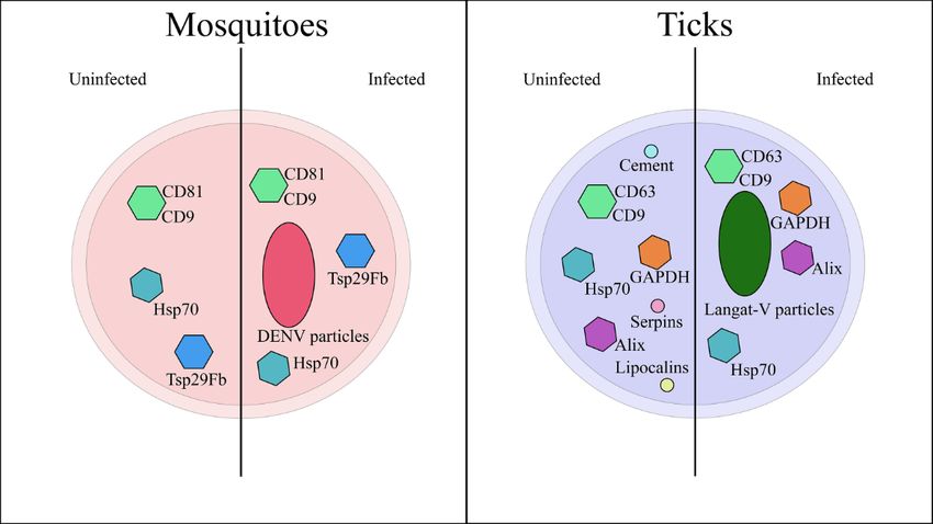
Encyclopedia 2022, 2                                                                                              874

                       lead to an increase in vesicle secretion or cause changes in the cargo packed within the
                       extracellular vesicles secreted by these cells. For example, pathogen-infected cells secrete
                       EVs that carry infectious cargo, such as viral RNA. These vesicles can enhance pathogen
                       transmission and replication [23–25]. EVs originating from infected vector cells can serve
                       as the source of infection for host cells in vitro [24,26–28]. In other cases, EVs produced by
                       virus-infected cells can inhibit pathogen transmission [29,30]. Nevertheless, the relevance
                       of these phenomena during in vivo pathogen transmission is undetermined. This entry
                       focuses on the function of EVs during arthropod feeding and their potential contribution in
                       pathogen transmission.

                       2. Hematophagy in Arthropods
                            Blood feeding behavior appeared in arthropods on six independent occasions through-
                       out evolution [31]. In the case of insects, hematophagy emerged on several separate
                       occasions driven by two potential scenarios: (1) The association of insects within the nest of
                       different vertebrate animals due to their attraction to the protected environment provided,
                       which increased the contact of insects with the vertebrate host. The constant interaction
                       with vertebrate animals may have led to behavioral and structural adaptations that al-
                       lowed these insects to access a higher nutritional substrate, such as blood. (2) Ancestral
                       insect lineages containing piercing mouthparts may have accidentally probed vertebrate
                       animals [32]. In the case of ticks, fluid feeding is characteristic of the Parasitiformes, the
                       major lineage that ticks belong to. Thus, it is probable that blood feeding arose as a result
                       of this adaptation to feed on fluids [33]. For arthropods to successfully feed on blood, they
                       had to acquire several molecular adaptations that allow them to overcome host immune
                       and wound healing responses, permit the detoxification of harmful molecules found within
                       blood, allow them to stop blood coagulation, and supplement nutritional factors that are
                       missing in blood. Understanding the adaptation of arthropods to hematophagy is impor-
                       tant due to the underlying connections with pathogen transmission [34]. As discussed in
                       the introduction, both mosquitos and ticks secrete salivary factors that dampen immune
                       responses, reduce coagulation, increase blood flow, alter the skin microbiome, and delay
                       wound healing. Herein, the authors will focus on the effect that certain salivary factors
                       have at the bite site.

                       2.1. Immune Modulation
                             Both mosquitos and ticks can diminish innate and adaptive immune responses. In the
                       case of the innate immune response, salivary components target immune cell migration,
                       the secretion of cytokines and chemokines, and the complement system. The complement
                       system comprises three complexes of plasma proteins and receptors that are part of the
                       innate immune system and assist in skin homeostasis [35]. The complement system is
                       initiated through the activation of the alternative, the lectin, or the classical pathway. Some
                       of the key components of the complement system are the proteins C3 and C5, which
                       form part of the alternative and lectin pathways. The classical pathway is initiated by the
                       interaction of C1 and an antibody bound to its specific antigen. Some mosquitos, such as
                       Anopheles (Nyssorhynchus) aquasalis, can inhibit the alternative pathway by blocking C3b
                       deposition [36]. Similarly, saliva from Rhipicephalus (Boophilus) microplus ticks can hinder
                       the activation of the classical and alternative pathways by affecting the cleavage of C4 by
                       C1b and preventing the action of the C3 convertase [37], suggesting that the control of the
                       complement system is a conserved mechanism within hematophagous arthropods.
                             For cell migration, mosquito and tick saliva have dissimilar effects. In mosquitos,
                       salivary gland extracts (SGE) from Aedes aegypti, the vector of dengue, increased the
                       permeability of endothelial cell monolayers in vitro and the leakage of the vasculature
                       of mice ear in vivo [38]. This augmented vascular leakage may explain the enhanced
                       migration of neutrophils, monocytes, and dendritic cells to the skin during inoculation of
                       dengue virus (DENV) and the Semliki Forest virus (SFV), a close relative of the chikungunya
                       virus [38,39]. In both cases, either the mosquito bite or the inoculation of SGE boosted the
Encyclopedia 2022, 2 875
severity of the viral infection in the skin. Although an increase in innate immune cells at
the bite site is also observed during tick feeding [16,40], tick saliva decreases the migration
of dendritic cells in vivo [41]. This effect on dendritic cell migration may be the result of the
decreased expression of adhesion proteins in subcutaneous tissue, as shown in vitro [42],
the reduced expression of chemokine receptors CCR5 and CCR7 and the secretion of TNFα
and IL12p40. Tick saliva also inhibited the differentiation of dendritic cells [43,44]. These
effects on differentiation and cytokine expression could be due to the induction of IL-10
secretion and the stimulation of the TLR2 receptor [45]. Thus, arthropods share some
common mechanism to dampen innate immune responses. However, depending on the
vector species, evident differences exist in how they affect signaling and cellular immunity.
These differences are probably the result of the unique selective pressures that mosquitos
and ticks face. The interaction of salivary compounds and immune modulation has been
previously described in detail in [46,47].
2.2. Antihemostatic and Wound Healing
Tissue repair, or wound healing response, takes place following an injury or abrasion
of the tissue. This process involves multiple stages starting from hemostasis, inflammation,
tissue proliferation, and remodeling [48]. Hemostasis includes the constriction of the
blood vessels to diminish blood lost, coagulation, and platelet aggregation. Blood sucking
arthropods have developed or acquired salivary molecules that restrict the hemostatic
process. One interesting example is the gain of a vertebrate vasodilator by soft ticks
of the genus Ornithodoros through horizontal gene transfer (HGT) [49]. Three isoforms
of an adrenomedullin (ADM)-like protein are found within the genome of Ornithodoros
moubata, O. parkeri, and O. coriaceus. Interestingly, the gene encoding this protein is missing
from other soft ticks and invertebrates. The closest homologs are present in amphibians
and fish, suggesting that this gene was acquired by an ancestor Ornithodoros probably
during feeding on ancient reptiles. Comparatively, SGE from two hard ticks, Rhipicephalus
appendiculatus and Dermacentor reticulatus, contain compounds that induce both vasodilation
and vasoconstriction, depending on the feeding time. During the rapid feeding phase,
male ticks that fed for 6 days showed vasodilatory properties, whereas females and males
that fed during the early and late stages showed vasoconstricting properties [50]. Thus,
this indicates that the vasoacitve properties of tick saliva may change depending on sex
and feeding phase. For mosquitoes, only vasodilatory compounds have been reported.
A. aegypti saliva contains proteomic compounds (susceptible to trypsin digestion) with
vasodilatory properties [51,52]. These proteins were identified as tachykinins, which have
also been identified in Aedes triseriatus [53]. Another vasodilator found within the saliva of
mosquitoes is catechol oxidase [53]. Other antihemostatic properties of hematophagous
arthropods are discussed in the review by D. Champagne [54].
Following the inflammatory process, the tissue proliferation and remodeling process
involves the restructuring of the extracellular matrix (ECM) of keratinocytes, fibroblast,
epithelium, and endothelium [48]. The growth and restructuring of the tissue require the
production and secretion of growth factors and the rearrangement of the cytoskeleton.
Tick saliva contains growth factor binding proteins that interact with transforming growth
factor beta (TGF-β1), platelet-derived growth factor (PDGF), fibroblast growth factor (FGF-
2), and hepatocyte growth factor (HGF), affecting cell proliferation and altering actin
polymerization during in vitro assays [55]. Another group of tick salivary proteins that act
by decreasing angiogenesis are serine protease inhibitors (Serpins). For a complete review
of tick salivary serpins, please refer to [56]. Proteins and factors that act on the wound
healing response during mosquito feeding have not been experimentally identified.
2.3. Control of Commensal Bacteria
Commensal microbes colonize the skin of humans and animals early in life. These
microbes form part of the skin microbiome, which interacts with immune and skin cells
to prime and shape the immune system [57]. Studies indicate that ticks can alter theEncyclopedia 2022, 2 876
skin microbiome by potentially killing Staphylococcus epidermidis through the degradation
of peptidoglycan by the salivary protein DaeIs [58]. This protein is secreted during the
bloodmeal to control the presence of bacteria that may be damaging to the tick midgut’s
integrity. Thus, this provides a fitness advantage to the tick.
3. Extracellular Vesicles in Mosquitoes
Mosquitoes are top vectors of many vector-borne pathogens that cause several human
diseases, such as malaria, dengue, West Nile, and Zika [5,59]. They feed by piercing through
the skin of their host using needle-like mouthparts (Figure 1A). Mosquito C6/36 cells se-
crete EVs in vitro [26,28]. Although the exact function of these vesicles during feeding
has not been determined, recent studies indicate that mosquito vesicles carry viral cargo,
which facilitates spread and potentially transmission. EVs that originated from Zika virus
(ZIKV) or DENV-infected cells promote viral spread by carrying viral RNA to receiving
naïve cells [26,28,60–62]. Along with the viral RNA, these vesicles also contain altered
host-derived cargo that could enhance pathogen transmission and replication. For example,
EVs from DENV3-infected monocyte-derived dendritic cells (Mo-DCs) contained mR-
NAs encoding the transcripts for Interferon-Induced Transmembrane Protein 1 (IFITM1),
Interferon-Induced Protein with Tetratricopeptide Repeats 1 (IFIT1), and DExD/H-Box
Helicase 58 (DDX58) involved in DENV infection [26,28]. These vesicles also transported
microRNA-4327, which is used as a marker for severe dengue, and the tetraspanins (CD9,
CD81, CD63) and Tsp29fb [61]. The modified vesicles may be involved in the transloca-
tion of DENV from DENV-infected C6/36 [26–28,61–63]. Nevertheless, EVs also inhibit
viral replication. For example, EVs secreted by DENV-infected C6/36 cells restricted viral
membrane fusion, inhibiting viral replication, and spread [28,30]. Thus, in the context of
dengue infection, EVs can facilitate or dampen viral spread. Whether these conflicting
Encyclopedia 2022, 2, FOR PEER REVIEW
effects are due to different vesicle populations being secreted by specific cells remains to5
be determined.
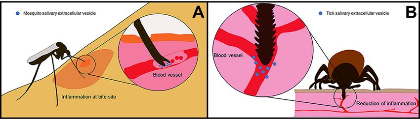
Figure 1. Extracellular vesicle secretion during arthropod feeding on vertebrate host. (A)
Figure 1. Extracellular vesicle secretion during arthropod feeding on vertebrate host. (A) Mosquitoes
Mosquitoes pierce the skin of their host using modified mouthparts. Their labella, labrum, and
pierce the skin of their host using modified mouthparts. Their labella, labrum, and labium form
labium form needle-like structures that can pierce directly to the blood vessels where they feed.
needle-like
Once insidestructures
the blood that can they
vessels, pierce directly
release to the blood vessels
saliva-containing wherepotentially
EVs, which they feed. interact
Once inside
with
the blood
blood cells.vessels, they
(B) Ticks, onrelease
the othersaliva-containing EVs, which
hand, use scissor-like potentially
structures interact with
called chelicerae blood
to cut cells.
through
(B) Ticks,
the on the
skin. They other hand,
introduce theiruse scissor-likewhich
hypostome, structures called
harbors chelicerae
several to cut
hook-like through the
structures thatskin.
allow They
the
introduce
ticks their hypostome,
to anchor themselves which
in the harbors several
skin while theyhook-like structures
feed. Another that allowthat
mechanism theallows
ticks toticks
anchorto
anchor
themselvesfirmlyin to
thethe host
skin skinthey
while is the secretion
feed. Anotherof cement proteins.
mechanism These cement
that allows ticks to proteins polymerize
anchor firmly to the
and
hostinteract
skin is thewith other proteins
secretion to form
of cement a cone-like
proteins. Thesestructure that presents
cement proteins antimicrobial
polymerize properties,
and interact with
facilitates tick attachment, seals the feeding lesion, and assists in pathogen transmission.
other proteins to form a cone-like structure that presents antimicrobial properties, facilitates Through
tick
the introduction of their hypostome, they damage blood vessels and cells in the dermis and
attachment, seals the feeding lesion, and assists in pathogen transmission. Through the introduction
epidermis. They secrete their saliva, containing EVs, into the pool of blood that forms at the bite site.
of their hypostome, they damage blood vessels and cells in the dermis and epidermis. They secrete
their saliva, containing EVs, into the pool of blood that forms at the bite site.
4. Extracellular Vesicles in Ticks
Ticks are hematophagous arthropods that, during feeding, secrete salivary
substances with anticoagulatory, anti-inflammatory, and vasodilatory functions [64].
Ticks cause significant damage to the skin of humans and animals (Figure 1B). Thus, these
substances function to circumvent the host’s immunological response and indirectly
facilitate the transmission of pathogens [65], including protozoan, bacterial, and viral
agents. Several studies have demonstrated the secretion of EVs within the saliva of IxodesEncyclopedia 2022, 2 877
During Zika virus infection, EVs can serve as the inoculum of infection [61]. EVs
secreted by infected C6/36 cells carrying viral E protein (ZIKV E-protein) are infectious
for human peripheral blood monocytes (THP-1) cells and endothelial vascular (HMEC-1)
cells [26,62]. Similarly, EVs secreted by ZIKV-infected C6/36 mosquito cells can activate a
pro-inflammatory response and induce an immunophenotype in human monocyte cells,
comparable to that observed during ZIKV infection [62]. When compared to mock cells, the
stimulation of healthy endothelial vascular cells with ZIKV C6/36 EVs also favored pro-
coagulant state and pro-inflammatory responses [26,62]. Despite the observed involvement
of mosquito derived vesicles in the spread of both DENV and ZIKV in vitro, whether
mosquito salivary EVs contain viral particles remains to be confirmed. Similarly, how
mosquito EVs may affect host immune responses at the bite site needs to be explored.
4. Extracellular Vesicles in Ticks
Ticks are hematophagous arthropods that, during feeding, secrete salivary substances
with anticoagulatory, anti-inflammatory, and vasodilatory functions [64]. Ticks cause sig-
nificant damage to the skin of humans and animals (Figure 1B). Thus, these substances
function to circumvent the host’s immunological response and indirectly facilitate the trans-
mission of pathogens [65], including protozoan, bacterial, and viral agents. Several studies
have demonstrated the secretion of EVs within the saliva of Ixodes scapularis, Amblyomma
maculatum, and Haemaphysalis longicornis ticks [16,17,66].
EVs transfer intracellular information such as proteins and miRNAs between cells or
tissues and mediate changes in cellular activity and pathways in the recipient cell [67,68].
The proteomic analysis of tick salivary EVs has identified several known effector pro-
teins with immunomodulatory properties, including lipocalins, serine protease inhibitors,
and cement [16,66], and several host proteins including histones, metabolic proteins, and
immunoglobulins [66]. Various known exosomal markers, such as heat shock protein
70 (HSP70), the tetraspanin CD63, ALG-2-Interacting Protein X (ALIX), and Tumor Sus-
ceptibility 101 (TSG101), have been detected in these exosomes through proteomics and
Western blot analysis [16,17,66].
Tick salivary EVs have also been shown to contain miRNAs, including novel miRNAS
and previously described small RNAs [69]. Several miRNAs showed an upregulation
during feeding, indicating a potential role in the tick’s response to host immunity. Salivary
exosomes carrying HSP70 and miRNAs potentially aid in fibrinogen degradation and
modulate host gene expression at the vector–host interface [70]. The vesicle’s cargo may
also regulate key homeostatic responses in the host [71]. In fact, Zhou et al., 2020 [17] made
the first report implicating tick salivary gland derived exosomes in the modulation of the
wound healing responses at the tick bite site [17]. Later, independent studies demonstrated
that tick EVs had a localized action on skin immunity by facilitating feeding [16]. EVs
may allow ticks to bypass the immune barriers present in the skin successfully to complete
their bloodmeal.
Due to their involvement in the manipulation of host immune responses, EVs are
suspected to aid in the transmission and establishment of tick-borne pathogens. Oliva
Chavez et al. [16] showed that the inoculation of the intracellular bacterium Anaplasma
phagocytophilum along with tick salivary EVs increased the frequency of bacterial infection
in the skin of naïve mice. Further, in 2018, Zhou et al. [28] demonstrated that cell lines
derived from I. scapularis ticks are capable of secreting EVs. Interestingly, the vesicles from
Langat virus-infected tick cells were shown to carry viral proteins and RNA and aid in the
viral spread to invertebrate and vertebrate host cells. Nevertheless, what exact function
EVs play during pathogen transmission remains to be defined.
5. Conclusions and Prospects
Blood feeding or hematophagy has evolved independently in around ~15,000 species
of arthropods [11,31,72]. To facilitate blood feeding, these arthropods have gained mech-
anisms to counteract blood coagulation [31], the presence of endosymbionts within theirEVs play during pathogen transmission remains to be defined.
5. Conclusions and Prospects
Blood feeding or hematophagy has evolved independently in around ~15,000 species
Encyclopedia 2022, 2 of arthropods [11,31,72]. To facilitate blood feeding, these arthropods have gained 878
mechanisms to counteract blood coagulation [31], the presence of endosymbionts within
their microbiome for the production of B vitamins [73], the ability to prevent damage by
heme molecules
microbiome for the resulting
productionfrom of the digestion
B vitamins ofthe
[73], hemoglobin [74], and
ability to prevent to escape
damage by hemehost
immune responses
molecules resulting [75,76].
from theAs a side effect
digestion of the immunomodulation
of hemoglobin [74], and to escape of host
host immune
immune
responses,[75,76].
responses arthropod saliva
As a side enhances
effect the establishment of
of the immunomodulation andhosttransmission
immune responses,of the
pathogens saliva
arthropod they carry [77]. Recent
enhances studies in ticks
the establishment and have demonstrated
transmission of thethat EVs secreted
pathogens they
carry
within[77].
tickRecent studies
saliva can in ticks
regulate thehave demonstrated
secretion of chemokinesthat EVs
andsecreted
cytokines,within
as welltickassaliva
affect
can regulate
specific immunethe secretion of chemokines
cell populations within theandskin
cytokines,
[16]. Theseas well as affect
vesicles specific
can also immune
affect wound
cell populations
healing responseswithin the skin [16].
[17]. Likewise, in vitroThese vesicles
studies usingcanboth also
tickaffect wound healing
and mosquito re-
cells lines
sponses [17]. Likewise,
have indicated in vitro studiesvesicles
that arthropod-derived using mayboth serve
tick and mosquito
in the cells lines
dissemination have
of viruses
indicated that arthropod-derived
from infected vesicles may
vector cells onto mammalian serve
host cellsin[28,62].
the dissemination
A graphicalofrepresentation
viruses from
infected vector
of the most cells onto
common mammalian
cargo found in host cells [28,62].
EVs derived fromAmosquito-cells
graphical representation of the
and tick salivary
most common cargo found in EVs derived from mosquito-cells and
glands is depicted in Figure 2. This figure also shows some of the modifications in EV tick salivary glands
is depicted
cargo in Figure
that occur 2. This
during figure
viral also shows
infection some
of vector of the
cells. modifications
Because in EV cargointhat
of the implications the
occur during viral infection of vector cells. Because of the implications
infectious process of vector-borne pathogens, EVs could serve as an alternative in the infectious
for the
process
design ofofvaccines
vector-borne pathogens, to
and therapeutics EVs could
stop serve as an alternative
the transmission for the design
of these pathogens. Similarly,of
vaccines and therapeutics
more studies should focustoonstop the transmission
defining the molecular of and
thesecellular
pathogens. Similarly,
mechanism more
by which
studies should
EVs affect focus on
signaling anddefining
immune theresponses
molecularatand thecellular
bite site.mechanism by which
This information mayEVslead
affect
to
signaling and immune responses at the bite site. This information
the discovery of novel mechanisms that arthropods have acquired to counteract host may lead to the discovery
of novel mechanisms that arthropods have acquired to counteract host responses.
responses.
Figure2.2. Extracellular
Figure Extracellular vesicle
vesicle cargo
cargo found
found within
within mosquito-
mosquito- and
andtick-derived
tick-derived vesicles.
vesicles. Mosquito-
Mosquito-
derived vesicles contain several well-known extracellular vesicle markers, such as tetraspanins
derived vesicles contain several well-known extracellular vesicle markers, such as tetraspanins (CD81
(CD81 and CD9) as well as Heat Shock Protein 70 (Hsp70). These proteins are present within vesicles
and CD9) as well as Heat Shock Protein 70 (Hsp70). These proteins are present within vesicles derived
derived from infected and uninfected cells. EVs secreted by DENV-infected cells contain viral
from infected and uninfected cells. EVs secreted by DENV-infected cells contain viral particles that
particles that can start an infection in naïve cells. Tick vesicles, on the other hand, contain EV
can start an infection in naïve cells. Tick vesicles, on the other hand, contain EV markers (CD9, CD63,
Alix, and Hsp79), metabolic proteins (GAPDH), and tick salivary effectors (Lipocalins, Cement, and
Serpins). Whether these salivary effectors are affected by bacterial or viral infection is unknown. EV
markers and viral particles are found within EVs secreted by virus-infected tick cells. In both systems,
arthropod-derived vesicles appear to facilitate viral spread and pathogen transmission.
Author Contributions: Writing—review and editing, B.L.-G., C.A.R. and A.O.C.; Figure design and
drawing, A.O.C.; funding acquisition, A.O.C. All authors have read and agreed to the published
version of the manuscript.
Funding: This work was funded by the cooperative agreement #58-3094-1-003 by the USDA-ARS and
by the National Institute of Food and Agriculture (NIFA) United States Department of Agriculture
Animal Health grant TEX09902 to AOC.
Conflicts of Interest: The authors declare no conflict of interest.Encyclopedia 2022, 2 879
References
1. Sutherst, R.W. Arthropods as disease vectors in a changing environment. Ciba Found. Symp. 1993, 175, 124–141. [CrossRef]
[PubMed]
2. World Health Organization. Vector-Borne Diseases. Available online: https://www.who.int/news-room/fact-sheets/detail/
vector-borne-diseases (accessed on 25 January 2022).
3. United States Department of Agriculture. Mitigating Impacts of Vector-Borne Diseases. Available online: https://www.ars.usda.
gov/research/annual-report-on-science-accomplishments/fy-2019/mitigating-impacts-of-vector-borne-diseases/ (accessed on
25 January 2022).
4. Crans, W.J. A classification system for mosquito life cycles: Life cycle types for mosquitoes of the northeastern United States. J.
Vector Ecol. 2004, 29, 1–10. [PubMed]
5. Jackman, J.A.; Olson, J.K. Mosquitoes and the Diseases they Transmit. Available online: https://texashelp.tamu.edu/wp-content/
uploads/2016/02/B6119-mosquitoes-and-the-diseases-they-transmit.pdf (accessed on 25 January 2022).
6. Roberts, L. Mosquitoes and disease. Science 2002, 298, 82–83. [CrossRef] [PubMed]
7. Foster, W.A. Mosquito sugar feeding and reproductive energetics. Annu. Rev. Entomol. 1995, 40, 443–474. [CrossRef] [PubMed]
8. Scott, T.W.; Takken, W. Feeding strategies of anthropophilic mosquitoes result in increased risk of pathogen transmission. Trends
Parasitol. 2012, 28, 114–121. [CrossRef]
9. Waage, J.K.; Nondo, J. Host behaviour and mosquito feeding success: An experimental study. Trans. R. Soc. Trop. Med. Hyg. 1982,
76, 119–122. [CrossRef]
10. Anderson, J.F.; Magnarelli, L.A. Biology of ticks. Infect. Dis. Clin. N. Am. 2008, 22, 195–215. [CrossRef]
11. Mans, B.J.; Neitz, A.W. Adaptation of ticks to a blood-feeding environment: Evolution from a functional perspective. Insect
Biochem. Mol. Biol. 2004, 34, 1–17. [CrossRef]
12. De la Fuente, J.; Estrada-Pena, A.; Venzal, J.M.; Kocan, K.M.; Sonenshine, D.E. Overview: Ticks as vectors of pathogens that cause
disease in humans and animals. Front. Biosci. 2008, 13, 6938–6946. [CrossRef]
13. Estrada-Peña, A.; Jongejan, F. Ticks feeding on humans: A review of records on human-biting Ixodoidea with special reference to
pathogen transmission. Exp. Appl. Acarol. 1999, 23, 685–715. [CrossRef]
14. Schwartz, A.M.; Kugeler, K.J.; Nelson, C.A.; Marx, G.E.; Hinckley, A.F. Use of Commercial Claims Data for Evaluating Trends in
Lyme Disease Diagnoses, United States, 2010–2018. Emerg. Infect. Dis. 2021, 27, 499–507. [CrossRef] [PubMed]
15. Center for Disease Control. How Many People Get Lyme Disease? Available online: https://www.cdc.gov/lyme/stats/
humancases.html (accessed on 6 March 2022).
16. Oliva Chávez, A.S.; Wang, X.; Marnin, L.; Archer, N.K.; Hammond, H.L.; Carroll, E.E.M.; Shaw, D.K.; Tully, B.G.; Buskirk, A.D.;
Ford, S.L.; et al. Tick extracellular vesicles enable arthropod feeding and promote distinct outcomes of bacterial infection. Nat.
Commun. 2021, 12, 3696. [CrossRef] [PubMed]
17. Zhou, W.; Tahir, F.; Wang, J.C.; Woodson, M.; Sherman, M.B.; Karim, S.; Neelakanta, G.; Sultana, H. Discovery of Exosomes From
Tick Saliva and Salivary Glands Reveals Therapeutic Roles for CXCL12 and IL-8 in Wound Healing at the Tick-Human Skin
Interface. Front. Cell Dev. Biol. 2020, 8, 554. [CrossRef] [PubMed]
18. Janas, T.; Janas, M.M.; Sapoń, K.; Janas, T. Mechanisms of RNA loading into exosomes. FEBS Lett. 2015, 589, 1391–1398. [CrossRef]
[PubMed]
19. Mathieu, M.; Martin-Jaular, L.; Lavieu, G.; Théry, C. Specificities of secretion and uptake of exosomes and other extracellular
vesicles for cell-to-cell communication. Nat. Cell Biol. 2019, 21, 9–17. [CrossRef] [PubMed]
20. Pegtel, D.M.; Gould, S.J. Exosomes. Annu. Rev. Biochem. 2019, 88, 487–514. [CrossRef]
21. Van Niel, G.; D’Angelo, G.; Raposo, G. Shedding light on the cell biology of extracellular vesicles. Nat. Rev. Mol. Cell Biol. 2018,
19, 213–228. [CrossRef]
22. Yu, X.; Odenthal, M.; Fries, J.W. Exosomes as miRNA Carriers: Formation-Function-Future. Int. J. Mol. Sci. 2016, 17, 2028.
[CrossRef]
23. Anderson, M.R.; Kashanchi, F.; Jacobson, S. Exosomes in Viral Disease. Neurotherapeutics 2016, 13, 535–546. [CrossRef]
24. Gioseffi, A.; Edelmann, M.J.; Kima, P.E. Intravacuolar Pathogens Hijack Host Extracellular Vesicle Biogenesis to Secrete Virulence
Factors. Front. Immunol. 2021, 12, 662944. [CrossRef]
25. Pegtel, D.M.; Cosmopoulos, K.; Thorley-Lawson, D.A.; van Eijndhoven, M.A.; Hopmans, E.S.; Lindenberg, J.L.; de Gruijl, T.D.;
Würdinger, T.; Middeldorp, J.M. Functional delivery of viral miRNAs via exosomes. Proc. Natl. Acad. Sci. USA 2010, 107,
6328–6333. [CrossRef] [PubMed]
26. Reyes-Ruiz, J.M.; Osuna-Ramos, J.F.; De Jesús-González, L.A.; Hurtado-Monzón, A.M.; Farfan-Morales, C.N.; Cervantes-Salazar,
M.; Bolaños, J.; Cigarroa-Mayorga, O.E.; Martín-Martínez, E.S.; Medina, F.; et al. Isolation and characterization of exosomes
released from mosquito cells infected with dengue virus. Virus Res. 2019, 266, 1–14. [CrossRef] [PubMed]
27. Reyes-Ruiz, J.M.; Osuna-Ramos, J.F.; De Jesús-González, L.A.; Palacios-Rápalo, S.N.; Cordero-Rivera, C.D.; Farfan-Morales,
C.N.; Hurtado-Monzón, A.M.; Gallardo-Flores, C.E.; Alcaraz-Estrada, S.L.; Salas-Benito, J.S.; et al. The Regulation of Flavivirus
Infection by Hijacking Exosome-Mediated Cell-Cell Communication: New Insights on Virus-Host Interactions. Viruses 2020,
12, 765. [CrossRef] [PubMed]Encyclopedia 2022, 2 880
28. Zhou, W.; Woodson, M.; Neupane, B.; Bai, F.; Sherman, M.B.; Choi, K.H.; Neelakanta, G.; Sultana, H. Exosomes serve as novel
modes of tick-borne flavivirus transmission from arthropod to human cells and facilitates dissemination of viral RNA and
proteins to the vertebrate neuronal cells. PLoS Pathog. 2018, 14, e1006764. [CrossRef]
29. Alenquer, M.; Amorim, M.J. Exosome Biogenesis, Regulation, and Function in Viral Infection. Viruses 2015, 7, 5066–5083.
[CrossRef]
30. Freitas, M.N.; Marten, A.D.; Moore, G.A.; Tree, M.O.; McBrayer, S.P.; Conway, M.J. Extracellular vesicles restrict dengue virus
fusion in Aedes aegypti cells. Virology 2020, 541, 141–149. [CrossRef]
31. Mans, B.J.; Louw, A.I.; Neitz, A.W. Evolution of hematophagy in ticks: Common origins for blood coagulation and platelet
aggregation inhibitors from soft ticks of the genus Ornithodoros. Mol. Biol. Evol. 2002, 19, 1695–1705. [CrossRef]
32. Lehane, M. The evolution of the blood-sucking habit. In Blood-Sucking in Insects, 2nd ed.; Cambridge University Press: Cambridge,
UK, 2005.
33. Walter, D.E.; Proctor, H.C. Feeding behaviour and phylogeny: Observations on early derivative Acari. Exp. Appl. Acarol. 2004, 22,
39–50. [CrossRef]
34. Nouzova, M.; Clifton, M.E.; Noriega, F.G. Mosquito adaptations to hematophagia impact pathogen transmission. Curr. Opin.
Insect Sci. 2019, 34, 21–26. [CrossRef]
35. Wang, T.; Li, K.; Xiao, S.; Xia, Y. A Plausible Role for Collectins in Skin Immune Homeostasis. Front. Immunol. 2021, 12, 594858.
[CrossRef]
36. Mendes-Sousa, A.F.; Vale, V.F.; Queiroz, D.C.; Pereira-Filho, A.A.; da Silva, N.C.S.; Koerich, L.B.; Moreira, L.A.; Pereira, M.H.;
Sant’Anna, M.R.; Araújo, R.N.; et al. Inhibition of the complement system by saliva of Anopheles (Nyssorhynchus) aquasalis. Insect
Biochem. Mol. Biol. 2018, 92, 12–20. [CrossRef] [PubMed]
37. Silva, N.C.; Vale, V.F.; Franco, P.F.; Gontijo, N.F.; Valenzuela, J.G.; Pereira, M.H.; Sant’Anna, M.R.; Rodrigues, D.S.; Lima, W.S.; Fux,
B.; et al. Saliva of Rhipicephalus (Boophilus) microplus (Acari: Ixodidae) inhibits classical and alternative complement pathways.
Parasites Vectors 2016, 9, 445. [CrossRef] [PubMed]
38. Schmid, M.A.; Glasner, D.R.; Shah, S.; Michlmayr, D.; Kramer, L.D.; Harris, E. Mosquito Saliva Increases Endothelial Permeability
in the Skin, Immune Cell Migration, and Dengue Pathogenesis during Antibody-Dependent Enhancement. PLoS Pathog. 2016,
12, e1005676. [CrossRef] [PubMed]
39. Pingen, M.; Bryden, S.R.; Pondeville, E.; Schnettler, E.; Kohl, A.; Merits, A.; Fazakerley, J.K.; Graham, G.J.; McKimmie, C.S. Host
Inflammatory Response to Mosquito Bites Enhances the Severity of Arbovirus Infection. Immunity 2016, 44, 1455–1469. [CrossRef]
[PubMed]
40. Glatz, M.; Means, T.; Haas, J.; Steere, A.C.; Müllegger, R.R. Characterization of the early local immune response to Ixodes ricinus
tick bites in human skin. Exp. Dermatol. 2017, 26, 263–269. [CrossRef]
41. Skallová, A.; Iezzi, G.; Ampenberger, F.; Kopf, M.; Kopecky, J. Tick saliva inhibits dendritic cell migration, maturation, and
function while promoting development of Th2 responses. J. Immunol. 2008, 180, 6186–6192. [CrossRef]
42. Maxwell, S.S.; Stoklasek, T.A.; Dash, Y.; Macaluso, K.R.; Wikel, S.K. Tick modulation of the in-vitro expression of adhesion
molecules by skin-derived endothelial cells. Ann. Trop. Med. Parasitol. 2005, 99, 661–672. [CrossRef]
43. Carvalho-Costa, T.M.; Mendes, M.T.; da Silva, M.V.; da Costa, T.A.; Tiburcio, M.G.; Anhê, A.C.; Rodrigues, V., Jr.; Oliveira, C.J.
Immunosuppressive effects of Amblyomma cajennense tick saliva on murine bone marrow-derived dendritic cells. Parasite Vectors
2015, 8, 22. [CrossRef]
44. Oliveira, C.J.; Cavassani, K.A.; Moré, D.D.; Garlet, G.P.; Aliberti, J.C.; Silva, J.S.; Ferreira, B.R. Tick saliva inhibits the chemotactic
function of MIP-1alpha and selectively impairs chemotaxis of immature dendritic cells by down-regulating cell-surface CCR5.
Int. J. Parasitol. 2008, 38, 705–716. [CrossRef]
45. Oliveira, C.J.; Carvalho, W.A.; Garcia, G.R.; Gutierrez, F.R.; de Miranda Santos, I.K.; Silva, J.S.; Ferreira, B.R. Tick saliva induces
regulatory dendritic cells: MAP-kinases and Toll-like receptor-2 expression as potential targets. Vet. Parasitol. 2010, 167, 288–297.
[CrossRef]
46. Nuttall, P.A. Tick saliva and its role in pathogen transmission. Wien. Klin. Wochenschr. 2019, 1–12. [CrossRef]
47. Pham, M.; Underwood, J.; Oliva Chávez, A.S. Changing the Recipe: Pathogen Directed Changes in Tick Saliva Components. Int.
J. Environ. Res. Public Health 2021, 18, 1806. [CrossRef] [PubMed]
48. Amiri, N.; Golin, A.P.; Jalili, R.B.; Ghahary, A. Roles of cutaneous cell-cell communication in wound healing outcome: An
emphasis on keratinocyte-fibroblast crosstalk. Exp. Dermatol. 2022, 31, 475–484. [CrossRef] [PubMed]
49. Iwanaga, S.; Isawa, H.; Yuda, M. Horizontal gene transfer of a vertebrate vasodilatory hormone into ticks. Nat. Commun. 2014,
5, 3373. [CrossRef] [PubMed]
50. Pekáriková, D.; Rajská, P.; Kazimírová, M.; Pecháňová, O.; Takáč, P.; Nuttall, P.A. Vasoconstriction induced by salivary gland
extracts from ixodid ticks. Int. J. Parasitol. 2015, 45, 879–883. [CrossRef] [PubMed]
51. Champagne, D.E.; Ribeiro, J.M. Sialokinin I and II: Vasodilatory tachykinins from the yellow fever mosquito Aedes aegypti. Proc.
Natl. Acad. Sci. USA 1994, 91, 138–142. [CrossRef]
52. Ribeiro, J.M. Characterization of a vasodilator from the salivary glands of the yellow fever mosquito Aedes aegypti. J. Exp. Biol.
1992, 165, 61–71. [CrossRef]
53. Ribeiro, J.M.; Nussenzveig, R.H.; Tortorella, G. Salivary vasodilators of Aedes triseriatus and Anopheles gambiae (Diptera: Culicidae).
J. Med. Entomol. 1994, 31, 747–753. [CrossRef]Encyclopedia 2022, 2 881
54. Champagne, D.E. Antihemostatic strategies of blood-feeding arthropods. Curr. Drug Targets Cardiovasc. Haematol. Disord. 2004, 4,
375–396. [CrossRef]
55. Hajnická, V.; Vančová-Štibrániová, I.; Slovák, M.; Kocáková, P.; Nuttall, P.A. Ixodid tick salivary gland products target host
wound healing growth factors. Int. J. Parasitol. 2011, 41, 213–223. [CrossRef]
56. Blisnick, A.A.; Foulon, T.; Bonnet, S.I. Serine Protease Inhibitors in Ticks: An Overview of Their Role in Tick Biology and
Tick-Borne Pathogen Transmission. Front. Cell. Infect. Microbiol. 2017, 7, 199. [CrossRef]
57. Dhariwala, M.O.; Scharschmidt, T.C. Baby’s skin bacteria: First impressions are long-lasting. Trends Immunol. 2021, 42, 1088–1099.
[CrossRef] [PubMed]
58. Hayes, B.M.; Radkov, A.D.; Yarza, F.; Flores, S.; Kim, J.; Zhao, Z.; Lexa, K.W.; Marnin, L.; Biboy, J.; Bowcut, V.; et al. Ticks Resist
Skin Commensals with Immune Factor of Bacterial Origin. Cell 2020, 183, 1562–1571.e12. [CrossRef] [PubMed]
59. Briscoe, M.S. Mosquitoes—Their Bionomics and Relation to Disease. J. Natl. Med. Assoc. 1957, 49, 136–137.
60. Correa, R.; Caballero, Z.; De León, L.F.; Spadafora, C. Extracellular Vesicles Could Carry an Evolutionary Footprint in Interking-
dom Communication. Front. Cell Infect. Microbiol. 2020, 10, 76. [CrossRef] [PubMed]
61. Martínez-Rojas, P.P.; Quiroz-García, E.; Monroy-Martínez, V.; Agredano-Moreno, L.T.; Jiménez-García, L.F.; Ruiz-Ordaz, B.H.
Participation of Extracellular Vesicles from Zika-Virus-Infected Mosquito Cells in the Modification of Naïve Cells’ Behavior by
Mediating Cell-to-Cell Transmission of Viral Elements. Cells 2020, 9, 123. [CrossRef] [PubMed]
62. Vora, A.; Zhou, W.; Londono-Renteria, B.; Woodson, M.; Sherman, M.B.; Colpitts, T.M.; Neelakanta, G.; Sultana, H. Arthropod
EVs mediate dengue virus transmission through interaction with a tetraspanin domain containing glycoprotein Tsp29Fb. Proc.
Natl. Acad. Sci. USA 2018, 115, E6604–E6613. [CrossRef]
63. Martins, S.T.; Kuczera, D.; Lötvall, J.; Bordignon, J.; Alves, L.R. Characterization of Dendritic Cell-Derived Extracellular Vesicles
During Dengue Virus Infection. Front. Microbiol. 2018, 9, 1792. [CrossRef]
64. Denisov, S.S.; Dijkgraaf, I. Immunomodulatory Proteins in Tick Saliva From a Structural Perspective. Front. Cell Infect. Microbiol.
2021, 11, 769574. [CrossRef]
65. Nuttall, P.A.; Paesen, G.C.; Lawrie, C.H.; Wang, H. Vector-host interactions in disease transmission. J. Mol. Microbiol. Biotechnol.
2000, 2, 381–386.
66. Nawaz, M.; Malik, M.I.; Zhang, H.; Hassan, I.A.; Cao, J.; Zhou, Y.; Hameed, M.; Hussain Kuthu, Z.; Zhou, J. Proteomic Analysis
of Exosome-Like Vesicles Isolated From Saliva of the Tick Haemaphysalis longicornis. Front. Cell. Infect. Microbiol. 2020, 10, 542319.
[CrossRef]
67. Yuan, D.; Zhao, Y.; Banks, W.A.; Bullock, K.M.; Haney, M.; Batrakova, E.; Kabanov, A.V. Macrophage exosomes as natural
nanocarriers for protein delivery to inflamed brain. Biomaterials 2017, 142, 1–12. [CrossRef] [PubMed]
68. Zhang, D.; Lee, H.; Zhu, Z.; Minhas, J.K.; Jin, Y. Enrichment of selective miRNAs in exosomes and delivery of exosomal miRNAs
in vitro and in vivo. Am. J. Physiol. Lung Cell. Mol. Physiol. 2017, 312, L110–L121. [CrossRef] [PubMed]
69. Nawaz, M.; Malik, M.I.; Zhang, H.; Gebremedhin, M.B.; Cao, J.; Zhou, Y.; Zhou, J. miRNA profile of extracellular vesicles isolated
from saliva of Haemaphysalis longicornis tick. Acta Trop. 2020, 212, 105718. [CrossRef]
70. Sultana, H.; Neelakanta, G. Arthropod exosomes as bubbles with message(s) to transmit vector-borne diseases. Curr. Opin. Insect
Sci. 2020, 40, 39–47. [CrossRef]
71. Hackenberg, M.; Langenberger, D.; Schwarz, A.; Erhart, J.; Kotsyfakis, M. In silico target network analysis of de novo-discovered,
tick saliva-specific microRNAs reveals important combinatorial effects in their interference with vertebrate host physiology. RNA
2017, 23, 1259–1269. [CrossRef]
72. Ribeiro, J.M. Blood-feeding arthropods: Live syringes or invertebrate pharmacologists? Infect. Agents Dis. 1995, 4, 143–152.
[PubMed]
73. Buysse, M.; Floriano, A.M.; Gottlieb, Y.; Nardi, T.; Comandatore, F.; Olivieri, E.; Giannetto, A.; Palomar, A.M.; Makepeace,
B.L.; Bazzocchi, C.; et al. A dual endosymbiosis supports nutritional adaptation to hematophagy in the invasive tick Hyalomma
marginatum. Elife 2021, 10, e72747. [CrossRef]
74. Graça-Souza, A.V.; Maya-Monteiro, C.; Paiva-Silva, G.O.; Braz, G.R.; Paes, M.C.; Sorgine, M.H.; Oliveira, M.F.; Oliveira, P.L.
Adaptations against heme toxicity in blood-feeding arthropods. Insect Biochem. Mol. Biol. 2006, 36, 322–335. [CrossRef]
75. Schroeder, H.; Skelly, P.J.; Zipfel, P.F.; Losson, B.; Vanderplasschen, A. Subversion of complement by hematophagous parasites.
Dev. Comp. Immunol. 2009, 33, 5–13. [CrossRef]
76. Titus, R.G.; Bishop, J.V.; Mejia, J.S. The immunomodulatory factors of arthropod saliva and the potential for these factors to serve
as vaccine targets to prevent pathogen transmission. Parasite Immunol. 2006, 28, 131–141. [CrossRef]
77. Gillespie, R.D.; Mbow, M.L.; Titus, R.G. The immunomodulatory factors of bloodfeeding arthropod saliva. Parasite Immunol. 2000,
22, 319–331. [CrossRef] [PubMed]You can also read