The Effects of NT-1044, a Novel AMPK Activator, on Endometrial Cancer Cell Proliferation, Apoptosis, Cell Stress and In Vivo Tumor Growth - Frontiers
←
→
Page content transcription
If your browser does not render page correctly, please read the page content below

ORIGINAL RESEARCH
                                                                                                                                                published: 05 August 2021
                                                                                                                                           doi: 10.3389/fonc.2021.690435

                                              The Effects of NT-1044, a Novel
                                              AMPK Activator, on Endometrial
                                              Cancer Cell Proliferation, Apoptosis,
                          Edited by:
                      Tonya J. Webb,
                                              Cell Stress and In Vivo Tumor Growth
    University of Maryland, Baltimore,
                        United States         Dario R. Roque 1†‡, Lu Zhang 2‡, Weiya Z. Wysham 1†, Jianjun Han 2, Wenchuan Sun 1,
                      Reviewed by:            Yajie Yin 1, James N. Livingston 3, Ken W. Batchelor 3, Chunxiao Zhou 1,4*
                   Dominique Bollino,         and Victoria L. Bae-Jump 1,4*
   University of Maryland, Baltimore,
                                              1 Division of Gynecologic Oncology, University of North Carolina at Chapel Hill, Chapel Hill, NC, United States, 2 Department
                        United States
                     Jocelyn Reader,          of Gynecologic Oncology, Shandong Cancer Hospital and Institute, Shandong First Medical University and Shandong
University of Maryland, United States         Academy of Medical Sciences, Jinan, China, 3 NovaTarg Therapeutics, First Flight Venture Center, Durham, NC, United States,
                                              4 Lineberger Comprehensive Cancer Center, University of North Carolina at Chapel Hill, Chapel Hill, NC, United States
                 *Correspondence:
                Victoria L. Bae-Jump
     victoria_baejump@med.unc.edu             Objectives: Anti-diabetic biguanide drugs such as metformin may have anti-tumorigenic
                       Chunxiao Zhou
                czhou@med.unc.edu
                                              effects by behaving as AMPK activators and mTOR inhibitors. Metformin requires organic
                  †
                  Present address:
                                              cation transporters (OCTs) for entry into cells, and NT-1044 is an AMPK activator
                     Dario R. Roque,          designed to have greater affinity for two of these transporters, OCT1 and OCT3. We
   Division of Gynecologic Oncology,
                                              sought to compare the effects of NT-1044 on cell proliferation in human endometrial
     Northwestern University Feiberg
     School of Medicine, Chicago, IL,         cancer (EC) cell lines and on tumor growth in an endometrioid EC mouse model.
                       United States
                  Weiya Z. Wysham,
                                              Methods: Cell proliferation was assessed in two EC cell lines, ECC-1 and Ishikawa, by
   Compass Oncology, Portland, OR,            MTT assay after exposure to NT-1044 for 72 hours of treatment. Apoptosis was analyzed
                       United States
                                              by Annexin V-FITC and cleaved caspase 3 assays. Cell cycle progression was evaluated
     ‡
      These authors have contributed
                                              by Cellometer. Reactive oxygen species (ROS) were measured using DCFH-DA and JC-1
                 equally to this work
                                              assays. For the in vivo studies, we utilized the LKB1 fl/flp53 fl/fl mouse model of
                   Specialty section:         endometrioid endometrial cancer. The mice were treated with placebo or NT-1044 or
         This article was submitted to        metformin following tumor onset for 4 weeks.
             Gynecological Oncology,
               a section of the journal       Results: NT-1044 and metformin significantly inhibited cell proliferation in a dose-
                 Frontiers in Oncology
                                              dependent manner in both EC cell lines after 72 hours of exposure (IC50 218 mM for
           Received: 02 April 2021
                                              Ishikawa; 87 mM for ECC-1 cells). Treatment with NT-1044 resulted in G1 cell cycle arrest,
           Accepted: 19 July 2021
         Published: 05 August 2021            induced apoptosis and increased ROS production in both cell lines. NT-1044 increased
                             Citation:        phosphorylation of AMPK and decreased phosphorylation of S6, a key downstream
  Roque DR, Zhang L, Wysham WZ,               target of the mTOR pathway. Expression of the cell cycle proteins CDK4, CDK6 and cyclin
 Han J, Sun W, Yin Y, Livingston JN,
           Batchelor KW, Zhou C and
                                              D1 decreased in a dose-dependent fashion while cellular stress protein expression was
 Bae-Jump VL (2021) The Effects of            induced in both cell lines. As compared to placebo, NT-1044 and metformin inhibited
  NT-1044, a Novel AMPK Activator,
                                              endometrial tumor growth in obese and lean LKB1fl/flp53fl/fl mice.
           on Endometrial Cancer Cell
  Proliferation, Apoptosis, Cell Stress       Conclusions: NT-1044 suppressed EC cell growth through G1 cell cycle arrest,
           and In Vivo Tumor Growth.
             Front. Oncol. 11:690435.
                                              induction of apoptosis and cellular stress, activation of AMPK and inhibition of the
     doi: 10.3389/fonc.2021.690435            mTOR pathway. In addition, NT-1044 inhibited EC tumor growth in vivo under obese

Frontiers in Oncology | www.frontiersin.org                                           1                                        August 2021 | Volume 11 | Article 690435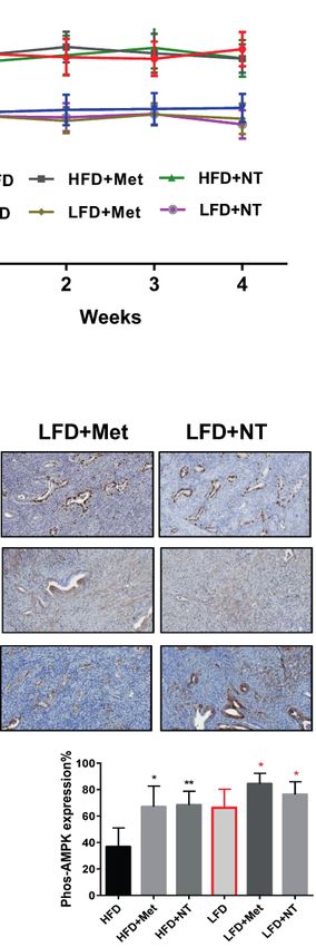
Roque et al.                                                                                                                      NT-1044 in Endometrial Cancer

                                              and lean conditions. More work is needed to determine if this novel biguanide will be
                                              beneficial in the treatment of women with EC, a disease strongly impacted by obesity and
                                              diabetes.
                                              Keywords: NT-1044, metformin, endometrial cancer, obesity, proliferation

INTRODUCTION                                                                         that metformin did not improve on the efficacy of standard-of-
                                                                                     care paclitaxel/carboplatin in advanced and recurrent
Endometrial cancer (EC) is known to be the most commonly                             endometrial cancer in NRG Oncology GOG286B (27), despite
diagnosed gynecologic malignancy in the United States (1). The                       promising pre-clinical data in endometrial cancer cell lines and
incidence of EC is rising in tandem with the changing in                             animal models (25, 26). Efforts are underway to develop new and
hormonal factors and increasing prevalence of obesity and                            improved pharmacologic versions of metformin for the
aging female population (2, 3). In 2021, it is estimated that                        treatment of diabetes mellitus, and it is logical that these same
66,570 women will be diagnosed with an endometrial carcinoma,                        drugs may also have greater anti-tumorigenic benefits than
and an estimated 12,940 patients will succumb to this disease (1).                   metformin. Metformin is a highly basic molecule with a pKa of
Traditionally, EC has been subdivided into Type I and Type II                        11.5 and is present at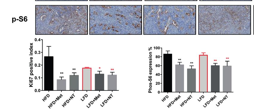
Roque et al.                                                                                                       NT-1044 in Endometrial Cancer

The ECC-1, Ishikawa and ES-T cells were grown in RPMI 1640                detected at Ex485/Em 485/530 nm by a Tecan plate reader.
medium (11 mM glucose) with 5% fetal bovine serum, MEM                    Each experiment was repeated at least three times for consistency
medium (5.5 mM glucose) with 5% fetal bovine serum and L-                 of response.
glutamine, and DMEM/F12 with 10% fetal bovine serum under 5%
CO2, respectively. All media included 100 units/ml penicillin and         Cleaved Caspase 3 Assays
100 microgram/ml streptomycin. NT-1044 was provided by                    Caspase activity assays were performed with modifications as
NovaTarg Therapeutics (Research Triangle Park, NC). All                   previously described (31). In brief, the ECC-1 and Ishikawa cells
primary and secondary antibodies were from Cell Signaling                 were plated in 6-well plates at a concentration of 2.5 × 10⁵ cells/
(Beverly, MA). Enhanced chemiluminescence reagents were                   well for 24 hours. The cells were treated with NT-1044 at
bought from Amersham Inc (Arlington Heights, IL).                         different concentrations for 12 hours. 150-180 ul 1X caspase
                                                                          lysis buffer was added to each well. Protein concentration was
Cell Proliferation Assays                                                 determined using the BSA assay. 10-15 ug lysates in a black clear
The Ishikawa, ECC-1 and ES-T cells (4000 cells/well) were                 bottom 96-well plate were incubated with reaction buffer and 200
cultured in 96-well plates and exposed to various                         uM of cleaved caspase 3 substrates for 30 min. The fluorescence
concentrations of metformin and NT1044 for 72 hours.                      of each well was determined using a Tecan microplate reader.
Following treatments, 5 µl MTT solution (5 mg/ml) was added               Each experiment was repeated three times to assess for
to each well, and the cells were cultured for an additional 1 hour.       consistency of results.
After aspiration of medium, 100 µl DMSO per well was used to
terminate the reactions. The results were read by measuring               Mitochondrial Membrane Potential Assay
absorption at 595 nm in a plate reader (Tecan, Cary, NC). The             Mitochondrial membrane potential was analyzed using the
effect of NT-1044 or metformin on cell proliferation was                  specific fluorescent probes JC-1 (32). The ECC-1 and Ishikawa
calculated as a percentage of control cell growth obtained from           cells were plated and treated with different concentrations of NT-
DMSO treated cells grown in the same 96-well plates. Each                 1044 for 12 hours. Treated cells were then incubated with 2 uM
experiment was performed in triplicate and repeated three times           JC-1 for 30 minutes at 37°C. The levels of the fluorescent probes
to assess for consistency of results.                                     were measured using a Tecan plate reader at two excitation/
                                                                          emission wavelength pairs. Each experiment was repeated three
Cell Cycle Analysis                                                       times to assess for consistency of results.
The ECC-1 and Ishikawa cells (2.5×10⁵ cells/ well) were cultured
with or without NT1044 for 24 hours. The cells were                       Ethidium Bromide Competition
subsequently collected by 0.05% Trypsin (Gibco), washed with              Assay for OCT1-3 Affinity
PBS, and fixed in a 90% methanol solution. On the day of                   HEK293 cells stably transfected with hOCT1, hOCT2, and
analysis, the cells were re-suspended in RNA A solution for 30            hOCT3 vectors were cultured for 24-48 hours in 24-well plates,
min at 37 °C, and then stained with PI staining solution for              respectively. After medium was aspirated, Ethidium Bromide
10 min in the dark. Cell cycle progression was analyzed by                (EtBr) or EtBr plus NT-1044 diluted in HBSS was added to each
Cellometer and analyzed by the FCS 4 Express Flow Cytometry               well and cultured for 2.5 min at 37°C. The plate was washed with
Software (De Novo Software, Glendale, CA, USA). The                       HBSS. Intracellular fluorescence was determined by a plate
experiments were repeated three times.                                    reader (Tecan) at 535/590-nm excitation and emission
                                                                          wavelengths. Nonspecific fluorescence in control cells was
Annexin V Staining Assay                                                  subtracted from fluorescence in hOCT-overexpressing cells to
Apoptotic cells were quantified by the Annexin-V FITC Kit                  yield data representing hOCT-specific fluorescence (33).
(Biovison). Briefly, ECC-1 and Ishikawa cells were seeded into 6
well plates at 2.5 × 10⁵ cells/well overnight, and then the cells         Western Immunoblotting
were treated with or without various concentrations of NT-1044            The Ishikawa and ECC-1 cells were seeded at 2.5 × 10⁵ cells/well
for 12 hours. The cells were harvested by 0.25% Trypsin and               in 6-well plates and then treated with various concentrations of
stained in 100 ul of Annexin-V and PI dual-stain solution for             NT-1044 for 24 hours. The treated cells were lysed in an RIPA
15 min. The expression of Annexin V was detected by                       buffer (1% NP40, 0.5 sodium deoxycholate and 0.1% SDS) and
Cellometer, and analyzed by FCS 4 software. Apoptotic cells               centrifuged at 12,000 rpm for 15 min to collect supernatants. The
were expressed as a percentage of the total number of                     protein concentration was determined using the BCA assay kit
cells stained.                                                            (Thermo Fisher Scientific). Equal amounts of protein were
                                                                          separated by SDS-PAGE gels and transferred to a PVDF
Reactive Oxygen Species (ROS) Assay                                       membrane for 2 hours. The membrane was blocked by 5%
Intracellular ROS production was detected using DCFH-DA                   milk and then incubated with the following primary
assay. The ECC-1 and Ishikawa cells were plated in the                    antibodies: phos-AMPK and S6, MCL-1, BCL-XL, CDK4,
appropriate media at 6000 cells/well in a 96-well plate                   CDK6, Bip, PERK and cyclin D1 at 4°C overnight. The
overnight, and then treated with various concentrations of NT-            membrane was washed with TBS-T and incubated with anti-
1044 for 12 hours. 10 ml of 200 mM of DCFH-DA was added into              rabbit or anti-mouse secondary antibodies for 1 hour. Enhanced
each well and mixed gently. The fluorescence intensity was                 chemiluminescence reagents was used to visualize protein

Frontiers in Oncology | www.frontiersin.org                           3                                 August 2021 | Volume 11 | Article 690435
Roque et al.                                                                                                           NT-1044 in Endometrial Cancer

binding through a gel imaging analysis system (Bio-Rad).                    100 and number 400 sieves. 8000 cells/well were seeded into 96-well
The band densitometry was performed using Image J software.                 plated and cell proliferation was measured with MTT assay 72 hours
The relative expression of target protein was normalized to the             after treatment.
expression of b-actin or a-Tubulin. Each experiment was
repeated three times to assess for consistency of results.                  Statistical Analysis
                                                                            All experiments were repeated a minimum of three times. Results
NT-1044 Treatment in a Transgenic Mouse                                     for experiments were normalized to the mean of the control and
Model of Endometrial Cancer                                                 analyzed using the Student t-test using GraphPad software (La Jolla,
For our in vivo studies, we utilized an LKB1fl/flp53fl/fl genetically           CA, USA). Differences were considered significant if the p value was
engineered mouse model. This model was developed in our lab,                less than 0.05 (p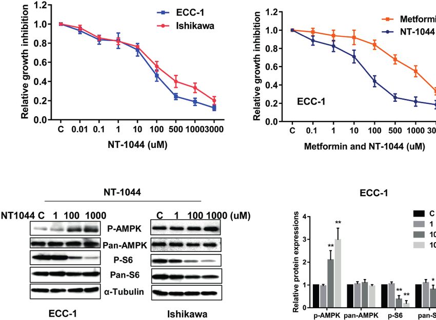
Roque et al.                                                                                                                         NT-1044 in Endometrial Cancer

                     A                                        B                                          C

                     D

 FIGURE 1 | NT-1044 inhibited cell proliferation and activated AMPK pathway in EC cells. The ECC-1 and Ishikawa cells were cultured in regular media for 24 hours,
 and then treated with the indicated concentrations of NT-1044 and metformin in 96-well plates for 72 hours. Cell proliferation was assessed by MTT assay. NT-1044
 and metformin significant inhibited cell proliferation in a dose dependent manner (A–C). IC50 value of NT-1044 for ECC-1 = 87 mM; Ishikawa=218 mM. The ECC-1
 and Ishikawa cells were treated with NT-1044 for 18 hours. Western blotting showed that NT-1044 increased phosphorylation of AMPK expression and decreased
 phosphorylation of S6 expression (D). The results are shown as the mean ± SE of triplicate samples and are representative of three independent experiments.
 *p < 0.05, **p < 0.01.

expression of phosphorylated S6 in both cell lines after 18 hours                    significantly increased Annexin V expression in a dose
of treatment with NT-1044. Expression of pan-AMPK was not                            dependent manner as demonstrated in Figure 3A (p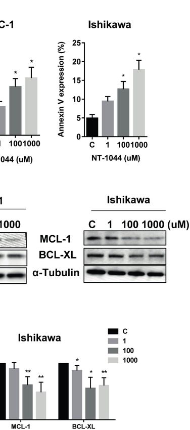
Roque et al.                                                                                                                       NT-1044 in Endometrial Cancer

                              A

                              B

 FIGURE 2 | NT-1044 induced cell cycle G1 arrest in EC cells. The ECC-1 and Ishikawa cells were treated with NT-1044 for 24 hours. Cell cycle progression was
 assessed by Cellometer. NT-1044 induced cell cycle G1 arrest in a dose-dependent manner in both cell lines (A). Western blotting showed that NT-1044 decreased
 the expression of CDK4, CDK6 and cyclin D in a dose-dependent manner in both cell lines after 24 hours of exposure (B). *p < 0.05, **p < 0.01.

(Bip), two proteins related to cell stress, after the cells were                   metformin. However, metformin and NT-1044 had a more
treated with different concentrations of NT-1044 for 24 hours.                     pronounced impact on the tumor growth of obese mice (70.3
The data showed that the expression of PERK and Bip increased                      and 75.8% reduction in tumor weight, respectively) compared to
with increasing doses of NT-1044 (Figure 4C). These results                        58.1 and 63.8% reduction in tumor weight with NT-1044 and
indicate that NT-1044 induces intracellular oxidative stress in                    metformin treatment in lean mice, respectively (p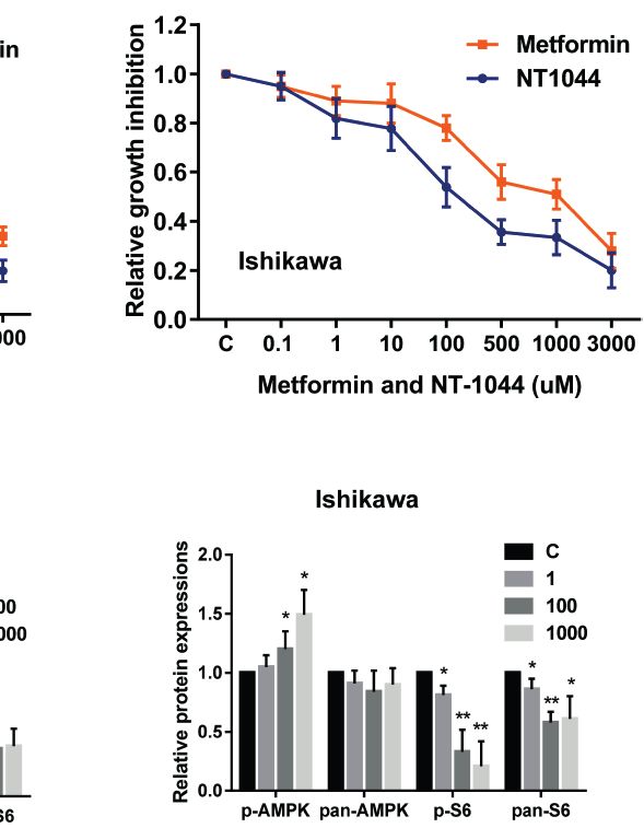
Roque et al.                                                                                                                       NT-1044 in Endometrial Cancer

                      A

                      B                                                     C

                                              D

 FIGURE 3 | NT-1044 induced apoptosis in EC cells. The ECC-1 and Ishikawa cells were cultured for 24 hours and then treated with NT-1044 for 12 hours. NT-
 1044 increased Annexin V expression in a dose-dependent manner in both cells (A). Both cell lines were treated with NT-1044 for 12 hours. Cleaved caspase 3 was
 measured by ELISA assay. The results showed that NT-1044 increased cleaved caspase 3 activity in both cell lines (B). Western blotting showed that NT-1044
 decreased the expression of BCL-XL and MCL-1 in a dose-dependent manner in both cell lines after 18 hours of exposure (C, D). *p < 0.05, **p < 0.01.

phosphorylated S6 and increased the expression of                                   anti-tumor activities in the LKB1fl/fl p53fl/fl mouse model of EC. In
phosphorylated AMPK in obese and lean mice compared with                            addition, we found that NT-1044 suppressed cell growth, induced
the untreated mice, suggesting that metformin and NT1044                            apoptosis and G1 cell cycle arrest, caused cellular stress, activated
inhibited tumor growth through the AMPK/mTORC1 pathway                              AMPK and inhibited mTOR pathways in vitro and in vivo.
in vivo (p
Roque et al. NT-1044 in Endometrial Cancer
A
B
C
FIGURE 4 | NT-1044 induced cell stress in EC cells. The ECC-1 and Ishikawa cells were cultured for 24 hours and then treated with NT-1044 for 12 hours.
Reactive Oxygen Species (ROS) was detected using the DCFH-DA Assay. Mitochondrial membrane potential was measured by JC-1 assay. NT-1044 significantly
increased ROS products (A) and decreased JC-1 levels (B) in a dose dependent manner in the both cells. Both cells were treated with NT-1044 for 24 hours.
Western blotting showed that NT-1044 increased the expression of BIP and PERK in the both cells (C). *p < 0.05, **p < 0.01.
(2-5 mM) (26). Lower doses of metformin had little effect on expression of p53 in sensitive cancer cells, which in turn leads
caspase-3 activity. Similar results have demonstrated that 10 mM to regulation of p53 downstream targets and to induction of
metformin significantly increased the proportion of Annexin V apoptosis (42). Given that metformin has poor lipophilicity and
positive cells and the expression of cleaved caspase-3 in Ishikawa its bio-distribution in cancer cells relies on OCT1, OCT2 and
cells (39). Whether metformin induces apoptosis is controversial OCT3, it is also possible that induction of apoptosis is related to
as it has also failed to induce apoptosis in prostate and breast OCT1/3 receptor expression and the biguanide potency. NT-
cancer cell lines at similar doses of treatment. The underlying 1044 has significantly higher affinity for both OCT1 (IC50 213
reason for this difference may be attributed to the duration and mM vs. 3,532 mM) and OCT3 (IC50 20 mM vs. 17,500 mM) when
concentration of metformin treatment as well as the status of compared to metformin (Table 1). Our preliminary studies on
wild type p53 (37, 41). Metformin may directly affect the transporter proteins have shown that OCT1 and OCT3 are
Frontiers in Oncology | www.frontiersin.org 8 August 2021 | Volume 11 | Article 690435Roque et al. NT-1044 in Endometrial Cancer
A B
C
FIGURE 5 | NT-1044 inhibited endometrial tumor growth in both the obese and lean LKB1fl/flp53fl/fl mice. LKB1fl/fl p53fl/fl mice were fed high fat diet (HFD) or low fat
diet (LFD) at 3 weeks of age to induce obesity. The mice were divided into six groups: obese, obese + metformin, obese+NT-1044, Lean, lean + metformin. Lean
+NT-1044. The obese and lean mice in both groups were treated with NT-1044 or metformin (200 mg/kg, oral gavage for NT1044 and drinking water for metformin)
or placebo for 4 weeks. Obesity promoted tumor growth in obese mice versus lean mice. Either metformin or NT-1044 significantly reduced tumor weight in the
obese and lean mice, with a greater impact on tumor weight in obese mice (A). During treatment, there were no significant changes in body weight in the six groups
(B). Change in expression of Ki-67, phosphorylated-S6 and phosphorylated-AMPK were assessed by immunohistochemistry in the endometrial cancer tissues. The
expression of Ki-67 and phosphorylated-S6 was reduced and phosphorylated-AMPK was increased in both groups after NT-1044 or metformin treatment (C).
*p < 0.05, **p < 0.01.
present in both endometrial cancer cell lines and endometrial including endometrial cancer (25, 26, 45–47). Since AMPK
tumors (Supplemental Figure 2). The higher potency of NT- negatively controls mTOR activation and mTOR is a
1044 as well as the higher affinity to the OCT1 and OCT3 downstream effector of AKT, Activation of AMPK is
transporter when compared to metformin, may explain why NT- considered to be a possible therapeutic target for cancers that
1044 is more effective at inducing apoptosis in our current study. contain high AKT activity (48). Given type I endometrial cancers
However, additional research is needed to determine if OCT1 exhibit a high frequency of PTEN deletions and mutations, leading
and OCT3 expression is in fact a predictor of treatment response to the activation of AKT, AMPK activation may have therapeutic
to biguanides including NT-1044. potential for this disease (49). In this study, ECC-1 and Ishikawa
Inhibition of cell proliferation by NT-1044 was accompanied cells have low expression of wild PTEN, but RL-95-2 cells, a PTEN
by inhibition of the AMPK/mTOR pathway. AMPK/mTOR mutant EC cell line, exhibited the similar sensitivity to metformin
pathway is well known to be regulated cancer cell growth and and NT1044 compared to ECC-1 and Ishikawa cells, suggesting
survival. The activation of AMPK reduces cell proliferation that PTEN status may not play a big role in inhibition of cell
through negatively regulates mTOR signaling (43). Recent growth in NT1044-treated endometrial cancer cells. Several
results showed that NT-1044 exhibited higher AMPK studies have confirmed that loss of PTEN significantly enhances
activation than metformin at the same dosage in differentiated the sensitivity to rapamycin, an mTOR inhibitor (50–52). The
human adipocytes (44).In its role as an AMPK activator, the results of the present study suggested that NT1044 may be
anti-tumorigenic activity of NT-1044 appears similar to that of promising therapy for type I endometrial cancer, which more
metformin, which has been shown to significantly decrease frequently have PTEN mutations, as well as type II endometrial
proliferation of several human cancer cell lines in vitro, cancers, which less frequently have PTEN mutations.
Frontiers in Oncology | www.frontiersin.org 9 August 2021 | Volume 11 | Article 690435Roque et al. NT-1044 in Endometrial Cancer
Lastly, we explored the activity of NT-1044 in vivo in an pathway well-known to be altered in both obesity and
endometrial cancer mouse model. We also assessed whether this endometrial cancer.
drug would have different activity in lean versus obese mice given
that obesity has been linked to both an increased risk of
developing endometrial cancer and an increased risk of DATA AVAILABILITY STATEMENT
mortality from the disease (53, 54). In the obese mice, NT-
The original contributions presented in the study are included in
1044 inhibited tumor growth by 76% (p=0.0065) whereas in the
the article/Supplementary Material. Further inquiries can be
lean mice, it inhibited tumor growth by 64% (p=0.0011),
directed to the corresponding authors.
suggesting that NT-1044 is more effective in the setting of
obesity. NT-1044 exhibited similar anti-tumor activity
compared to metformin. These findings are not surprising ETHICS STATEMENT
given the association between obesity, insulin resistance and
endometrial cancer. In a pre-operative window study, metformin The animal study was reviewed and approved by Institutional
was shown to have increased activity against endometrial cancer Animal Care and Use Committee, UNC.
in the subset of patients with metabolomic profiles consistent
with increased insulin resistance (55).
AUTHOR CONTRIBUTIONS
DR, LZ, WW, JH, and WS performed the experiments in vitro.
LZ, JH, and YY performed animal experiments. DR, LZ, and
CONCLUSION WW participated in analyzing and interpreting the data. DR
As our work has demonstrated, NT-1044 has greater potency and CZ wrote the manuscript. JL and KB provided NT-1044. CZ
than metformin in vitro and comparable effects to metformin and VB-J designed experiments, revised the manuscript and
in vivo under obese and lean conditions. In addition, we provided financial support. All authors contributed to the
recently found that NT1044 inhibited cell proliferation in article and approved the submitted version.
normal endometrial stromal and epithelial cells, suggesting
that NT1044 may have potential to treat non-malignant FUNDING
hyperplastic diseases in uterus (Supplemental Figure 1). In
pharmacologic studies of NT-1044 for diabetes treatment, This work is supported by: (1) VB-J: American Cancer Society
NT-1044 demonstrated the anticipated pharmacologic (ACS) Research Scholar Grant - RSG CCE 128826. (2) VB-J:
activity of a biguanide but at just 1/5 of the metformin dose NIH/NCI - R37CA226969.
(data not shown). Therefore, NT-1044 represents a novel
biguanide with improved pharmacologic features over
metformin that include increased selectivity of transporters ACKNOWLEDGMENTS
and a longer plasma half-life, although this did not result in an
The NovaTarg Therapeutics provided NT-1044.
increased benefit in its anti-tumorigenic effects in the LKB1 fl/fl
p53fl/fl. Thus, future work will focus on expansion of the NT-
1044 treatment studies into additional patient-derived SUPPLEMENTARY MATERIAL
xenograft mouse models of endometrial cancer of varying
genomic backgrounds to assess for potential biomarkers of The Supplementary Material for this article can be found online at:
NT-1044 response that may also align with obesity status, in https://www.frontiersin.org/articles/10.3389/fonc.2021.690435/
particular mutations that affect the AMPK/mTOR pathway, a full#supplementary-material
7. Friberg E, Mantzoros CS, Wolk A. Diabetes and Risk of Endometrial Cancer:
REFERENCES A Population-Based Prospective Cohort Study. Cancer Epidemiol Biomarkers
1. Siegel RL, Miller KD, Fuchs HE, Jemal A. Cancer Statistics, 2021. CA Cancer J Prev (2007) 16:276–80. doi: 10.1158/1055-9965.EPI-06-0751
Clin (2021) 71:7–33. doi: 10.3322/caac.21654 8. Friberg E, Orsini N, Mantzoros CS, Wolk A. Diabetes Mellitus and Risk of
2. Fader AN, Arriba LN, Frasure HE, von Gruenigen VE. Endometrial Cancer Endometrial Cancer: A Meta-Analysis. Diabetologia (2007) 50:1365–74. doi:
and Obesity: Epidemiology, Biomarkers, Prevention and Survivorship. 10.1007/s00125-007-0681-5
Gynecol Oncol (2009) 114:121–7. doi: 10.1016/j.ygyno.2009.03.039 9. Soliman PT, Wu D, Tortolero-Luna G, Schmeler KM, Slomovitz BM, Bray
3. von Gruenigen VE, Gil KM, Frasure HE, Jenison EL, Hopkins MP. The MS, et al. Association Between Adiponectin, Insulin Resistance, and
Impact of Obesity and Age on Quality of Life in Gynecologic Surgery. Am J Endometrial Cancer. Cancer (2006) 106:2376–81. doi: 10.1002/cncr.21866
Obstet Gynecol (2005) 193:1369–75. doi: 10.1016/j.ajog.2005.03.038 10. Chia VM, Newcomb PA, Trentham-Dietz A, Hampton JM. Obesity, Diabetes, and
4. Prat J, Gallardo A, Cuatrecasas M, Catasus L. Endometrial Carcinoma: Pathology Other Factors in Relation to Survival After Endometrial Cancer Diagnosis. Int J
and Genetics. Pathology (2007) 39:72–87. doi: 10.1080/00313020601136153 Gynecol Cancer (2007) 17:441–6. doi: 10.1111/j.1525-1438.2007.00790.x
5. lHecht JL, Mutter GL. Molecular and Pathologic Aspects of Endometrial 11. Cust AE, Kaaks R, Friedenreich C, Bonnet F, Laville M, Lukanova A, et al.
Carcinogenesis. J Clin Oncol (2006) 24:4783–91. doi: 10.1200/JCO.2006.06.7173 Plasma Adiponectin Levels and Endometrial Cancer Risk in Pre- and
6. Sherman ME. Theories of Endometrial Carcinogenesis: A Multidisciplinary Postmenopausal Women. J Clin Endocrinol Metab (2007) 92:255–63. doi:
Approach. Mod Pathol (2000) 13:295–308. doi: 10.1038/modpathol.3880051 10.1210/jc.2006-1371
Frontiers in Oncology | www.frontiersin.org 10 August 2021 | Volume 11 | Article 690435Roque et al. NT-1044 in Endometrial Cancer
12. Evans JM, Donnelly LA, Emslie-Smith AM, Alessi DR, Morris AD. Metformin 31. Choudhary GS, Al-Harbi S, Almasan A. Caspase-3 Activation Is a Critical
and Reduced Risk of Cancer in Diabetic Patients. Bmj (2005) 330:1304–5. doi: Determinant of Genotoxic Stress-Induced Apoptosis. Methods Mol Biol
10.1136/bmj.38415.708634.F7 (2015) 1219:1–9. doi: 10.1007/978-1-4939-1661-0_1
13. Libby G, Donnelly LA, Donnan PT, Alessi DR, Morris AD, Evans JM. New 32. Perry SW, Norman JP, Barbieri J, Brown EB, Gelbard HA. Mitochondrial
Users of Metformin Are at Low Risk of Incident Cancer: A Cohort Study Membrane Potential Probes and the Proton Gradient: A Practical Usage
Among People With Type 2 Diabetes. Diabetes Care (2009) 32:1620–5. doi: Guide. Biotechniques (2011) 50:98–115. doi: 10.2144/000113610
10.2337/dc08-2175 33. Lee WK, Reichold M, Edemir B, Ciarimboli G, Warth R, Koepsell H, et al.
14. Bowker SL, Majumdar SR, Veugelers P, Johnson JA. Increased Cancer-Related Organic Cation Transporters OCT1, 2, and 3 Mediate High-Affinity
Mortality for Patients With Type 2 Diabetes Who Use Sulfonylureas or Transport of the Mutagenic Vital Dye Ethidium in the Kidney Proximal
Insulin: Response to Farooki and Schneider. Diabetes Care (2006) 29:1990–1. Tubule. Am J Physiol Renal Physiol (2009) 296:F1504–13. doi: 10.1152/
doi: 10.2337/dc06-0997 ajprenal.90754.2008
15. Hinke SA, Martens GA, Cai Y, Finsi J, Heimberg H, Pipeleers D, et al. Methyl 34. Lim S, Kaldis P. Cdks, Cyclins and CKIs: Roles Beyond Cell Cycle Regulation.
Succinate Antagonises Biguanide-Induced AMPK-Activation and Death of Development (2013) 140:3079–93. doi: 10.1242/dev.091744
Pancreatic Beta-Cells Through Restoration of Mitochondrial Electron 35. Grossmann ME, Yang DQ, Guo Z, Potter DA, Cleary MP. Metformin
Transfer. Br J Pharmacol (2007) 150:1031–43. doi: 10.1038/sj.bjp.0707189 Treatment for the Prevention and/or Treatment of Breast/Mammary
16. Hawley SA, Gadalla AE, Olsen GS, Hardie DG. The Antidiabetic Drug Tumorigenesis. Curr Pharmacol Rep (2015) 1:312–23. doi: 10.1007/s40495-
Metformin Activates the AMP-Activated Protein Kinase Cascade via an 015-0032-z
Adenine Nucleotide-Independent Mechanism. Diabetes (2002) 51:2420–5. 36. Dowling RJ, Lam S, Bassi C, Mouaaz S, Aman A, Kiyota T, et al. Metformin
doi: 10.2337/diabetes.51.8.2420 Pharmacokinetics in Mouse Tumors: Implications for Human Therapy. Cell
17. Gallagher EJ, LeRoith D. Epidemiology and Molecular Mechanisms Tying Metab (2016) 23:567–8. doi: 10.1016/j.cmet.2016.03.006
Obesity, Diabetes, and the Metabolic Syndrome With Cancer. Diabetes Care 37. Fatehi Hassanabad A, MacQueen KT. Molecular Mechanisms Underlining
(2013) 36 Suppl 2:S233–9. doi: 10.2337/dcS13-2001 the Role of Metformin as a Therapeutic Agent in Lung Cancer. Cell Oncol
18. Quinn BJ, Kitagawa H, Memmott RM, Gills JJ, Dennis PA. Repositioning (Dordr) (2021) 44:1–18. doi: 10.1007/s13402-020-00570-0
Metformin for Cancer Prevention and Treatment. Trends Endocrinol Metab 38. Jin DH, Kim Y, Lee BB, Han J, Kim HK, Shim YM, et al. Metformin Induces
(2013) 24:469–80. doi: 10.1016/j.tem.2013.05.004 Cell Cycle Arrest at the G1 Phase Through E2F8 Suppression in Lung Cancer
19. Noto H, Goto A, Tsujimoto T, Noda M. Cancer Risk in Diabetic Patients Cells. Oncotarget (2017) 8:101509–19. doi: 10.18632/oncotarget.21552
Treated With Metformin: A Systematic Review and Meta-Analysis. PloS One 39. Takahashi A, Kimura F, Yamanaka A, Takebayashi A, Kita N, Takahashi K,
(2012) 7:e33411. doi: 10.1371/journal.pone.0033411 et al. Metformin Impairs Growth of Endometrial Cancer Cells via Cell Cycle
20. Gehrig PA, Bae-Jump VL. Promising Novel Therapies for the Treatment of Arrest and Concomitant Autophagy and Apoptosis. Cancer Cell Int (2014)
Endometrial Cancer. Gynecol Oncol (2010) 116:187–94. doi: 10.1016/ 14:53. doi: 10.1186/1475-2867-14-53
j.ygyno.2009.10.041 40. Safe S, Nair V, Karki K. Metformin-Induced Anticancer Activities: Recent
21. Dedes KJ, Wetterskog D, Ashworth A, Kaye SB, Reis-Filho JS. Emerging Insights. Biol Chem (2018) 399:321–35. doi: 10.1515/hsz-2017-0271
Therapeutic Targets in Endometrial Cancer. Nat Rev Clin Oncol (2011) 8:261– 41. Buzzai M, Jones RG, Amaravadi RK, Lum JJ, DeBerardinis RJ, Zhao F, et al.
71. doi: 10.1038/nrclinonc.2010.216 Systemic Treatment With the Antidiabetic Drug Metformin Selectively
22. Cheung LW, Hennessy BT, Li J, Yu S, Myers AP, Djordjevic B, et al. High Impairs P53-Deficient Tumor Cell Growth. Cancer Res (2007) 67:6745–52.
Frequency of PIK3R1 and PIK3R2 Mutations in Endometrial Cancer doi: 10.1158/0008-5472.CAN-06-4447
Elucidates a Novel Mechanism for Regulation of PTEN Protein Stability. 42. Hsieh Li SM, Liu ST, Chang YL, Ho CL, Huang SM. Metformin Causes Cancer
Cancer Discovery (2011) 1:170–85. doi: 10.1158/2159-8290.CD-11-0039 Cell Death Through Downregulation of P53-Dependent Differentiated Embryo
23. Kandoth C, Schultz N, Cherniack AD, Akbani R, Liu Y, Shen H, et al. Chondrocyte 1. J BioMed Sci (2018) 25:81. doi: 10.1186/s12929-018-0478-5
Integrated Genomic Characterization of Endometrial Carcinoma. Nature 43. Inoki K, Zhu T, Guan KL. TSC2 Mediates Cellular Energy Response to
(2013) 497:67–73. doi: 10.1038/nature12113 Control Cell Growth and Survival. Cell (2003) 115:577–90. doi: 10.1016/
24. Salvesen HB, Carter SL, Mannelqvist M, Dutt A, Getz G, Stefansson IM, et al. S0092-8674(03)00929-2
Integrated Genomic Profiling of Endometrial Carcinoma Associates 44. Arner P, Kulyte A, Batchelor K, Laurencikiene J, Livingston J, Ryden M.
Aggressive Tumors With Indicators of PI3 Kinase Activation. Proc Natl Mapping of Biguanide Transporters in Human Fat Cells and Their Impact on
Acad Sci USA (2009) 106:4834–9. doi: 10.1073/pnas.0806514106 Lipolysis. Diabetes Obes Metab (2018) 20:2416–25. doi: 10.1111/dom.13395
25. Guo H, Kong W, Zhang L, Han J, Clark LH, Yin Y, et al. Reversal of Obesity- 45. Zakikhani M, Dowling RJ, Sonenberg N, Pollak MN. The Effects of
Driven Aggressiveness of Endometrial Cancer by Metformin. Am J Cancer Res Adiponectin and Metformin on Prostate and Colon Neoplasia Involve
(2019) 9:2170–93. Activation of AMP-Activated Protein Kinase. Cancer Prev Res (Phila)
26. Cantrell LA, Zhou C, Mendivil A, Malloy KM, Gehrig PA, Bae-Jump VL. (2008) 1:369–75. doi: 10.1158/1940-6207.CAPR-08-0081
Metformin Is a Potent Inhibitor of Endometrial Cancer Cell Proliferation– 46. Di Matteo S, Nevi L, Overi D, Landolina N, Faccioli J, Giulitti F, et al. Metformin
Implications for a Novel Treatment Strategy. Gynecol Oncol (2010) 116:92–8. Exerts Anti-Cancerogenic Effects and Reverses Epithelial-to-Mesenchymal
doi: 10.1016/j.ygyno.2009.09.024 Transition Trait in Primary Human Intrahepatic Cholangiocarcinoma Cells.
27. Bae-Jump V, Sill M, Gehrig PA, Moxley K, Hagemann AR, Waggoner SE, Sci Rep (2021) 11:2557. doi: 10.1038/s41598-021-81172-0
et al. A Randomized Phase II/III Study of Paclitaxel/Carboplatin/Metformin 47. Hampsch RA, Wells JD, Traphagen NA, McCleery CF, Fields JL, Shee K, et al.
Versus Paclitaxel/Carboplatin/Placebo as Initial Therapy for Measurable Stage AMPK Activation by Metformin Promotes Survival of Dormant ER(+) Breast
III or IVA, Stage IVB, or Recurrent Endometrial Cancer: An NRG Oncology/ Cancer Cells. Clin Cancer Res (2020) 26:3707–19. doi: 10.1158/1078-
GOG Study. 51st Annual Meeting of the Society of Gynecologic Oncology, 0432.CCR-20-0269
April 2020, Virtual Meeting Due to COVID-19. Gynecol Oncol (2020) 159 48. Faria J, Negalha G, Azevedo A, Martel F. Metformin and Breast Cancer:
(Supplement 1):7 doi: 10.1016/j.ygyno.2020.06.013 Molecular Targets. J Mammary Gland Biol Neoplasia (2019) 24:111–23. doi:
28. Graham GG, Punt J, Arora M, Day RO, Doogue MP, Duong JK, et al. Clinical 10.1007/s10911-019-09429-z
Pharmacokinetics of Metformin. Clin Pharmacokinet (2011) 50:81–98. doi: 49. Leskela S, Perez-Mies B, Rosa-Rosa JM, Cristobal E, Biscuola M, Palacios-
10.2165/11534750-000000000-00000 Berraquero ML, et al. Molecular Basis of Tumor Heterogeneity in Endometrial
29. Aoki M, Terada T, Kajiwara M, Ogasawara K, Ikai I, Ogawa O, et al. Kidney- Carcinosarcoma. Cancers (Basel) (2019) 11(7):964. doi: 10.3390/
Specific Expression of Human Organic Cation Transporter 2 (OCT2/ cancers11070964
SLC22A2) is Regulated by DNA Methylation. Am J Physiol Renal Physiol 50. Podsypanina K, Lee RT, Politis C, Hennessy I, Crane A, Puc J, et al. An
(2008) 295:F165–70. doi: 10.1152/ajprenal.90257.2008 Inhibitor of mTOR Reduces Neoplasia and Normalizes P70/S6 Kinase
30. Scheen AJ. Clinical Pharmacokinetics of Metformin. Clin Pharmacokinet Activity in Pten+/- Mice. Proc Natl Acad Sci USA (2001) 98:10320–5. doi:
(1996) 30:359–71. doi: 10.2165/00003088-199630050-00003 10.1073/pnas.171060098
Frontiers in Oncology | www.frontiersin.org 11 August 2021 | Volume 11 | Article 690435Roque et al. NT-1044 in Endometrial Cancer
51. Grunwald V, DeGraffenried L, Russel D, Friedrichs WE, Ray RB, Hidalgo M. The remaining authors declare that the research was conducted in the absence of
Inhibitors of Mtor Reverse Doxorubicin Resistance Conferred by PTEN Status any commercial or financial relationships that could be construed as a potential
in Prostate Cancer Cells. Cancer Res (2002) 62:6141–5. conflict of interest.
52. Neshat MS, Mellinghoff IK, Tran C, Stiles B, Thomas G, Petersen R, et al.
Enhanced Sensitivity of PTEN-Deficient Tumors to Inhibition of FRAP/mTOR.
Proc Natl Acad Sci USA (2001) 98:10314–9. doi: 10.1073/pnas.171076798 Publisher’s Note: All claims expressed in this article are solely those of the authors
53. Calle EE, Rodriguez C, Walker-Thurmond K, Thun MJ. Overweight, Obesity, and do not necessarily represent those of their affiliated organizations, or those of
and Mortality From Cancer in a Prospectively Studied Cohort of U. S. adults. the publisher, the editors and the reviewers. Any product that may be evaluated in
New Engl J Med (2003) 348:1625–38. doi: 10.1056/NEJMoa021423 this article, or claim that may be made by its manufacturer, is not guaranteed or
54. Lu KH, Broaddus RR. Endometrial Cancer. N Engl J Med (2020) 383:2053–64. endorsed by the publisher.
doi: 10.1056/NEJMra1514010
55. Schuler KM, Rambally BS, DiFurio MJ, Sampey BP, Gehrig PA, Makowski L, Copyright © 2021 Roque, Zhang, Wysham, Han, Sun, Yin, Livingston, Batchelor,
et al. Antiproliferative and Metabolic Effects of Metformin in a Preoperative Zhou and Bae-Jump. This is an open-access article distributed under the terms of the
Window Clinical Trial for Endometrial Cancer. Cancer Med (2015) 4:161–73. Creative Commons Attribution License (CC BY). The use, distribution or
doi: 10.1002/cam4.353 reproduction in other forums is permitted, provided the original author(s) and the
copyright owner(s) are credited and that the original publication in this journal is
Conflict of Interest: JL and KB are employees and shareholders of NovaTarg cited, in accordance with accepted academic practice. No use, distribution or
Therapeutics. reproduction is permitted which does not comply with these terms.
Frontiers in Oncology | www.frontiersin.org 12 August 2021 | Volume 11 | Article 690435You can also read