CTBP2 INTERACTS WITH TGIF TO PROMOTE THE PROGRESSION OF ESOPHAGEAL SQUAMOUS CELL CANCER THROUGH THE WNT/Β CATENIN PATHWAY
←
→
Page content transcription
If your browser does not render page correctly, please read the page content below
ONCOLOGY REPORTS 47: 29, 2022

                    CtBP2 interacts with TGIF to promote the
                  progression of esophageal squamous cell cancer
                       through the Wnt/β‑catenin pathway
                               QIANQIAN JU1,2, MAORONG JIANG2,3, WENXIN HUANG4,
                                 QINGBO YANG5, ZHENGHONG LUO5 and HUI SHI1,5

                  1
                  School of Basic Medical Sciences, Nanjing Medical University, Nanjing, Jiangsu 211166;
          2
          Key Laboratory for Neuroregeneration, Medical College of Nantong University, Nantong, Jiangsu 226001;
          3
           School of Life Sciences, Nantong University, Nantong, Jiangsu 226019; 4Department of General Surgery,
             Shanghai General Hospital, Shanghai Jiao Tong University School of Medicine, Shanghai 200080;
             5
              Department of Thoracic Surgery, Shanghai Tenth People's Hospital, Shanghai 200072, P.R. China

                                       Received July 8, 2021; Accepted October 13, 2021

                                                   DOI: 10.3892/or.2021.8240

Abstract. C‑terminal‑binding protein 2 (CtBP2), a transcrip‑       these proteins were co‑localized in the nucleus. CtBP2 and
tional co‑repressor, plays a main role in tumorigenesis and in     TGIF mRNA and protein expression levels were robustly
the development of multiple tumors. Transforming growth            and simultaneously increased in both ESCC tissues and cell
interacting factor (TGIF) is involved in a number of cellular      lines. There was a direct correlation between CtBP2 and TGIF
signal transduction pathways and is related to tumor occurrence    expression levels in ESCC tissues, and both were significantly
and development. In the present study, the proteins interacting    associated with metastasis and survival. The TGIF and CtBP2
with CtBP2 were identified and the mechanisms underlying           expression levels were significantly increased or decreased
the biological activity of CtBP2 in esophageal squamous cell       simultaneously, in ECA109 cells transfected with LV‑CtBP2
carcinoma (ESCC) were investigated. The Search Tool for            or sh‑CtBP2, and vice versa. According to the results of CCK‑8
the Retrieval of Interacting Genes (STRING) database was           assay, EdU staining and Transwell assay, CtBP2 promoted the
used to search for known proteins interacting with CtBP2,          proliferation, migration and invasion of ECA109 cells through
and co‑immunoprecipitation (Co‑IP) assay was performed to          the Wnt/β‑catenin pathway. On the whole, the present study
validate the interactions. Reverse transcription‑quantitative      demonstrates that CtBP2 interacts with TGIF and promotes
PCR (RT‑qPCR), immunohistochemistry (IHC) and western              the malignant progression of ESCC through the Wnt/β‑catenin
blot analysis were performed to examine the expression             pathway.
levels of CtBP2 and TGIF in ESCC. The correlation between
CtBP2 and TGIF was analyzed using Gene Expression                  Introduction
Profiling Interactive Analysis (GEPIA) by Pearson's correla‑
tion analysis, and the co‑localization of CtBP2 with TGIF in       A high incidence of esophageal cancer (EC) has been reported
the ECA109 cells was identified using immunofluorescence           in China (1,2). Recent surveys have demonstrated that there
staining. XAV939 treatment, CCK‑8, 5‑ethynyl‑2'‑deoxyuri‑          are ~450,000 new cases of EC each year worldwide, with
dine (EdU) staining, wound healing and Transwell assays            more than half of these cases originating from China, ranking
were performed to investigate the signaling pathways involved      sixth among the list malignant tumors in China (3,4). EC is
in the biological activity of CtBP2 in ECA109 cells. According     an invasive from of cancer, with the two main pathological
to the results obtained from STRING and Co‑IP analysis,            types being esophageal squamous cell carcinoma (ESCC) and
an interaction between CtBP2 and TGIF was indicated, and           esophageal adenocarcinoma (EA) (5). ESCC has been reported
                                                                   as the main histological type of EC in China, accounting
                                                                   for >95% of reported cases (6,7). The 5‑year survival rate
                                                                   of patients with EC is only ~10%, and the recurrence and
                                                                   mortality rates remain high (8). Radiotherapy, chemotherapy
Correspondence to: Dr Hui Shi, School of Basic Medical Sciences,
Nanjing Medical University, 101 Longmian Avenue, Jiangning,        and targeted therapy have been developed in recent years
Nanjing, Jiangsu 211166, P.R. China                                for the treatment of EC. However, the clinical efficacy and
E‑mail: disney1982@163.com                                         prognosis of patients have not been satisfactory (9,10). This
                                                                   is due to the lack of an in‑depth understanding of the patho‑
Key words: esophageal squamous cell carcinoma, CtBP2, TGIF,        genesis of EC (11). Therefore, a more detailed elucidation of
Wnt/β‑catenin, proliferation, migration, invasion                  the pathogenesis of EC at the molecular level may lead to
                                                                   the discovery of an ideal molecular target or other effective
                                                                   therapeutic drugs for EC.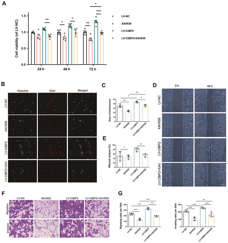
2              ju et al: CtBP2 INTERACTING WITH TGIF PROMOTES THE PROCESSION OF ESCC VIA THE Wnt PATHWAY

    The transcriptional co‑repressor C‑terminal binding            it was hypothesized that an in‑depth study of the regulatory
protein (CtBP) has been originally named thus, since it binds      mechanisms between CtBP2 and TGIF may be of utmost
to the five amino acid domains (PLDLS) at the C‑terminus           significance, in order to clarify the roles of CtBP2 and TGIF
of the adenovirus early region 1A (E1A) protein (12‑14).           in the occurrence and development of ESCC.
The CtBP protein, including the CtBP1 and CtBP2 protein
isoforms, is an evolutionarily conserved transcriptional           Materials and methods
repressor that has been reported to specifically bind to DNA
or protein targets and function as a bridge molecule between       Patients and tissue samples. A total of 108 patients with ESCC
DNA‑binding proteins and transcriptional repressors, thereby       were identified and enrolled from 2015 to 2019 at the Affiliated
inhibiting gene transcription (15). In addition, CtBP has been     Hospital of Nantong University (Nantong, China). None of the
revealed to promote epithelial‑mesenchymal transition (EMT)        patients had previously received radiotherapy, chemotherapy,
by inhibiting E‑cadherin expression (14,16). Furthermore, it       or immunotherapy prior to surgery. All fresh tissues (ESCC
may function as a transcriptional co‑repressor, negatively         tissues and matched adjacent tissues) were collected following
regulating certain tumor suppressors, thereby promoting the        surgical resection, were immediately washed with sterile
occurrence and development of tumors (17,18). Recent studies       phosphate‑buffered saline (PBS) and immediately fixed in 4%
have revealed that the CtBP family plays a crucial role in the     paraformaldehyde (PFA) for 12 h before being embedded in
occurrence and development of breast, colon, ovarian and           paraffin or stored at ‑80˚C. Patient written informed consent
prostate cancer (19‑21).                                           was obtained before the commencement of the study, according
    A previous study by the authors found that the expres‑         to the guidelines of the Ethics Committee of the Affiliated
sion of CtBP2 was significantly increased in ESCC tissues,         Hospital of Nantong University, and ethics approval was also
and was positively associated with the tumor histological          provided from the respective ethics committee (2015 L132).
grade, and negatively associated with p16 tumor suppressor
gene expression (22). In addition, CtBP2 has been demon‑           Cells and cell culture. The human ESCC cell lines,
strated to promote ECA109 cell proliferation and migration,        ECA109 (CC‑Y1150), TE‑1 (TCHu 89) and KYSE‑150
and reduce cell susceptibility to cisplatin (23). Moreover,        (TCHu236), and the human normal esophageal epithelial
cyclin H/cyclin‑dependent kinase 7 (CCNH/CDK7) has                 cell line, HEEC (CL0420), were provided by the Cell Bank
been previously reported to competitively bind to home‑            of Type Culture Collection of the Chinese Academy of
odomain‑interacting protein kinase 2 (HIPK2) and CtBP2,            Sciences and were cultured in high‑glucose DMEM (Gibco;
thereby inhibiting the phosphorylation and dimerization of         Thermo Fisher Scientific, Inc.) supplemented with 10% FCS
CtBP2, ultimately regulating its stability in breast cancer        (Shanghai Shuangru Biotechnology Co., Ltd.) and 1% peni‑
cells (24). CtBP2 has been also revealed to promote the            cillin/streptomycin antibiotic solution (Beijing Solarbio
proliferation and migration, as well as inhibit the apoptosis of   Science & Technology Co., Ltd.). All cell lines were cultured
ESCC cells, through the regulation of its downstream target        at 37˚C with 5% CO2.
molecule, basic fibroblast growth factor (FGF2) (25). These
results suggest that CtBP2 is involved in the occurrence and       Lentiviral transduction. To knockdown or overexpress CtBP2
development of ESCC; however, the underlying mechanisms            and TGIF, recombinant lentiviral vectors (sh‑CtBP2, sh‑TGIF,
remain unknown. Therefore, it was hypothesized that CtBP2,         LV‑CtBP2 and LV‑TGIF, respectively) were constructed (Vigen
as a transcriptional co‑repressor, may also interact with other    Biotechnology) (23). The coding sequence of CtBP2 or TGIF
proteins and participate in the development of ESCC.               was cloned as an overexpression vector, into a GV492 vector
    The transforming growth‑interacting factor (TGIF) gene is      (Vigen Biotechnology). The shRNA of CtBP2 or TGIF, whose
located on chromosome 18p11 and encodes a nuclear protein          target sequence was 5'‑GCGCCTTGGTCAGTAATAG‑3'
composed of 272 amino acid residues with a molecular weight        or 5'‑AGCTTCTAGTGGATGTTGC‑3', was cloned into a
of ~30 kDa (26). It belongs to the family of three amino acid      GV248 vector, respectively. The lentiviral particles were
loop extensions (TALEs), which are expressed in various cells      obtained from Vigen Biotechnology Co. Ltd. Briefly, 293T
and tissues (27,28). TGIF has been reported to be involved in      packaging cells (The Cell Bank of Type Culture Collection of
a number of cellular signal transduction pathways, particularly    the Chinese Academy of Sciences) were co‑transfected with
the TGF‑β pathway (29). It has been demonstrated that TGIF         shutter plasmids and packaging vectors using polyethylenei‑
can bind to Smad2 and Smad3, possibly changing chromatin           mine (PEI; Shanghai life ilab Biotechnology; http://life‑ilab.
structure from loose to dense through the recruitment of histone   com/) and incubated in 5% CO2, 37˚C incubator for overnight.
deacetylase (HDAC), thereby inhibiting the transcription of        The following day, the 293T cells were supplemented with
target genes mediated by TGF‑β (29). TGIF can also inhibit         fresh medium. The supernatant was collected at 48 and 72 h
the TGF‑β signaling pathway through a Smad‑independent             and post‑transfection filtered through 0.45‑µm filter, and then
mechanism, by recruiting HDAC (30). Previous research has          concentrated with lentivirus concentration solution. Briefly,
confirmed that TGIF expression is increased in various tumors      the supernatant from 293T cells co‑transfected with pMD2G,
and may be related to the occurrence and development of            psPAX2 and shutter plasmids was collected and centrifuged at
tumors (26,31,32). In addition, it was revealed in a previously    2,000 x g for 10 min and then filtered through 0.45‑µm filters
published study that high levels of TGIF were associated           to remove cells and debris. In total, four volumes of clarified
with high levels of Wnt signaling pathway components               supernatant were mixed with one volume of concentration
(Axin1, Axin2 and β ‑catenin) and a poor survival rate of          reagent. The mixture was incubated at 4˚C overnight followed
patients with triple‑negative breast cancer (33). Therefore,       by centrifugation at 1,500 x g for 45 min at 4˚C. Following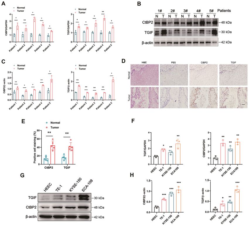
ONCOLOGY REPORTS 47: 29, 2022                                                       3

centrifugation, the off‑white pellet was re‑suspended by           tion with enhanced chemiluminescence detection reagent
PBS. The ECA109 cells were then transfected with recom‑            (SuperSignal™ West Pico PLUS Chemiluminescent Substrate,
binant lentiviral vectors and negative control viruses at a        34577) (Thermo Fisher Scientific, Inc.) at room temperature
multiplicity of infection (MOI) of 5. The following formula        for 5 min, and the bands were quantified using ImageJ (v1.48)
was used to calculate to volume of virus to be added: Virus        software (National Institutes of Health).
volume = MOI x cell number/virus titer. In addition, 1 µg/ml
polybrene was added, to improve the transduction efficiency.       Immunohistochemistry (IHC). Immunohistochemical
The mixture was centrifuged at 800 x g for 50 min at 32˚C.         staining was performed as previously described (22). Briefly,
Following centrifugation, the cells were seeded into 6‑well        4% PFA‑fixed ESCC tissue or normal tissue sections were
culture dish and incubated at 37˚C overnight. Transfected          deparaffinized in xylene, rehydrated in a graded alcohol series,
ECA109 cells were selected using 2.5 µg/ml puromycin for           and finally washed three times with 0.1 M PBS. The slides were
1 week. The efficiency of lentivirus‑mediated knockdown or         submerged in Tris‑EDTA buffer (100˚C, 20 min) for antigen
overexpression of CtBP2 or TGIF was verified using reverse         retrieval and cooled naturally at room temperature. The IHC kit
transcription‑quantitative PCR (RT‑qPCR) and western blot          was purchased from Zhongshan Golden Bridge Biotechnology
analysis.                                                          Co., Ltd. (cat. no. SP‑9000). The sections were blocked with
                                                                   10% goat serum (containing 0.1% triton‑100, reagent 2) for
RT‑qPCR. Total RNA extraction and reverse transcription were       30 min at 37˚C following treatment with reagent 1 (endogenous
performed as previously described (23). Total RNA from cells       peroxidase blockers) for 5 min, followed by incubation with
or tumor tissues was extracted using TRIzol® LS reagent            CtBP2 (sc‑17759; 1:200; Santa Cruz Biotechnology, Inc.) or
(Invitrogen; Thermo Fisher Scientific, Inc.) according to the      TGIF (ab52955; 1:1,000; Abcam) antibody overnight at 4˚C.
manufacturer's instructions and was quantified using the           After washing three times with 0.1 M PBS, the sections were
NanoDrop ND‑1000 spectrophotometer (Thermo Fisher                  incubated with reagent 3 [HRP‑conjugated goat anti‑rabbit
Scientific, Inc.). cDNA was synthesized by reverse transcription   IgG (A8919; 1:1,000) or HRP‑conjugated rabbit anti‑mouse
using the PrimeScript RT reagent kit (Takara Bio, Inc.). qPCR      IgG (A9044; 1:1000, Sigma‑Aldrich; Merck KGaA) secondary
was performed using SYBR Premix Ex Taq II (Tli RNaseH              antibodies] for 1 h at 37˚C, and subsequently incubated with
Plus; Takara Bio Inc.) on a LightCycler 96 system (Roche           reagent 4 at 37˚C for 1 h. Subsequently, hematoxylin (Beyotime
Diagnostics). Primers were synthesized by Sangon Biotech Co.,      Institute of Biotechnology) was used for re‑staining at room
Ltd. The thermocycling conditions were as follows: Firstly 95˚C    temperature for 2 h, and finally the sections were dehydrated in
for 10 min, followed by 95˚C for 10 sec, 60˚C for 15 sec and       graded alcohol until they were transparent in xylene. The scoring
72˚C for 20 sec, for 40 cycles. Data were analyzed using the       criteria (semi‑quantitative method) were comprehensive and
2‑ΔΔCq method with GAPDH as the internal reference control (34).   determined by the staining intensity and proportion of positively
All results are expressed as the mean ± standard deviation of      stained cells. The staining intensity score was as follows: 0 points
three independent experiments. The following primer sequences      for no staining, 1 point for weak staining (light yellow), 2 points
were used: GAPDH sense, 5'‑GACCTGACCTGCCGTCTA‑3'                   for moderate staining (yellowish‑brown), and 3 points for strong
and antisense, 5'‑AGGAGTGGGTGTCGCTGT‑3'; CtBP2                     staining (brown). The score for the proportion of positive cells was
sense, 5'‑CTGAGTTCCTGGCCTTTCTG‑3' and antisense,                   as follows: 0 for ≤5%, 1 for 5‑25%, 2 for 25‑50%, 3 for 50‑75%,
5'‑GACTTGATATCCGCGTCCTC‑3'; TGIF sense, 5'‑GGA                     4 for >75%. A final score was obtained by multiplying the two
TGAGGACAGCATGGACA‑3' and antisense, 5'‑AGGCAT                      scores, 0‑4 indicated negative, 4‑8 was weakly positive, and
TGTAACGGTGCT CA‑3'.                                                >8 was strongly positive. CtBP2 and TGIF expression in ESCC
                                                                   tissues and normal tissues of patients were detected according
Protein extraction and western blot analysis. Cells or tissues     to the aforementioned method and score. Low expression was
were lysed with ice‑cold lysis buffer (Beyotime Institute of       observed in the negative and weak positive groups, whereas
Biotechnology), as previously described (23). The protein          high expression was observed in the strongly positive group.
concentration of each sample was determined by BCA assay           After staining, five fields were randomly selected in each section
(Thermo Scientific, Inc.) according to the manufacturer's          (magnification, x40) under a microscope (Axio Imager 2; Carl
instructions. Proteins were separated via 10% SDS‑PAGE             Zeiss AG). The digitized images of immunohistochemistry were
(50 µg protein/lane), and then blotted onto PVDF membranes         quantitatively analyzed using Image‑Pro Plus 6.0 software (IPP
(Bio‑Rad, Hercules, CA). The membranes were blocked with           6.0; Media Cybernetics, Inc.).
Tris‑buffered saline and 0.1% Tween‑20 (TBST) supplemented
with 5% non‑fat milk for 2 h at room temperature, and the          H&E staining. H&E staining was used to distinguish tissue
PVDF membranes were incubated with primary antibodies              morphology as per the manufacturer's instructions (Beyotime
including, rabbit anti‑TGIF (ab52955; 1:1,000; Abcam), mouse       Institute of Biotechnology). ESCC tissue or normal tissue
anti‑CtBP2 (sc‑17759; 1:200; Santa Cruz Biotechnology, Inc.)       sections (4% PFA‑fixed) were deparaffinized in xylene, then
and rabbit anti‑β‑catenin (ab68183; 1:1,000; Abcam) overnight      rehydrated in a graded alcohol series. Nuclear staining was
at 4˚C. Rabbit anti‑β‑actin (ab8227; 1:1,000; Abcam) was used      performed with hematoxylin at room temperature for 10 min
as an internal control. The membranes were then incubated          following by flushing with running water to yield the color blue.
with HRP‑conjugated goat anti‑rabbit IgG (A8919; 1:1,000)          The sections were then differentiated with 1% hydrochloric
or rabbit anti‑mouse IgG (A9044; 1:1000) (Sigma‑Aldrich;           acid ethanol for 3 sec and then washing with running water
Merck KGaA) secondary antibodies at room temperature for           was continued. The cytoplasm was stained with eosin at room
2 h. Finally, western blot images were visualized by incuba‑       temperature for 30 sec, followed by 95% ethanol twice for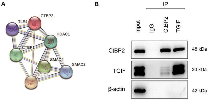
4              ju et al: CtBP2 INTERACTING WITH TGIF PROMOTES THE PROCESSION OF ESCC VIA THE Wnt PATHWAY

5 min, 100% ethanol twice for 5 min, 100% ethanol + xylene         facturer's instructions in three independent experiments. The
for 5 min (1:1), xylene for 5 min twice, and finally sealed with   transfected ECA109 cells at a density of 5x10³ cells/well were
neutral gum. H&E staining was observed under a microscope          seeded in 96‑well plates with 100 µl 10% FBS. After culturing
(Axio Imager 2; Carl Zeiss AG).                                    the cells for 24, 48 and 72 h with (10 and 100 nM, and 1, 10 and
                                                                   100 µM) XAV939 (Wnt signaling pathway inhibitor; Merck
Immunofluorescence. Staining reagents included CtBP2               KGaA) at 37˚C, 10 µl CCK‑8 reagent were added to each well
(sc‑17759; 1:200; Santa Cruz Biotechnology, Inc.) and TGIF         and incubated at 37˚C for a further 4 h. Cell viability was then
(ab52955; 1:1,000; Abcam) primary antibodies, anti‑mouse           measured based on the absorbance at 450 nm wavelength
(SAB3701092) or anti‑rabbit (F4890) secondary antibodies           (OD450) using a microplate reader (BioTek Instruments, Inc.).
(MilliporeSigma), and Hoechst 33342 (Beyotime Institute
of Biotechnology). The cells were digested with trypsin and        5‑Ethynyl‑2'‑deoxyuridine (EdU) staining. Cell proliferation
pipetted vigorously to make a single‑cell suspension. The cells    of the transfected ECA109 cells upon XAV939 treatment was
were placed on glass coverslips (24‑well plates), incubated at     investigated using EdU staining assay. Briefly, 1x10⁵ trans‑
37˚C and fixed with 4% PFA for 20 min at 15‑25˚C. The glass        fected ECA109 cells were resuspended in 200 µl DMEM,
coverslips were then washed three times with PBS for 10 min        and then seeded into 24‑well plates. Following incubation for
each and blocked with 10% bovine serum albumin (BSA) for           48 h with 10 µM XAV939, the cells were stained with EdU
2 h at 15‑25˚C. Subsequently, all cells were incubated with        (Beyotime Institute of Biotechnology) at room temperature for
primary antibody (1:200) overnight at 4˚C. The following day,      30 min against exposure to light. The cells were re‑stained
the cells were incubated with secondary antibody (1:2,000) for     with Hoechst 33342 for 10 min in the dark, followed by
2 h at 15‑25˚C. Finally, the cells were stained with Hoechst       washing with PBS. The number of EdU‑positive cells was
33258 (Beyotime Institute of Biotechnology) at room tempera‑       photographed, and five randomly selected fields were counted,
ture for 10 min, fixed with anti‑fade solution and imaged under    using a fluorescence microscope (Carl Zeiss AG) (magnifica‑
a fluorescence microscope (Zeiss AG).                              tion, x200). The experiments were performed in triplicate.

Co‑immunoprecipitation (Co‑IP) assay. The ECA109 cells             Wound healing assay. The migration of the transfected
were transfected with lentiviruses expressing CtBP2 or             ECA109 cells upon XAV939 treatment was investigated using
TGIF. At 1 week post‑transfection, the cells were subjected        a wound healing assay, as previously described (35). To ensure
to co‑immunoprecipitation assay using a commercial kit             the consistency of each initial scratch, a scratch chamber was
(Prod#26149; Thermo Fisher Scientific, Inc.). Briefly, firstly,    used to carry out the experiment. Briefly, 5x10⁴ transfected
for antibody immobilization, ultrapure water, 20X Coupling         ECA109 cells were seeded into the scratch chamber (70 µl
Buffer and 10 µg affinity‑purified antibody were added             cell suspension for each side of the scratch chamber), and then
directly to the agarose (50% protein A/G agarose with ratio        cultured overnight to adhere, followed by the removal of the
of 100 µl for a 1 ml sample) in the spin column. Secondly, for     scratch chamber. The cells were then washed twice with PBS,
the lysis of the cell cultures, the culture medium was carefully   in order to remove any non‑adherent cells. Before scratching,
removed from the cells. The cells were then washed once with       the cells were cultured in high‑glucose DMEM containing
1X modified Dulbecco's PBS. This was followed by the addi‑         10% FBS and supplemented with 1% penicillin/streptomycin
tion of 400 µl per well ice‑cold IP lysis/wash buffer (2X 50 ml,   mixture (36). To ensure the consistency of each initial scratch,
0.025 M Tris, 0.15 M NaCl, 0.001 M EDTA, 1% NP‑40,                 ibidi Culture‑Inserts (ibidi GmbH) were used to carry out
5% glycerol; pH 7.4) to the cells cultured in a 6‑well plate.      the experiment. Briefly, 5x10⁴ transfected ECA109 cells were
The cells were then incubated on ice for 5 min with periodic       seeded into the ibidi Culture‑Inserts (70 µl cell suspension for
mixing. The lysate was then transferred to a microcentrifuge       each side of the scratch chamber), and then cultured overnight
tube and centrifuged at ~13,000 x g at 4˚C for 10 min to           to adhere, followed by the removal of the ibidi Culture‑Inserts.
pellet the cell debris. The agarose and cell proteins were then    Following the removal of the ibidi Culture‑Inserts, the scratch
mixed and appropriate experimental controls were prepared,         wound was created. After scratching was complete, the cells
followed by rocking overnight at 4˚C. The column was then          were cultured in high‑glucose DMEM containing 2% FBS and
centrifuged at 1,000 x g at 4˚C for 1 min. The protocol uses       supplemented with 1% penicillin/streptomycin mixture. The
the IP Lysis/Wash Buffer (1M NaCl) for coupling and washing        migration (wound closure) of the indicated cells was monitored
the immune complex. The spin column was then placed in a           and photographed randomly at 0, 24 and 48 h. The wound
new collection tube. This was followed by the addition of 50 µl    healing rate (%) = (x h scratch area ‑ 0 h scratch area)/0 h
elution buffer and centrifugation. The tube was centrifuged at     scratch area x100. Independent experiments were performed
1,000 x g at 4˚C for 1 min and the flow‑through was collected      at least three times.
and analyze for protein. The resulting immuno‑complex
was analyzed using western blot analysis, with an anti‑TGIF        Transwell assay. The cell migratory and invasive abilities were
(ab52955; 1:1,000; Abcam) or anti‑CtBP2 (sc‑17759; 1:200;          examined using a Transwell assay, as previously described (35).
Santa Cruz Biotechnology, Inc.) antibody. To eliminate heavy       Briefly, for the migration assay, 1x10⁵ transfected ECA109
chain signals, a light chain‑specific secondary antibody (cat.     cells were resuspended in 200 µl DMEM, and then seeded into
no. 58802; Cell Signaling Technology, Inc.) was used.              the upper chamber of the Transwell (8 µm pore size; Corning,
                                                                   Inc.) in 24‑well plates. For the invasion assay, the upper cham‑
CCK‑8 assay. Cell viability was measured using CCK‑8 assay         bers were coated with Matrigel matrix (50 µl, BD Biosciences)
(Dojindo Molecular Technologies, Inc.) according to the manu‑      before seeding the cells, and 500 µl of 10% FBS were added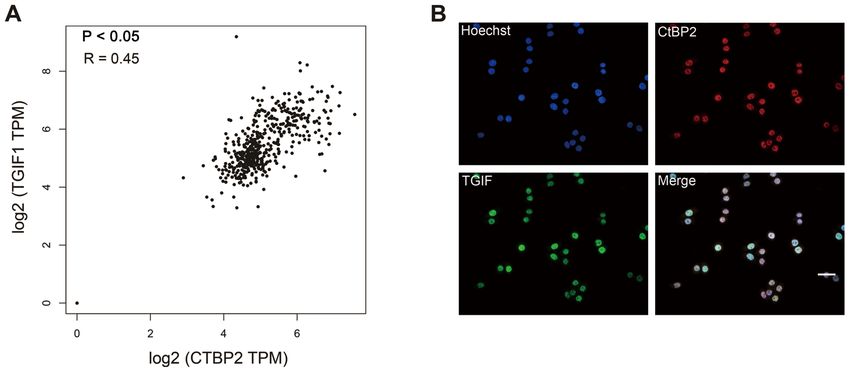
ONCOLOGY REPORTS 47: 29, 2022                                                                  5

Figure 1. Interaction between CtBP2 and TGIF. (A) Visualization of CtBP2 protein network based on STRING database analysis. (B) Western blot analysis of
immuno‑complexes. The cells were lysed, and immunoprecipitation was performed with anti‑TGIF or anti‑CtBP2 antibody. The immuno‑complexes were then
subjected to western blot analysis. IgG was used as the negative control. IP, immunoprecipitation; CtBP2, C‑terminal‑binding protein 2; TGIF, transforming
growth interacting factor; STRING, Search Tool for the Retrieval of Interacting Genes.

to the lower chamber. Following incubation at 37˚C for 48 h                    protein network is illustrated in Fig. 1A. As a predictive result,
with 10 µM XAV939, the non‑migrating cells were removed                        an interaction was detected between CtBP2 and TGIF.
using a cotton swab. However, the migrated or invaded cells                        Co‑IP was performed to validate the interaction between
on the underside were fixed with 4% PFA and stained with                       CtBP2 and TGIF. Following ECA109 cell transfection with
0.1% crystal violet (Beyotime Institute of Biotechnology) at                   the with the CtBP2 or TGIF lentiviral transduction vector,
room temperature for ~40 min. The number of migrating and                      the cells were subjected to Co‑IP assay with anti‑CtBP2 or
invading cells was photographed, and cells in five randomly                    anti‑TGIF antibody. Immuno‑complexes of CtBP2 and TGIF
selected fields were counted using a phase contrast microscope                 were observed using western blot analysis (Fig. 1B).
(Leica DM IL LED) (magnification, x200). All experiments
were performed in triplicate.                                                  Expression levels of CtBP2 and TGIF in ESCC tissues and
                                                                               cells. RT‑qPCR and western blot analysis were performed to
Bioinformatics analysis. The interactions between CtBP2 and                    examine the expression levels of CtBP2 and TGIF in ESCC
predictive proteins were analyzed using the STRING database                    tissues and cells. The mRNA expression levels of CtBP2 and
(https://string‑db.org). Gene Expression Profiling Interactive                 TGIF were robustly increased simultaneously in the tumor
Analysis (GEPIA) (http://gepia.cancer‑pku.cn) was used to                      tissues of representative patients with ESCC (Fig. 2A). Western
analyze the correlation between the expression of two inter‑                   blot analysis and IHC were performed to further confirm the
esting genes in a given tissue (37).                                           CtBP2 and TGIF expression levels in ESCC tissues. Compared
                                                                               with the adjacent normal tissues, the expression levels of
Statistical analysis. Statistical analysis was performed using                 CtBP2 and TGIF were significantly upregulated simultane‑
GraphPad Prism version 8.0 (GraphPad Software, Inc.) or                        ously in ESCC tissues (Fig. 2B‑E). RT‑qPCR and western blot
SPSS 23.0 (IBM Corp.). χ2 tests were performed to assess the                   analysis were performed to evaluate the mRNA and protein
clinical association between CtBP2 and TGIF expression and                     expression levels of CtBP2 and TGIF in the ESCC cell lines.
other tumor characteristics. Survival analysis was performed                   In comparison with the human normal esophageal epithelial
using the Kaplan‑Meier method and the log‑rank test. A Cox                     cell line, HEEC, the mRNA and protein expression levels of
proportional hazards regression model was established to                       both CtBP2 and TGIF were increased simultaneously in the
assess the factors independently associated with patient                       ESCC cell lines. In ECA109 cells, the levels of CtBP2 and
survival. Differences between two groups were compared                         TGIF were the highest (Fig. 2F‑H). These results revealed that
using an unpaired or paired Student's t‑test. To compare more                  CtBP2 and TGIF expression levels were significantly simulta‑
than two groups, one‑way ANOVA with post hoc Holm‑Sidak                        neously increased in ESCC tissues and cells.
correction for multiple comparisons was performed. All
experimental data are presented as the mean ± SD. P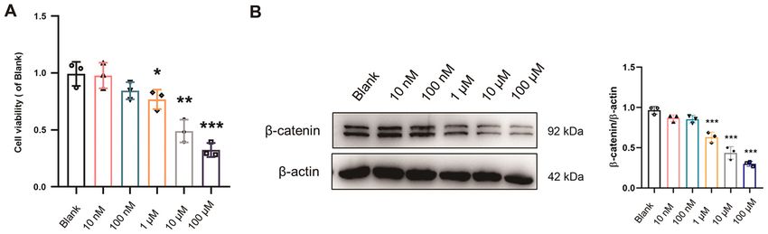
6                 ju et al: CtBP2 INTERACTING WITH TGIF PROMOTES THE PROCESSION OF ESCC VIA THE Wnt PATHWAY

Figure 2. Expression levels of CtBP2 and TGIF in tissues or cells of ESCC. (A) mRNA expression levels of CtBP2 and TGIF in ESCC tissues were examined
by RT‑qPCR. (B) Expression levels of CtBP2 and TGIF in ESCC tissues were tested by western blot analysis. N, normal tissues; T, tumor tissues. (C) Statistical
analysis of western blot analysis results. (D) Expression levels of CtBP2 and TGIF in ESCC tissues were tested by IHC. PBS was used as the negative control.
Scale bar=200 µm. (E) Statistical analysis of IHC results. (F) Expression levels of CtBP2 and TGIF in the human normal esophageal epithelial cell line, HEEC,
and the human ESCC cell lines, ECA109, TE‑1 and KYSE‑150, were examined using western blot analysis. (G) mRNA expression levels of CtBP2 and TGIF
in HEEC and ECA109, TE‑1 and KYSE‑150 cells were examined using RT‑qPCR. (H) Statistical analysis of the western blots in panel F. Data are presented
as the mean ± SD, and the Student's t‑test and one‑way ANOVA were performed. *P
ONCOLOGY REPORTS 47: 29, 2022                                                                           7

Table I. Association between CtBP2 and TGIF expression and the clinicopathological characteristics of patients with ESCC.

                                                         CtBP2                                                                 TGIF
                                           ‑‑‑‑‑‑‑‑‑‑------------------------‑‑‑‑‑‑‑‑‑‑                         ‑‑‑‑‑‑‑‑‑‑-----------------------‑‑‑‑‑‑‑‑‑‑
Characteristic                              Low                             High              P‑value           Low                             High          P‑value

Age (years)			                      0.912			                                                                                                                  0.742
8 ju et al: CtBP2 INTERACTING WITH TGIF PROMOTES THE PROCESSION OF ESCC VIA THE Wnt PATHWAY
Table II. Univariate analyses of various prognostic parameters in patients with ESCC using Cox regression analysis.
Univariate analysis
‑‑‑‑‑‑‑‑‑‑---------------------------------------------------------------------------------------------------‑‑‑‑‑‑‑‑‑‑
Parameter Hazard ratio 95% confidence interval P‑value
Age 1.473 0.867‑2.503 0.152
Sex 0.763 0.43‑1.354 0.355
Clinical stage 2.088 1.349‑3.232 0.001a
Histological differentiation 0.959 0.683‑1.346 0.807
Tumor diameter 2.381 1.299‑4.364 0.005a
T classification 1.497 1.049‑2.137 0.026a
N classification 1.684 1.240‑2.287 0.001a
Metastasis 1.240 0.747‑2.058 0.405
Depth 1.239 0.952‑1.613 0.112
CtBP2 2.361 1.288‑4.329 0.005a
TGIF 4.194 2.317‑7.590 0.001a
Statistical analyses were performed using the log‑rank test. aP90% (Fig. 5A, B, D and E). RT‑qPCR was performed
revealed that CtBP2 (P= 0.005), TGIF (P= 0.001), clinical to further examine CtBP2 and TGIF mRNA expression in the
stage (P= 0.001), tumor diameter (P= 0.005), T classification ECA109 cells transfected with the vectors. In comparison with
(P= 0.026) and N classification (P= 0.001) were independent the negative control (NC), the CtBP2 and TGIF mRNA expres‑
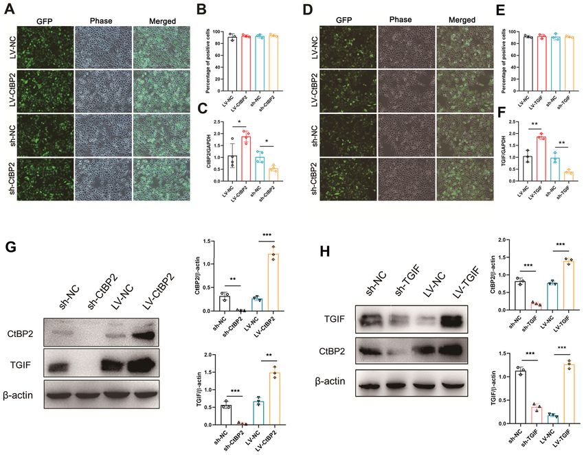
prognostic indicators of overall survival (Table II). sion levels were significantly increased or decreased (PONCOLOGY REPORTS 47: 29, 2022 9 Figure 5. Overexpression or knockdown of CtBP2 and TGIF in ECA109 cells. (A and D) ECA109 cells were observed under a fluorescence and phase contrast microscope following transfection. The recombinant lentiviral vectors contained the gene encoding the green fluorescent protein. Scale bar, 500 µm. (B and E) statistical analysis of percentage of positive cells in panels A and D. (C and F) mRNA expression levels of CtBP2 and TGIF in ECA109 cells following transfection was examined using RT‑qPCR. (G and H) Expression levels of CtBP2 and TGIF in ECA109 cells following transfection were examined using western blot analysis. Data are presented as the mean ± SD, and the Student's t‑test and one‑way ANOVA were performed. *P
10                ju et al: CtBP2 INTERACTING WITH TGIF PROMOTES THE PROCESSION OF ESCC VIA THE Wnt PATHWAY

Figure 7. Signaling pathway involved in the biological activity of CtBP2 in ECA109 cells. (A) Viability of ECA109 cells transfected with LV‑CtBP2 and
treated with 10 µM XAV939 for 24, 48 and 72 h, was measured using CCK‑8 assay. (B) The proliferation of ECA109 cells transfected with LV‑CtBP2 and
treated with 10 µM XAV939 for 48 h was measured using EdU staining. Scale bar, 50 µm. (C) Statistical analysis of EdU staining results. (D) The migration of
ECA109 cells transfected with LV‑CtBP2 and treated with 10 µM XAV939 for 48 h was measured using wound healing assay. Scale bar, 100 µm. (E) Statistical
analysis of the wound healing assay results. (F) The migration and invasion of ECA109 cells transfected with LV‑CtBP2 and treated with 10 µM XAV939
for 48 h was measured using Transwell assay. Scale bar, 100 µm. (G) Statistical analysis of the Transwell assay results. Data are presented as the mean ± SD,
and the Student's t‑test and one‑way ANOVA were performed. *P
ONCOLOGY REPORTS 47: 29, 2022                                                  11

demonstrated that the number of EdU‑positive cells was               and the survival of patients with ESCC. The results indicated
significantly decreased (P
12              ju et al: CtBP2 INTERACTING WITH TGIF PROMOTES THE PROCESSION OF ESCC VIA THE Wnt PATHWAY

and HS contributed to data analysis and interpretation. QJ and          14. Zhao LJ, Subramanian T, Vijayalingam S and Chinnadurai G:
                                                                            PLDLS‑dependent interaction of E1A with CtBP: Regulation
HS confirmed the authenticity of all the raw data. QJ, MJ and               of CtBP nuclear localization and transcriptional functions.
HS wrote the manuscript. All authors have read and approved                 Oncogene 26: 7544‑7551, 2007.
the final manuscript.                                                   15. Jecrois AM, Dcona MM, Deng X, Bandyopadhyay D, Grossman SR,
                                                                            Schiffer CA and Royer WE Jr: Cryo‑EM structure of CtBP2
                                                                            confirms tetrameric architecture. Structure 29: 310‑319.e5, 2021.
Ethics approval and consent to participate                              16. Ma Y, Sekiya M, Kainoh K, Matsuda T, Iwasaki H, Osaki Y,
                                                                            Sugano Y, Suzuki H, Takeuchi Y, Miyamoto T, et al: Transcriptional
                                                                            co‑repressor CtBP2 orchestrates epithelial‑mesenchymal tran‑
Patient written informed consent was obtained before the                    sition through a novel transcriptional holocomplex with OCT1.
study, according to the guidelines of the Ethics Committee                  Biochem Biophys Res Commun 523: 354‑360, 2020.
of the Affiliated Hospital of Nantong University, and ethics            17. Wang DP, Gu LL, Xue Q, Chen H and Mao GX: CtBP2 promotes
                                                                            proliferation and reduces drug sensitivity in non‑small cell lung
approval has been also provided from the respective ethics                  cancer via the Wnt/β‑catenin pathway. Neoplasma 65: 888‑897,
committee (2015 L132).                                                      2018.
                                                                        18. Thio SS, Bonventre JV and Hsu SI: The CtBP2 co‑repressor
                                                                            is regulated by NADH‑dependent dimerization and possesses
Patient consent for publication                                             a novel N‑terminal repression domain. Nucleic Acids Res 32:
                                                                            1836‑1847, 2004.
Not applicable.                                                         19. Zhao Z, Hao D, Wang L, Li J, Meng Y, Li P, Wang Y, Zhang C,
                                                                            Zhou H, Gardner K, et al: CtBP promotes metastasis of breast
                                                                            cancer through repressing cholesterol and activating TGF‑ β
Competing interests                                                         signaling. Oncogene 38: 2076‑2091, 2019.
                                                                        20. Dcona MM, Damle PK, Zarate‑Perez F, Morris BL, Nawaz Z,
                                                                            Dennis MJ, Deng X, Korwar S, Singh SJ, Ellis KC, et al:
The authors declare that they have no competing interests.                  Active‑site tryptophan, the target of antineoplastic C‑terminal
                                                                            binding protein inhibitors, mediates inhibitor disruption of CtBP
References                                                                  oligomerization and transcription coregulatory activities. Mol
                                                                            Pharmacol 96: 99‑108, 2019.
                                                                        21. Thomas G, Jacobs KB, Yeager M, Kraft P, Wacholder S, Orr N,
 1. Lin Y, Totsuka Y, Shan B, Wang C, Wei W, Qiao Y, Kikuchi S,             Yu K, Chatterjee N, Welch R, Hutchinson A, et al: Multiple loci
    Inoue M, Tanaka H and He Y: Esophageal cancer in high‑risk              identified in a genome‑wide association study of prostate cancer.
    areas of China: Research progress and challenges. Ann                   Nat Genet 40: 310‑315, 2008.
    Epidemiol 27: 215‑221, 2017.                                        22. Guan C, Shi H, Wang H, Zhang J, Ni W, Chen B, Hou S, Yang X,
 2. Yu C, Tang H, Guo Y, Bian Z, Yang L, Chen Y, Tang A, Zhou X,            Shen A and Ni R: CtBP2 contributes to malignant development
    Yang X, Chen J, et al; China Kadoorie Biobank Collaborative             of human esophageal squamous cell carcinoma by regulation of
    Group: Hot tea consumption and its interactions with alcohol and        p16INK4A. J Cell Biochem 114: 1343‑1354, 2013.
    tobacco use on the risk for esophageal cancer: a population‑based   23. Shi H, Mao Y, Ju Q, Wu Y, Bai W, Wang P, Zhang Y and Jiang M:
    cohort study. Ann Intern Med 168: 489‑497, 2018.                        C‑terminal binding protein‑2 mediates cisplatin chemoresistance
 3. Lin Y, Totsuka Y, He Y, Kikuchi S, Qiao Y, Ueda J, Wei W,               in esophageal cancer cells via the inhibition of apoptosis. Int J
    Inoue M and Tanaka H: Epidemiology of esophageal cancer in              Oncol 53: 167‑176, 2018.
    Japan and China. J Epidemiol 23: 233‑242, 2013.                     24. Wang Y, Liu F, Mao F, Hang Q, Huang X, He S, Wang Y, Cheng C,
 4. Yang S, Lin S, Li N, Deng Y, Wang M, Xiang D, Xiang G,                  Wang H, Xu G, et al: Interaction with cyclin H/cyclin‑dependent
    Wang S, Ye X, Zheng Y, et al: Burden, trends, and risk factors          kinase 7 (CCNH/CDK7) stabilizes C‑terminal binding protein 2
    of esophageal cancer in China from 1990 to 2017: An up‑to‑date          (CtBP2) and promotes cancer cell migration. J Biol Chem 288:
    overview and comparison with those in Japan and South Korea.            9028‑9034, 2013.
    J Hematol Oncol 13: 146, 2020.                                      25. Shi H, Xu J, Zhao R, Wu H, Gu L and Chen Y: FGF2 regulates
 5. Anandavadivelan P and Lagergren P: Cachexia in patients with            proliferation, migration, and invasion of ECA109 cells through
    oesophageal cancer. Nat Rev Clin Oncol 13: 185‑198, 2016.               PI3K/Akt signalling pathway in vitro. Cell Biol Int 40: 524‑533,
 6. Gao QY and Fang JY: Early esophageal cancer screening in                2016.
    China. Best Pract Res Clin Gastroenterol 29: 885‑893, 2015.         26. Shah A, Melhuish TA, Fox TE, Frierson HF Jr and Wotton D:
 7. Chen W, Li H, Zheng R, Ren J, Shi J, Cao M, Sun D, Sun X,               TGIF transcription factors repress acetyl CoA metabolic gene
    Cao X, Zhou J, et al: An initial screening strategy based on            expression and promote intestinal tumor growth. Genes Dev 33:
    epidemiologic information in esophageal cancer screening:               388‑402, 2019.
    A prospective evaluation in a community‑based cancer                27. Wang Y, Shi L, Li J, Li L, Wang H and Yang H: Long‑term
    screening cohort in rural China. Gastrointest Endosc 93:                cadmium exposure promoted breast cancer cell migration and
    110‑118.e2, 2021.                                                       invasion by up‑regulating TGIF. Ecotoxicol Environ Saf 175:
 8. Chen W, Li H, Ren J, Zheng R, Shi J, Li J, Cao M, Sun D, He S,          110‑117, 2019.
    Sun X, et al: Selection of high‑risk individuals for esophageal     28. Wotton D and Taniguchi K: Functions of TGIF homeodomain
    cancer screening: A prediction model of esophageal squamous             proteins and their roles in normal brain development and
    cell carcinoma based on a multicenter screening cohort in rural         holoprosencephaly. Am J Med Genet C Semin Med Genet 178:
    China. Int J Cancer 148: 329‑339, 2021.                                 128‑139, 2018.
 9. Liu M, He Z, Guo C, Xu R, Li F, Ning T, Pan Y, Li Y, Ding H,        29. Nakashima H, Tsujimura K, Irie K, Ishizu M, Pan M, Kameda T and
    Zheng L, et al: Effectiveness of intensive endoscopic screening         Nakashima K: Canonical TGF‑β signaling negatively regulates
    for esophageal cancer in China: a community‑based study. Am J           neuronal morphogenesis through TGIF/Smad complex‑mediated
    Epidemiol 188: 776‑784, 2019.                                           CRMP2 suppression. J Neurosci 38: 4791‑4810, 2018.
10. Kelly RJ: Emerging multimodality approaches to treat localized      30. Sharma A, Sinha NR, Siddiqui S and Mohan RR: Role of
    esophageal cancer. J Natl Compr Canc Netw 17: 1009‑1014, 2019.          5'TG3'‑interacting factors (TGIFs) in Vorinostat (HDAC
11. Borggreve AS, Kingma BF, Domrachev SA, Koshkin MA,                      inhibitor)‑mediated Corneal Fibrosis Inhibition. Mol Vis 21:
    Ruurda JP, van Hillegersberg R, Takeda FR and Goense L:                 974‑984, 2015.
    Surgical treatment of esophageal cancer in the era of multimo‑      31. Du R, Shen W, Liu Y, Gao W, Zhou W, Li J, Zhao S, Chen C,
    dality management. Ann N Y Acad Sci 1434: 192‑209, 2018.                Chen Y, Liu Y, et al: TGIF2 promotes the progression of lung
12. Chen L, Wang L, Qin J and Wei DS: CtBP2 interacts with                  adenocarcinoma by bridging EGFR/RAS/ERK signaling to
    ZBTB18 to promote malignancy of glioblastoma. Life Sci 262:             cancer cell stemness. Signal Transduct Target Ther 4: 60,
    118477, 2020.                                                           2019.
13. Wang H, Xiao Z, Zheng J, Wu J, Hu XL, Yang X and Shen Q:            32. Liu ZM, Tseng HY, Tsai HW, Su FC and Huang HS: Transforming
    ZEB1 represses neural differentiation and cooperates with               growth factor β‑interacting factor‑induced malignant progression
    CTBP2 to dynamically regulate cell migration during neocortex           of hepatocellular carcinoma cells depends on superoxide
    development. Cell Rep 27: 2335‑2353.e6, 2019.                           production from Nox4. Free Radic Biol Med 84: 54‑64, 2015.
ONCOLOGY REPORTS 47: 29, 2022 13
33. Zhang MZ, Ferrigno O, Wang Z, Ohnishi M, Prunier C, Levy L, 39. Huang X, Zhou X, Hu Q, Sun B, Deng M, Qi X and Lü M: Advances
Razzaque M, Horne WC, Romero D, Tzivion G, et al: TGIF in esophageal cancer: A new perspective on pathogenesis asso‑
governs a feed‑forward network that empowers Wnt signaling ciated with long non‑coding RNAs. Cancer Lett 413: 94‑101, 2018.
to drive mammary tumorigenesis. Cancer Cell 27: 547‑560, 40. Szklarczyk D, Morris JH, Cook H, Kuhn M, Wyder S,
2015. Simonovic M, Santos A, Doncheva NT, Roth A, Bork P, et al: The
34. Livak KJ and Schmittgen TD: Analysis of relative gene expression STRING database in 2017: Quality‑controlled protein‑protein
data using real‑time quantitative PCR and the 2(‑Delta Delta association networks, made broadly accessible. Nucleic Acids
C(T)) method. Methods 25: 402‑408, 2001. Res 45 (D1): D362‑D368, 2017.
35. Shi H, Shi J, Zhang Y, Guan C, Zhu J, Wang F, Xu M, Ju Q, 41. Wang Y, Shi L, Li J, Wang H and Yang H: The roles of TG‑interacting
Fang S and Jiang M: Long non‑coding RNA DANCR promotes factor in cadmium exposure‑promoted invasion and migration of
cell proliferation, migration, invasion and resistance to apoptosis lung cancer cells. Toxicol In Vitro 61: 104630, 2019.
in esophageal cancer. J Thorac Dis 10: 2573‑2582, 2018. 42. Zhang LN, Zhao L, Yan XL and Huang YH: Loss of G3BP1
36. Mo Y, Wang Y, Zhang S, Xiong F, Yan Q, Jiang X, Deng X, suppresses proliferation, migration, and invasion of esophageal
Wang Y, Fan C, Tang L, et al: Circular RNA circRNF13 inhibits cancer cells via Wnt/β ‑catenin and PI3K/AKT signaling
proliferation and metastasis of nasopharyngeal carcinoma via pathways. J Cell Physiol 234: 20469‑20484, 2019.
SUMO2. Mol Cancer 20: 112, 2021. 43. Yu F, Yu C, Li F, Zuo Y, Wang Y, Yao L, Wu C, Wang C and
37. Tang Z, Li C, Kang B, Gao G, Li C and Zhang Z: GEPIA: A web Ye L: Wnt/β‑catenin signaling in cancers and targeted therapies.
server for cancer and normal gene expression profiling and inter‑ Signal Transduct Target Ther 6: 307, 2021.
active analyses. Nucleic Acids Res 45 (W1): W98‑W102, 2017.
38. Shetti D, Zhang B, Fan C, Mo C, Lee BH and Wei K: Low dose This work is licensed under a Creative Commons
of paclitaxel combined with XAV939 attenuates metastasis, Attribution-NonCommercial-NoDerivatives 4.0
angiogenesis and growth in breast cancer by suppressing Wnt International (CC BY-NC-ND 4.0) License.
signaling. Cells 8: 8, 2019.You can also read