Taxonomy, Phylogeny, Molecular Dating and Ancestral State Reconstruction of Xylariomycetidae (Sordariomycetes)
←
→
Page content transcription
If your browser does not render page correctly, please read the page content below
Taxonomy, Phylogeny, Molecular Dating and Ancestral State Reconstruction of
Xylariomycetidae (Sordariomycetes)
Milan C. Samarakoon
School of Life Science and Technology, Center for Informational Biology, University of Electronic Science and Technology of China, Chengdu 611731, P.R.
China
Kevin D Hyde ( kdhyde3@gmail.com )
Center of Excellence in Fungal Research
Sajeewa S. N. Maharachchikumbura
School of Life Science and Technology, Center for Informational Biology, University of Electronic Science and Technology of China, Chengdu 611731, P.R.
China
Marc Stadler
Institute of Microbiology, Technische Universität Braunschweig, Spielmannstraβe 7, 38106 Braunschweig, Germany
E. B. Gareth Jones
Department of Botany and Microbiology, College of Science, King Saud University, P.O Box 2455, Riyadh 11451, Kingdom of Saudi Arabia
Itthayakorn Promputtha
Department of Biology, Faculty of Science, Chiang Mai University, Chiang Mai 50200, Thailand
Nakarin Suwannarach
Department of Biology, Faculty of Science, Chiang Mai University, Chiang Mai 50200, Thailand
Erio Camporesi
A.M.B. Gruppo, Micologico Forlivese "Antonio Cicognani", Via Roma 18, Forlí, Italy
Timur S. Bulgakov
Department of Plant Protection, Federal Research Centre the Subtropical Scientific Centre of the Russian Academy of Sciences, Yana Fabritsiusa Street 2/28,
Sochi 354002, Krasnodar Region, Russia
Jian-Kui Liu
School of Life Science and Technology, Center for Informational Biology, University of Electronic Science and Technology of China, Chengdu 611731, P.R.
China
Research Article
Keywords: 33 new taxa, Amphisphaeriales, Appendicosporaceae, Evolution, Stromata, Xylariales
Posted Date: October 1st, 2021
DOI: https://doi.org/10.21203/rs.3.rs-935829/v1
License: This work is licensed under a Creative Commons Attribution 4.0 International License. Read Full License
Page 1/87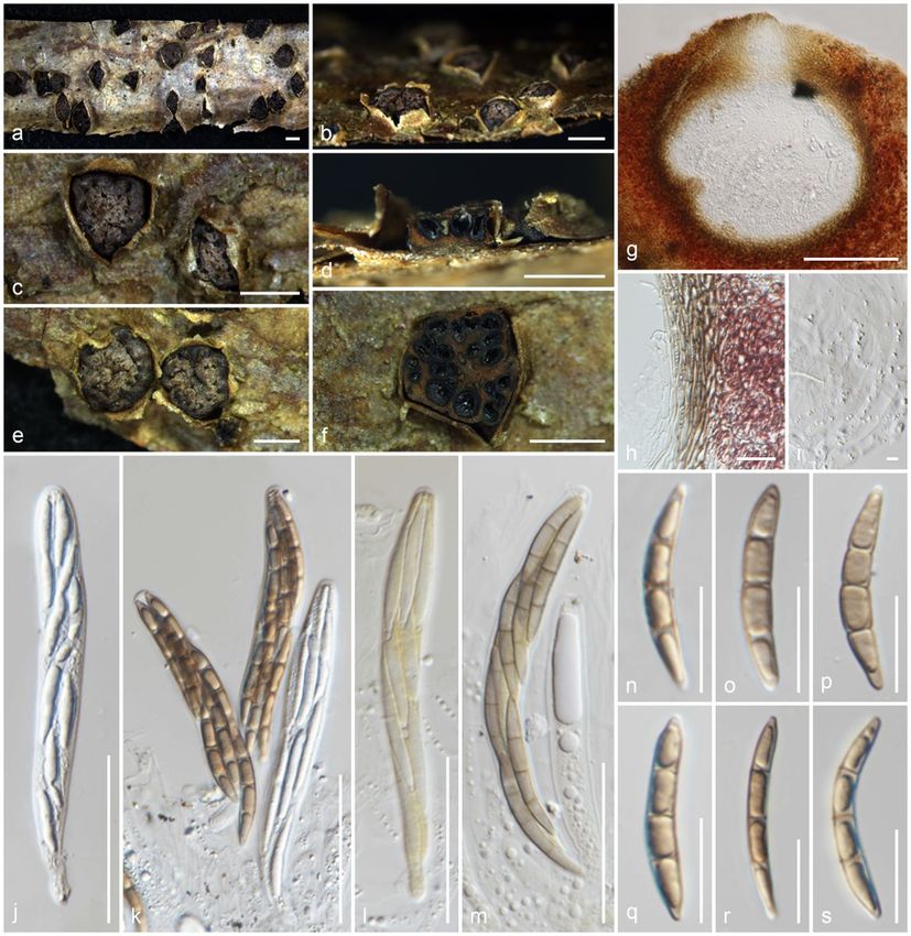
Abstract
Xylariomycetidae ( Ascomycota ) is a highly diversified group with variable stromatic characters. Our research focused on inconspicuous stromatic xylarialean
taxa from China, Italy, Russia, Thailand and the United Kingdom. Detailed morphological descriptions, illustrations and combined ITS-LSU- rpb 2- tub 2- tef 1
phylogenies revealed 38 taxa from our collections belonging to Amphisphaeriales and Xylariales . A new family ( Appendicosporaceae ), five new genera (
Magnostiolata , Melanostictus , Neoamphisphaeria , Nigropunctata and Paravamsapriya ), 27 new species ( Acrocordiella photiniicola , Allocryptovalsa
sichuanensis , Amphisphaeria parvispora , Anthostomella lamiacearum , Apiospora guiyangensis , Ap. sichuanensis , Biscogniauxia magna , Eutypa camelliae
, Helicogermslita clypeata , Hypocopra zeae , Magnostiolata mucida , Melanostictus longiostiolatus , Me. thailandicus , Nemania longipedicellata , Ne.
delonicis , Ne. paraphysata , Ne. thailandensis , Neoamphisphaeria hyalinospora , Neoanthostomella bambusicola , Nigropunctata bambusicola , Ni.
nigrocircularis , Ni. thailandica , Occultitheca rosae , Paravamsapriya ostiolata , Peroneutypa leucaenae , Seiridium italicum and Vamsapriya mucosa ) and
seven new host/geographical records are introduced and reported. Divergence time estimates indicate that Delonicicolales diverged from Amphisphaeriales +
Xylariales at 161 (123–197) MYA. Amphisphaeriales and Xylariales diverged 154 (117–190) MYA with a crown age of 127 (92–165) MYA and 147 (111–184)
MYA, respectively. Appendicosporaceae ( Amphisphaeriales ) has a stem age of 89 (65–117) MYA. Ancestral character state reconstruction indicates that
astromatic, clypeate ascomata with aseptate, hyaline ascospores that lack germ slits may probably be ancestral Xylariomycetidae having plant-fungal
endophytic associations. The Amphisphaeriales remained mostly astromatic with common septate, hyaline ascospores. Stromatic variations may have
developed mostly during the Cretaceous period. Brown ascospores are common in Xylariales , but they first appeared in Amphisphaeriaceae ,
Melogrammataceae and Sporocadaceae during the early Cretaceous. The ascospore germ slits appeared only in Xylariales during the Cretaceous after the
divergence of Lopadostomataceae . Hyaline, filiform and apiospores may have appeared as separate lineages providing the basis to Xylariaceae , which may
have diverged independently. The future classification of polyphyletic xylarialean taxa will not be based on stromatic variations, but the type of ring, the colour
of the ascospores, and the presence or absence of the type of germ slit.

Introduction
Eriksson and Winka (1997) introduced Xylariomycetidae with a single order Xylariales, based on morphology and SSU phylogeny. Eriksson (1983) suggested
to treat Amphisphaeriales as accommodating Amphisphaeriaceae, Cainiaceae, Clypeosphaeriaceae and Hyponectriaceae based on ascospore morphology.
Barr (1990), however, arranged Amphisphaeriales, Diatrypales, Phyllachorales and Trichosphaeriales in Xylariales. Kang et al. (1998) revived Amphisphaeriales
based on 5.8S-ITS2 molecular phylogeny and coelomycetous asexual morphs and accepted Clypeosphaeriaceae and Cainiaceae in the order. Subsequently,
Kang et al. (2002) revised their previous conclusion, and Amphisphaeriaceae and Xylariaceae were accepted in Xylariales based on a combined SSU-ITS
molecular phylogeny. Kirk et al. (2008) documented Xylariales as the only order in Xylariomycetidae with nine families (Amphisphaeriaceae, Cainiaceae,
Clypeosphaeriaceae, Diatrypaceae, Graphostromataceae, Hyponectriaceae, Iodosphaeriaceae, Myelospermataceae and Xylariaceae). A revision with additional
collections and the morpho-phylogenetic study revealed that Xylariomycetidae consisted of two orders; Amphisphaeriales (Amphisphaeriaceae, Bartaliniaceae,
Clypeosphaeriaceae, Discosiaceae, Pestalotiopsidaceae and Phlogicylindriaceae) and Xylariales (Apiosporaceae, Cainiaceae, Coniocessiaceae, Diatrypaceae,
Graphostromataceae, Hyponectriaceae, Iodosphaeriaceae, Lopadostomataceae, Melogrammataceae, Pseudomassariaceae, Vialaeaceae and Xylariaceae)
(Senanayake et al. 2015). In an outline of Sordariomycetes, Maharachchikumbura et al. (2016) treated Amphisphaeriales as a synonym of Xylariales due to
inadequate statistical support in the phylogenetic analyses by Senanayake et al. (2015), and accepted 22 families in Xylariales based on morphology and
combined LSU-SSU-tef1-rpb2 phylogeny. With divergence time estimates as additional information for the standardizing of higher ranks, Samarakoon et al.
(2016) and Hongsanan et al. (2017) accepted Xylariales and Amphisphaeriales in Xylariomycetidae, which may have diverged around 152–187 MYA.

Perera et al. (2017) introduced Delonicicolales as the third order in Xylariomycetidae, which diverged from the Amphisphaeriales+Xylariales clade at 181 (133–
234) MYA. Following several consecutive morpho-molecular studies, Hyde et al. (2020b) provided an outline for the Sordariomycetes, including
Xylariomycetidae with three orders as Amphisphaeriales (17 families), Delonicicolales (2 families) and Xylariales (15 families). In addition,
Myelospermataceae is treated as a Xylariomycetidae families incertae sedis due to lack of molecular data.

Species in Xylariomycetidae are distributed worldwide with dynamic nutritional relationships as endophytes (U’Ren et al. 2016; Rashmi et al. 2019), pathogens
and saprobes (Zhang et al. 2006; Daranagama et al. 2018; Hyde et al. 2020b). Xylariomycetidae comprises species with conspicuous and inconspicuous,
superficial or immersed stromata, usually black and thick-walled ascomata with periphysate, papillate ostioles; unitunicate or rarely bitunicate-like asci with or
without apical ring bluing in Melzer’s reagent, and mostly pigmented ascospores in their sexual state and hyphomycetous or coelomycetous asexual morphs
(Smith et al. 2003; Wang et al. 2004; Zhang et al. 2006; Jaklitsch and Voglmayr 2012; Senanayake et al. 2015; Hyde et al. 2020b). Conspicuous massive,
stalked or sessile stromata are commonly found among xylarialean taxa (Daranagama et al. 2016b). Daranagama et al. (2016b) reviewed the stromatic
diversity of xylarialean taxa and considered the collection of taxa with inconspicuous form to be sparse.

As a result, the taxonomic placement of many taxa lacking distinct stromata are uncertain (Daranagama et al. 2018; Wendt et al. 2018). Several recent studies
have focused on the morphology and phylogeny of inconspicuous xylarialean taxa, including the re-examination of herbarium specimens (Daranagama et al.
2018). Those studies not only focused on providing morpho-molecular information but also placed them in higher ranks (e.g. Barrmaeliaceae, Induratiaceae,
Fasciatisporaceae and Oxydothidaceae) (Konta et al. 2016; Voglmayr et al. 2018; Hyde et al. 2020a; Samarakoon et al. 2020c). The genera, which have been
introduced in new families were previously accepted with uncertain morphologies and phylogenies. They are morphologically unique in having inconspicuous,
immersed ascomata, that do not have key characters for delimiting higher ranks as compared to conspicuous stromatic xylarialean taxa. However, the asci
and ascospore morphologies were cardinal characters coupled with molecular phylogenies towards establishing new higher ranks. There are many taxonomic
uncertainties of xylarialean taxa that are not yet resolved.

We are researching xylarialean taxa towards resolving taxonomic uncertainties. Here we provided new collections with their morphology, analysed their DNA
sequences and investigated their phylogenetic relationships to better identify and classify them. We evaluated different stromatic characters of selected taxa

                                                                           Page 2/87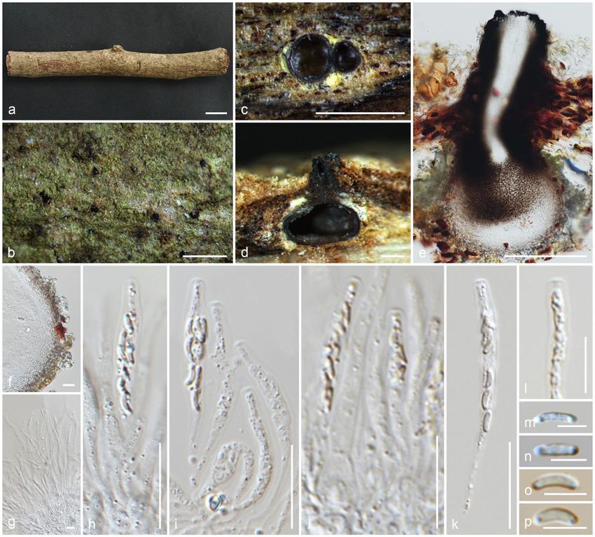
in Xylariomycetidae to reconstruct the ancestral state. In addition, ascospore characters i.e. colour, septation and the presence or absence of a germ slit were
evaluated to understand the ancestral state of the xylarialean taxa.

Materials And Methods
Collection, isolation and morphological studies

Fresh specimens were collected and received from China, Italy, Russia, Thailand and the United Kingdom during 2016–2020. External examinations were
made as described in Samarakoon et al. (2020b). Indian ink, Congo red and Melzer’s reagent were used where necessary. The Tarosoft (R) Image Frame Work
(v 0.9.7) program and Adobe Photoshop CS6 software (Adobe Systems, USA) were used for measuring and processing images.

Axenic cultures were obtained from single spores or tissues by the method described in Senanayake et al. (2020). Germinating spores were observed with a
Motic SMZ 168 Stereo Zoom microscope and transferred to potato dextrose agar (PDA; 39 g/l distilled water, Difco potato dextrose). The cultures were
incubated at 25–30°C for 4–6 weeks, with frequent observations. The herbarium specimens were deposited in the Mae Fah Luang University Herbarium
(MFLU), Chiang Rai, Thailand and the Cryptogamic Herbarium of Kunming Institute of Botany Academia Sinica (HKAS), Chinese Academy of Sciences,
Kunming, China. Ex-type cultures were deposited in the Mae Fah Luang University Culture Collection (MFLUCC), the Guizhou Culture Collection (GZCC),
Guizhou, and China General Microbiological Culture Collection Center (CGMCC), Institute of Microbiology Chinese Academy of Sciences, Beijing, China. New
taxa were linked with Facesoffungi and Index Fungorum databases as explained in Jayasiri et al. (2015) and Index Fungorum (http://www.indexfungorum.org;
accessed at 9 September 2021).

In addition, selected cultures deposited in MFLUCC were loaned for regenerating missing sequences. Subcultures were obtained and incubated them at 25–
30°C for 4–6 weeks with frequent observations. Those cultures were used for total DNA extraction and PCR amplification.

DNA extraction, PCR amplification and sequencing

Fresh mycelium was scraped from the margins of colonies on PDA plates, incubated at 25–30°C for four weeks. When fungi failed to grow in culture, DNA
was extracted directly from the fruiting bodies. Total DNA extraction kits were used according to the manufacturer’s instructions [Sangon Biotech (Shanghai)
Co. Ltd. China]. The primers and PCR protocols are summarised in Table 1. The total volume of 25 μl containing 12.5 μl of 2× PCR Master Mix with dye [0.1 U
Taq Polymerase/μl, 500 μM dNTP each, 20 mM Tris-HCl (pH 8.3), 100 mM KCl, 3 mM MgCl2], 1 μl of each primer, 9.5 μl of double-distilled water and 1 μl (100–
500 ng) of DNA template. All the PCR products were immediately subjected to 4°C and were visualised on 1% agarose electrophoresis gels stained with
GoldView I nuclear staining dye (1 µL/10 mL of agarose) with D2000 DNA ladder (Realtimes Biotech, Beijing, China). DNA sequencing was performed at
Sangon Biotech (Shanghai) Co. Ltd., China.

Phylogenetic analyses

All the assembled sequences were used for BLAST search (https://www.ncbi.nlm.nih.gov) (Altschul et al. 1990). Related sequences for newly obtained
sequences were downloaded from the GenBank (Supplementary Table 1). Individual loci were aligned using FFT-NS-2 Tree-based progressive method, 20
PAM/ k = 2 Scoring matrix for nucleotide sequences and 1.0 Gap opening penalty settings of MAFFT V.7.036 (http://mafft.cbrc.jp/alignment/server/) (Katoh
et al. 2019) and improved manually when necessary, using BioEdit v. 7.0 (Hall 1999). ITS and LSU sequences were trimmed with TrimAl [(v.1.0) Gappyout
option] (Capella-Gutierrez et al. 2009). Exon regions of rpb2, tub2 and tef1 were extracted with reference to Amphirosellinia nigrospora (HAST 91092308) and
Graphostroma platystomum (CBS 270.87).

Characters were assessed to be unordered and equally weighted. MrModeltest 2.3 was performed for each locus to estimate the best-fit evolutionary model
under the Akaike Information Criterion (AIC) (Nylander 2004). Phylogenies were generated using maximum-likelihood (ML) and Bayesian Inference (BI)
analyses using single and ITS-LSU-rpb2-tub2 and ITS-LSU-rpb2-tub2-tef1 combined alignments. For future studies, all the newly generated sequences were
deposited in GenBank (Dissanayake et al. 2020) (Table 2).

The ML analyses were performed with IQ-TREE (Nguyen et al. 2015, Trifinopoulos et al. 2016) using the ML+rapid bootstrap setting with 1,000 replicates. The
Bayesian tree was generated using MCMC sampling in MrBayes v3.1.2 (Huelsenbeck and Ronquist 2001; Zhaxybayeva and Gogarten 2002) for 10,000,000
MCMC generations using four chains and partition analysis with 100 sample frequencies. The first 25,000 (25% from total) trees were in the burn-in phase and
were discarded. The remaining 75,000 trees were used to calculate the posterior probability (PP). The resulting trees were viewed with FigTree v.1.4.0
(Rambaut 2012), and the final layout was done with Adobe Illustrator® CS5 (Version 15.0.0, Adobe®, San Jose, CA). The final alignment and tree were
registered in TreeBASE (http://www.treebase.org/) under the submission ID: XXXX.

Divergence time estimation

Divergence time estimation among the families in Xylariomycetidae was performed using the BEAST.v1.10.4 program. Combined LSU, ITS, rpb2, tub2 and tef1
DNA loci were used for the analysis, representing 240 taxa (Supplementary Table 2). The XML file was obtained, including the partitioned alignment, using the
BEAUti (BEAST package). The crown age of Xylariomycetidae was used as the secondary calibration node (mean = 168 MYA, SD = 16, Normal distribution)
(Samarakoon et al. 2016; Hongsanan et al. 2017). The analysis was performed for 80,000,000 generations using BEAST.v1.10.4 (Suchard et al. 2018),
obtaining logging parameters and trees for every 5000 generations. Effective sample sizes (ESS) of parameters were checked using Tracer v.1.6 (Rambaut et
al. 2013) (ESS > 200). The first 20% trees were discarded based on the ESS values, and the remaining trees were used to generate a maximum clade credibility
tree by using TreeAnnotator v1.10.4. The resulted tree was viewed with FigTree v.1.4.0 (Rambaut 2012), and the final layout was done with Adobe Illustrator®
CS5 (Version 15.0.0, Adobe®, San Jose, CA). The geographical timescale was followed as in Walker (2019).

                                                                          Page 3/87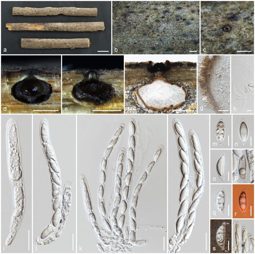
Ancestral character state analyses

Bayesian Binary MCMC was performed in RASP 3.2.1 (Reconstruct Ancestral State in Phylogenies) to construct ancestral character state (Yu et al. 2015).
Time-calibrated maximum clade credibility tree reconstructed in BEAST was used for the analysis and exported to RASP 3.2.1. Each terminal in the tree was
coded for seven stromatic characters (Table 3), and undetermined sexual morphs were treated as separate undetermined characters for the family level. In
addition, characters of ascospore septation (aseptate/septate/apiosporous/undetermined), ascospore colour (hyaline/brown/undetermined) and ascospore
germ slit (presence/absence/undetermined) were evaluated (Supplementary Table 2). Bayesian Binary MCMC trees were performed and visualised in RASP
3.2.1 using default settings as follows: 1,010,000 iterations for BayesTraits with a burnin of 10,000, sampling 1000 trees and with 10 ML trees; 50,000
generations for Bayesian Binary MCMC, with 10 chains, a sampling frequency of 100, a temperature of 0.1, state frequencies fixed (JC), and among-site rate
variation equal.

Results
Phylogenetic analyses

All gene regions resulted in GTR+I+G model. Maximum likelihood tree topologies for each gene dataset and combined datasets were compared, and the
overall tree topology was congruent to those obtained from the combined dataset. The RAxML analysis of the combined ITS-LSU-rpb2-tub2-tef1 dataset
yielded the best-scoring tree (Fig. 1). Bayesian posterior probabilities from MCMC were evaluated with a final average standard deviation of split frequencies
less than 0.01.

Delonicicolales clusters as basal to the Amphisphaeriales and Xylariales clades, with 100%/1.00 PP statistical support. Amphisphaeriales and Xylariales form
distinct clades with 99%/0.92 PP statistical support, similar to a previous study in Hyde et al. (2020b). Amphisphaeriales comprised 27 clades (Clade Am)
including 21 families, while Xylariales (Clade Xy) comprised 31 clades including 16 families. Uncertain clades with a single or few taxa are identified as six in
Amphisphaeriales and 15 in Xylariales. Forty-nine of newly generated sequences from our study group with Xylariales and nine with Amphisphaeriales.

One of our collections (HKAS 107015) is similar to Appendicospora hongkongensis and is introduced here as a reference specimen. Two isolates (MFLU 19-
2131, HKAS 106988), Neoamphisphaeria hyalinospora gen. et sp. nov. form a sister clade to Appendicospora, and here we introduce Appendicosporaceae
fam. nov. in Amphisphaeriales to accommodate Appendicospora and Neoamphisphaeria. New species for the families Amphisphaeriaceae (Amphisphaeria
parvispora sp. nov. MFLU 18-0767), Apiosporaceae (Apiospora guiyangesnsis sp. nov. HKAS 102403, Ap. sichuanensis sp. nov. HKAS 107008) and
Sporocadaceae (Seiridium italicum sp. nov. MFLU 16-1315) are introduced with high statistical support.

Seven newly generated sequences group in Diatrypaceae and identify them as, Allocryptovalsa sichuanensis sp. nov. (HKAS 107017), Diatrype disciformis
(MFLU 17-1549), Eutypa camelliae sp. nov. (HKAS 107022, MFLU 20-0182HT), Melanostictus longiostiolatus gen. et sp. nov. (MFLU 19-2146), Me.
thailandicus sp. nov. (MFLU 19-2123) and Peroneutypa leucaenae sp. nov. (MFLU 18-0816). Twelve taxa cluster in Xylariaceae sensu stricto. Helicogermslita
clypeata sp. nov. (MFLU 18-0852, HKAS 102321) clusters in Astrocystis+Collodiscula clade. Four Nemania species, Ne. longipedicellata sp. nov. (MFLU 18-
0819), Ne. delonicis sp. nov. (MFLU 19-2124), Ne. paraphysata sp. nov. (MFLU 19-2121) and Ne. thailandensis sp. nov. (MFLU 19-2122, MFLU 19-2117) are
supported with high statistical support. A single taxon, MFLU 18-0809, clusters with Stromatoneurospora phoenix (BCC 82040) sister to the Hypocopra clade.
Three newly generated sequences cluster in Vamsapriyaceae. Paravamsapriya ostiolata gen. et sp. nov. (MFLU 18-0761, MFLU 18-0813) and Vamsapriya
mucosa sp. nov. (MFLU 18-0103) are described here.

Anthostomella-like taxa collected in this study group in seven clades (Xy3, Xy4, Xy22, Xy23, Xy24, Xy25 and Xy26) in Xylariales. Magnostiolata mucida gen. et
sp. nov. (MFLU 19-2133) and Occultitheca rosae sp. nov. (HKAS 102393) cluster between Clypeosphaeriaceae and Induratiaceae as distinct clades.
Neoanthostomella bambusicola sp. nov. (MFLU 18-0796) is accommodated in Clade Xy22 with Neo. pseudostromatica, the generic type with high statistical
support (100%/1.00 PP). Clade Xy23 comprises Calceomyces, Ceratocladium, Circinotrichum, Gyrothrix and Xenoanthostomella. Our new collection, MFLU 18-
0840, clusters as a sister to Xe. chromolaenae (MFLUCC 17-1484) with 100%/1.00 PP statistical support. Anthostomella lamiacearum, “Neoathostomella fici”
and “Neo. viticola” cluster group in Clade Xy24 (Anthostomella helicofissa clade), which is distinct from Neo. pseudostromatica. Our new collections,
Anthostomella lamiacearum sp. nov. (MFLU 18-0101, HKAS 102325) clustered in Clade Xy24.

Several Anthostomella, Alloanthostomella and Pseudoanthostomella taxa cluster in Clade Xy25. Three newly generated sequences cluster in
Pseudoanthostomella. Based on morphological similarities and phylogeny, we accepted those collections as Ps. pini-nigrae. Clade Xy26 accommodates five
taxa (three species): Nigropunctata bambusicola gen. et sp. nov. (MFLU 19-2134, MFLU 19-2145), Ni. nigrocircularis sp. nov. (MFLU 19-2130) and Ni.
thailandica sp. nov. (MFLU 19-2118, HKAS 106975). Thirteen sequences of our collections clustered in other families’; viz. Coniocessiaceae (Paraxylaria
xylostei MFLU 17-1645, MFLU 17-1636, HKAS 102313), Fasciatisporaceae (Fasciatispora cocoes MFLU 19-2143, HKAS 107000), Graphostromataceae
(Biscogniauxia magna sp. nov. MFLU 18-0850, Bi. Petrensis HKAS 102388, Camillea tinctor MFLU 18-0786), Lopadostomataceae (Lopadostoma quercicola
MFLU 17-0843, MFLU 17-0731, MFLU 17-0940) and Requienellaceae (Acrocordiella photiniicola sp. nov. MFLU 17-1552, HKAS 102287).

Divergence time estimation

Three clades were obtained in Xylariomycetidae, including 39 families representing the orders Amphisphaeriales, Delonicicolales and Xylariales (Fig. 2).
According to the estimates, Delonicicolales diverged from Amphisphaeriales+Xylariales 161 (123–197) MYA. Amphisphaeriales and Xylariales diverged 154
(117–190) MYA with a crown age of 127 (92–165) MYA and 147 (111–184) MYA, respectively, with similar results to Hyde et al. (2020b) and Samarakoon et
al. (2020c). The new family Appendicosporaceae diverged from Hyponectriaceae and Nothodactylariaceae 89 (65–117) MYA.

Character analysis
                                                                          Page 4/87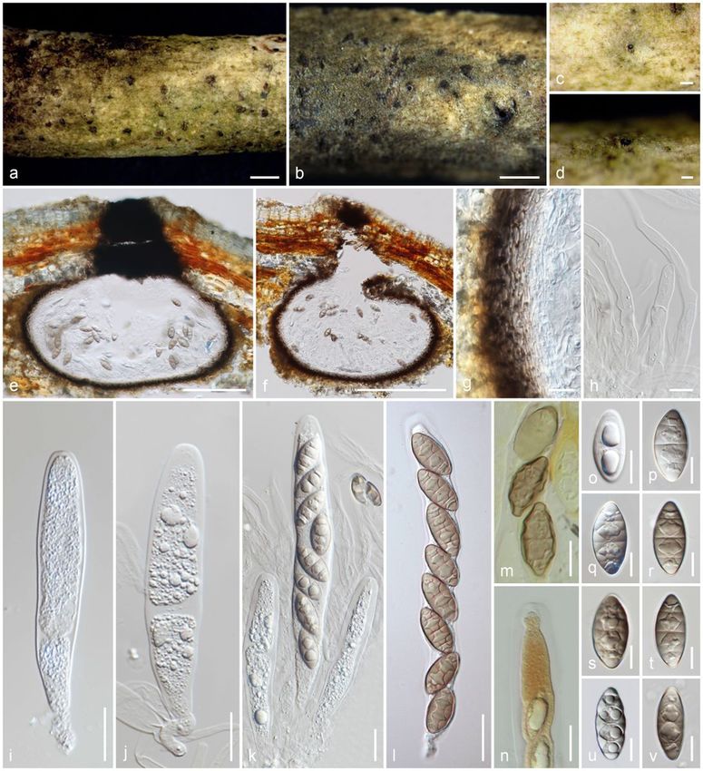
Ancestral character state analyses resulting from Bayesian Binary MCMC (BBM) are shown in Figs. 2 and 3. Xylariomycetidae was reconstructed as derived
from inconspicuous, immersed or semi-immersed ascomata with a prominent or rudimentary carbonaceous clypeus (Character 6) and shared a high
percentage among Amphisphaeriales, Delonicicolales and Xylariales. Xylariaceae includes highly variable stromatic characters, and is a diversified group as
compared to all the other families in Xylariomycetidae. Hypoxylaceae was reconstructed as having a conspicuous, unipartite, carbonaceous stroma (Character
3) and diversified into a conspicuous, stalked or sessile, carbonaceous stroma (Character 1). The conspicuous, erumpent, bipartite, carbonaceous stromatic
development (Character 2) and semi-immersed, erumpent or superficial, pseudostromatic development (Character 4) were mostly distributed in
Graphostromataceae and Diatrypaceae, respectively. Even though the sexual morphs of Beltraniaceae and Castanediellaceae are undetermined, there is a high
possibility that they will have inconspicuous, immersed or semi-immersed ascomata with a prominent or rudimentary carbonaceous clypeus stromata
(Character 6), based on evidence from recent ancestors of the clade. It is therefore possible to predict characters of the sexual morphs in some families that
lack known sexual morphs through their ancestral characters. In addition, septate, hyaline ascospores and the absence of a germ slit are ancestral characters
of Xylariomycetidae. Apiospores have evolved independently in several clades. Brown ascospores are often found in Xylariales, while Induratiaceae and
Vamsapriyaceae have hyaline apiospores. Several xylarialean taxa have ascospores with germ slits, but these are not found in Amphisphaeriales and
Delonicicolales. The Amphisphaeriales clade comprises a variety of characters and several groups with undetermined sexual morphs.

Taxonomy

In this paper, we follow the classifications in the studies of Hyde et al. (2020b) and Wijayawardene et al. (2020), and are updated according to recent relevant
literature.

Ascomycota R.H. Whittaker, Quarterly Review of Biology 34: 220 (1959)

Sordariomycetes O.E. Erikss. & Winka, Myconet 1: 10 (1997)

XylariomycetidaeO.E. Erikss & Winka

Notes: For the latest treatments of this subclass, we follow Hyde et al. (2020b) and Wijayawardene et al. (2020). Myelospermataceae is accepted in
Xylariomycetidae families incertae sedis due to lack of molecular data.

AmphisphaerialesD. Hawksw. & O.E. Erikss.

Amphisphaeriaceae G. Winter [as 'Amphisphaerieae'], Rabenh. Krypt.-Fl., Edn 2 (Leipzig) 1.2: 259 (1885)

Amphisphaeriaceae was introduced by Winter (1887), which mainly consists of saprobes in terrestrial, aquatic and marine habitats and occasionally
hemibiotrophic or necrotrophic species (Wang et al. 2004; Senanayake et al. 2015; Jaklitsch et al. 2016). Hyde et al. (2020b) and Wijayawardene et al. (2020)
accepted Amphisphaeriaceae in Amphisphaeriales with three genera as Amphisphaeria, Griphosphaerioma, and Lepteutypa. Samarakoon et al. (2020b)
revised the morphology and phylogeny of Amphisphaeria and Lepteutypa, and synonymised Lepteutypa under Amphisphaeria. In addition, the monospecific
genus Trochilispora, which had been accepted in Amphisphaeriaceae, is revised and synonymised under Hymenopleella (Sporocadaceae) (Samarakoon et al.
2020b). As a result of these studies, only Amphisphaeria and Griphosphaerioma are accepted in Amphisphaeriaceae.

Amphisphaeria Ces. & De Not., Comm. Soc. crittog. Ital. 1(4): 223 (1863)

Notes: Amphisphaeria is the type genus of Amphisphaeriaceae, with A. umbrina as the type species (Cesati and de Notaris 1863). Amphisphaeria species are
saprobes on woody branches and some monocotyledons, including grasses (Wang et al. 2004). Amphisphaeria accommodates 27 species (Samarakoon et
al. 2020b), which are characterised by solitary or aggregated ascomata under a poorly-developed clypeus or clypeus lacking; unitunicate asci with J+ or J-,
apical rings and light brown to dark brown, ellipsoid to fusiform, 1–3-septate ascospores and coelomycetous asexual morphs.

Amphisphaeria parvispora Samarak. & K.D. Hyde, sp. nov.

Index Fungorum number: IF558710; Facesoffungi number: FoF 10186; Fig. 4

Etymology: The specific epithet reflects the small ascospores.

Holotype: MFLU 18-0767

Saprobic on a dead branch. Sexual morph: Ascomata 230–260 × 300–400 μm (x̄ = 245 × 360 μm, n = 10), immersed, visible as raised, black dots, solitary, in
cross-section, conical with mostly flattened base. Ostioles centric, prominent, conical, wide, ostiolar canal periphysate. Peridium 8.5–23 μm (x̄ = 16.5 μm, n =
10) wide, wider at the apex, multi-layered, outer layer comprising reddish brown, thick-walled cells of textura angularis, inner layer composed of hyaline, thin-
walled cells of textura angularis. Paraphyses 2.5–5 μm (x̄ = 3.6 μm, n = 15) wide, longer than asci, cellular, septate, constricted at septa, guttulate, embedded
in a gelatinous matrix. Asci 62–105 × 5–6.5 μm (x̄ = 83.5 × 5.7 μm, n = 20), 8-spored, unitunicate, cylindrical, with a bifurcate pedicel, with a 0.7–0.9 × 1.8–2.2
μm (x̄ = 0.8 ×2 μm, n = 5), discoid, apical ring, J+ in Melzer’s reagent, apically rounded. Ascospores 9.5–11.5 × 3–4 μm (x̄ = 10.5 × 3.5 μm, n = 30), L/W 3,
uniseriate, hyaline when young, light brown to grayish when mature, ellipsoid, 1-septate, constricted at septa, bi-guttulate, smooth-walled, lack of mucilaginous
sheath. Asexual morph: Undetermined.

Material examined: Thailand, Phayao Province, Phu Sang, on dead branch, 20 July 2017, M.C. Samarakoon, SAMC060 (MFLU 18-0767, holotype), (HKAS
102328, isotype).

                                                                           Page 5/87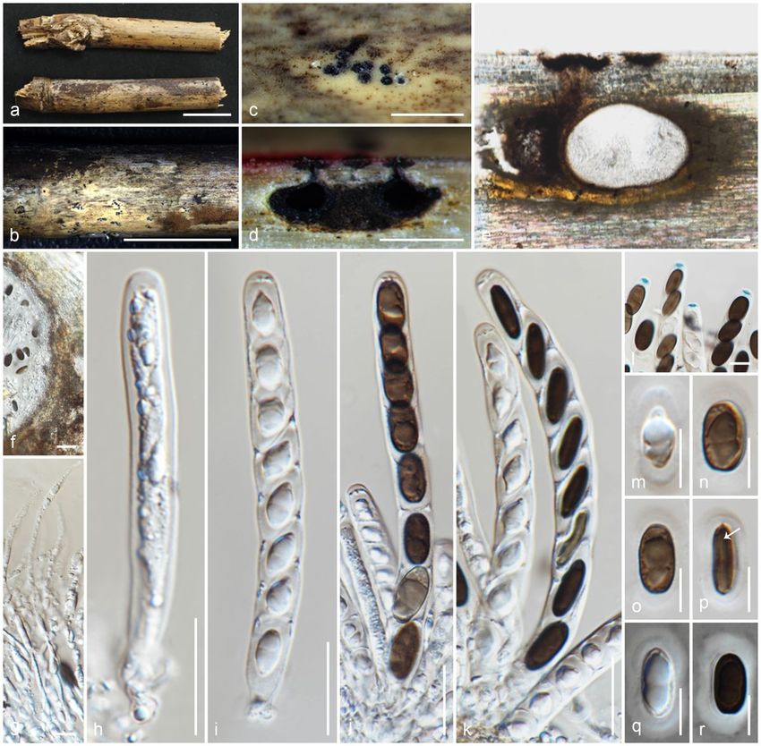
Notes: Amphisphaeria parvispora shares similar morphologies to other species in the genus in having solitary, immersed ascomata with two-layered peridium,
unitunicate asci with J+, discoid, apical ring, and brown ascospores. Our novel taxon is similar to Am. curvaticonidia, Am. thailandica (Thailand) and Am. sorbi
(Italy) in having conical to subglobose, solitary ascomata with a short, periphysate and a narrow ostiolar canal. Amphisphaeria curvaticonidia possesses 2-
distoseptate ascospores with a median, slightly constricted euseptum and thin mucilaginous sheath, while Am. parvispora possesses 1-septate ascospores
lacking a mucilaginous sheath. Amphisphaeria thailandica and Am. sorbi have J-, apical rings, while Am. parvispora has a J+, apical ring. Compared to all
Amphishaeria species, our new collection has the smallest asci (83.5 × 5.7 μm) and ascospores (10.5 × 3.5 μm) among J+, apical ring bearing species. The
LSU sequence of Am. parvispora is similar to Am. curvaticonidia MFLU 18-0789 (98.5%, 4/732 gaps), Am. fuckelii CBS 140409 (98%, 1/873 gaps) and Am.
thailandica MFLU 18-0794 (98%, 0/869 gaps), while rpb2 is similar to Am. fuckelii CBS 140409 (87%, 0/1067 gaps), Am. qujingensis KUMCC 19-0187 (86%,
0/1067 gaps) and Am. curvaticonidia MFLU 18-0789 (85%, 2/880 gaps). In combined gene phylogeny, Am. parvispora clusters with Am. sorbi (MFLUCC 13-
0721) and Am. thailandica (MFLU 18-0767), as a basal clade with 84% statistical support. Based on distinct morphology and phylogeny, Am. parvispora is
introduced as a new species.
Apiosporaceae K.D. Hyde, J. Fröhl., Joanne E. Taylor & M.E. Barr, in Hyde, Fröhlich & Taylor, Sydowia 50(1): 23 (1998)
Hyde et al. (1998) established Apiosporaceae with five genera as Appendicospora, Arthrinium (= Apiospora), Dictyoarthrinium, Endocalyx and Spegazzinia
based only on morphology. Species accommodated in Apiosporaceae are saprobic, pathogenic or endophytic on plant tissues, lichens, and marine algae,
occasionally infecting humans or isolated from soil (Hyde et al. 2020b). Tanaka et al. (2015) provided the phylogenetic affinity of Spegazzinia in
Didymosphaeriaceae (Pleosporales). A taxonomic and phylogenetic revision of Nigrospora showed that the genus has a close affinity to Apiosporaceae
(Wang et al. 2017). Samarakoon et al. (2020a) revised the morphology and phylogeny of Dictyoarthrinium (D. musae and D. sacchari), and transferred it into
Didymosphaeriaceae (Pleosporales) sister to Spegazzinia. Moreover, Konta et al. (2021) introduced Endocalyx metroxyli and transferred Endocalyx to
Cainiaceae based on morphology and multigene phylogeny. Pintos and Alvarado (2021) re-evaluated the multigene phylogeny and the morphology of
Arthrinium and suggested accepting Arthrinium sensu stricto and Apiospora as independent lineages within Apiosporaceae. Appendicosporaceae is
introduced as a new family to accommodate Appendicospora in this study. At present, only the genera Apiospora, Arthrinium and Nigrospora remain in
Apiosporaceae (Hyde et al. 2020b; Samarakoon et al. 2020a; Konta et al. 2021).
Apiospora Sacc., Atti Soc. Veneto-Trent. Sci. Nat., Padova, Sér. 4 4: 85 (1875)
Notes: Crous and Groenewald (2013) re-evaluated the morphology and phylogeny of Arthrinium (= Apiospora). Arthrinium species have densely arranged
perithecial ascomata in a longitudinal stroma; clavate to broadly cylindrical asci and apiospores in the sexual and coelomycetous or hyphomycetous asexual
morphs. The genus is widely distributed as endophytes, epiphytes, saprobes and plant pathogens on commercial crops and ornamentals (Crous and
Groenewald 2013; Hyde et al. 2020b). Arthrinium was expanded with abundant sampling and isolation with morpho-phylo studies while accepting > 70
species in recent years (Hyde et al. 2020b). Pintos and Alvarado (2021) provided molecular data for the type species Ar. cariciola and accepted two genera as
Apiospora and Arthrinium. Arthrinium species have variously shaped conidia and inhabit Cyperaceae or Juncaceae in temperate, cold or alpine habitats.
Apiospora species have rounded/lenticular conidia and inhabit mainly on Poaceae (and many other plant host families) in a wide range of habitats, including
tropical and subtropical regions. Nearly 20 Apiospora/Arthrinium species have been recorded from China (Senanayake et al. 2020; Farr and Rossman 2021;
Feng et al. 2021).
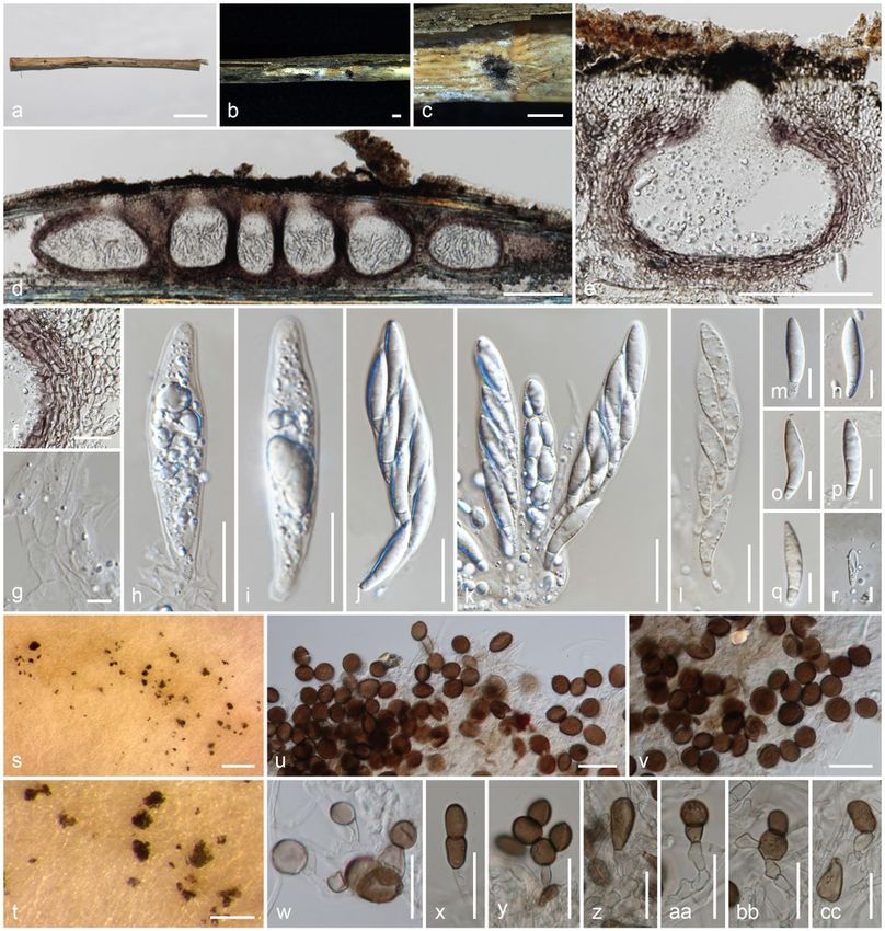
Apiospora guiyangensisSamarak., Jian K. Liu & K.D. Hyde, sp. nov.
Index Fungorum number: IF558711; Facesoffungi number: FoF 10187; Fig. 5
Etymology: The specific epithet reflects the location, Guiyang, from where the species was first collected.
Holotype: HKAS 102403
Saprobic on dead culm of grass. Sexual morph: Stromata 3.6–6 × 0.9–4.6 × 0.16–1.2 mm (x̄ = 4.4 × 2.2 × 0.4 mm), scattered to gregarious, partially
immersed, becoming erumpent to superficial, raised, dark brown, in linear rows, with a slit-like opening, multi-loculate. Ascomata 150–210 × 100–230 μm (x̄ =
170 × 180 μm, n = 10), with 2–12 ascomata forming groups immersed in stromata, arranged in rows, clustered, gregarious, to erumpent through host surface,
dark brown, in cross-section ellipsoidal to subglobose. Ostioles centric, ostiolar canal periphysate. Peridium 20–30 μm (x̄ = 23.7 μm, n = 15) wide, multi-
layered, outer layer comprising dark brown or reddish brown to lightly pigmented cells of textura angularis, inner layer very thin, composed of hyaline cells of
textura angularis. Paraphyses 3.5–6 μm (x̄ = 4.5 μm, n = 20) wide, septate, branched, smooth-walled, constricted at septa, embedded in a gelatinous
matrix. Asci 80–110 × 12–15 μm (x̄ = 94 × 13.5 μm, n = 20), 8-spored, unitunicate, clavate, with short basal pedicel, thin-walled, lacking an apical ring, with
obtusely rounded apex. Ascospores 26–29 × 5.5–7 μm (x̄ = 28 × 6.5 μm, n = 25), L/W 4.3, 2–3-seriate, hyaline, ellipsoid to reniform, straight to curved,
apiosporous, not constricted at septa, large cell 21–24 μm (x̄ = 22.7 μm) long, small cell 5–5.6 μm (x̄ = 5.2 μm) long, covered with a 4–8 μm (x̄ = 6 μm, n = 10)
wide mucilaginous sheath. Asexual morph: On PDA, Hyphae 1.5–3.5 μm (x̄ = 2.4 μm, n = 10) wide, branched, septate, hyaline. Conidiophores reduced to
conidiogenous cells. Conidiogenous cells 3.5–7.5 × 3–6 (x̄ = 5.3 × 4.5 μm, n = 8), solitary on hyphae, integrated, branched, ampuliform, cylindrical, hyaline to
brown. Conidia 10–13 × 7–10.5 (x̄ = 11.3 × 8.9 μm, n = 15), brown, smooth, guttulate, globose to ellipsoid in surface view, lenticular with a paler equatorial slit
in side view. Sterile cells 13–20 × 6–11 (x̄ = 16.7 × 8.7 μm, n = 10), elongated, mixed among conidia.
Culture characteristics: Colonies on PDA reaching 55 mm diam. after two weeks at 25°C, cottony, flat, spreading, with moderate aerial mycelium, circular,
dense, entire margin, and light brown; reverse brown at center and dirty white.
Material examined: China, Guizhou, Guiyang, Guizhou Academy of Agricultural Sciences (GZAAS) premises, on dead culm of grass (Poaceae), 7 July 2018,
M.C. Samarakoon, SAMC173 (HKAS 102403, holotype), (MFLU 19-2113, isotype); ex-type living cultures GZCC 21-0041 = CGMCC3.20365.
Page 6/87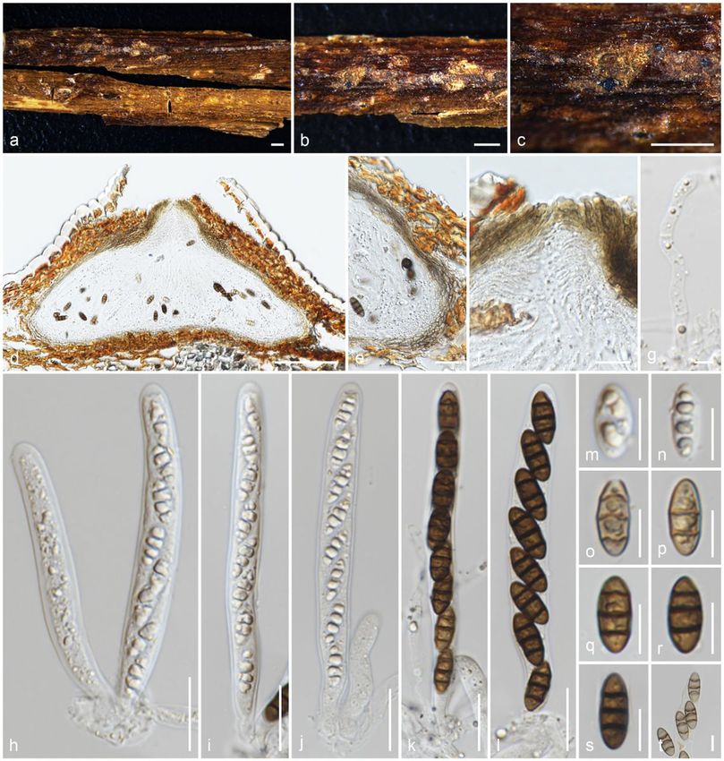
Notes: Apiospora guiyangensis differs from Ap. cyclobalanopsidis in its small conidiogenous cells (3.5–7.5 × 3–6 µm vs 6–19 × 2.5–7 µm). Apiospora
guiyangensis clustered in a distinct well-supported clade closely related to Ap. camelliae-sinensis CGMCC 3.18333 (98% sequence similarity in ITS, 2/584
gaps; 92% in tub2, 11/760 gaps), Ap. cyclobalanopsidis (99% sequence similarity in ITS, 1/572 gaps; 93% in tub2, 11/786 gaps) and Ap. jiangxiense (97%
sequence similarity in ITS; 3/541 gaps, 93% in tub2; 6/736 gaps).

Apiospora sichuanensis Samarak., Jian K. Liu & K.D. Hyde, sp. nov.

Index Fungorum number: IF558712; Facesoffungi number: FoF 10188; Fig. 6

Etymology: The specific epithet reflects the location, Sichuan, from where the species was first collected.

Holotype: HKAS 107008

Saprobic on dead culm of grass. Sexual morph: Stromata 1.1–5.1 × 0.3–0.7 × 0.2–0.4 mm (x̄ = 2.2 × 0.47 × 0.28 mm, n=10), scattered to gregarious, partially
immersed, becoming erumpent to superficial, raised, dark brown, in linear rows, with a slit-like opening, multi-loculate. Ascomata 160–205 × 205–270 μm (x̄ =
182.5 × 241.6 μm, n = 10), with 5–15 ascomata forming in groups immersed in stromata, arranged in rows, clustered, gregarious, to erumpent through host
surface, dark brown, in cross-section ellipsoidal to subglobose. Ostioles centric, ostiolar canal periphysate. Peridium 11–20 μm (x̄ = 16.7 μm, n = 15) wide,
thinner at the base, multi-layered, outer layer comprising dark brown or reddish brown to lightly pigmented cells of textura angularis, inner layer very thin,
composed of hyaline cells of textura angularis. Paraphyses 3.6–6.5 μm (x̄ = 5.1 μm, n = 20) wide, septate, branched, smooth-walled, constricted at septa,
embedded in a gelatinous matrix. Asci 72–125 × 18–30 μm (x̄ = 100.2 × 23.7 μm, n = 20), 8-spored, unitunicate, clavate, with short basal pedicel, thin-walled,
lacking an apical ring, with obtusely rounded apex. Ascospores 29–48 × 7–10.5 μm (x̄ = 39.7 × 9 μm, n = 25), L/W 4.4, 2–3-seriate, hyaline, ellipsoid to
reniform, straight to curved, apiosporous, not constricted at septa, large cell 34–43 μm (x̄ = 38 μm) long, small cell 3.5–7.5 μm (x̄ = 5.3 μm) long, covered with
a 13–24 μm (x̄ = 19 μm, n = 10) wide, up to 30 μm, mucilaginous sheath. Asexual morph: Undetermined.

Material examined: China, Sichuan, Chengdu, Flowing Water Park, Huaxing Road 5, Jinjiang, on dead culm of grass (Poaceae), 1 October 2019, M.C.
Samarakoon, SAMC241 (HKAS 107008, holotype), (MFLU HT20-0168, isotype).

Notes: Apiospora sichuanensis clustered with Ap. pseudoparenchymatica in the combined gene phylogeny. Morphological comparison is not possible due to
the lack of a similar morph for both species (Wang et al. 2018). The ITS sequence of Ap. sichuanensis is similar to Ap. pseudoparenchymatica CGMCC
3.18336 (97.5%, 3/559 gaps) and Ap. hyphopodii MFLUCC 15-0003 (93%, 13/585 gaps), and tub2 to Ap. pseudoparenchymatica CGMCC 3.18336 (94.5%,
17/705 gaps) and Ap. marii (86%, 30/959 gaps).

Appendicosporaceae Samarak. & K.D. Hyde, fam. nov.

Index Fungorum number: IF558713; Facesoffungi number: FoF 06297

Etymology: Named after the type genus, Appendicospora.

Saprobic on dead rachis/fronds of palms and dicotyledonous twigs. Sexual morph: Ascomata immersed, under slightly raised areas, visible as brown or black
dots, solitary or aggregated in clusters, in cross-section, conical to subglobose with mostly flattened base. Ostioles centric, ostiolar canal periphysate or filled
with white amorphous tissues. Peridium multi-layered, outer layer comprising brown, thick-walled, flattened cells of textura angularis, inner layer composed of
hyaline, thin-walled cells of textura angularis. Paraphyses wider at the base, septate, embedded in a gelatinous matrix. Asci 8-spored, unitunicate, clavate to
cylindrical, short pedicellate or sessile, lacking an apical ring, apically rounded. Ascospores uniseriate or 2–3-seriate, hyaline, clavate to broadly ellipsoidal, 1-
septate, not constricted at septa, with or without appendages at one end. Asexual morph: Undetermined.

Type genus: Appendicospora K.D. Hyde

Notes: Appendicospora shares similar morphologies with Apiospora and Pseudomassaria with an uncertain taxonomic placement (Hyde 1995; Bahl 2006).
Several morpho-phylo studies suggested that Appendicospora consistently grouped with Hyponectria and was best placed within the Hyponectriaceae,
although further work to confirm the taxonomic placement was suggested (Wang and Hyde 1999; Smith et al. 2003; Bahl 2006). The only available LSU
sequence of Appendicospora sp. (HKUCC 1120) links the morphology and phylogeny of this group. Combined gene phylogeny in our study shows that
Appendicospora forms a distinct clade to Apiosporaceae in Amphisphaeriales. In addition, an inconspicuous taxon introduced as Neoamphisphaeria in this
study clustered with Appendicospora with high statistical support (100%/1.00 PP). Appendicosporaceae clustered in the clade comprising
Anungitiomycetaceae, Iodosphaeriaceae and Pseudosporidesmiaceae with strong statistical support (95%). In addition, the divergence time estimates show
that Appendicosporaceae has diverged at 89 (65–117) MYA (Amphisphaeriales), which is comparable with the common divergence trend in family level (50–
150 MYA) as described in Hyde et al. (2017). Based on distinct morphologies, phylogeny and divergence time estimates, we introduce Appendicosporaceae
with the type genus Appendicospora and tentatively accommodate Neoamphisphaeria.

Appendicospora K.D. Hyde, Sydowia 47(1): 31 (1995)

Notes: Appendicospora was introduced by Hyde (1995), which is distinguished from Apiospora by ascospores with basal bifurcate appendages.
Appendicospora coryphae (≡ Apiosporella coryphae), the generic type, was described on dead rachides of Corypha elata from the Philippines. Hyde and
Fröhlich (1997) introduced the second species as App. hongkongensis, occurring on fronds of Livistona chinensis in Hong Kong.

Appendicospora hongkongensis Yanna, K.D. Hyde & J. Fröhl., Mycoscience 38(4): 395 (1997)

                                                                            Page 7/87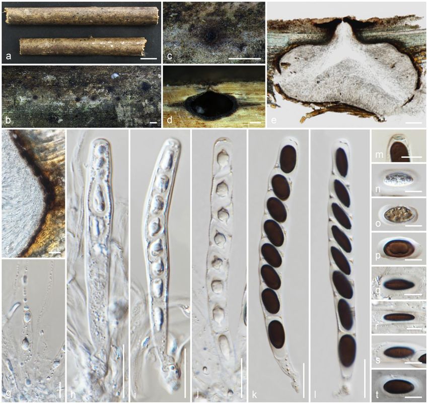
Index Fungorum number: IF442936; Facesoffungi number: FoF 10189; Fig. 7

Reference specimen: HKAS 107015 designated here

Saprobic on dead frond of Livistona chinensis. Sexual morph: Ascomata 70–125 × 105–145 μm (x̄ = 95 × 125 μm, n = 10), immersed in the host tissue
(subepidermal) under slightly raised areas, irregular in outline, individually light brown in the middle and dark at the periphery, solitary or aggregated in
clusters, or evenly distributed, in cross-section conical to subglobose with mostly flattened base. Ostioles centric, ostiolar canal periphysate, filled with bright
yellow pigmented drops when immature, blackish when mature, deteriorating when overmature. Peridium 5.5–13 μm (x̄ = 9.2 μm, n = 10) wide, multi-layered,
outer layer comprising light brown, flattened cells of textura angularis, inner layer composed of hyaline cells of textura angularis. Paraphyses 5–9 μm (x̄ = 6.5
μm, n = 20) wide, septate, smooth-walled, constricted at septa, difficult to distinguish, embedded in a gelatinous matrix. Asci 55–70 × 15–19.5 μm (x̄ = 60 ×
17.5 μm, n = 15), few, 8-spored, unitunicate, clavate, short pedicellate, or pedicel lacking, thin-walled, lacking an apical ring, deliquescing early and releasing
spores, developing from the base and lower sides of the ascomata, apically rounded. Ascospores 18–24.5 × 6–7 μm (x̄ = 22 × 6.5 μm, n = 25), L/W 3.4, 2–3-
seriate, hyaline, clavate, unequally 2-celled, not constricted at septa, large cell 10.5–18 μm (x̄ = 14 μm) long, small cell 10.5–18 μm (x̄ = 14 μm) long with a
bifurcated (moustache-shaped) appendage, lacking a mucilaginous sheath. Asexual morph: Undetermined.

Culture characteristics: Colonies on PDA reaching 45 mm diam. after three weeks at 25°C, flat, powdery, with an outer radiating margin, hyphae embedded in
the media, greenish white; reverse yellowish brown in the center, greenish brown marginal area, media becoming light brown.

Material examined: China, Sichuan Province, Chengdu, University of Electronic Science and Technology of China (Qingshuihe Campus), on dead frond of
Livistona chinensis (Arecaceae), 30 September 2019, M.C. Samarakoon, SAMC247 (HKAS 107015, reference specimen designated here), (MFLU HT20-0175);
living cultures GZCC 21-0044 = CGMCC3.20364.

Notes: The type of Appendicospora hongkongensis (HKU(M) 5301) was collected on fronds of Livistona chinensis in Hong Kong. Our specimen is similar to
App. hongkongensis with overlapping size of height of ascomata (70–125 μmvs 108–128 μm), asci (55–70 × 15–19.5 μmvs 70–80 × 16–24 μm),
ascospores (18–24.5 × 6–7 μmvs 70–80 × 17–24 × 5–8 μm) and brown peridium. Appendicospora coryphae possesses a hyaline peridium, which allows for
discrimination of the species from App. hongkongensis (Hyde 1995; Hyde and Fröhlich 1997). Apart from similar morphology, both (HKU(M) 5301) and our
specimen were collected from the same host and similar geography. In the multigene phylogeny, our strain clusters with an Appendicospora sp. (HKUCC
1120), which has only LSU sequence data (Smith et al. 2003). However, HKU(M) 5301 does not have molecular data. Based on similar morphology, host and
geographical distribution, here we propose a reference specimen for App. hongkongensis on the dead frond of Livistona chinensis from Sichuan, China.

Neoamphisphaeria Samarak. & K.D. Hyde, gen. nov.

Index Fungorum number: IF558714; Facesoffungi number: FoF 10190

Etymology: After its morphological similarities to Amphisphaeria.

Saprobic on dead twigs. Sexual morph: Ascomata immersed, slightly raised, visible as black dots, solitary, in cross-section conical with mostly flattened base
or less globose. Ostioles centric, filled with white amorphous tissue. Peridium multi-layered, outer layer comprising reddish brown, thick-walled cells of textura
angularis, inner layer composed of hyaline, thin-walled cells of textura angularis. Paraphyses wider at the base, long, septate, branched. Asci 8-spored,
unitunicate, cylindrical, short pedicel, with a bilobed or dome-shaped apical ring, J- in Melzer’s reagent, apically rounded. Ascospores uniseriate, hyaline,
broadly ellipsoidal, aseptate when immature, 1-septate when mature, guttulate, lacking a mucilaginous sheath. Asexual morph: Undetermined.

Type: Neoamphisphaeria hyalinospora Samarak. & K.D. Hyde

Notes: Neoamphisphaeria is similar to Amphisphaeria in having immersed ascomata with a brown peridium, long hyaline paraphyses, cylindrical asci and
ellipsoid, 1-septate, mature ascospores. The distinct characters of Neoamphisphaeria are the ostiolar canal filled with amorphous hyaline cells, asci with a
bilobed or dome-shaped apical ring and hyaline ascospores. In the phylogeny, Neoamphisphaeria clusters with Appendicospora with 100%/1.00 PP statistical
support, which has a periphysate ostiolar canal, clavate asci and 2–3-seriate, hyaline, clavate ascospores with a bifurcated (moustache-shaped) appendage.
With inconspicuous, immersed ascomata, asci with short or lacking pedicels with J-, apical ring and 2-celled hyaline ascospores and strong phylogenetic
evidence, we accept Neoamphisphaeria as a new genus in Appendicosporaceae.

Neoamphisphaeria hyalinosporaSamarak. & K.D. Hyde, sp. nov.

Index Fungorum number: IF558715; Facesoffungi number: FoF 10191; Fig. 8

Etymology: The specific epithet reflects the hyaline ascospores.

Holotype: MFLU 19-2131

Saprobic on dead twigs. Sexual morph: Ascomata 220–280 × 335–365 μm (x̄ = 250 × 350 μm, n = 8), immersed, under slightly raised areas, visible as black
dots, solitary, in cross-section conical with mostly flattened base or subglobose. Ostioles prominent, centric, filled with white amorphous tissues. Peridium 25–
34 μm (x̄ = 30 μm, n = 10) wide, wider in upper regions, multi-layered, outer layer comprising reddish brown, thick-walled cells of textura angularis, inner layer
composed of hyaline, thin-walled cells of textura angularis. Paraphyses 5–9 μm (x̄ = 6.5 μm, n = 15) wide, wider at the base, long, cellular, septate, branched,
guttulate, constricted at septa, embedded in a gelatinous matrix. Asci 105–130 × 7.5–9.5 μm (x̄ = 118 × 8.5 μm, n = 20), 8-spored, unitunicate, cylindrical, with
a short pedicel, with a bilobed or dome-shaped apical ring, J- in Melzer’s reagent, apically rounded. Ascospores 14.5–17.5 × 6–8 μm (x̄ = 16 × 6.5 μm, n = 30),

                                                                            Page 8/87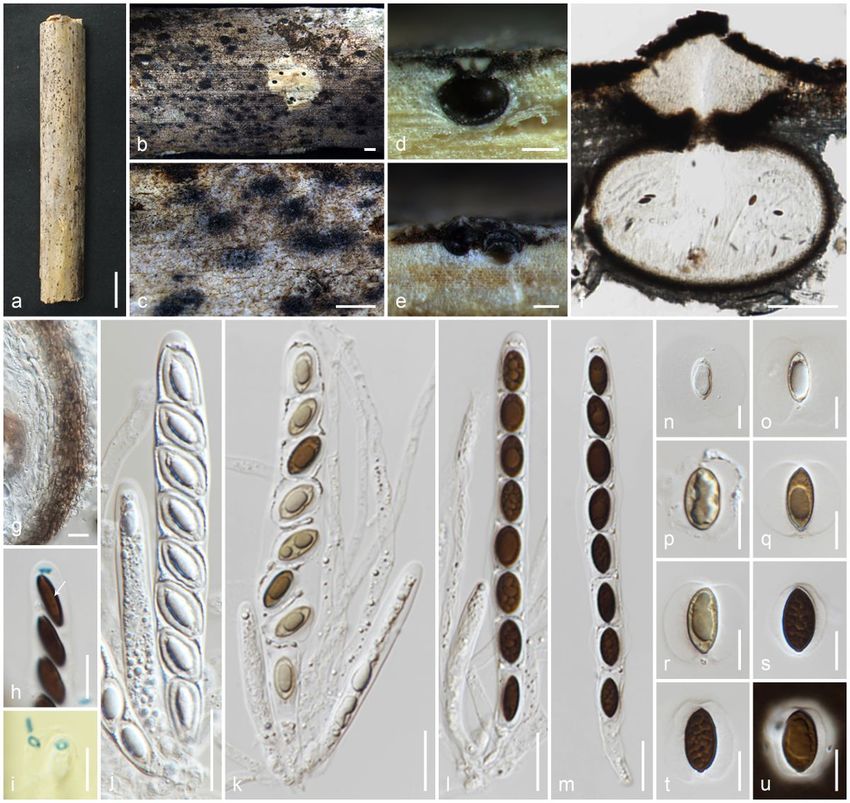
L/W 2.5, uniseriate, hyaline, broadly ellipsoidal, aseptate when immature, 1-septate when mature, guttulate, lacking a mucilaginous sheath or any appendage.
Asexual morph: Undetermined.

Material examined: Thailand, Phrae Province, on dead twigs, 24 January 2019, M.C. Samarakoon, SAMC209 (MFLU 19-2131, holotype), (HKAS 106988,
isotype).

Notes: Neoamphisphaeria hyalinospora is similar to Amphisphaeria with its subglobose to conical ascomata, cylindrical asci with a short pedicel and 2-celled
ascospores, but differs in the amorphous cells in the ostiolar canal and hyaline ascospores. Asci and ascospores size and shape of Neoa. hyalinospora are
similar to Keissleriella hyalinospora (formerly known as Am. hyalinospora) (Müller and von Arx 1962). However, Am. hyalinospora has dark brown, sparsely
bristly hairs in the ostiolar canal, whereas Neoa. hyalinospora has hyaline amorphous cells. Combined gene phylogenies showed that Neoamphisphaeria is
not related to Amphisphaeria, but clustered with Appendicospora with high statistical support (100%/1.00 PP). Based on unique morphology and distinct
phylogeny, we introduce Neoa. hyalinospora as a new species.

Melogrammataceae G. Winter [as 'Melogrameae'], Rabenh. Krypt.-Fl., Edn 2 (Leipzig) 1.2: 797 (1886)

Melogrammataceae was introduced by Winter (1887) to accommodate Melogramma. Species of the family are saprobes or hemibiotrophs on the bark of
woody plants. Based on morphology and phylogenetic revisions, Jaklitsch and Voglmayr (2012) accepted Melogrammataceae in Xylariales. Senanayake et al.
(2015) re-evaluated the phylogeny of Melogrammataceae and accepted it in Amphispheriales, and this has been confirmed in the later studies by Hongsanan
et al. (2017) and Hyde et al. (2020b).

Melogramma Fr., Summa veg. Scand., Section Post. (Stockholm): 386 (1849)

Notes: Melogramma was established by Fries (1849) with Mel. campylosporum as the type species, which is a commonly and abundantly reported species.
Melogramma is characterised by reddish brown stromata, unitunicate asci with J-, apical ring and 3-septate, brown ascospores, and a coelomycetous asexual
morph (Jaklitsch and Voglmayr 2012; Maharachchikumbura et al. 2016). Seventeen Melogramma epithets are accepted (Hyde et al. 2020b).

Melogramma campylosporum Fr., Summa veg. Scand., Sectio Post. (Stockholm): 386 (1849)

Index Fungorum number: IF150772; Facesoffungi number: FoF 00841; Fig. 9

Saprobic dead aerial branch of Corylus avellana. Sexual morph: Stromata 1–2.5 × 0.6–1.4 × 0.3–1 mm (x̄ = 1.6 × 1 × 0.7 mm, n=10), erumpent from bark,
solitary, scattered or aggregated, pulvinate or discoid, smooth to slightly velutinous, surface brown with slightly papillate black ostiolar dots. Ascomata 315–
400 × 225–360 μm (x̄ = 341 × 274 μm, n = 10), 2–12 per stroma, immersed, in cross-section globose to subglobose. Ostioles centric, ostiolar canal
periphysate. Peridium 21–30 μm (x̄ = 25 μm, n = 15) wide, multi-layered, outer layer comprising reddish brown, flattened cells of textura angularis, thin inner
layer comprising hyaline cells of textura angularis. Paraphyses 3–5 μm (x̄ = 4.2 μm, n = 20) wide, numerous, septate, smooth-walled, apically blunt. Asci 100–
140 × 13–16 μm (x̄ = 122 × 14.5 μm, n = 25), 8-spored, unitunicate, clavate or fusoid, straight, curved or sigmoid, with short and narrow pedicel, lacking an
apical ring, apically rounded. Ascospores 38–50 × 4–6 μm (x̄ = 44 × 5 μm, n = 25), L/W 8.8, 2–3-seriate, hyaline, straight and 0–1-septate when immature,
brown, 3 equidistant septa when mature, end cells slightly lighter entirely or only at their tips, falcate, often strongly curved or slightly straight, tips narrowly
rounded to subacute, with a smooth narrow hyaline perispore, often with one large guttule in each cell. Asexual morph: Undetermined.

Culture characteristics: Colonies on PDA, very slow growing, reaching 9 mm diam. after three months at 25°C, convex and a papillate surface, compact, lobate
with zonate margin, hyphae embedded in the media, dark greenish brown; reverse dark brown in the center, yellowish brown marginal area, media becoming
reddish brown.

Material examined: Italy, Province of Forlì-Cesena, Tontola di Predappio, on the dead aerial branch of Corylus avellana(Betulaceae), 5 February 2017, E.
Camporesi, IT3241 (MFLU 17-0348, HKAS 102324); living culture MFLUCC 17-2674. Russia, Krasnodar region, Sochi, Central city district, park “Riviera”, on
dead aerial branchs and twigs of Corylus avellana (Betulaceae), 10 October 2016, T.S. Bulgakov, SC-101 (MFLU 18-0778, HKAS 102312); living culture
MFLUCC 18-0612.

Notes: Melogramma campylosporum has been described on Alnus glutinosa subsp. glutinosa (Turkey), Carpinus betulus (Austria, Poland, Sweden), Carpinus
sp. (Ukraine, United Kingdom), Corylus avellana (Austria, Poland) and Fagus sylvatica (Italy) (Farr and Rossman 2021). LSU and ITS sequences of our two
collections are 100% identical to the ITS-LSU of acc. JF440978 (CBS 141086). The microscopic characters of our two collections are similar to Mel.
campylosporum,described by Jaklitsch and Voglmayr (2012). This is the first record of Mel. campylosporum on Corylus avellane in Italy.

Sporocadaceae Corda [as 'Sporocadeae'], Icon. fung. (Prague) 5: 34 (1842)

Sporocadaceae species typically possess appendage bearing conidia and are important as saprobes or pathogens on leaves, twigs, branches, fruits of
flowering plants and gymnosperms, and as endophytes or parasites on humans and animals (Liu et al. 2019; Hyde et al. 2020b). Following the recent revision
of the morphology and multigene phylogeny, there are 23 genera in Sporocadaceae (Liu et al. 2019).

Seiridium Nees, Syst. Pilze (Würzburg): 22 (1816) [1816-17]

Notes: Seiridium was introduced by Nees (1816) with the type species Se. marginatum. The sexual morph of Seiridium is characterised by immersed to semi-
erumpent ascomata, a dark peridium, centric, slightly papillate, periphysate ostiolar canals, cylindrical, 8-spored asci with J+, apical rings and cylindrical-

                                                                            Page 9/87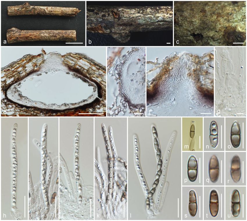
oblong, euseptate, yellow to dark brown ascospores (Bonthond et al. 2018). The coelomycetous asexual morph of Seiridium differs from closely related
Nothoseiridium and Nonappendiculata in having versicolorous, 5-septate conidia with appendages. There are 44 Seiridium species (Bonthond et al. 2018).

Seiridium italicum Samarak., Camporesi & K.D. Hyde, sp. nov.

Index Fungorum number: IF558716; Facesoffungi number: FoF 10192; Fig. 10

Etymology: The specific epithet reflects Italy, from where the species was first collected.

Holotype: MFLU 16-1315

Saprobic on dead aerial branch of Laurus nobilis. Sexual morph: Ascomata 185–205 × 275–450 μm (x̄ = 195 × 365 μm, n = 10), immersed, visible as raised,
black dots, solitary or aggregated, in cross-section conical with mostly flattened base or sub-globose. Ostioles prominent, centric or eccentric, conical, wide,
ostiolar canal periphysate. Peridium 13–21 μm (x̄ = 17.3 μm, n = 10) wide, multi-layered, outer layer comprising yellowish brown, thick-walled cells of textura
angularis, inner layer composed of hyaline, thin-walled cells of textura angularis. Paraphyses 3.3–5.4 μm (x̄ = 4 μm, n = 20) wide, longer than asci, cellular,
septate, rarely branched, guttulate, constricted at septa, embedded in a gelatinous matrix. Asci 110–130 × 8–11 μm (x̄ = 120 × 10 μm, n = 20), 8-spored,
unitunicate, cylindrical, short pedicellate, J- in Melzer’s reagent, apically rounded. Ascospores 14.8–18.5 × 5.5–8 μm (x̄ = 16.3 × 7.3 μm, n = 30), L/W 2.3,
uniseriate, hyaline, light to dark brown when mature, ellipsoid, 2–3-septate, constricted at septa, lacking a mucilaginous sheath. Asexual morph: Undetermined.

Culture characteristics: Colonies on PDA reaching 23–25 mm diam. after four weeks at 25°C, circular, flat, entire margin, white and yellowish brown as
concentric zones; yellowish orange center, salmon pink marginal area.

Material examined: Italy, Forlì-Cesena Province, Camposonaldo - Santa Sofia, on dead aerial branch of Laurus nobilis, 6 April 2016, E. Camporesi, IT2945
(MFLU 16-1315, holotype), (HKAS 102342, isotype); ex-type living culture MFLUCC 18-0510.

Notes: Seiridium italicum is only known from its sexual morph and is similar to the generic description except in having J-, apical rings (Bonthond et al. 2018).
However, the morphological comparisons are difficult due to the availability of sexual characteristics. In addition, Nonappendiculata and Nothoseiridium are
close to Se. italicum in phylogeny but lack sexual morphologies (Liu et al. 2019; Crous et al. 2020). ITS sequence of Se. italicum is close to Se. papillatum CBS
340.97 (93%, 22/580 gaps), Se. persooniae CBS 143445 (93%, 19/568 gaps) and Se. podocarpi CBS 137995 (91%, 35/594 gaps), while the LSU sequence is
close to Se. phylicae CPC 19962 (99%), Se. rosarum MFLUCC 17-0654 (99%, 1/888 gaps) and Notho. podocarpi CPC 36967 (99%, 0/866 gaps). Combined
gene phylogenies show that Se. italicum clusters apart from other species, and here we introduce Se. italicum as a new species.

Xylariales Nannf.

Coniocessiaceae Asgari & Zare, Mycol. Progr. 10(2): 195 (2011)

Coniocessiaceae was introduced by Asgari and Zare (2011) to accommodate Coniocessia, which comprises saprobes in plants, soil and dung.
Coniocessiaceae is placed in Xylariales based on morphology and phylogeny and divergence time estimations (Asgari and Zare 2011; Maharachchikumbura
et al. 2016; Hyde et al. 2020).

Paraxylaria Wanas., Gafforov, E.B.G. Jones & K.D. Hyde, in Wanasinghe et al., Fungal Diversity: 89; 1-236, [200] (2018)

≡ Rosellinia subgen. Amphisphaerella Sacc., Syll. fung. (Abellini) 1: 262 (1882)

= Amphisphaerella (Sacc.) Kirschst., Trans. Br. mycol. Soc. 18(4): 306 (1934) [1933] (Nom. illegit., Art. 53.1), non-Amphisphaerella Henn., Hedwigia 41: 13
(1902)

Paraxylaria was introduced by Wanasinghe et al. (2018) to accommodate Pa. rosacearum, a saprobe on trunks and branches of Rosa sp. from Uzbekistan.
Paraxylaria has asci with J+, apical ring and uniseriate, ellipsoid, mostly symmetrical, aseptate, brown ascospores, which lack germ slits (Wanasinghe et al.
2018). We re-examined the holotype and observed germ pores in ascospores of Pa. rosacearum (Fig. 136, Wanasinghe et al. 2018). Molecular phylogeny also
supported Paraxylaria as a basal clade, but distinct from Coniocessia in Coniocessiaceae.

Kirschstein (1934) introduced Amphisphaerella to accommodate Amp. amphisphaerioides. Amphisphaerella was introduced to accommodate a micro-fungal
group, which is characterised by immersed to semi-immersed ascomata, often beneath a clypeus, globose to subglobose in section, 8-spored, cylindrical,
pedicellate asci with apically rounded, J+/J-, apical rings and ellipsoidal, brown, aseptate ascospores with two to six germ pores arranged equatorially or
scattered or in groups towards the poles (Kirschstein 1934; Barr 1994). However, this introduction is invalid according to the Art. 53.1 since Hennings (1902)
introduced Amphisphaerella with the type of Amp. hypoxyloides. Petrini (2013) synonymised and accepted the monospecific Amphisphaerella (Amp.
hypoxyloides) in Rosellinia (Ro. hypoxyloides) based on morphology. Only the invalidly published Amphisphaerella has been considered thereafter. Munk
(1953) proposed Amphisphaerellaceae for Amphisphaerella. However, Eriksson (1966), Barr (1990) and Lumbsch and Huhndorf (2010) retained
Amphisphaerella in Amphisphaeriaceae until more information was available. There are 13 Amphisphaerella epithets that have been accepted. Lu and Hyde
(2000) observed that the holotype of Amp. dispersella and ascospores with germ pores are identified as a unique character of the genus. Here we accept
Paraxylaria as the valid name to accommodate species similar to Amphisphaerella.

Paraxylaria xylostei (Pers.) Samarak. & K.D. Hyde, comb. nov.

≡ Sphaeria xylostei Pers., Neues Mag. Bot. 1: 84 (1794)

                                                                           Page 10/87
You can also read