Supplement of Fragile protein folds: sequence and environmental factors affecting the equilibrium of two interconverting, stably folded protein ...
←
→
Page content transcription
If your browser does not render page correctly, please read the page content below

Supplement of Magn. Reson., 2, 63–76, 2021                                                     Open Access
https://doi.org/10.5194/mr-2-63-2021-supplement
© Author(s) 2021. This work is distributed under
the Creative Commons Attribution 4.0 License.

Supplement of
Fragile protein folds: sequence and environmental factors affecting
the equilibrium of two interconverting, stably folded protein
conformations
Xingjian Xu et al.

Correspondence to: Kevin H. Gardner (kgardner@gc.cuny.edu)

The copyright of individual parts of the supplement might differ from the CC BY 4.0 License.
Supplementary Figures

Supplementary Figure S1 – Sequence comparison and changes in backbone hydrogen bonding
between WT and SLIP conformations. (a) Sequence alignment (EMBOSS stretcher (Madeira et al.,
2019)) of WT (WT conformation) and F444Q/F446A/Y456T (SLIP conformation-locked) variant, with
locations of key secondary structures labeled. (b) Comparison of the five antiparallel b-strands between
WT and F444Q/F446A/Y456T variant. External-orienting residues (bold) and internal-orienting residues
(italic) are marked. Some residues on the two ends of strands are not labeled as they are transitioning into
connecting loops. The Ib-strand of the mutant shows a 3-residue slip and an inversion of topology. (c)
Backbone H-bonds between b-strands are shown for both WT and F444Q/F446A/Y456T variant.
Changes are highlighted in the panel showing the H-bonds of the mutant (red).

                                                                                                           2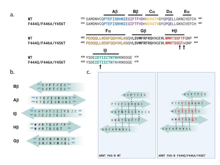
Supplementary Figure S2 – Temperature dependence of the WT:SLIP conformational equilibrium
in ARNT PAS-B Y456T. (a) Variable temperature 15N/1H HSQC spectra of ARNT PAS-B Y456T,
recorded in 5 degree increments between 278K and 333K. Inset shows the temperature-dependent
chemical shift changes of a pair of peaks representing the two conformations adopted by residue E403.
(b) Expansion of the E403 signal in 15N/1H HSQC spectra at indicated temperatures (288K – 313K).
Relative populations are indicated on the edge of each figure, showing intensities of slices through the
center of the WT peak (black) and the SLIP peak (red).

                                                                                                           3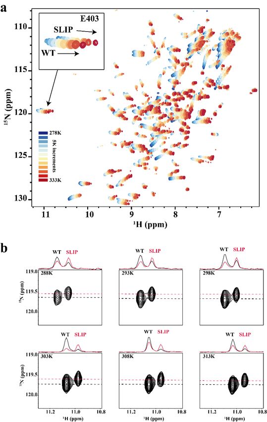
Supplementary Figure S3 – pH and salt concentration have negligible effects on WT:SLIP
equilibrium in ARNT PAS-B Y456T. (a) The WT:SLIP equilibrium remains unchanged between pH 7.0
and 9.0 in Tris (black circles, red line) or between pH 6.0 and 7.5 in PIPES (blue circles, blue line), with
17 mM NaCl present at all points. The lines represent linear regressions to the data points. (b) The
WT:SLIP equilibrium is chiefly unchanged throughout a NaCl titration between 50 and 200 mM (black
circles, red line), all at pH 7.5. The line represents a linear regression to the data points.
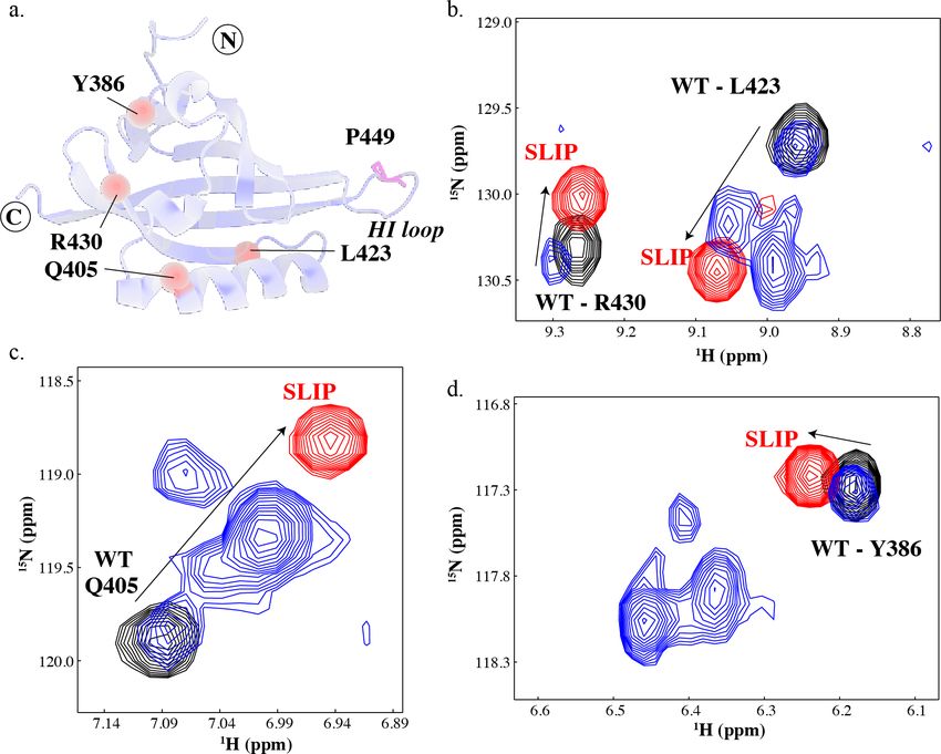
4Supplementary Figure S4 – P449A point mutation generates multiple conformations of ARNT
PAS-B. (a) A schematic of the solution structure of wildtype ARNT PAS-B domain (PDB:1X0O (Card et
al., 2005)), highlighting the location of the P449 residue (magenta sticks) and residues indicated in panels
b-d. (b-d) Widespread peak multiplicity in the P449A mutant is observed throughout 15N/1H HSQC
spectra as probed at residues Y386, Q405, L423, and R430. Four major species are observed: One at the
WT location (14% intensity), and three additional ones at locations which do not overlap the SLIP
location. Approximate populations of these peaks are 60%, 14%, and 12%; assignments were made by
assuming that the largest peak for each residue corresponded to the dominant (60%) state, the next state
corresponded to the peak closest to the WT-SLIP vector, followed by peaks further from this vector.
Proteins: wildtype (black), F444Q/F446A/Y456T (red) and P449A (blue).
5Supplementary Figure S5 – 15N/1H HSQC spectra of ARNT PAS-B sequence variants presented in
Table 1. Spectra of ARNT PAS-B variants P449G/Y456T, F444Q/F446A/P449A/Y456T, and Y456T +
TEV (pre- and post-cleavage) are shown (blue). 15N/1H HSQC spectra of WT (black) and
F444Q/F446A/Y456T (red) are also shown as reference. Insets show the relative population of residue
E403 in each variant.
6Supplementary Figure S6 – ARNT PAS-B variants have similar stabilities as probed by Trp
fluorescence during urea denaturation. The peak emission wavelength of ARNT PAS-B wildtype
(black circles), Y456T (red triangles), and F444A/F446Q/Y456T (blue squares) red shifted with
increasing concentrations of urea as the proteins unfolded. Comparable stabilities of all three variants
were observed.
78
9
Supplementary Figure S7 – Counter-screen of ligands that bind to F444Q/F446A/Y456T triple
mutant (TRIP). Ligands that bind to ARNT PAS-B WT (Guo et al., 2013) were tested for binding
against the TRIP mutant. Shown are 15N/1H HSQC spectra of WT ARNT PAS-B and TRIP mutant
without (black) or with 500 µM of ligands added (red) as indicated, in the presence of 2% DMSO.

                                                                                                  10
Supplementary Figure S8 – KG-548 and KG-655 binding titration analysis using 13C/1H HSQC. (a)
Example peaks from 15N/1H HSQC spectra of KG-655 titration series (0, 500, 1000, 2500, 5000, 10000
µM) against 250 µM ARNT PAS-B Y456T. Peaks associated with the WT conformation show chemical
shift changes, while peaks associated with the SLIP conformation do not, indicating selective binding.
Unlike KG-548, many analyzable residues (where both WT and SLIP peaks are assigned) are directly
involved with ligand binding and showing mixed fast-intermediate exchange characteristics in NMR
spectra. (b) 13C/1H HSQC titration experiments of KG-548 (0, 500, 1000, 2000, 3000, 4000 µM) and KG-
655 (0, 500, 1000, 2500, 5000, 10000 µM) against 250 µM ARNT PAS-B Y456T, focusing on the L391
11d1 methyl signals. Both WT and SLIP peaks are well resolved at all compound concentrations with no
sign of peak broadening. Chemical shift changes to the WT conformation are minimal in the presence of
both KG-548 and KG-655, suggesting the residue is not directly involved in the binding of either
compounds. (c) KG-548 and KG-655 binding to ARNT PAS-B Y456T as monitored by peak volumes of
L391 d1 from 13C/1H HSQC NMR spectra. Data are fit to Eq. 1 to extract dissociation constant Kd and
maximum binding Bmax; resulting values are Kd = 414 ± 7.1 µM and Bmax = 192 ± 0.96 µM for KG-548,
and Kd = 1947 ± 152 µM and Bmax = 172 ± 5.7 µM for KG-655. Uncertainties were estimated using
bootstrapping. Noises with mean of 0 and variance of the standard error were added to the experimental
data. Generated datasets (n = 30) were fit to obtain the 95% confidence interval.
12Supplementary Figure S9 – KG-655 loses surface binding to the WT conformation of ARNT PAS-B
Y456T while retaining the internal binding mode. Shown here is the 13C/1H HSQC titration series of
KG-655 (0 – 10 mM) against ARNT PAS-B Y456T, zoomed in on the methyl region. As previously
reported, KG-655 binds to wildtype ARNT PAS-B via two binding modes (Gagné, 2020). The surface
binding of KG-655 involves sidechains of residue I364 and I458, resulting in chemical shift changes that
are observed when titrated against wildtype ARNT PAS-B, but are missing from these spectra even at the
highest ligand concentration (black dotted squares). Methyl groups with changes in chemical shifts (I396,
L408, and M439, blue dotted squares) are all on sidechains oriented inward (inset PDB:4EQ1 (Guo et al.,
2013)), to the internal cavity of the protein, suggesting the internal binding mode is retained.
13References
Card, P. B., Erbel, P. J. A., and Gardner, K. H.: Structural basis of ARNT PAS-B dimerization: use of a
common beta-sheet interface for hetero- and homodimerization, J. Mol. Biol., 353, 664-677,
10.1016/j.jmb.2005.08.043, 2005.
Gagné, D. Azad, R.; Edupuganti, U.R.; Williams, J.; Aramini, J.M.; Akasaka, K., and Gardner, K.H.: Use
of high pressure NMR spectroscopy to rapidly identify internal ligand-binding voids in proteins, preprint
on bioRxiv, 10.1101/2020.08.25.267195, 2020.
Guo, Y., Partch, C. L., Key, J., Card, P. B., Pashkov, V., Patel, A., Bruick, R. K., Wurdak, H., and
Gardner, K. H.: Regulating the ARNT/TACC3 axis: multiple approaches to manipulating protein/protein
interactions with small molecules, ACS Chem. Biol., 8, 626-635, 10.1021/cb300604u, 2013.
Madeira, F., Park, Y. M., Lee, J., Buso, N., Gur, T., Madhusoodanan, N., Basutkar, P., Tivey, A. R. N.,
Potter, S. C., Finn, R. D., and Lopez, R.: The EMBL-EBI search and sequence analysis tools APIs in
2019, Nucleic Acids Res., 47, W636-W641, 10.1093/nar/gkz268, 2019.
14You can also read