Safety, patient acceptance and diagnostic accuracy of ultrasound core needle biopsy of parotid or submandibular glands in primary Sjögren's ...
←
→
Page content transcription
If your browser does not render page correctly, please read the page content below

Sjögren syndrome
RMD Open: first published as 10.1136/rmdopen-2021-001901 on 7 February 2022. Downloaded from http://rmdopen.bmj.com/ on June 24, 2022 by guest. Protected by copyright.
ORIGINAL RESEARCH
Safety, patient acceptance and
diagnostic accuracy of ultrasound core
needle biopsy of parotid or
submandibular glands in primary
Sjögren’s syndrome with suspected
salivary gland lymphoma
Ivan Giovannini,1 Michele Lorenzon,2 Valeria Manfrè,1 Sara Zandonella Callegher,1,3
Enrico Pegolo,4 Chiara Zuiani,2 Rossano Girometti,2 Alojzija Hocevar ,5
Christian Dejaco ,3,6 Luca Quartuccio,1 Salvatore De Vita,1 Alen Zabotti 1
To cite: Giovannini I, ABSTRACT
Lorenzon M, Manfrè V, et al. Background Enlargement of the major salivary glands
Key messages
Safety, patient acceptance (SGs) is a major risk factor for B-cell lymphoma among
and diagnostic accuracy of
patients with primary Sjögren’s syndrome (pSS).
What is already known about this subject?
ultrasound core needle biopsy of ► Salivary gland enlargement in primary Sjögren’s
Ultrasound-guided core needle biopsy (US-guided CNB)
parotid or submandibular glands syndrome (pSS) is a main risk of lymphoma devel-
in primary Sjögren’s syndrome could be a novel technique to manage SG enlargement
among patients with pSS. opment, whose diagnosis must be pathologically
with suspected salivary
Objective Accordingly, this study’s main aim was to evaluate proven through histological analysis. In this scenar-
gland lymphoma. RMD Open
the safety, patient tolerance and diagnostic accuracy of US- io, ultrasound-guided core needle biopsy (US-guided
2022;8:e001901. doi:10.1136/
rmdopen-2021-001901 guided CNB procedure for patients with pSS with major SG CNB) has been proposed as a diagnostic tool, but
enlargement. evidence for its safety, tolerance and procedural
► Additional supplemental Methods Patients with clinical diagnosis of pSS and a clinical standardisation remains limited.
material is published online only. indication for SG biopsy consecutively underwent US-guided
What does this study add?
To view, please visit the journal CNB between September 2019 and June 2021. These patients ► This study provides insights into the technical as-
online (http://dx.d oi.org/10. were evaluated clinically 1, 2 and 12 weeks after US-guided
1136/rmdopen-2021-0 01901). pects and anatomical issues of the US- guided
CNB. Patients were asked to complete a questionnaire about
CNB procedure, the safety and patients’ tolerability
postprocedural complications as well as periprocedural
through objective measures (ie, pain visual analogue
pain, using the Visual Analogue Scale. Complications were
Received 27 August 2021 scale).
Accepted 20 December 2021 categorised as transient (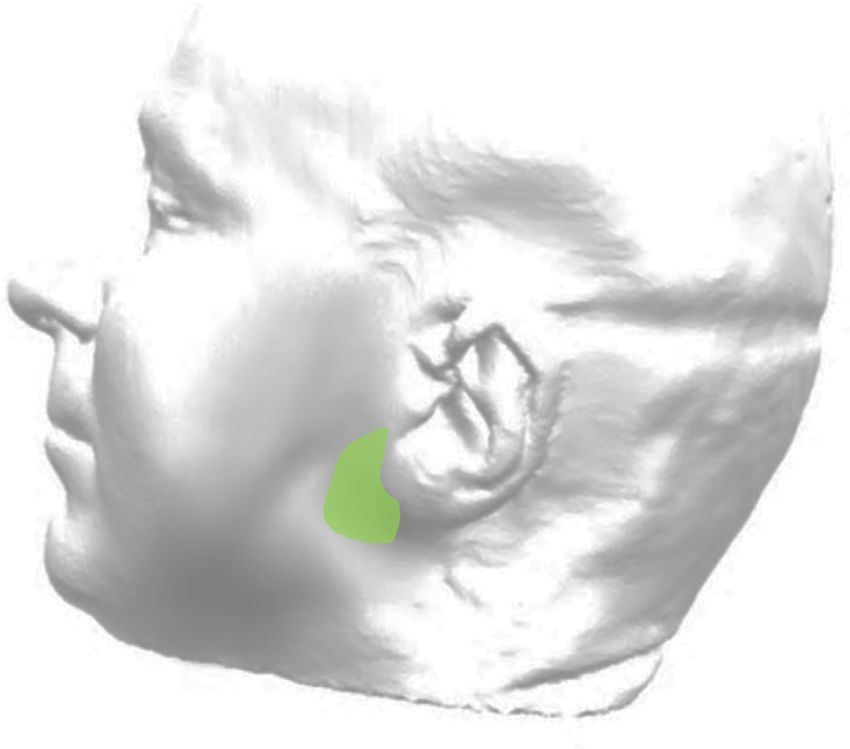
RMD Open
RMD Open: first published as 10.1136/rmdopen-2021-001901 on 7 February 2022. Downloaded from http://rmdopen.bmj.com/ on June 24, 2022 by guest. Protected by copyright.
Figure 2 The ‘safety area’ or ‘safety zone’ (green area) for a
parotid biopsy.
Figure 1 Sonographic images of a focal lesion (the white
arrows) in the salivary gland parenchyma (A); a peculiar
appearance of the salivary gland, showing confluent procedure’s safety, patient tolerance and standardisa-
hypoechoic areas (B). tion remains limited.23 The most feared complication
is the injury of the facial nerve, which presents a strict
anatomical relation with the parotid gland. In interven-
Persistent major salivary gland (SG) enlargement is tional procedures on the parotid glands, a ‘safety zone’
a major clinical risk factor for B-cell lymphoma, and it or ‘safety area’ has been identified. This region is located
may signify an established lymphoma stage, a prelympho- between 1 cm anterior and 1 cm below the ear lobe,
matous stage or even a completely different disease.6 7 where the facial nerve runs in the glandular parenchyma
The parotid glands are the main site of B-cell lymphoma protected by the superficial lobe of the parotid for a
development among patients with pSS, and biopsies length of approximately 2 cm (figure 2).
of the major SGs are reserved for patients with pSS To overcome these limitations, the current prospective
suspected of having glandular lymphoma, mainly based study aimed to (1) strengthen our preliminary experi-
on SG swelling. ence,15 (2) describe the US-guided CNB procedure in
An ultrasonographic examination of the major SGs detail and (3) evaluate the procedure’s safety, patient
could assist clinicians in assessing the gland’s typical tolerance and the diagnostic accuracy for lymphoprolif-
structural abnormalities and detecting parenchyma erative disease of US-guided CNB among a cohort of defi-
lesions,8–10 suggesting that SG ultrasonography (SGUS) nite patients with pSS or patients suspected to have pSS
may come to be used as a stratification (scoring system) with persistent parotid or submandibular gland swelling.
and prognostic tool.11–13
The presence of a focal lesion within altered glan-
dular parenchyma (figure 1A) or severe glandular inho- METHODS
mogeneity (eg, diffuse, large- confluent, hypoechoic Patients
areas; figure 1B) or both are the typical sonographic Our study included consecutive patients referring to the
patterns of SG lymphoma among patients with pSS.14–16 Institute of Rheumatology, University Hospital of Udine
Nevertheless, currently, lymphoma is mainly suspected (Italy), from September 2019 to June 2021, with a clin-
among patients with pSS due to clinical characteristics, ical diagnosis of pSS, who underwent US-guided CNB
rather than imaging features, and the diagnosis of B-cell due to suspected major SG lymphoma. The suspicion of
lymphoma must be pathologically proven.17 18 lymphoma development was clinically based on parotid
An open surgical biopsy of the parotid gland is and/or submandibular gland swelling, defined as either
the recommended procedure in case of SG enlarge- (1) chronic (>12 months), unilateral or bilateral parotid
ment;19 20 however, the need of a skilled surgeon and or submandibular gland swelling or (2) recurrent glan-
the risk of potential complications limit its generalis- dular swelling of at least 2 months of duration in one
ability.20 21 Recently, ultrasound (US)-guided core needle episode.24 25
biopsy (US-guided CNB) has been proposed as a diag- Written informed consent was obtained from each
nostic procedure for patients with pSS with suspected patient in accordance with the Declaration of Helsinki
glandular lymphoma.17 22 However, evidence of this and with local guidelines for good clinical practice.26
2 Giovannini I, et al. RMD Open 2022;8:e001901. doi:10.1136/rmdopen-2021-001901Sjögren syndrome
RMD Open: first published as 10.1136/rmdopen-2021-001901 on 7 February 2022. Downloaded from http://rmdopen.bmj.com/ on June 24, 2022 by guest. Protected by copyright.
Demographic, clinical and laboratory data
Patients’ clinical data were collected from their medical
charts, including their age, gender, disease duration,
previous unstimulated sialometry and Schirmer’s tests
and evidence of serum antibodies, as well as antinuclear,
anti-Ro/SSA and anti-La/SSB antibodies. Furthermore,
the presence of lymphoma development risk factors for
patients with pSS was noted at the time of participants’
biopsy procedures (glandular swelling, lymphadenop-
athy, cryoglobulinemia, a serum monoclonal compo-
nent, rheumatoid factor, low serum C4 and leucopenia).6
The US-guided CNB procedure for the major SGs
In this study, US-guided CNB was performed by a radiolo-
gist (ML) with extensive experience (ie, 10 years of prac-
tice) in ultrasound-guided biopsies. The procedures were
conducted under real-time US guidance with linear high-
frequency transducers (RS85, probe LM4-15B (Samsung,
Seoul, South Korea) or Affiniti 70G, probe L18-5 (Philips,
Amsterdam, the Netherlands)). The procedures were
performed with an aseptic, free-hand technique under
real-time US guidance in a non-operating room.
Biopsies were performed with a 14-gauge, semiauto- Figure 3 (A): A patient’s supine positioning for a parotid
matic CNB system (Precisa 14G, HS Hospital Service, gland CNB, with their shoulders slightly lifted (and a pillow
Aprilia, Italy), with a sampling length set on 10 mm or, below their upper back), slight hyperextension of their neck
more frequently, 20 mm, depending on the size of the and facing towards the contralateral side of the target gland.
target area. US-guided CNB was performed on partici- (B): Local anaesthetic injected under ultrasound guidance
pants’ clinically most swollen gland. The biopsy was into the subcutaneous tissue and the posterior, superficial
part of the parotid gland while moving the needle in a
performed on the area with the most suspicious ultra-
caudocranial direction. (C): A semi-automatic needle inserted
sonographic appearance (severe inhomogeneity of into the ‘safety area’ of the left parotid gland. (D): A semi-
the glandular parenchyma or focal lesion, whenever automatic needle inserted into the left submandibular gland,
present). For deep lesions, the procedure’s risks and (E): The needle’s sonographic appearance in a focal lesion of
benefits were weighed by the radiologist and the clini- the parotid gland. (F): The needle’s sonographic appearance
cians. The optimal procedure technique was adapted in a peculiar appearance of the salivary gland, in the absence
to participants’ SG type (ie, parotid vs submandibular of focal lesion. CNB, core needle biopsy.
glands) and lesion type (ie, localised vs diffuse).
During their biopsies, patients were positioned in a
supine position.27 Their shoulders were slightly lifted of the parotid gland, with their heads fully turned to the
(usually with a pillow below their upper back), and their opposite side. Patients’ submandibular glands were accessed
necks were slightly hyperextended, turned towards the anteriorly or posteriorly, depending on patients’ coopera-
contralateral side of the target gland (figure 3A). tion, operator preferences and the eventual US detection of
For patients with parotid focal lesions, the spatial rela- a focal lesion, as described above, in this case, nerve injury
tionship between the ‘safety area’ and the lesions them- was not a concern. Attention was paid also on localising and
selves was primarily assessed to determine the procedure’s then avoiding the path of the facial artery, which represents
technical feasibility and safety. After the radiologist anal- the major noble anatomical structure in strict contact with
ysed the safety and feasibility, the procedure targeted the gland.
lesions posteriorly through the ‘safety area’, maintaining The asepsis of the procedure was guaranteed by accurate
the shortest path and the most superficial needle posi- disinfection of the skin with gluconate chlorhexidine or povi-
tion possible within a depth of 1–1.5 cm from the glan- done–iodine solution and the use of single use prove covers.
dular surface. A local anaesthetic was injected under US guidance with a
For patients with diffusely inhomogeneous paren- fine needle (23 G) through the skin and subcutaneous fat
chyma without focal lesions, parotid biopsies targeted (figure 3B). Shortly after this local anaesthetic injection, a
the posterior- caudal part of the gland in a caudocra- small incision of the anesthetised skin was made with a scalpel.
nial direction. The needle entered within the ‘safety The biopsy was then performed with a 14 G semiautomatic
area’, maintaining the most superficial needle direction needle under US guidance (figure 3B,C). One to five needle
possible within 1–1.5 cm of the glandular surface. passes were performed through the same skin incision
For US-guided submandibular gland biopsies, patients (figure 3E,F). The biopsy samples were fixed in formalin and
were positioned like patients receiving a US-guided biopsy sent for histological analysis. After this procedure, patients
Giovannini I, et al. RMD Open 2022;8:e001901. doi:10.1136/rmdopen-2021-001901 3RMD Open
RMD Open: first published as 10.1136/rmdopen-2021-001901 on 7 February 2022. Downloaded from http://rmdopen.bmj.com/ on June 24, 2022 by guest. Protected by copyright.
compressed their puncture sites and remained under obser- women. Patients’ mean age at the time of the biopsies was
vation for at least 30 min. 59.8 years (SD: 13.2 years). Moreover, 23 of the study’s 30
patients (76.6%) met the American College of Rheuma-
Evaluation of postbiopsy complications and peri-procedural tology (ACR) and EULAR classification criteria for pSS,28
pain and 24 (80%) had anti-Ro/SSA antibodies. Among the seven
This study’s participants were evaluated clinically 1, 2 and patients who did not fulfil the 2016 ACR/EULAR criteria,
12 weeks after their US-guided CNB and beyond the 12 pSS was highly suspected based on clinical grounds in five
weeks in case of persistent complications. All patients were patients, while in two patients, the clinical picture did not
asked to complete a questionnaire, reporting any postproce- allow one particular disease as much more likely. They all
dural complications (online supplemental figure 1) as well refused a minor SG biopsy performed concomitantly with
as assessing their intraprocedural and postprocedural pain US-guided CNB of the major SGs.
using the Visual Analogue Scale (VAS 0–10). Complications Additionally, 22 patients (73.3%) had parotid gland
were categorised as transient (lastingSjögren syndrome
RMD Open: first published as 10.1136/rmdopen-2021-001901 on 7 February 2022. Downloaded from http://rmdopen.bmj.com/ on June 24, 2022 by guest. Protected by copyright.
lesion among 19 of 30 (63.3%) patients, whereas 11 less than 2 hours. More details about these complications
patients (36.7%) showed inhomogeneous glandular are presented in figure 4A.
parenchyma, suggesting lymphoproliferative disease Intraprocedural pain, evaluated using the VAS, was
in the absence of glandular focal lesions. All biopsied assessed via a questionnaire and found to be low (mean
glands showed a sonographic OMERACT (Outcome VAS: 1.67±2.47), like postprocedural pain (mean VAS:
Measures in Rheumatology) score ≥2 (grade 2 in 4 out of 1.23±2.3). Specifically, 17 of 30 patients (56.67%) reported
30 patients and grade 3 in 26 out of 30 patients). Overall, no intraprocedural pain, eight patients (26.67%) reported
22 parotid glands (73.33%) and 8 submandibular glands mild pain, two patients (6.67%) reported moderate pain
(26.67%) were biopsied. and three patients (10%) reported severe pain (maximum
Out of 22 US-guided CNBs of the parotid glands, 14 VAS pain: 8/10). Postprocedural pain did not occur for 21
(63.6%) were performed on focal lesions, and in all cases, of 30 patients (70%), while 5 patients (16.67%) experienced
the procedure was technically and safely feasible through mild postprocedural pain, two patients (6.67%) experienced
the ‘safety area’. For biopsies of the submandibular moderate postprocedural pain and two patients (6.67%)
glands, five of eight (62.5%) procedures were performed experienced severe postprocedural pain. Figure 4B presents
on focal lesions. At least two samples were taken in all more details about participants’ procedure-related pain.
suspected cases. During the US- guided parotid gland Overall, for the 9 of 30 patients (30%) who reported post-
CNB, the facial nerve was not clearly identifiable by SGUS procedural pain, their median postprocedural pain duration
in none of 22 patients (100%). was only 2 days (Q25–75: 1–3 days). No statistically significant
difference was found between parotid and submandibular
Safety and patient acceptance US-guided CNB in pain assessment. Importantly, patients
US-guided CNB was well tolerated, and patients reported did not report any surgical wounds or scar formation after
no long-term complications during the follow-up period the procedures’ skin incisions. The entire duration of the
of mean±SD: (10.6±7.3 months). Only transient compli- US-guided CNB procedure, from patients’ entrance into the
cations (lastingRMD Open
RMD Open: first published as 10.1136/rmdopen-2021-001901 on 7 February 2022. Downloaded from http://rmdopen.bmj.com/ on June 24, 2022 by guest. Protected by copyright.
an effective treatment of pSS is lacking; however, the assessable via ultrasound.37 To the best of our knowledge,
recent availability of biologic target therapies might offer none of the studies was able to assess by US of the facial nerve
new treatment options.29 on its extracranial emergence when it enters the parotid
SGUS is frequently used to assess the structural abnor- parenchyma from a longitudinal or transverse view of way.
malities typical of pSS.8 Furthermore, recent reports have In the current authors’ experience, the sonographic assess-
suggested that the sonographic detection of focal lesions ment of the facial nerve as it exits the stylomastoid foramen
or a peculiar sonographic appearance of SGs (eg, large, has not been feasible, particularly in cases where the disease
confluent, hypoechoic areas spread over the gland) could affects the parotid gland, leading to ultrasonographic glan-
indicate glandular B-cell lymphoma,17 18 which has yet to be dular impairment. Therefore, we performed US- guided
proven by histology. parotid gland biopsies in the ‘safety zone’ for our patients.
For patients without pSS who present SGUS-detected In the current study, we described our US-guided approach
focal lesions of the major SGs, fine- needle aspiration and the technique used to safely perform a US-guided CNB
cytology (FNAC) is performed to differentiate between in SGs of patients with pSS in detail, focusing on parotid
benign and malignant lesions. FNAC is a safe technique, gland biopsies in the ‘safety zone’. In this area, the facial
but it frequently fails to provide material sufficient nerve runs deeply in the glandular parenchyma, and the
for a diagnosis.30 In patients with pSS at a high risk of parotid gland’s superficial lobe protects the facial nerve at a
lymphoma development, a histological, rather than length of approximately 2 cm (with some variability between
cytological, sampling is usually needed. Therefore, such individuals), minimising the risk of nerve damage.
other procedures as open surgical biopsy or US-guided The few reports available for submandibular US-guided
CNB may play a role.18 CNB among patients with pSS17 38 have described the proce-
Open surgical biopsy of the major SGs is a safe tech- dure’s efficacy in providing sufficient material for diagnosis
nique when performed by expert surgeons but presents and a good safety profile. Our patient cohort reported no
several disadvantages, such as the need for an operating long-term complications, confirming US- guided CNB’s
room and possible adverse effects (such as facial nerve safety profile for both parotid and submandibular glands.
damage), which could limit its accessibility. Importantly,
Overall, in our opinion, US-guided CNB offers several
the focal lesions identified by SGUS may not be precisely
advantages, the main being its safety and feasibility: in fact,
identified during surgical biopsy procedures; therefore,
it provides relatively easy access to both the parotid gland
this approach cannot accurately perform biopsies of
and the submandibular gland without a need for surgery,
SGUS-identified focal lesions. Furthermore, as reported
being less invasive and more patient friendly. In the future,
in the preliminary experience by Zabotti et al,15 the open
the US-guided CNB approach might be performed directly
surgical biopsy approach showed a higher number of
by rheumatologists with expertise in sonography and bioptic
persistent complication, compared with US-guided CNB
procedures, as is now common practice for synovial biopsies.
approach.
Second, this procedure allows for the targeting of glandular
US-guided CNB could overcome both FNAC and
areas with different sonographic patterns in both the parotid
surgical limitations. Recent evidence suggests that, for
patients with pSS with major SG enlargement—a signif- and submandibular glands. According to our study, major
icant risk factor for B- cell lymphoma6—US-guided SG biopsies may provide adequate samples for the early
CNB can provide sufficient sampling for pathological detection of suspected SG lymphoma, thus playing a role in
examination.31 the follow-up of suspected SG lymphoma in pSS.24 39 More-
The current study completed a preliminary study15 demon- over, the procedure allowed us to improve differential diag-
strating that US-guided CNB of the SGs is accurate and safe noses for our patients initially suspected to have pSS (such
for patients with pSS suspected to have lymphoma, providing as granulomatous sialadenitis consistent with sarcoidosis and
sufficient material for a histopathological diagnosis in most IgG4-related disease). These findings emphasise the impor-
cases. The safety of US-guided CNB of the parotid glands tance of SG biopsies—especially in patients with pSS with
was recently assessed in a meta-analysis,19 which reported glandular swelling—in order to detect not only lymphopro-
high diagnostic accuracy for both sensitivity and specificity liferative disease but also less common diseases that would
as well as a very low complication rate and facial nerve paral- otherwise be underdiagnosed.40–42
ysis as the most severe complication. The identification of Furthermore, this new sampling technique might provide
the facial nerve via ultrasound is challenging. Currently, histological material from the major SGs that offers possible
few data are available on ultrasonographic scanning of the future research implications for tissue sampling.21 It may also
facial nerve. High- resolution ultrasonography might play provide superior diagnostic accuracy while improving prog-
a role in the assessment and scanning of the facial nerve.32 nostic value,39 allowing for the monitoring of disease activity
To identify the facial nerve, Tawfik et al described the facial and tissue damage10 43 as well as patients’ response to treat-
nerve’s sonographic appearance, providing reference values ment.44 45 Further studies should better assess US-guided
from 50 healthy volunteers.33 Meanwhile, other authors34–36 CNB’s diagnostic accuracy and safety for patients with pSS,
have tried to scan the facial nerve in order to manage Bell’s as well as more researches should focus on the relationship
palsy. However, the extracranial part of the facial nerve after between major SG and minor SG histology, and between sali-
its emergence from the stylomastoid foramen is only partly vary histology and sonographic appearances in pSS.
6 Giovannini I, et al. RMD Open 2022;8:e001901. doi:10.1136/rmdopen-2021-001901Sjögren syndrome
RMD Open: first published as 10.1136/rmdopen-2021-001901 on 7 February 2022. Downloaded from http://rmdopen.bmj.com/ on June 24, 2022 by guest. Protected by copyright.
CONCLUSION 8 van Ginkel MS, Glaudemans AWJM, van der Vegt B, et al. Imaging in
Primary Sjögren’s Syndrome. J Clin Med 2020;9:2492.
US-guided CNB represents a novel approach for the manage- 9 Zabotti A, Zandonella Callegher S, Gandolfo S, et al. Hyperechoic
ment of patients with pSS. This procedure has shown remark- bands detected by salivary gland ultrasonography are related to
able patient safety and tolerance, allowing for adequate tissue salivary impairment in established Sjögren's syndrome. Clin Exp
Rheumatol 2019;37 Suppl 118:146–52.
sampling and definite diagnoses for almost all patients who 10 Zandonella Callegher S, Zabotti A, Giovannini I, et al. Normal-
participated in this study. Appearing Salivary Gland Ultrasonography Identifies a
Milder Phenotype of Primary Sjögren’s Syndrome. Front Med
2020;7:602354.
Author affiliations 11 De Vita S, Lorenzon G, Rossi G, et al. Salivary gland echography in
1
Rheumatology Clinic, Department of Medical and Biological Sciences, University primary and secondary Sjögren's syndrome. Clin Exp Rheumatol
Hospital 'Santa Maria della Misericordia' c/o University of Udine, Udine, Italy 1992;10:351–6.
2
Institute of Radiology, Department of Medicine, University Hospital 'Santa Maria 12 Jousse-Joulin S, D'Agostino MA, Nicolas C, et al. Video clip
della Misericordia' c/o University of Udine, Udine, Italy assessment of a salivary gland ultrasound scoring system in
3 Sjögren's syndrome using consensual definitions: an OMERACT
Department of Rheumatology, Hospital of Bruneck, Bruneck, Italy
4 ultrasound Working group reliability exercise. Ann Rheum Dis
Institute of Anatomic Pathology, University Hospital 'Santa Maria della 2019;78:967–73.
Misericordia' c/o University of Udine, Udine, Italy 13 Zabotti A, Zandonella Callegher S, Tullio A, et al. Salivary Gland
5
Department of Rheumatology, Division of Internal Medicine, University Medical Ultrasonography in Sjögren’s Syndrome: A European Multicenter
Centre Ljubljana, Ljubljana, Slovenia Reliability Exercise for the HarmonicSS Project. Front Med
6
Department of Rheumatology, Medical University Graz, Graz, Austria 2020;7:581248.
14 Di Franco FT, Lorenzon M, Zabotti A. Main sonographic features of
malt lymphoma in major salivary glands in Sjögren syndrome: our
Correction notice This article has been amended since it was first published experience. EcR 2021 EPOS. Available: https://epos.myesr.org/
online. Luca Quartuccio was spelt Quartuccio Luca. poster/esr/ecr2021/C-10893 [Accessed 23 Aug 2021].
Contributors Conceptualisation: AZ, IG, SZC, CD, AH, ML, EP. Methodology: AZ, 15 Zabotti A, Zandonella Callegher S, Lorenzon M, et al. Ultrasound-
guided core needle biopsy compared with open biopsy: a new
ML, EP. Investigation: IG, SZC, VM. Data curation: IG, SZC, VM, AZ, EP. Writing—
diagnostic approach to salivary gland enlargement in Sjögren’s
original draft preparation: IG, AZ. Writing—review and editing: IG, VM, AZ, SDV, ML.
syndrome? Rheumatology 2021;60:1282–90.
Supervision: SDV, LQ, CD, CZ, RG, AH. Guarantors: AZ. All authors have read and 16 Lorenzon M, Tulipano Di Franco F, Zabotti A, et al. Sonographic
agreed to the published version of the manuscript. features of lymphoma of the major salivary glands diagnosed with
Funding The authors have not declared a specific grant for this research from any ultrasound-guided core needle biopsy in Sjögren's syndrome. Clin
funding agency in the public, commercial or not-for-profit sectors. Exp Rheumatol 2021;39 Suppl 133:175–83.
17 Baer AN, Grader‐Beck T, Antiochos B, et al. Ultrasound‐Guided
Competing interests None declared. Biopsy of Suspected Salivary Gland Lymphoma in Sjögren’s
Patient consent for publication Not applicable. Syndrome. Arthritis Care Res 2021;73:849–55.
18 Baldini C, Zabotti A, Filipovic N, et al. Imaging in primary Sjögren's
Ethics approval The study was conducted according to a protocol approved by syndrome: the 'obsolete and the new'. Clin Exp Rheumatol 2018;36
the Regional Ethical Committee (CEUR-2017-Os-027- ASUIUD). This study involves Suppl 112:215–21.
human participants and was approved by Ethics Committee of Friuli Venezia Giulia. 19 Haldar S, Sinnott JD, Tekeli KM, et al. Biopsy of parotid masses:
Participants gave informed consent to participate in the study before taking part. review of current techniques. World J Radiol 2016;8:501.
Provenance and peer review Not commissioned; externally peer reviewed. 20 Colella G, Cannavale R, Vicidomini A, et al. Salivary gland biopsy:
a comprehensive review of techniques and related complications.
Data availability statement Data are available upon reasonable request. Rheumatology 2010;49:2117–21.
Open access This is an open access article distributed in accordance with the 21 Pijpe J, Kalk WWI, van der Wal JE, et al. Parotid gland biopsy
compared with labial biopsy in the diagnosis of patients with primary
Creative Commons Attribution Non Commercial (CC BY-NC 4.0) license, which
Sjogren’s syndrome. Rheumatology 2007;46:335–41.
permits others to distribute, remix, adapt, build upon this work non-commercially, 22 Manfrè V, Giovannini I, Zandonella Callegher S, et al. Ultrasound and
and license their derivative works on different terms, provided the original work is Bioptic investigation of patients with primary Sjögren's syndrome. J
properly cited, appropriate credit is given, any changes made indicated, and the Clin Med 2021;10:1171.
use is non-commercial. See: http://creativecommons.org/licenses/by-nc/4.0/. 23 Witt BL, Schmidt RL. Ultrasound-Guided core needle biopsy of
salivary gland lesions: a systematic review and meta-analysis.
ORCID iDs Laryngoscope 2014;124:695–700.
Alojzija Hocevar http://orcid.org/0000-0002-7361-6549 24 De Vita S, De Marchi G, Sacco S, et al. Preliminary classification
Christian Dejaco http://orcid.org/0000-0002-0173-0668 of nonmalignant B cell proliferation in Sjögren's syndrome:
Alen Zabotti http://orcid.org/0000-0002-0573-464X perspectives on pathobiology and treatment based on an integrated
Clinico-pathologic and molecular study approach. Blood Cells Mol
Dis 2001;27:757–66.
REFERENCES 25 Quartuccio L, Salvin S, Fabris M, et al. BLyS upregulation in
1 Goules AV, Tzioufas AG. Primary Sjögren's syndrome: clinical Sjogren’s syndrome associated with lymphoproliferative disorders,
phenotypes, outcome and the development of biomarkers. Immunol higher ESSDAI score and B-cell clonal expansion in the salivary
Res 2017;65:331–44. glands. Rheumatology 2013;52:276–81.
2 Goules AV, Argyropoulou OD, Pezoulas VC, et al. Primary Sjögren's 26 HarmonicSS – HARMONIzation and integrative analysis of regional,
syndrome of early and late onset: distinct clinical phenotypes and national and international Cohorts on primary Sjögren’s Syndrome
lymphoma development. Front Immunol 2020;11:594096. (pSS) towards improved stratification, treatment and health policy
3 Zintzaras E, Voulgarelis M, Moutsopoulos HM. The risk of lymphoma making. Available: https://www.harmonicss.eu/ [Accessed 15 Sep
development in autoimmune diseases: a meta-analysis. Arch Intern 2021].
Med 2005;165:2337–44. 27 Di Franco FT, Lorenzon M, Zabotti A. Feasibility and safety issues
4 Nocturne G, Pontarini E, Bombardieri M, et al. Lymphomas of ultrasound-guided core biopsy of focal lesions of major salivary
complicating primary Sjögren's syndrome: from autoimmunity to glands: our experience. EcR 2021 EPOS, 2021. Available: https://
lymphoma. Rheumatology 2019. doi:10.1093/rheumatology/kez052. epos.myesr.org/poster/esr/ecr2021/C-12973 [Accessed 23 Aug
[Epub ahead of print: 05 Mar 2019]. 2021].
5 Nakamura S, Ponzoni M. Marginal zone B-cell lymphoma: lessons 28 Shiboski CH, Shiboski SC, Seror R, et al. 2016 American College of
from Western and eastern diagnostic approaches. Pathology Rheumatology/European League against rheumatism classification
2020;52:15–29. criteria for primary Sjögren's syndrome: a consensus and data-
6 De Vita S, Gandolfo S. Predicting lymphoma development in patients driven methodology involving three international patient cohorts.
with Sjögren's syndrome. Expert Rev Clin Immunol 2019;15:929–38. Arthritis Rheumatol 2017;69:35–45.
7 Alunno A, Leone MC, Bartoloni E, et al. Novel insights on lymphoma 29 Vivino FB, Bunya VY, Massaro-Giordano G, et al. Sjogren's
and lymphomagenesis in primary Sjögren's syndrome. Panminerva syndrome: an update on disease pathogenesis, clinical
Med 2021;63:491-498. manifestations and treatment. Clin Immunol 2019;203:81–121.
Giovannini I, et al. RMD Open 2022;8:e001901. doi:10.1136/rmdopen-2021-001901 7RMD Open
RMD Open: first published as 10.1136/rmdopen-2021-001901 on 7 February 2022. Downloaded from http://rmdopen.bmj.com/ on June 24, 2022 by guest. Protected by copyright.
30 Fernandes H, D'souza CRS, Khosla C, et al. Role of FNAC in the 39 De Vita S, Boiocchi M, Sorrentino D, et al. Characterization of
preoperative diagnosis of salivary gland lesions. J Clin Diagn Res prelymphomatous stages of B cell lymphoproliferation in Sjögren's
2014;8:FC01–3. syndrome. Arthritis Rheum 1997;40:318–31.
31 Parker SH, Jobe WE, Dennis MA, et al. US-guided automated large- 40 Fragoulis GE, Zampeli E, Moutsopoulos HM. Igg4-Related
core breast biopsy. Radiology 1993;187:507–11. sialadenitis and Sjögren's syndrome. Oral Dis 2017;23:152–6.
32 Wegscheider H, Volk GF, Guntinas-Lichius O, et al. High-Resolution 41 Hong X, Li W, Xie X-Y, et al. Differential diagnosis of IgG4-
ultrasonography of the normal extratemporal facial nerve. Eur Arch related sialadenitis, primary Sjögren syndrome, and chronic
Otorhinolaryngol 2018;275:293–9. obstructive submandibular sialadenitis. Br J Oral Maxillofac Surg
33 Tawfik EA. Sonographic characteristics of the facial nerve in healthy 2017;55:179–84.
volunteers. Muscle Nerve 2015;52:767–71. 42 Brito-Zerón P, Pérez-Alvarez R, Feijoo-Massó C, et al. Coexistence
34 Baek S-H, Kim YH, Kwon Y-J, et al. The Utility of Facial Nerve of immune-mediated diseases in sarcoidosis. frequency and clinical
Ultrasonography in Bell’s Palsy. Otolaryngology–Head and Neck significance in 1737 patients. Joint Bone Spine 2021;88:105236.
Surgery 2020;162:186–92. 43 Baldini C, Luciano N, Seghieri C. Early damage assessment and
35 Tawfik EA, Walker FO, Cartwright MS. A Pilot Study of Diagnostic prediction of damage accrual in primary Sjögren’s syndrome using
Neuromuscular Ultrasound in Bell’s Palsy. J Neuroimaging salivary gland ultrasonography during 2 years of follow-up. Ann
2015;25:564–70. Rheum Dis 2017;76:589.
36 Lo YL, Fook-Chong S, Leoh TH, et al. High-resolution ultrasound 44 Delli K, Haacke EA, Kroese FGM, et al. Towards personalised
in the evaluation and prognosis of Bell's palsy. Eur J Neurol treatment in primary Sjögren's syndrome: baseline parotid
2010;17:885–9. histopathology predicts responsiveness to rituximab treatment. Ann
37 Gruber H, Kovacs P. Sonographic Anatomy of the Peripheral Rheum Dis 2016;75:1933–8.
Nervous System. In: Peer S, Bodner G, eds. High-Resolution 45 Jousse-Joulin S, Devauchelle-Pensec V, Cornec D. Ultrasonographic
Sonography of the Peripheral Nervous System. Medical Radiology salivary glands response to rituximab in primary Sjögren syndrome
Diagnostic Imaging. Berlin, Heidelberg: Springer, 2003: 13–36. patients in the tolerance and efficacy of rituximab in primary Sjogren
38 Kim HJ, Kim JS. Ultrasound-Guided core needle biopsy in salivary syndrome study is not associated with the anatomopathology
glands: a meta-analysis. Laryngoscope 2018;128:118–25. changes. Arthritis Rheum 2013;65:S1236.
8 Giovannini I, et al. RMD Open 2022;8:e001901. doi:10.1136/rmdopen-2021-001901Correction
Correction: Safety, patient acceptance and diagnostic
accuracy of ultrasound core needle biopsy of parotid or
submandibular glands in primary Sjögren’s syndrome with
suspected salivary gland lymphoma
Giovannini I, Lorenzon M, Manfrè V, et al. Safety, patient acceptance and diagnostic
accuracy of ultrasound core needle biopsy of parotid or submandibular glands in
primary Sjögren’s syndrome with suspected salivary gland lymphoma. RMD Open
2022;8:e001901. doi: 10.1136/rmdopen-2021-001901
The author name Luca Quartuccio was incorrectly spelt as Quartuccio Luca.
Open access This is an open access article distributed in accordance with the Creative Commons Attribution Non
Commercial (CC BY-NC 4.0) license, which permits others to distribute, remix, adapt, build upon this work non-
commercially, and license their derivative works on different terms, provided the original work is properly cited,
appropriate credit is given, any changes made indicated, and the use is non-commercial. See: http://creativecommons.
org/licenses/by-nc/4.0/.
© Author(s) (or their employer(s)) 2022. Re-use permitted under CC BY-NC. No commercial re-use. See rights and
permissions. Published by BMJ.
RMD Open 2022;8:e001901corr1. doi:10.1136/rmdopen-2021-001901corr1
RMD Open 2022;8:e001901corr1. doi:10.1136/rmdopen-2021-001901corr1 1You can also read