Role of integrin linked kinase in static compressive stress induced autophagy via phosphatidylinositol 3 kinase in human periodontal ligament cells
←
→
Page content transcription
If your browser does not render page correctly, please read the page content below
INTERNATIONAL JOURNAL OF MOlecular medicine 48: 167, 2021

                  Role of integrin‑linked kinase in static compressive
                  stress‑induced autophagy via phosphatidylinositol 3
                       kinase in human periodontal ligament cells
              RUI ZOU1‑3*, SHIYANG WU1‑3*, YIJIE WANG1‑3, XUEPING KANG1‑3, SHUYANG ZHAO3,4,
           HAOYU SHI3,4, DANQING ZHENG1‑3, BEI GAO1‑3, SHUYU MA1‑3, LIN NIU1‑3 and YUNAN GAO1‑3

            1
            Key Laboratory of Shaanxi Province for Craniofacial Precision Medicine Research, College of Stomatology,
          Xi'an Jiaotong University; 2Clinical Research Centre of Shaanxi Province for Dental and Maxillofacial Diseases,
               College of Stomatology, Xi'an Jiaotong University; 3College of Stomatology, 4Health Science Centre,
                                   Xi'an Jiaotong University, Xi'an, Shaanxi 710004, P.R. China

                                        Received December 16, 2020; Accepted May 27, 2021

                                                     DOI: 10.3892/ijmm.2021.5000

Abstract. Orthodontic tooth movement (OTM) is achieved                  understanding the mechanism behind OTM. In the present
by using mechanical stimuli, which lead to the remodeling of            study, human periodontal ligament cells (hPDLCs) were
periodontal tissues. Previous findings have demonstrated that           embedded into a collagen‑alginate complex hydrogel for
autophagy may be one of the cell responses to mechanical                three‑dimensional (3D) culturing. Static compressive stress
stress. As a key structure in the integrin pathway, integrin            (2.5 g/cm 2) was loaded using the uniform weight method for
linked‑kinase (ILK) may play a role in the transmission of              5, 15, 30, and 60 min. The autophagy of hPDLCs was detected
these mechanical signals. In addition, ILK is an important              by the expression of Beclin‑1 (BECN1) and ATG‑5 using
upstream molecule that regulates autophagy, under the influ‑            RT‑qPCR and LC3, respectively, using immunofluorescence.
ence of phosphatidylinositol 3 kinase (PI3K). Therefore,                The results showed that the level of autophagy and gene
exploring the effect of mechanical stress on autophagy and              expression of ILK increased significantly under static
the associated role of ILK/PI3K is of utmost significance to            compressive stress. In ILK‑silenced cells, static compressive
                                                                        stress could also upregulate ILK expression and increase the
                                                                        levels of autophagy. After PI3K inhibition, the increase in
                                                                        the autophagy level and the upregulation of ILK expression
Correspondence to: Professor Lin Niu or Dr Yunan Gao, Key               disappeared. These findings suggest that static compressive
Laboratory of Shaanxi Province for Craniofacial Precision Medicine      stress can induce autophagy in hPDLCs in a rapid, transient
Research, College of Stomatology, Xi'an Jiaotong University,            process, regulated by ILK and PI3K. Moreover, this static
98 Xiwu Road, Xi'an, Shaanxi 710004, P.R. China                         stress can upregulate ILK expression in a PI3K‑dependent
E‑mail: niulin@mail.xjtu.edu.cn                                         manner.
E‑mail: 55495131@qq.com
                                                                        Introduction
*
    Contributed equally

Abbreviations: 2D, two‑dimensional; 3D, three‑dimensional;              The cell microenvironment, including mechanical forces, has
AKT, kinase B; CCK‑8, cell counting kit‑8; DAPI, 4‑6‑diamidino‑         emerged as a key determinant of cell behavior and function.
2‑phenylindole;     DGL,      D‑(+)‑Glucono‑1,5‑lactone;   DMEM,        The physiological function of cells in vivo is closely related
Dulbecco's modified Eagle's medium; DMSO, dimethyl sulfoxide;           to the various types of mechanical forces present. The peri‑
ECM, extracellular matrix; FBS, fetal bovine serum; GAPDH,              odontal ligament (PDL) is the supporting tissue that connects
glyceraldehyde‑3‑phosphate dehydrogenase; hPDLC, human                  the teeth to the alveolar bone in the oral cavity. As a result, the
periodontal ligament cell; ILK, integrin linked‑kinase; mTOR,           PDL is frequently subjected to mechanical stimulation owing
mammalian target of rapamycin; OA, osteoarthritis; OD, optical          to mastication, biting, or orthodontic forces. Orthodontic
density; OTM, orthodontic tooth movement; PBS, phosphate‑buffered
                                                                        tooth movement (OTM) causes periodontal tissue remodeling,
saline; PDL, periodontal ligament; PI3K, phosphatidylinositol 3
                                                                        which involves complex biochemical reactions and cellular
kinase; PIP3, phosphatidylinositol (3,4,5) triphosphate; RT‑qPCR,
reverse transcription‑quantitative polymerase chain reaction; SEM,      signal transduction pathways (1). Human periodontal ligament
standard error of the mean                                              cells (hPDLCs) are the main cells residing in the PDL that
                                                                        participate in the restoration and remodeling of periodontal
Key words: static compressive stress, autophagy, human periodontal      tissue. Over the past two decades, various forces such as fluid
ligament cells, integrin‑linked kinase, phosphatidylinositol 3 kinase   shear, centrifugal force, and tension have been applied to
                                                                        hPDLCs to explore the effect of orthodontic forces on peri‑
                                                                        odontal tissues (2‑4). However, the cells used in those studies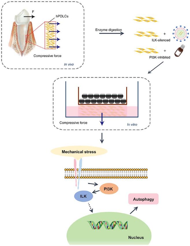
2                         ZOU et al: MECHANISM OF STATIC STRESS-INDUCED AUTOPHAGY IN PDL CELLS

were often cultured in two dimensions, which is inconsistent        Materials and methods
with the dynamic three‑dimensional microenvironment found
in vivo (5). Thus, it is crucial to find a suitable mechanical      Cell isolation and culture. The culture and usage of hPDLCs
stress model to explore the effects of cellular mechanics on the    in the present study were approved by the Ethics Committee of
regulation of cell fate.                                            Xi'an Jiaotong University (no. 2019‑1282), and informed consent
    Previous studies have found that mechanical stress can          was obtained from all the patients. hPDLCs were isolated from
regulate a variety of cellular behaviors, such as cell prolif‑      premolar teeth, which were extracted for orthodontic reasons,
eration, apoptosis, and differentiation (6,7). However, there       from teenagers between the ages of 12 and 15 years. After
are insufficient data on the process of autophagy. Autophagy        rinsing the teeth with phosphate‑buffered saline (PBS) supple‑
is a process during which a cell degrades its damaged organ‑        mented with 100 U/ml penicillin and 100 U/ml streptomycin,
elles and proteins using lysosomes (8,9). It is an important        the periodontal tissue was gently collected from the middle of
mechanism for maintaining intracellular homeostasis and             the root. The tissue was then digested with type I collagenase
integrity. As a dominant catabolic mechanism, autophagy             (Sigma‑Aldrich; Merck KGaA) and dispase (Beijing Solarbio
is involved in the physiological and pathological processes         Science & Technology Co., Ltd.) at 37˚C, for 40 min. The
of cells (10). Increasing evidence suggests that there is a         cells were then re‑suspended in Dulbecco's modified Eagle's
close relationship between autophagy and mechanical stress.         medium (DMEM; Gibco; Thermo Fisher Scientific, Inc.)
King et al (11) found that mammalian cells can respond              culture medium containing 10% fetal bovine serum (FBS;
to mechanical stress by rapidly inducing the formation of           Gibco; Thermo Fisher Scientific, Inc.). Cells at 3‑5 passages
autophagosomes. This indicates that autophagy can be acti‑          were selected for this experiment (25). The verification methods
vated when cells adapt to mechanical stress. Ma et al (12)          are described in the immunofluorescence staining section
suggested that autophagy may be a key response by nucleus           below. After blocking using 5% bull serum albumin (Wuhan
pulposus cells resisting mechanical overload. Mechanical            Boster Biological Technology, Ltd.), hPDLCs were subjected
stress triggers autophagy and excessive autophagy leads to          to immunofluorescence with primary antibodies to vimentin
cell death. This process involves complex mechanical signal         and cytokeratin (1:300, BM0135 and BM0030; Wuhan Boster
transduction pathways.                                              Biological Technology, Ltd.). Cells used in the present study
    The extracellular matrix‑integrin‑cytoskeleton has been         were derived from one individual. The results of characteriza‑
reported to be an important mechanical signal transduction          tion of the hPDLCs are presented in Fig. S1.
pathway (13,14) Integrin‑linked kinase (ILK), the cytoplasmic
domain of β1 integrin, performs a central role in cell growth,      Construction of the 3D static stress loading model for
survival, and differentiation (15‑17). It can promote actin         hPDLCs using collagen‑alginate hydrogel. The dissolved
rearrangement and participate in the maturation of focal            type I collagen solution (extracted from 8‑week‑old female
adhesions (15). A lack of ILK results in less adhesion of           Sprague‑Dawley rats weighing 280‑320 g without any specific
fibroblasts to the ECM, prevents defective cell extension,          treatment), DMEM (Gibco; Thermo Fisher Scientific, Inc.),
and delays the formation of adhesion sites (18). In addition,       sodium alginate solution (Sigma‑Aldrich; Merck KGaA),
ILK can also exhibit kinase activity and can activate the           D‑(+)‑glucono‑1,5‑lactone (DGL, Thermo Fisher Scientific,
kinase B (AKT)/mammalian target of rapamycin (mTOR), a              Inc.), CaSO4 (Tianjin Kemiou Chemical Reagent Co. Ltd.) and
key regulatory pathway of autophagy, with the assistance of         NaOH (Tianjin Tianli Chemical Reagent Co. Ltd.) solution
phosphatidylinositol 3 kinase (PI3K) (19,20). Sosa et al (21)       were mixed in a predetermined order and proportion. Next,
proposed that hyperphosphatemia may activate mTOR and               the hPDLC suspension obtained after digestion with trypsin
reduce autophagy in myoblasts through ILK activation. In            (Beijing Solarbio Science & Technology Co., Ltd.) was added.
recent studies it was confirmed that ILK is a key molecule          The mixed solution was added to the pre‑designed mold and
involved in the effect of mechanical stress on the proliferation,   placed in an incubator (37˚C) for 1 h. The mold was then
apoptosis, and differentiation of hPDLCs (22,23). Therefore,        removed, the formed hydrogel was placed into a 6 mm petri
ILK/PI3K may be important connecting molecules that                 dish, and 3 ml of DMEM containing 10% FBS was placed
transmit mechanical signals to downstream pathways, thereby         over the gel; it was then placed back to incubator (37˚C). The
mediating the effect of mechanical forces on autophagy in           culture medium was changed every other day (Fig. 2A) and
hPDLCs.                                                             static stress was loaded after 72 h.
    In the present study, a collagen‑alginate composite
hydrogel was used to create a 3D cell culture in vitro to           Cell counting kit‑8 (CCK‑8) assay. The cells were mixed
simulate the periodontal microenvironment. The uniform              with liquid hydrogel or seeded in DMEM, containing various
weight method (24) was used to generate unidirectional              concentrations (0, 25, 50 and 100 mmol/l) of inhibitors
static compressive stress that is similar to orthodontic forces,    (LY294002, Echelon Biosciences), and transferred to a 96‑well
to explore the effect of mechanical stress on autophagy             culture plate. A set of five wells in each group were used as
in hPDLCs. Models of ILK silencing and PI3K‑specific                controls. After the cells were cultured for 12 h at 37˚C in a
inhibitory hPDLCs were also established to verify the role          5% CO2 incubator, 10 µl of CCK‑8 solution (Beijing Solarbio
of ILK/PI3K in this process. A schematic diagram of the             Science & Technology Co., Ltd.) was added to each well in the
methods for developing this study is presented in Fig. 1. The       dark every 12 h. A microplate reader (Multiskan FC; Thermo
aim of the present study was to provide insight into exploring      Fisher Scientific, Inc.) was used to measure the optical density
the molecular mechanisms of periodontal remodeling and              (OD) value of each well at a wavelength of 450 nm, at 0, 12,
OTM.                                                                24, 48 and 72 h.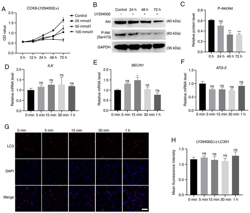
INTERNATIONAL JOURNAL OF MOlecular medicine 48: 167, 2021 3 Figure 1. Schematic diagram of the method for exploring the synergistic effects of ILK and PI3K in the role of static compressive stress on autophagy of human periodontal ligament cells (hPDLCs). hPDLCs were obtained from the human periodontal ligament and divided into untreated, ILK silencing, and PI3K inhibitory groups. The compressive force of hPDLCs was simulated by applying gravity on the collagen‑alginate hydrogel in vitro. The effects of mechanical stress on hPDLC autophagy and the potential modulatory mechanisms were then demonstrated by a series of corresponding assays. ILK, integrin‑linked kinase; PI3K, phosphatidylinositol 3 kinase. Transduction of ILK short hairpin (shRNA) lentiviral vectors. (containing 1 µl/ml of Polybrene) at 50‑60% hPDLC confluency. Two shRNAs targeting the human ILK mRNA (target sequence of The medium‑containing lentiviruses were removed and replaced ILK siRNA1, 5'‑GTGGTTGAGATGTTGATCATG‑3') and one with a fresh medium following 48 h of incubation (37˚C). The negative control shRNA (target sequence of ILK siRNA2, 5'‑TTC transfected cells were seeded in hydrogels and incubated for 48 h TCCGAACGTGTCACGT‑3') were designed and synthesized (37˚C) and were finally collected for experiments. by Shanghai GenePharma Co., Ltd. The hPDLCs were seeded in 6‑well plates at a density of 1.2x10⁵ cells/well. Lentiviruses PI3K specific‑inhibitory assay. For the present study, stock (multiplicity of infection, 50) were diluted into the culture medium solutions of LY294002 (a specific inhibitor of PI3K, Echelon)
4                         ZOU et al: MECHANISM OF STATIC STRESS-INDUCED AUTOPHAGY IN PDL CELLS

dissolved in dimethyl sulfoxide (DMSO, Sigma‑Aldrich;               preparations. The mRNA expression value was calculated
Merck KGaA), were diluted and added to the culture medium.          using the 2‑ΔΔCq method (ΔCq=the mean cycle threshold Cq of
Based on the results of the CCK‑8 assay, the final concentra‑       the target gene ‑ the mean Cq of GAPDH; ΔΔCq=ΔCq of the
tion of LY294002 was determined as 50 mmol/l. The control           experimental group ‑ ΔCt of the control group) (27).
group was treated with the same amount of DMSO. After
72 h, the cells were harvested for western blotting to verify the   Western blot analysis. The abovementioned method was used
inhibitory effect of LY294002 on PI3K.                              to obtain cells for western blot analysis. Total cellular proteins
                                                                    were extracted by adding a RIPA lysis buffer (Beyotime
Uniform weight method to exert static stress on hPDLCs. A           Institute of Biotechnology) containing 1% (v/v) phenylmeth‑
3D printer (Objet Eden260VS Dental Advantage, Stratasys)            anesulfonylfluoride (Beyotime) on ice. Protein concentrations
was used to produce a series of pre‑designed molds. Weighed         were determined with a BCA protein assay kit (Beyotime
to 3, 5, 7 and 10 g, the mold was loaded with different sizes       Institute of Biotechnology). Equal protein amounts (20 µg)
and number of steel balls and was placed on the hydrogel with       were separated by sodium dodecyl sulfate polyacrylamide gel
hPDLCs embedded, pressed for 5, 15 and 30 min, and 1 h, and         electrophoresis (SDS‑PAGE) on 12‑15% gels and were trans‑
then removed. A schematic diagram of static stress loading is       ferred to polyvinylidene difluoride (PVDF) membranes. The
shown in Fig. 2B.                                                   membranes were blocked in 5% non‑fat dry milk for 1 h at
                                                                    room temperature and incubated overnight with phospho‑AKT
Immunofluorescence staining. After washing the compressed           (mAb no. 4060, Ser473; Cell Signaling Technology, Inc.;
hydrogel with PBS, three pieces of hydrogel with a thickness        1:1,000), AKT (mAb no. 4691, Cell Signaling Technology, Inc.;
of 1 mm and a size of 2x2 mm were randomly cut using a              1:1,000) and GAPDH antibodies (1:5,000, bs‑12257R, BIOSS),
surgical blade and fixed in 4% paraformaldehyde, for 15 min.        at 4˚C. The following day, the membranes were washed with
Fixed hydrogels were permeabilized in 0.1% Triton X‑100             Tris‑buffered saline with 0.05% Tween‑20 (Wuhan Boster
(Beijing Solarbio Science & Technology Co., Ltd) for 20 min         Biological Technology, Ltd.) and then incubated with rabbit IgG
before being washed three times with PBS. The pieces were           horseradish peroxidase (HRP)‑linked secondary antibodies
blocked in 5% bull serum albumin (Wuhan Boster Biological           (BA1054, 1:5,000; Wuhan Boster Biological Technology,
Technology, Ltd.) for 30 min at room temperature and                Ltd.), for 1 h at room temperature (28). Finally, the bands
incubated overnight with a primary antibody against LC3             were visualized using the Omega Lum G gel imaging system
(EPR18709, 1:200; Abcam) at 4˚C. Then, the pieces were              (Aplegen). ImageJ software (Version 1.48; National Institutes
incubated with CY3‑labeled anti‑rat secondary antibodies            of Health) was used for densitometric analysis.
(BA1035, 1:50; Wuhan Boster Biological Technology, Ltd.) at
room temperature, for 2 h. After washing with PBS, the nuclei       Statistical analysis. GraphPad prism software (Version 8.0;
were stained with 4‑6‑diamidino‑2‑phenylindole (DAPI)               GraphPad, Inc.) was used for statistical analysis. Values are
for 5 min and were evaluated under a confocal microscope            expressed as mean ± standard error of the mean (SEM).
(Olympus Corporation). ImageJ software (Version 1.48;               One‑way analysis of variance (ANOVA) followed by Tukey's
National Institutes of Health) was used to analyze the images.      multiple comparisons test were used to analyse the statistical
Background corrections and contrast/brightness enhance‑             significance of differences. P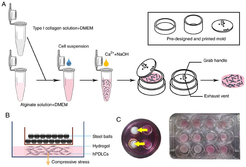
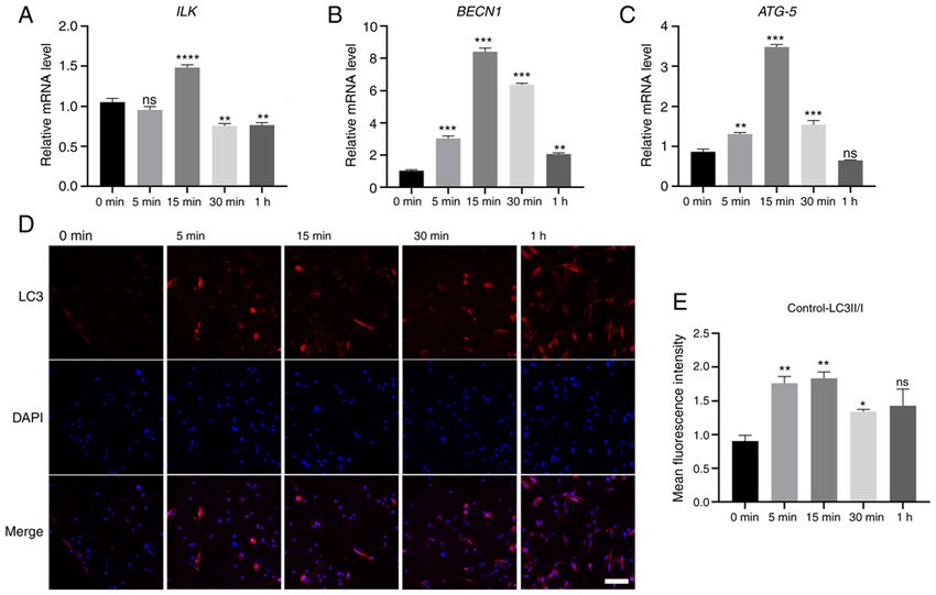
INTERNATIONAL JOURNAL OF MOlecular medicine 48: 167, 2021                                                           5

Figure 2. Three‑dimensional (3D) culture of hPDLCs and the loading of compressive stress. (A) Schematic diagram of the preparation of the collagen‑alginate
complex hydrogel. We mixed collagen, alginate solution, and Dulbecco's modified Eagle's medium in certain proportions and added the cell suspension,
calcium‑crosslinked solution [composed of D‑(+)‑Glucono‑1,5‑lactone and CaSO4], and NaOH solution successively. (B) Schematic diagram of the uniform
weight method. Cells were embedded in the hydrogel, and steel balls of certain weights were placed in the mould above it, which simulated a certain static
compressive stress to hPDLCs using gravity. (C) Hydrogels under and after force loading. The yellow arrows show the load of a certain weight. hPDLCs, human
periodontal ligament cells.

weight method and the hydrogel before and after compres‑                       The red speckled fluorescence representing LC3II could be
sion is shown in Fig. 2B and C. Following compression,                         observed after 5 min of force‑loading, indicating the appear‑
the hydrogel had flattened out. Based on a previous study,                     ance of autophagosomes (Fig. 4D). ImageJ software was used
four compressive forces, including 1.5, 2.5, 3.5 and 5 g/cm 2                  to analyze the average fluorescence intensity of both LC3I and
were loaded to determine the lightest force that can induce                    LC3II, the ratio of which was considered a classic indicator
the most active autophagic response. RT‑qPCR was used to                       of autophagy levels. The results were consistent with those of
detect the expression of the autophagy‑related genes, BECN1                    RT‑qPCR (Fig. 4E). With the extension of the loading time,
and ATG‑5, to reflect the level of autophagy. Compared with                    the ratio of LC3II/I gradually increased, then decreased at
the 2.5 and 3.5 g/cm2 groups, the expression levels of BECN1                   30 min. Compared with the control group, the difference was
and ATG‑5 in the cells were decreased at 5 g/cm 2 loading                      significant (P
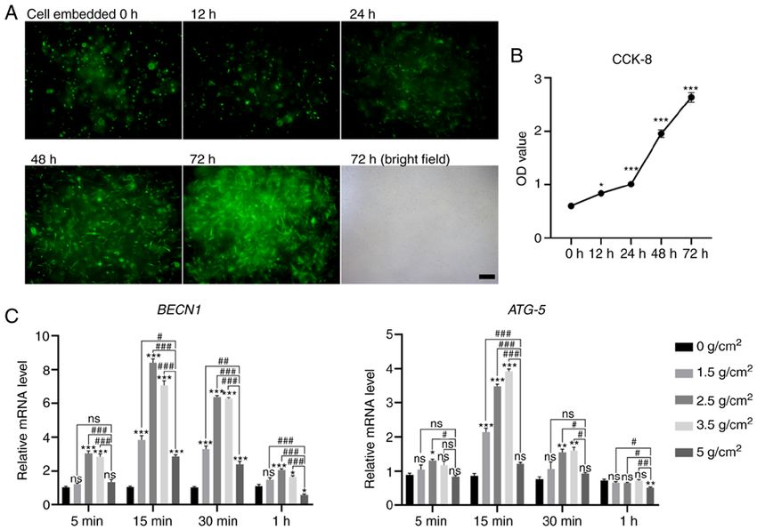
6                              ZOU et al: MECHANISM OF STATIC STRESS-INDUCED AUTOPHAGY IN PDL CELLS

Figure 3. Growth and proliferation of three‑dimensional cultured hPDLCs and the autophagy level with increasing intensity of the mechanical stimuli (0, 1.5,
2.5, 3.5 and 5 g/cm2) applied for different amounts of time (5, 15, 30 min and 1 h). (A) The spreading of hPDLCs (green fluorescence: GFP; scale bar, 200 µm).
The cells appeared as rounded bright spots at 1 h, spindles at 12 h, and increased significantly after 24 h. The morphology of hPDLCs embedded for 72 h in the
bright field is also shown. (B) Optical density value. (C) Gene expression of Beclin‑1 (BECN1) and ATG‑5. Data are presented as mean ± standard error of the
mean. *P
INTERNATIONAL JOURNAL OF MOlecular medicine 48: 167, 2021                                                             7

Figure 4. Effect of static compressive stress (2.5 g/cm 2) on gene expression of (A) ILK, (B) Beclin‑1 (BECN1) and (C) ATG‑5. The expression of LC3 in
hPDLCs under compressive stress was observed by (D) immunofluorescence, red fluorescence: LC3; blue fluorescence: DAPI; scale bar, 200 µm. (E) The ratio
of LC3II/LC3I. Data are presented as mean ± standard error of the mean., *P
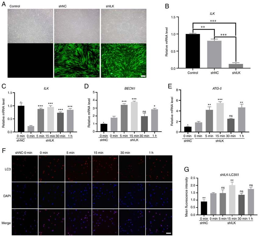
8                              ZOU et al: MECHANISM OF STATIC STRESS-INDUCED AUTOPHAGY IN PDL CELLS

Figure 5. Loading static stress following the gene silencing of ILK. (A) Significant GFP expression was observed in both shNC and shILK groups (scale bar,
200 µm). (B) ILK gene expression in shILK decreased significantly in comparison with shNC and control groups (P
INTERNATIONAL JOURNAL OF MOlecular medicine 48: 167, 2021                                                              9

Figure 6. Loading static stress after PI3K inhibition. (A) Optical density value under different concentrations of drugs. (B) Protein expression level of AKT,
P‑AKT (Ser473) and GAPDH. (C) The ratio of P‑AKT/total AKT level. (D) ILK gene expression. No significant change was found between groups (P>0.05).
(E) Beclin‑1 (BECN1) gene expression was slightly increased at 15 min (P0.05).
(F) ATG‑5 gene expression remained unchanged between groups (P>0.05). (G) The expression of LC3 (red fluorescence: LC3; blue fluorescence: DAPI; scale
bar, 200 µm). (H) The ratio of LC3II/LC3I. Data are presented as mean ± standard error of the mean. *P
10                        ZOU et al: MECHANISM OF STATIC STRESS-INDUCED AUTOPHAGY IN PDL CELLS

of mechanical stress on ILK disappeared after PI3K was              Competing interests
inhibited. Our results confirmed that mechanical stress
can upregulate the expression of ILK in a PI3K‑dependent            The authors declare that they have no competing interests.
manner. This may provide evidence of the transduction
mechanism of mechanical signals and the interaction with            References
the role of ILK and PI3K in OTM.
    However, the extraction of total protein of hPDLCs               1. Berry S, Javed F, Rossouw PE, Barmak AB, Kalogirou EM and
under 3D culture needs to be performed after the hydrogel               Michelogiannakis D: Influence of thyroxine supplementation on
                                                                        orthodontically induced tooth movement and/or inflammatory
is dissolved, and following protein degradation, which may              root resorption: A systematic review. Orthod Craniofac Res 24:
affect the accuracy of the results. Therefore, the western blot         206‑213, 2020.
assay of LC‑3I/II and other autophagy‑associated proteins            2. Jin Y, Li J, Wang YT, Ye R, Feng XX, Jing Z and Zhao Z:
                                                                        Functional role of mechanosensitive ion channel Piezo1 in
were absent in this study. Ideal hydrogel that can be rapidly           human periodontal ligament cells. Angle Orthodontist 85:
degraded and does not affect cells is required. In conclusion,          87‑94, 2015.
the current findings confirmed that static compressive stress        3. Kikuta J, Yamaguchi M, Shimizu M, Yoshino T and Kasai K:
                                                                        Notch signaling induces root resorption via RANKL and IL‑6
can induce autophagy within a certain range of force that is            from hPDL cells. J Dental Res 94: 140‑147, 2015.
related to the ILK/PI3K signaling pathway. The inhibition            4. Chang ML, Lin H, Fu HD, Wang BK, Han GL and Fan MW:
of PI3K can invalidate the upregulation of ILK induced by               MicroRNA‑195‑5p regulates osteogenic differentiation of
                                                                        periodontal ligament cells under mechanical loading. J Cell
mechanical stress. Further studies are required to clarify              Physiology 232: 3762‑3774, 2017.
the downstream targeting pathway of mechanically induced             5. Duval K, Grover H, Han LH, Mou Y, Pegoraro AF, Fredberg J
autophagy and the mechanism involving mechanical stress,                and Chen Z: Modeling physiological events in 2D vs. 3D cell
                                                                        culture. Physiology (Bethesda) 32: 266‑277, 2017.
PI3K and ILK.                                                        6. Zhao Y, Wang C, Li S, Song H, Wei F, Pan K, Zhu K, Yang P, Tu Q
                                                                        and Chen J: Expression of Osterix in mechanical stress‑induced
Acknowledgements                                                        osteogenic differentiation of periodontal ligament cells in vitro.
                                                                        Eur J Oral Sci 116: 199‑206, 2008.
                                                                     7. Chen JL, Zhang W, Backman LJ, Kelk P and Danielson P:
Not applicable.                                                         Mechanical stress potentiates the differentiation of periodontal
                                                                        ligament stem cells into keratocytes. Brit J Ophthalmol 102:
                                                                        562‑569, 2018.
Funding                                                              8. Banerjee A and Bandopadhyay R: Autophagy: Nobel prize in
                                                                        physiology or medicine' 16 to the intra‑cellular suicidal process.
The present study was supported by the Xi'an Science and                Natl Acad Sci Lett 40: 461‑465, 2017.
                                                                     9. Klionsky DJ and Emr SD: Autophagy as a regulated pathway of
Technology Project, China [no. 201805100YX8SF34(2)],                    cellular degradation. Science 290: 1717‑1721, 2000.
the General project from the field of social Development, in        10. Ott C, König J, Höhn A, Jung T and Grune T: Macroautophagy
Department of Science and Technology of Shaanxi Province,               is impaired in old murine brain tissue as well as in senescent
                                                                        human fibroblasts. Redox Biol 10: 266‑273, 2016.
China (no. 2019SF‑081), and by the National Natural Science         11. King JS, Veltman DM and Insall RH: The induction of autophagy
Foundation, China (no. 8197032023).                                     by mechanical stress. Autophagy 7: 1490‑1499, 2011.
                                                                    12. Ma KG, Shao ZW, Yang SH, Wang J, Wang BC, Xiong LM, Wu Q
                                                                        and Chen SF: Autophagy is activated in compression‑induced
Availability of data and materials                                      cell degeneration and is mediated by reactive oxygen species
                                                                        in nucleus pulposus cells exposed to compression. Osteoarthr
All data generated or analyzed during this study are included           Cartilage 21: 2030‑2038, 2013.
                                                                    13. Iskratsch T, Wolfenson H and Sheetz MP: Appreciating force
in this published article.                                              and shape ‑ the rise of mechanotransduction in cell biology. Nat
                                                                        Rev Mol Cell Bio 15: 825‑833, 2014.
Authors' contributions                                              14. Kechagia JZ, Ivaska J and Roca‑Cusachs P: Integrins as biome‑
                                                                        chanical sensors of the microenvironment. Nat Rev Mol Cell
                                                                        Bio 20: 457‑473, 2019.
RZ, YW and YG conceived and designed the experiments.               15. Zheng CC, Hu HF, Hong P, Zhang QH, Xu WW, He QY and
SW, XK and DZ drafted the manuscript and performed the                  Li B: Significance of integrin‑linked kinase (ILK) in tumorigen‑
                                                                        esis and its potential implication as a biomarker and therapeutic
experiments. SW and YW participated in data analysis and                target for human cancer. Am J Cancer Res 9: 186‑197, 2019.
edited the manuscript. SZ, HS, BG and SM were involved in           16. Zheng QM, Chen XY, Bao QF, Yu J and Chen LH: ILK enhances
the discussion and interpretation of the results. LN participated       migration and invasion abilities of human endometrial stromal
                                                                        cells by facilitating the epithelial‑mesenchymal transition.
in data analysis and provided technical guidance. RZ and SW             Gynecol Endocrinol 34: 1091‑1096, 2018.
confirm the authenticity of all the raw data. All authors have      17. Zhu XY, Liu N, Liu W, Song SW and Guo KJ: Silencing of
read and approved the final manuscript.                                 the integrin‑linked kinase gene suppresses the proliferation,
                                                                        migration and invasion of pancreatic cancer cells (Panc‑1). Genet
                                                                        Mol Biol 35: 538‑544, 2012.
Ethics approval and consent to participate                          18. Sakai T, Li S, Docheva D, Grashoff C, Sakai K, Kostka G,
                                                                        Braun A, Pfeifer A, Yurchenco PD and Fässler R: Integrin‑linked
                                                                        kinase (ILK) is required for polarizing the epiblast, cell adhe‑
The present study was approved by the Ethics Committee of               sion, and controlling actin accumulation. Genes Dev 17: 926‑940,
Stomatology Hospital of Xi'an Jiaotong University College of            2003.
Medicine.                                                           19. Jung CH, Ro SH, Cao J, Otto NM and Kim DH: mTOR regula‑
                                                                        tion of autophagy. FEBS Lett 584: 1287‑1295, 2010.
                                                                    20. Delcommenne M, Tan C, Gray V, Rue L, Woodgett J and
Patient consent for publication                                         Dedhar S: Phosphoinositide‑3‑OH kinase‑dependent regula‑
                                                                        tion of glycogen synthase kinase 3 and protein kinase B/AKT
                                                                        by the integrin‑linked kinase. Proc Natl Acad Sci USA 95:
Not applicable.                                                         11211‑11216, 1998.
INTERNATIONAL JOURNAL OF MOlecular medicine 48: 167, 2021 11
21. Sosa P, Alcalde‑Estevez E, Plaza P, Troyano N, Alonso C, 35. Branco da Cunha C, Klumpers DD, Li WA, Koshy ST, Weaver JC,
Martínez‑Arias L, Evelem de Melo Aroeira A, Rodriguez- Chaudhuri O, Granja PL and Mooney DJ: Influence of the stiff‑
Puyol D, Olmos G, López‑Ongil S and Ruíz‑Torres MP: ness of three‑dimensional alginate/collagen‑I interpenetrating
Hyperphosphatemia promotes senescence of myoblasts by networks on fibroblast biology. Biomaterials 35: 8927‑8936, 2014.
impairing autophagy through Ilk overexpression, a possible 36. Li D, Lu Z, Xu Z, Ji J, Zheng Z, Lin S and Yan T: Spironolactone
mechanism involved in sarcopenia. Aging Dis 9: 769‑784, 2018. promotes autophagy via inhibiting PI3K/AKT/mTOR signalling
22. Wan WT, He C, Du CY, Wang YJ, Wu SY, Wang TR and pathway and reduce adhesive capacity damage in podocytes
Zou R: Effect of ILK on small‑molecule metabolism of human under mechanical stress. Biosci Rep 36: e00355, 2016.
periodontal ligament fibroblasts with mechanical stretching. 37. Xu HG, Yu YF, Zheng Q, Zhang W, Wang CD, Zhao XY,
J Periodontal Res 55: 229‑237, 2020. Tong WX, Wang H, Liu P and Zhang XL: Autophagy protects
23. Wang Y, Du C, Wan W, He C, Wu S, Wang T, Wang F and Zou R: end plate chondrocytes from intermittent cyclic mechanical
shRNA knockdown of integrin‑linked kinase on hPDLCs migra‑ tension induced calcification. Bone 66: 232‑239, 2014.
tion, proliferation, and apoptosis under cyclic tensile stress. Oral 38. Ghatak S, Morgner J and Wickstrom SA: ILK: A pseudokinase
Dis 26: 1747‑1754, 2020. with a unique function in the integrin‑actin linkage. Biochem
24. Li M, Zhang C and Yang Y: Effects of mechanical forces on Soc Trans 41: 995‑1001, 2013.
osteogenesis and osteoclastogenesis in human periodontal liga‑ 39. Vautrin‑Glabik A, Botia B, Kischel P, Ouadid‑Ahidouch H
ment fibroblasts: A systematic review of in vitro studies. Bone and Rodat‑Despoix L: IP3 R3 silencing induced actin cytoskel‑
Joint Res 8: 19‑31, 2019. etal reorganization through ARHGAP18/RhoA/mDia1/FAK
25. Yang J, Zhou J, Cui B and Yu T: Evaluation of hypoxia on pathway in breast cancer cell lines. Biochim Biophys Acta Mol
the expression of miR‑646/IGF‑1 signaling in human peri‑ Cell Res 1865: 945‑958, 2018.
odontal ligament cells (hPDLCs). Med Sci Monit 24: 5282‑5291, 40. Memmel S, Sisario D, Zoller C, Fiedler V, Katzer A, Heiden R,
2018. Becker N, Eing L, Ferreira FLR, Zimmermann H, et al: Migration
26. Brazvan B, Farahzadi R, Mohammadi SM, Saheb SM, pattern, actin cytoskeleton organization and response to PI3K‑,
Shanehbandi D, Schmied L, Soleimani Rad J, Darabi M and mTOR‑, and Hsp90‑inhibition of glioblastoma cells with different
Nozad Charoudeh H: Key immune cell cytokines affects the invasive capacities. Oncotarget 8: 45298‑45310, 2017.
telomere activity of cord blood cells in vitro. Adv Pharm Bull 6: 41. Boppart MD and Mahmassani ZS: Integrin signaling: Linking
153‑161, 2016. mechanical stimulation to skeletal muscle hypertrophy. Am
27. Livak KJ and Schmittgen TD: Analysis of relative gene expres‑ J Physiol Cell Physiol 317: C629‑C641, 2019.
sion data using real‑time quantitative PCR and the 2(‑Delta Delta 42. Mousavizadeh R, Hojabrpour P, Eltit F, McDonald PC, Dedhar S,
C(T)) method. Methods 25: 402‑408, 2001. McCormack RG, Duronio V, Jafarnejad SM and Scott A: β1 inte‑
28. Farahzadi R, Fathi E and Vietor I: Mesenchymal stem cells could grin, ILK and mTOR regulate collagen synthesis in mechanically
be considered as a candidate for further studies in cell‑based loaded tendon cells. Sci Rep 10: 12644, 2020.
therapy of Alzheimer's disease via targeting the signaling 43. Fruman DA, Chiu H, Hopkins BD, Bagrodia S, Cantley LC and
pathways. ACS Chem Neurosci 11: 1424‑1435, 2020. Abraham RT: The PI3K pathway in human disease. Cell 170:
29. Moraes C, Sun Y and Simmons CA: (Micro)managing the 605‑635, 2017.
mechanical microenvironment. Integr Biol (Camb) 3: 959‑971, 44. Porter KM, Jeyabalan N and Liton PB: MTOR‑independent
2011. induction of autophagy in trabecular meshwork cells subjected
30. Wozniak MA, Modzelewska K, Kwong L and Keely PJ: Focal to biaxial stretch. Biochim Biophys Acta 1843: 1054‑1062, 2014.
adhesion regulation of cell behavior. Biochim Biophys Acta 1692: 45. Nishimura T and Tooze SA: Emerging roles of ATG proteins and
103‑119, 2004. membrane lipids in autophagosome formation. Cell Discov 6: 32,
31. Cukierman E, Pankov R, Stevens DR and Yamada KM: Taking 2020.
cell‑matrix adhesions to the third dimension. Science 294: 46. Wang X, Zhang Y, Feng T, Su G, He J, Gao W, Shen Y and Liu X:
1708‑1712, 2001. Fluid shear stress promotes autophagy in hepatocellular carci‑
32. Oh SA, Lee HY, Lee JH, Kim TH, Jang JH, Kim HW and noma cells. Int J Biol Sci 14: 1277‑1290, 2018.
Wall I: Collagen three‑dimensional hydrogel matrix carrying 47. Dedhar S: Cell‑substrate interactions and signaling through ILK.
basic fibroblast growth factor for the cultivation of mesenchymal Curr Opin Cell Biol 12: 250‑256, 2000.
stem cells and osteogenic differentiation. Tissue Eng Part A 18:
1087‑1100, 2012.
33. Rajan N, Habermehl J, Cote MF, Doillon CJ and Mantovani D:
Preparation of ready‑to‑use, storable and reconstituted type I This work is licensed under a Creative Commons
collagen from rat tail tendon for tissue engineering applications. Attribution-NonCommercial-NoDerivatives 4.0
Nat Protoc 1: 2753‑2758, 2006. International (CC BY-NC-ND 4.0) License.
34. Yuan T, Zhang L, Feng L, Fan H and Zhang X: Chondrogenic
differentiation and immunological properties of mesenchymal
stem cells in collagen type I hydrogel. Biotechnol Prog 26:
1749‑1758, 2010.You can also read