Sensitizing TRAIL resistant A549 lung cancer cells and enhancing TRAIL induced apoptosis with the antidepressant amitriptyline
←
→
Page content transcription
If your browser does not render page correctly, please read the page content below
ONCOLOGY REPORTS 46: 144, 2021
Sensitizing TRAIL‑resistant A549 lung cancer cells and enhancing
TRAIL‑induced apoptosis with the antidepressant amitriptyline
K.M.A. ZINNAH1,2 and SANG‑YOUEL PARK1
1
Biosafety Research Institute, College of Veterinary Medicine, Jeonbuk National University, Iksan, Jeonbuk 54596,
Republic of Korea; 2Department of Animal and Fish Biotechnology, Faculty of Biotechnology and Genetic Engineering,
Sylhet Agricultural University, Sylhet 3100, People's Republic of Bangladesh
Received December 14, 2020; Accepted April 16, 2021
DOI: 10.3892/or.2021.8095
Abstract. Tumor necrosis factor‑related apoptosis‑inducing Introduction
ligand (TRAIL) is a cytokine with the potential to induce
cancer cell‑specific apoptosis with minimal toxicity to normal Lung cancer is the most common cause of cancer‑related
cells. Therefore, the resistance of certain cancer cells to deaths worldwide (1). In a study in 2018 in the U.S., lung cancer
TRAIL is a major concern and agents that can either enhance was the second most common cancer diagnosis by sex and
TRAIL capabilities or overcome TRAIL resistance are neces‑ was newly diagnosed in 14% of men and 13% of women (2).
sary for the development of cancer treatments. The present Typically, patients with non‑small cell lung cancer (NSCLC)
study investigated whether the antidepressant drug amitripty‑ are identified with advanced cancer and approximately 16.43%
line could sensitize TRAIL‑resistant A549 lung cancer cells of patients survive for five years (3,4). Lung cancer treatments
and enhance TRAIL‑induced apoptosis. Antidepressants are include surgery, radiotherapy, and chemotherapeutic drugs and
usually prescribed to cancer patients to relieve emotional their combinations (5,6). Specific combination strategies with
distress, such as depression or dysthymia. The present study potent chemotherapeutic drugs may be a potential approach to
revealed for the first time, to the best of our knowledge, that cancer treatment (7,8).
amitriptyline increased death receptor (DR) 4 and 5 expres‑ Tumor necrosis factor‑related apoptosis‑inducing ligand
sion, a requirement for TRAIL‑induced cell death. Genetic (TRAIL) is a highly attractive anticancer treatment that selec‑
inhibitors of DR4 and DR5 significantly reduced amitripty‑ tively kills cancer cells without causing toxicity to normal
line‑enhanced TRAIL‑mediated apoptosis. Additionally, the cells (9). TRAIL binds to death receptor (DR)4/DR5 to initiate
present study explored whether blocking autophagy increased apoptotic cell death. Along with A549 lung cancer cells, a
DR4 and DR5 expression. Blocking autophagy flux with the large number of cancer cells are resistant to TRAIL due to the
final stage autophagy inhibitor chloroquine (CQ) also upregu‑ insufficient expression of death receptors DR4/DR5 and the
lated DR4 and DR5 expression. TRAIL in combination with extreme expression of decoy receptors, as well as the mutation
amitriptyline or CQ significantly increased the expression of of TRAIL receptors (10‑12). Interestingly, it is possible to over‑
apoptosis‑indicator proteins cleaved caspase‑8 and caspase‑3. come TRAIL resistance with suitable pharmacological agents
The expression levels of LC3‑II and p62 were significantly that can enhance the expression of TRAIL receptors (13,14).
higher in amitriptyline‑treated cells, which confirmed that Autophagy plays an important role in maintaining
amitriptyline blocks autophagy by inhibiting the fusion of cellular homeostasis (15). Autophagy suppresses tumors by
autophagosomes with lysosomes. Overall, the present results maintaining cellular homeostasis; however, these tumors can
contributed to understanding the mechanism responsible for play a survival role when cancer has already developed (16).
the synergistic anticancer effect of amitriptyline and TRAIL Tumor cells fulfill their energy demands by using autophagy
and also presented a novel mechanism involved in DR4 and and through this process develop treatment resistance (17).
DR5 upregulation. Numerous previous studies have demonstrated that blocking
autophagy flux by inhibiting autophagosome‑lysosome fusion
can be an encouraging approach for cancer therapy (18,19).
Consequently, pha r macological agents that induce
autophagosome accumulation by inhibiting lysosomal fusion
Correspondence to: Professor Sang‑Youel Park, Biosafety
Research Institute, College of Veterinary Medicine, Jeonbuk
and increase TRAIL receptors can be an effective approach to
National University, Iksan, Jeonbuk 54596, Republic of Korea overcome TRAIL resistance.
E‑mail: sypark@chonbuk.ac.kr Depression is a common psychological disorder in cancer
patients. Continuous depression reduces the antitumor immune
Key words: amitriptyline, tumor necrosis factor‑related apoptosis‑ response and creates a favorable environment for tumor
inducing ligand, death receptor‑4/5, apoptosis, autophagy growth (20). Animal model studies have shown that behav‑
ioral stress induces the rapid development of prostate (20),
ovarian (21), pancreatic (22), and breast cancer (22), as well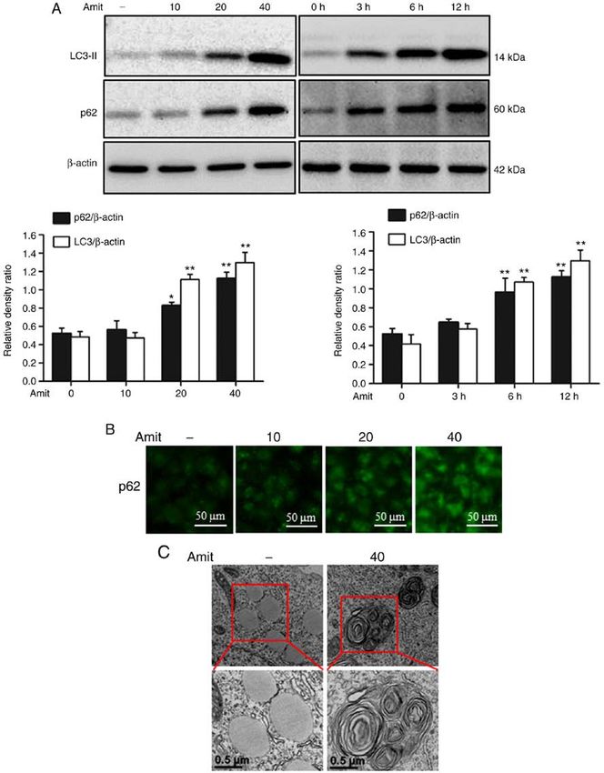
2                         ZINNAH and PARK: AMITRIPTYLINE ENHANCES TRAIL-MEDIATED APOPTOSIS

as carcinomas and malignant melanomas (23). Several studies         LDH cytotoxicity detection kit (Takara Bio, Inc.) following
have recommended that amitriptyline is a productive option to       the manufacturer's protocol. LDH activity was measured
control cancer‑associated depression, anxiety, and pain (24,25).    at 490 nm using a microplate reader (Spectra Max M2;
     Amitriptyline is a psychoactive tricyclic antidepressant       Molecular Devices, LLC).
(TCA) drug. The drug has been revealed to markedly exert
effective anticancer effects on a large number of cancer cell       Colony‑formation assay. Cells were plated in 6‑well plates
types, including colon, prostate, glioma osteosarcoma, skin,        at 37˚C and treated with the indicated doses of amitriptyline
squamous carcinoma, and multiple myeloma (26). Another              (40 µM), CQ (20 µM) and TRAIL (100 ng/ml). Two days later,
study revealed that amitriptyline induced p53 expression,           the culture medium was changed with new medium without
activated caspase‑3, and decreased anti‑apoptotic proteins          amitriptyline, CQ and TRAIL, and the culture continued for
Bcl‑2 and Mcl‑1 in multiple myeloma. In combination with            7 days. Colonies were fixed for 20 min at RT in 4% para‑
bortezomib, amitriptyline induced apoptosis in multiple             formaldehyde, stained with 0.05% (w/v) crystal violet for
myeloma (27). Amitriptyline has also been studied as a poten‑       10 min at RT, and counted under an inverted light microscope
tial candidate for oxidative therapy for its cytotoxicity in H460   (Nikon Corporation).
lung cancer cells, which may be more effective than other
chemotherapeutic drugs (28).                                        Flow cytometric analysis of apoptosis. Apoptosis was evalu‑
     In the present study, it was demonstrated that amitriptyline   ated cells (50 cells/µl) using Annexin V‑FITC Assay Kit (Santa
could sensitize TRAIL‑resistant lung cancer cells to induce         Cruz Biotechnology, Inc.), for flow cytometry according to the
TRAIL‑mediated apoptosis. The molecular mechanism                   manufacturer's instructions (Guava EasyCyte HT System;
underlying the anticancer effects of amitriptyline in combina‑      EMD Millipore). The fluorescence was measured at 488 nm
tion with TRAIL and, specifically, the role of autophagy in         of excitation and 525/30 emission using Guava® InCyte and
lung cancer treatment was also investigated.                        GuavaSuite Software.

Materials and methods                                               Western blot analysis. The cells were lysed in lysis buffer
                                                                    [25 mM HEPES (pH 7.4), 100 mM ethylenediaminetet‑
Cells and culture systems. A549 lung cancer cells were              raacetic acid (EDTA), 5 mM MgCl 2, 0.1 mM dithiothreitol
acquired from the American Type Culture Collection (ATCC).          (DTT), and a protease inhibitor cocktail], and sonicated to
The cells were cultured in Roswell Park Memorial Institute          prepare cell lysates. Equal amounts (40 µg) of proteins were
(RPMI)‑1640 medium (Gibco BRL; Thermo Fisher Scientific,            separated by 8‑15% sodium dodecyl sulfate‑polyacrylamide
Inc.) supplemented with 10% (v/v) fetal bovine serum                gel electrophoresis (SDS) and transferred onto polyvinylidene
(Sigma‑Aldrich; Merck KGaA) and antibiotics (100 µg/ml              fluoride (PVDF) membranes. The membranes were blocked
penicillin‑streptomycin; Sigma‑Aldrich; Merck KGaA) at 37˚C         at 25˚C for 1 h, and then incubated with the indicated concen‑
in a 5% CO2 incubator.                                              trations of primary antibodies at 25˚C for 1 h, and then they
                                                                    were blotted with anti‑mouse IgG (Alexa Fluor 647 conju‑
Reagents. Amitriptyline was purchased from Cayman                   gate) secondary antibodies (product. no. 4410; 1:2,000; Cell
Chemical Company, and chloroquine (CQ) (20 µM) was                  Signaling Technology, Inc.) at 25˚C for 1 h. The membranes
obtained from Sigma‑Aldrich; Merck KGaA. Human recom‑               were developed with enhanced chemiluminescence reagents
binant TRAIL (100 ng/ml) was purchased from AbFrontier.             (ECL; GE Healthcare Life Sciences). Primary antibodies used
                                                                    for the immunoblotting included: DR4 (product. code. ab8414;
Cell viability assay. Cell viability was assessed with MTT and      1:1,000), DR5 (product. code. ab181846; 1:10,000) (both
crystal violet staining assays. The cells were plated in 12‑well    from Abcam), LC3 (product. no. 3868; 1:1,000), p62
plates at a density of 1.0x10⁴ cells/well and incubated at 37˚C     (cat. no. 5114; 1:1,000), cleaved caspase‑3 (product. no. 9661;
for 24 h. The cells were pretreated with different concentra‑       1:500), p‑AMPKα (product. no. 2531; 1:1,000) all from
tions of amitriptyline (0, 10, 20 and 40 µM) or CQ for 12 h and     Cell Signaling Technology, Inc., cleaved caspase‑8
then exposed to recombinant TRAIL (100 ng/ml) for 3 h. Cell         (cat. no. 551242; 1:1,000, BD Pharmingen; BD Biosciences),
morphology was observed under an inverted light microscope          and β‑actin (cat. no. A2228; 1:2,000, Sigma‑Aldrich; Merck
(magnification, x100; Nikon Corporation). Cell viability was        KGaA). The bands were visualized and captured with a
assessed by adding 50 µl of 5 mg/ml methyl‑thiazolyl tetra‑         Fusion‑FX7 using easy‑to‑use FusionCapt V16.07 Software
zolium (MTT) to each well and incubating them at 37˚C for           (both Vilber Lourmat).
2 h. After incubation, the MTT solution was removed and the
cells were treated with 500 µl of dimethyl sulfoxide and the        Immunocytochemistry. The cells (~1x10⁶ cells) were grown
absorbance was measured at 570 nm with a spectrophotometer          on glass coverslips, then treated with amitriptyline, washed
(Bio‑Rad Laboratories). For the crystal violet assay, the cells     with 1% PBS, and fixed with 4% paraformaldehyde in PBS
were stained with a staining solution (0.5% crystal violet in       at RT for 15 min. They were then washed twice with ice‑cold
30% ethanol and 3% formaldehyde) for 10‑20 min at room              PBS and incubated at RT for 10 min in PBS containing
temperature (RT), washed 3‑4 times with phosphate‑buffered          0.25% Triton X‑100. After the incubation, the cells were
saline (PBS), and then imaged.                                      washed three times with PBS and blocked with 1% BSA
                                                                    in PBST for 30 min. The cells were then incubated with a
Lactate dehydrogenase (LDH) assay. Cytotoxicity was                 primary antibody [anti‑p62 (1:1,000; product. no. 5114; Cell
analyzed in the collected supernatant and determined by an          Signaling Technology, Inc.) and DR4/5 diluted with 1% BSA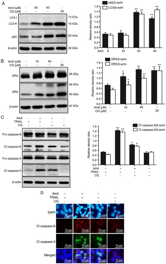
ONCOLOGY REPORTS 46: 144, 2021 3
in PBST] in a 5% CO2 incubator for 3 h at 37˚C. After incu‑ Laboratories, Inc.), following the manufacturer's instructions
bation, the cells were washed three times with PBS. Next, at 85˚C for 5 sec, 37˚C for 10 min and 4˚C for 15 min. Gene
the cells were incubated with a secondary antibody [(Alexa primers (1 µl) and SYBR‑Green (Bio‑Rad Laboratories, Inc.)
Fluor ® 488‑conjugate; donkey polyclonal anti‑rabbit, 1:500; contained in a total reaction volume of 20 µl were used to
cat. no. A‑21206; Thermo Fisher Scientific, Inc.), diluted with conduct the RT‑qPCR . The reaction protocols were as follows:
1% BSA in PBST] in the dark for 2 h at RT. The solution was Predenaturation at 95˚C for 30 sec, 40 cycles of denaturation
removed and the cells were washed 3‑4 times with PBS. The at 95˚C for 5 sec and annealing at 60˚C for 30 sec. GAPDH
cells were treated with DAPI (4',6‑diamidino‑2‑phenylindole, were used as the respective internal control. The sequences
D9564; Sigma‑Aldrich;Merck KGaA) and incubated for of the primers used were: DR4 forward, 5'‑GGGACAG CA
10 min at 25˚C. The cells were washed three times, then CGGACCCAGTG‑3' and reverse, 5'‑ATCCTTGACCTTGAC
mounted with fluorescent mounting medium and the images CATCC‑3'; DR5 forward, 5'‑GCG GTCCTG CTGT TG GTC
were captured using a fluorescence microscope (Nikon TC‑3' and reverse, 5‑GCT T CT GTC CAC ACG C TC AG‑3';
ECLIPSE 80i; magnification, x400; Nikon Corporation). and GAPDH as an internal control forward, 5'‑TGCACCACC
AACTGCT TAG‑3' and reverse, 5'‑GGATGCAGGGATGAT
Transmission electron microscopy. Trypsinized cells were GTT‑3'. All data were evaluated using Bio‑Rad CFX manager,
fixed with 2% glutaraldehyde (Electron Microscopy Sciences) version 2.1 analysis software (Bio‑Rad Laboratories, Inc.).
for 2 h at 4˚C in PBS, followed by 2% osmium tetroxide The collected data from three independent experiments were
(Electron Microscopy Sciences), and dehydrated with an analyzed using the 2‑ΔΔCq method (29).
ethanol series (25, 50, 70, 90 and 100%) for 5 min each.
After dehydration, the samples were embedded in epoxy Statistical analysis. The data are expressed as the mean ± stan‑
resin (Embed 812; Electron Microscopy Sciences) for 48 h dard deviation (SD) from three independent experiments.
at 60˚C according to the manufacturer's instructions. Ultrathin The significance of the differences between the treatments
sections (60 nm) were prepared using an LKB III ultratome was analyzed using one‑way analysis of variance (ANOVA),
(Leica Microsystems GmbH) and stained with 0.5% uranyl followed by the Tukey‑Kramer post hoc test. Statistical
acetate (Electron Microscopy Sciences) for 20 min and 0.1% analyses were executed using GraphPad Prism 7 (GraphPad
lead citrate (Electron Microscopy Sciences) for 7 min at RT. Software, Inc). P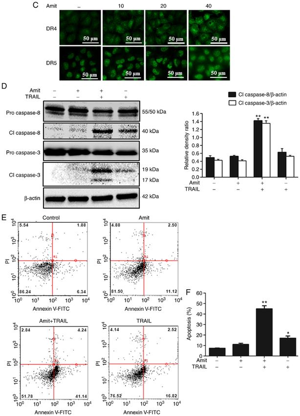
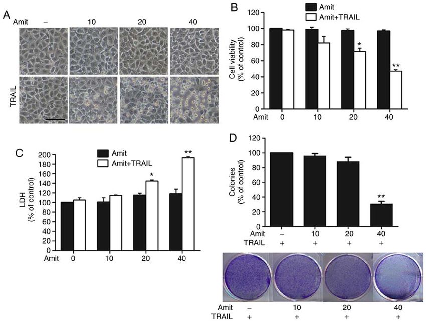
4                               ZINNAH and PARK: AMITRIPTYLINE ENHANCES TRAIL-MEDIATED APOPTOSIS

Figure 1. Amitriptyline enhances TRAIL‑induced apoptosis in lung cancer cells. A549 cells were preincubated with the designated concentrations of
amitriptyline for 12 h and then co‑treated with 100 ng/ml of TRAIL for 3 h. (A) Cell morphologies were captured and examined under a light microscope
(magnification x100; scale bar, 50 µm). (B) MTT assays were used to reveal cell viability (bar graph). (C) Cytotoxicity was measured by LDH released from
co‑treatment. (D) Colonies were fixed with paraformaldehyde and stained with crystal violet dye and the number of colonies were counted under a light micro‑
scope. Statistically significant differences between the control and each indicated treatment group are presented as *P
ONCOLOGY REPORTS 46: 144, 2021 5
Figure 2. Continued.
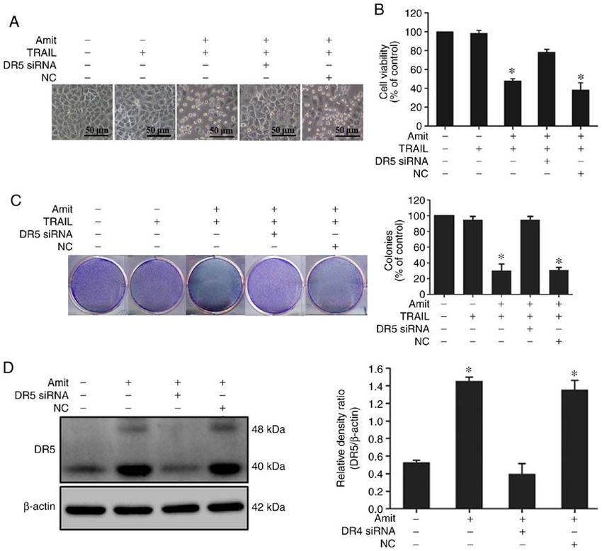
was blocked after siRNA transfection compared to the images also demonstrated the increased expression of p62 in
non‑transfected cells (Figs. 3D and 4D). These experimental a dose‑dependent manner (Fig. 5B). Transmission electron
findings confirmed that the upregulation of DR4 and DR5 is microscopy revealed the higher accumulation of autophagic
required in attenuating TRAIL resistance. vacuoles compared to the control, confirming autophagy flux
inhibition by amitriptyline (Fig. 5C). These results indicated
Amitriptyline blocks autophagy by inhibiting autophagosome‑ that amitriptyline blocked autophagy flux at the final stage of
lysosome fusion. To investigate the role of amitriptyline in autophagy.
autophagy flux, the well‑known autophagy markers LC3‑II and
p62 were analyzed. Western blot analysis revealed the conver‑ Blocking autophagy induces DR4 and DR5 upregulation and
sion of LC3I to LC3‑II, indicating the formation of complete enhances TRAIL‑mediated apoptosis. The role of autophagy
autophagosomes. However, p62 is a cargo adaptor protein that blocking in death receptor expression was investigated using
depends on lysosomes or proteasomes for degradation (31). an autophagy inhibitor. Blocking autophagy flux with a final
The expression of LC3‑II and p62 was increased following stage autophagy inhibitor CQ upregulated both DR4 and DR5
amitriptyline treatment, indicating the blocking of autophagy expression, leading to an increase in apoptosis. The cells were
flux by inhibiting autophagosome‑lysosome fusion in the late treated with or without 20 µM CQ and the indicated doses
stage of autophagy (Fig. 5A). The immunocytochemistry of amitriptyline for 12 h. Western blot analysis revealed that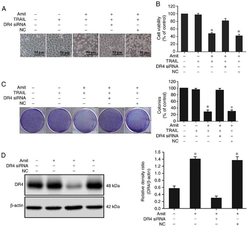
6                              ZINNAH and PARK: AMITRIPTYLINE ENHANCES TRAIL-MEDIATED APOPTOSIS

Figure 2. DR4 and DR5 enhancement are required by amitriptyline for TRAIL‑mediated apoptosis. A549 cells were preincubated with the designated doses
of amitriptyline for 12 h. (A) Harvested cell lysates were collected and subjected to western blotting to determine the dose‑ and time‑dependent expression of
DR4 and DR5. (B) DR4 and DR5 mRNA expression assessed by reverse transcription‑quantitative polymerase chain reaction. GAPDH was used as an internal
control to indicate equivalent RNA loading. (C) The immunocytochemistry results revealed the significant expression of DR4 and DR5 in dose‑dependent
manners (scale bar, 50 µm). (D) Amitriptyline (40 µM)‑treated cells were incubated for 12 h and additionally exposed to 100 ng/ml TRAIL protein for 3 h.
Intracellular apoptosis regulatory proteins cleaved caspase‑8 and cleaved caspase‑3 were analyzed by western blotting. β ‑actin was detected as a protein
loading control. (E) Cells were treated with Annexin V‑FITC and PI, which binds to phosphatidylserine to the plasma membrane and nuclei during apoptosis.
(F) Bar graph showing the averages of the Annexin V‑positive cells. Values represent the mean ± SD (n=3). Statistically significant differences between the
control and each indicated treatment group are presented as *P
ONCOLOGY REPORTS 46: 144, 2021                                                                    7

Figure 3. Silencing of DR4 expression negatively controls amitriptyline‑induced TRAIL‑mediated apoptosis. DR4 siRNA and control siRNA (40 nM) were
transfected for 24 h, then the cells were treated with amitriptyline (40 µM) for 12 h and finally, 100 ng/ml of TRAIL protein was added for 3 h. (A) Images of
the cells were captured and morphological variations were examined under a light microscope (magnification, x100; scale bar, 50 µm). (B) MTT assays were
performed to reveal cell viability percentages (bar graph). (C) Cell colonies were stained with crystal violet dye and the number of colonies were counted.
Statistically significant differences between the control and each indicated treatment group are presented as *P
8                              ZINNAH and PARK: AMITRIPTYLINE ENHANCES TRAIL-MEDIATED APOPTOSIS

Figure 4. Silencing of DR5 expression negatively controls amitriptyline‑induced TRAIL‑mediated apoptosis. DR5 siRNA and control siRNA (40 nM) were
transfected for 24 h, then the cells were treated with amitriptyline (40 µM) for 12 h and finally, 100 ng/ml of TRAIL protein was added for 3 h. (A) Images of
the cells were captured and morphological variations were examined under a light microscope (magnification, x100; scale bar, 50 µm). (B) Cell colonies were
stained with crystal violet dye and the number of colonies were counted. (C) MTT assays were performed to reveal the cell viability percentages (bar graph).
Statistically significant differences between the control and each indicated treatment group are presented as *P
ONCOLOGY REPORTS 46: 144, 2021                                                                    9

Figure 5. Amitriptyline blocks autophagy by inhibiting autophagosome‑lysosome fusion. A549 cells were incubated with designated doses of amitriptyline
for 12 h or 40 µM amitriptyline for the indicated time‑points. Whole‑cell lysates were collected and analyzed by western blotting to determine (A) LC3
conversion, and p62 expression assessed at dose‑ and time‑dependent manner. (B) The immunocytochemistry results displayed the significant upregulation
of p62 expression in a dose‑dependent fashion (scale bar, 50 µm). (C) The transmission electron microscopy results indicated the compact gathering of
autophagosomes (scale bar, 0.5 µm). Statistically significant differences between the control and each indicated treatment group are presented as *P
10                             ZINNAH and PARK: AMITRIPTYLINE ENHANCES TRAIL-MEDIATED APOPTOSIS

Figure 6. Blocking autophagy induces DR4/5 upregulation and enhances TRAIL‑mediated apoptosis. The cells were incubated with or without CQ (20 µM)
and amitriptyline (40 µM) for 12 h. (A) LC3 and p62 were evaluated by western blotting. (B) DR4 and DR5 were evaluated by immunoblotting. (C) The cells
were incubated with or without CQ (20 µM) and amitriptyline (40 µM) for 12 h and finally, with or without 100 ng/ml TRAIL protein for 2 h. Western blotting
was used to evaluate the expression of apoptosis‑associated cleaved caspase‑8 and cleaved caspase‑3. (D) The immunocytochemistry results also indicated the
activation of caspase‑8 and cleaved caspase‑3 (scale bar, 50 µm). Statistically significant differences between the control and each indicated treatment group
are presented as *P
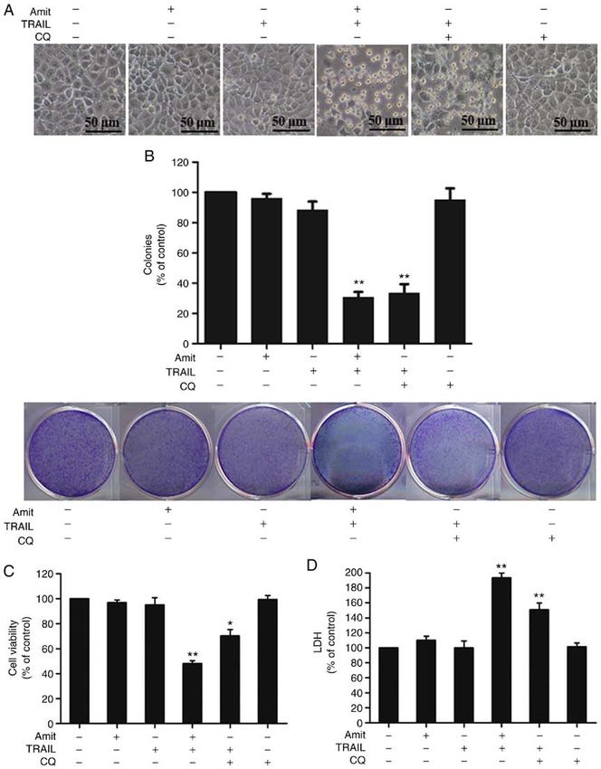
ONCOLOGY REPORTS 46: 144, 2021                                                                   11

Figure 7. Blocking autophagy by amitriptyline aggravates TRAIL‑mediated apoptosis. A549 cells were preincubated with or without CQ (20 µM) and
amitriptyline (40 µM) for 12 h and finally, with or without 100 ng/ml TRAIL for 3 h. (A) Cell morphology images were captured under light microscopy
(magnification, x100; scale bar, 50 µm). (B) Cell colonies were stained with crystal violet dye and the number of colonies were counted. (C) MTT assays were
performed to display cell viability (bar graph). (D) Secretion of LDH into the collected supernatant. Statistically significant differences between the control
and each indicated treatment group are presented as *P
12                       ZINNAH and PARK: AMITRIPTYLINE ENHANCES TRAIL-MEDIATED APOPTOSIS

    The present findings demonstrated that the genetic inhib‑   Patient consent for publication
itor of DR4 and DR5 decreased the effect of amitriptyline on
TRAIL‑mediated apoptosis. These results indicated that DR4      Not applicable.
and DR5 were essential for the combined effect. Additionally,
these findings revealed for the first time that amitriptyline   Competing interests
promoted DR4 and DR5 expression via autophagy inhibition.
Cancer cell death was promoted by autophagy inhibition,         The authors declare that they have no competing interests.
while autophagy played a cell‑protective role in anticancer
treatment (49,50). Under such conditions, the aforementioned    References
findings confirmed that amitriptyline increases autophago‑
some formation, indicated by LC3‑II accumulation, and            1. Jemal A, Bray F, Center MM, Ferlay J, Ward E and Forman D:
inhibits lysosomal fusion resulting in the accumulation of          Global cancer statistics. CA Cancer J Clin 61: 69‑90, 2011.
p62, causing the inhibition of autophagy flux by blocking        2. Siegel RL, Miller KD and Jemal A: Cancer statistics, 2018. CA
                                                                    Cancer J Clin 68: 7‑30, 2018.
autophagosome‑lysosome fusion.                                   3. Ettinger DS, Akerley W, Borghaei H, Chang AC, Cheney RT,
    The combined effect of TRAIL with amitriptyline or CQ           Chi r ieac LR, D'A m ico TA, Dem my T L, Ganti A K,
increased cell death unlike the individual treatments. The          Govindan R, et al: Non‑Small cell lung cancer. J Natl Compr
                                                                    Canc Netw 10: 1236‑1271, 2012.
inhibition of autophagy by amitriptyline and the well‑known      4. Kanitkar AA, Schwartz AG, George J and Soubani AO: Causes
autophagy inhibitor CQ resulted in DR4 and DR5 upregulation         of death in long‑term survivors of non‑small cell lung cancer: A
and improved TRAIL‑mediated caspase‑dependent cell death            regional surveillance, epidemiology, and end results study. Ann
                                                                    Thorac Med 13: 76‑81, 2018.
confirmed by the enhanced caspase cascade. Amitriptyline         5. Heinzmann K, Nguyen QD, Honess D, Smith DM, Stribbling S,
is a psychoactive TCA drug. In this study, only amitriptyline       Brickute D, Barnes C, Griffiths J and Aboagye E: Depicting
among the numerous antidepressant drugs was used to reveal          changes in tumor biology in response to cetuximab monotherapy
                                                                    or combination therapy by apoptosis and proliferation imaging
the enhancing effect with TRAIL. Further studies using other        using 18 F‑ICMT‑11 and 18 F‑FLT PET. J Nucl Med 59: 1558‑1565,
antidepressant drugs are required to support or demonstrate         2018.
the sensitization to TRAIL and anticancer effect by treatment    6. Thomas PA: Stage IIIA N2 non‑small‑cell lung cancer: Current
                                                                    controversies in combined‑modality therapy. Eur J Cardiothorac
of TCA drugs.                                                       Surg 36: 431‑432, 2009.
    Collectively, these findings contributed to the mecha‑       7. Nowak‑Sliwinska P, Scapozza L and Altaba AR: Drug repur‑
nistic evidence that amitriptyline sensitized lung cancer           posing in oncology: Compounds, pathways, phenotypes and
                                                                    computational approaches for colorectal cancer. Biochim
cells to TRAIL and the sensitization was mediated through           Biophys Acta Rev Cancer 1871: 434‑454, 2019.
DR4 and DR5 upregulation and autophagy inhibition. These         8. Jia Y, Yun CH, Park E, Ercan D, Manuia M, Juarez J, Xu C,
results provide an understanding of the anticancer effect of        Rhee K, Chen T, Zhang H, et al: Overcoming EGFR(T790M)
                                                                    and EGFR(C797S) resistance with mutant‑selective allosteric
amitriptyline and suggest further evaluation is required to         inhibitors. Nature 534: 129‑132, 2016.
develop possible therapeutic regimens against lung cancer and    9. Nesterov A, Ivashchenko Y and Kraft AS: Tumor necrosis
cancer‑associated depression.                                       factor‑related apoptosis‑inducing ligand (TRAIL) triggers apop‑
                                                                    tosis in normal prostate epithelial cells. Oncogene 21: 1135‑1140,
                                                                    2002.
Acknowledgements                                                10. Trivedi R and Mishra DP: Trailing TRAIL resistance: Novel
                                                                    targets for TRAIL sensitization in cancer Cells. Front Oncol 5:
                                                                    69, 2015.
Not applicable.                                                 11. Marsters SA, Sheridan JP, Pitti RM, Huang A, Skubatch M,
                                                                    Baldwin D, YuanJ, Gurney A, Goddard AD, Godowski P and
Funding                                                             Ashkenazi A: A novel receptor for Apo2L/TRAIL contains a
                                                                    truncated death domain. Curr Biol 7: 1003‑1006, 1997.
                                                                12. Jin CY, Moon DO, Lee JD, Heo MS, Choi YH, Lee CM,
This study was supported by a grant from the National               Park YM and Kim GY: Sulforaphane sensitizes tumor necrosis
Research Foundation of Korea (NRF) funded by the Ministry           factor‑related apoptosis‑inducing ligand‑mediated apoptosis
                                                                    through downregulation of ERK and akt in lung adenocarcinoma
of Education (grant. no. 2019R1A6A1A03033084).                      A549 cells. Carcinogenesis 28: 1058‑1066, 2007.
                                                                13. Thorburn A, Behbakht K and Ford H: TRAIL receptor‑targeted
Availability of data and materials                                  therapeutics: Resistance mechanisms and strategies to avoid
                                                                    them. Drug Resist Updat 11: 17‑24, 2008.
                                                                14. Mérino D, Lalaoui N, Morizot A, Solary E and Micheau O:
All datasets generated or analyzed during the present study         TRAIL in cancer therapy: Present and future challenges. Expert
are available from the corresponding author upon reasonable         Opin Ther Targets 11: 1299‑1314, 2007.
                                                                15. Hale AN, Ledbetter DJ, Gawriluk TR and Rucker EB III:
request.                                                            Autophagy: Regulation and role in development. Autophagy 9:
                                                                    951‑972, 2013.
Authors' contributions                                          16. Rouschop KM and Wouters BG: Regulation of autophagy
                                                                    through multiple independent hypoxic signaling pathways. Curr
                                                                    Mol Med 9: 417‑424, 2009.
KMAZ and SYP designed and performed the study, analyzed         17. Thorburn A, Thamm DH and Gustafson DL: Autophagy and
data and wrote the manuscript. Both authors have read and           cancer therapy. Mol Pharmacol 85: 830‑838, 2014.
                                                                18. Sui X, Chen R, Wang Z, Huang Z, Kong N, Zhang M, Han W,
approved the final manuscript.                                      Lou F, Yang J, Zhang Q, et al: Autophagy and chemotherapy
                                                                    resistance: A promising therapeutic target for cancer treatment.
Ethics approval and consent to participate                          Cell Death Dis 4: e838‑e838, 2013.
                                                                19. Zinnah KMA and Park SY: Duloxetine enhances TRAIL‑mediated
                                                                    apoptosis via AMPK‑mediated inhibition of autophagy flux in
Not applicable.                                                     lung cancer cells. Anticancer Res 39: 6621‑6633, 2019.
ONCOLOGY REPORTS 46: 144, 2021 13
20. Di Rosso ME, Sterle HA, Cremaschi GA and Genaro AM: 42. Cheng H, Hong B, Zhou L, Allen JE, Tai G, Humphreys R,
Beneficial effect of fluoxetine and sertraline on chronic Dicker DT, Liu YY and El‑Deiry WS: Mitomycin C potentiates
stress‑induced tumor growth and cell dissemination in a mouse TRAIL‑induced apoptosis through p53‑independent upregula‑
model of lymphoma: Crucial role of antitumor immunity. Front tion of death receptors: Evidence for the role of c‑Jun N‑terminal
Immunol 9: 1341, 2018. kinase activation. Cell Cycle 11: 3312‑3323, 2012.
21. Thaker PH, Han LY, Kamat AA, Arevalo JM, Takahashi R, Lu C, 43. Dolloff NG, Mayes PA, Hart LS, Dicker DT, Humphreys R
Jennings NB, Armaiz‑Pena G, Bankson JA, Ravoori M, et al: and El‑Deiry WS: Off‑target lapatinib activity sensitizes colon
Chronic stress promotes tumor growth and angiogenesis in a cancer cells through TRAIL death receptor up‑regulation. Sci
mouse model of ovarian carcinoma. Nat Med 12: 939‑944, 2006. Transl Med 3: 86ra50, 2011.
22. Kim‑Fuchs C, Le CP, Pimentel MA, Shackleford D, Ferrari D, 44. Maiuri MC, Zalckvar E, Kimchi A and Kroemer G: Self‑Eating
Angst E, Hollande F and Sloan EK: Chronic stress acceler‑ and self‑killing: Crosstalk between autophagy and apoptosis. Nat
ates pancreatic cancer growth and invasion: A critical role for Rev Mol Cell Biol 8: 741‑752, 2007.
beta‑adrenergic signaling in the pancreatic microenvironment. 45. Mizushima N, Levine B, Cuervo AM and Klionsky DJ:
Brain Behav Immun 40: 40‑47, 2014. Autophagy fights disease through cellular self‑digestion.
23. Hasegawa H and Saiki I: Psychosocial stress augments tumor Nature 451: 1069‑1075, 2008.
development through beta‑adrenergic activation in mice. 46. Amin A, Bajbouj K, Koch A, Gandesiri M and Schneider‑Stock R:
Jpn J Cancer Res 93: 729‑735, 2002. Defective autophagosome formation in p53‑null colorectal
24. Fann JR, Fan MY and Unützer J: Improving primary care for cancer reinforces crocin‑induced apoptosis. Int J Mol Sci 16:
older adults with cancer and depression. J Gen Intern Med 24 1544‑1561, 2015.
(Suppl 2): S417‑S424, 2009. 47. Wu YT, Tan HL, Huang Q, Kim YS, Pan N, Ong WY, Liu ZG,
25. Laird B, Colvin L and Fallon M: Management of cancer pain: Ong CN and Shen HM: Autophagy plays a protective role during
Basic principles and neuropathic cancer pain. Eur J Cancer 44: zVAD‑induced necrotic cell death. Autophagy 4: 457‑466, 2008.
1078‑1082, 2008. 48. White E: Autophagic cell death unraveled: Pharmacological inhi‑
26. Frick LR and Rapanelli M: Antidepressants: Influence on cancer bition of apoptosis and autophagy enables necrosis. Autophagy 4:
and immunity? Life Sci 92: 525‑532, 2013. 399‑401, 2008.
27. Zhang Z, Du X, Zhao C, Cao B, Zhao Y and Mao X: The antide‑ 49. Vucicevic L, Misirkic M, Janjetovic K, Vilimanovich U, Sudar E,
pressant amitriptyline shows potent therapeutic activity against Isenovic E, Prica M, Harhaji‑Trajkovic L, Kravic‑Stevovic T,
multiple myeloma. Anticancer Drugs 24: 792‑798, 2013. Bumbasirevic V and Trajkovic V: Compound C induces protective
28. Cordero MD, Sánchez‑Alcázar JA, Bautista‑Ferrufino MR, autophagy in cancer cells through AMPK inhibition‑independent
Carmona‑López MI, Illanes M, Ríos MJ, Garrido‑Maraver J, blockade of Akt/mTOR pathway. Autophagy 7: 40‑50, 2011.
Alcudia A, Navas P and de Miguel M: Acute oxidant damage 50. Shen S, Zhang Y, Wang Z, Zhang R and Gong X: Bufalin induces the
promoted on cancer cells by amitriptyline in comparison with some interplay between apoptosis and autophagy in glioma cells through
common chemotherapeutic drugs. Anticancer Drugs 21: 932‑944, endoplasmic reticulum stress. Int J Biol Sci 10: 212‑224, 2014.
2010. 51. Klionsky DJ, Abdelmohsen K, Abe A, Abedin MJ, Abeliovich H,
29. Livak KJ and Schmittgen TD: Analysis of relative gene expres‑ Arozena AA, Adachi H, Adams CM, Adams PD and Adeli K:
sion data using real‑time quantitative PCR and the 2(‑Delta Delta Guidelines for the use and interpretation of assays for monitoring
C(T)) method. Methods 25: 402‑408, 2001. autophagy (3rd edition). Autophagy 12: 1‑222, 2016.
30. Yuan X, Gajan A, Chu Q, Xiong H, Wu K and Wu GS: Developing 52. Gómez‑Sánchez R, Yakhine‑Diop SMS, Rodríguez‑Arribas M,
TRAIL/TRAIL death receptor‑based cancer therapies. Cancer Bravo‑San Pedro JM, Martínez‑Chacón G, Uribe‑Carretero E,
Metastasis Rev 37: 733‑748, 2018. de Castro DC, Pizarro‑Estrella E, Fuentes JM and González‑
31. Islam MA, Sooro MA and Zhang P: Autophagic regulation of Polo RA: mRNA and protein dataset of autophagy markers (LC3
p62 is critical for cancer therapy. Int J Mol Sci 19: 1405, 2018. and p62) in several cell lines. Data Brief 7: 641‑647, 2016.
32. Wiley SR, Schooley K, Smolak PJ, Din WS, Huang CP, 53. Mauthe M, Orhon I, Rocchi C, Zhou X, Luhr M, Hijlkema KJ,
Nicholl JK, Sutherland GR, Smith TD, Rauch C, Smith CA, et al: Coppes RP, Engedal N, Mari M and Reggiori F: Chloroquine
Identification and characterization of a new member of the TNF inhibits autophagic flux by decreasing autophagosome‑lysosome
family that induces apoptosis. Immunity 3: 673‑682, 1995. fusion. Autophagy 14: 1435‑1455, 2018.
33. Walczak H, Miller RE, Ariail K, Gliniak B, Griffith TS, Kubin M, 54. Nordstrøm LU, Sironi J, Aranda E, Maisonet J, Perez‑Soler R,
Chin W, Jones J, Woodward A, Le T, et al: Tumoricidal activity of Wu P and Schwartz EL: Discovery of autophagy inhibitors with
tumor necrosis factor‑related apoptosis‑inducing ligand in vivo. antiproliferative activity in lung and pancreatic cancer cells. ACS
Nat Med 5: 157‑163, 1999. Med Chem Lett 6: 134‑139, 2015.
34. Aggarwal BB, Bhardwaj U and Takada Y: Regulation of 55. Pan H, Wang Y, Na K, Wang Y, Wang L, Li Z, Guo C, Guo D and
TRAIL‑induced apoptosis by ectopic expression of antiapoptotic Wang X: Autophagic flux disruption contributes to Ganoderma
factors. Vitam Horm 67: 453‑483, 2004. lucidum polysaccharide‑induced apoptosis in human colorectal
35. Wang S: TRAIL: A sword for killing tumors. Curr Med Chem 17: cancer cells via MAPK/ERK activation. Cell Death Dis 10: 456,
3309‑3317, 2010. 2019.
36. Chaudhary PM, Eby M, Jasmin A, Bookwalter A, Murray J and 56. Gąsiorkiewicz BM, Koczurkiewicz‑Adamczyk P, Piska K and
Hood L: Death receptor 5, a new member of the TNFR family, Pękala E: Autophagy modulating agents as chemosensitizers for
and DR4 induce FADD‑dependent apoptosis and activate the cisplatin therapy in cancer. Invest New Drugs 39: 538‑563, 2020.
NF‑kappaB pathway. Immunity 7: 821‑830, 1997. 57. Nazim UM, Yin H and Park SY: Downregulation of c‑FLIP and
37. Pan G, O'Rourke K, Chinnaiyan AM, Gentz R, Ebner R, Ni J upregulation of DR‑5 by cantharidin sensitizes TRAIL‑mediated
and Dixit VM: The receptor for the cytotoxic ligand TRAIL. apoptosis in prostate cancer cells via autophagy flux. Int J Mol
Science 276: 111‑113, 1997. Med 46: 280‑288, 2020.
38. Cretney E, Takeda K and Smyth MJ: Cancer: Novel therapeutic 58. Park EJ, Min Kj, Choi KS, Kubatka P, Kruzliak P, Kim DE and
strategies that exploit the TNF‑related apoptosis‑inducing ligand Kwon TK: Chloroquine enhances TRAIL‑mediated apoptosis
(TRAIL)/TRAIL receptor pathway. Int J Biochem Cell Biol 39: through up‑regulation of DR5 by stabilization of mRNA and
280‑286, 2007. protein in cancer cells. Sci Rep 6: 22921, 2016.
39. Danial NN and Korsmeyer SJ: Cell death: Critical control points. 59. Shin GC, Kang HS, Lee AR and Kim KH: Hepatitis B virus‑triggered
Cell 116: 205‑219, 2004. autophagy targets TNFRSF10B/death receptor 5 for degradation to
40. Plummer R, Attard G, Pacey S, Li L, Razak A, Perrett R, limit TNFSF10/TRAIL response. Autophagy 12: 2451‑2466, 2016.
Barrett M, Judson I, Kaye S, Fox NL, et al: Phase 1 and phar‑ 60. Twomey JD and Zhang B: Circulating tumor cells develop resis‑
macokinetic study of lexatumumab in patients with advanced tance to TRAIL‑induced apoptosis through autophagic removal
cancers. Clin Cancer Res 13: 6187‑6194, 2007. of death receptor 5: Evidence from an in vitro model. Cancers
41. Hotte SJ, Hirte HW, Chen EX, Siu LL, Le LH, Corey A, (Basel) 11: 94, 2019.
Iacobucci A, MacLean M, Lo L, Fox NL and Oza AM: A phase 1
study of mapatumumab (fully human monoclonal antibody to This work is licensed under a Creative Commons
TRAIL‑R1) in patients with advanced solid malignancies. Clin Attribution-NonCommercial-NoDerivatives 4.0
Cancer Res 14: 3450‑3455, 2008. International (CC BY-NC-ND 4.0) License.You can also read