COPD monocytes demonstrate impaired migratory ability
←
→
Page content transcription
If your browser does not render page correctly, please read the page content below
Ravi et al. Respiratory Research (2017) 18:90
DOI 10.1186/s12931-017-0569-y

 RESEARCH                                                                                                                                      Open Access

COPD monocytes demonstrate impaired
migratory ability
Arjun K Ravi1,3*, Jonathan Plumb1, Rosemary Gaskell1, Sarah Mason1, Caroline S Broome1,3, George Booth1,
Matthew Catley2, Jørgen Vestbo1 and Dave Singh1,3

  Abstract
  Background: Increased lung macrophage numbers in COPD may arise from upregulation of blood monocyte
  recruitment into the lungs. CCR5 is a monocyte chemokine receptor regulated by interleukin-6 (IL-6); the concentration
  of CCR5 ligands are known to be elevated in COPD lungs. The objective of this study was to investigate mechanisms of
  monocyte recruitment to the lung in COPD, including the role of CCR5 signalling.
  Methods: Ninety one COPD patients, 29 smokers (S) and 37 non-smokers (NS) underwent sputum induction, plasma
  sampling (to measure IL-6 and soluble IL-6 receptor [sIL-6R] by immunoassay), monocyte characterization (by flow
  cytometry) and monocyte isolation for cell migration and quantitative polymerase chain reaction studies. Lung
  tissue was used for immunohistochemistry.
  Results: Plasma IL-6 and sIL-6R levels were increased in COPD. Greater proportions of COPD CD14++CD16+
  monocytes expressed CCR5 compared to controls. Monocyte stimulation with IL-6 and sIL-6R increased CCR5
  gene expression. COPD monocytes demonstrated impaired migration towards sputum supernatant compared
  to NS (% migration, 4.4 vs 11.5, respectively; p < 0.05). Pulmonary microvessels showed reduced monocyte recruitment
  (% marginated cells) in COPD compared to NS, (9.3% vs 83.1%, respectively). The proportion of replicating Ki67+ alveolar
  macrophages was reduced in COPD compared to NS. All alveolar macrophages from COPD and S expressed the anti-
  apoptosis marker BCL2; this protein was not present in non-smokers or COPD ex-smokers.
  Conclusion: COPD monocytes show decreased migratory ability despite increased CCR5 expression. Increased COPD
  lung macrophage numbers may be due to delayed apoptosis.
  Keywords: COPD, Monocytes, CCR5, Chemotaxis, Interleukin-6

Background                                                                          from blood monocytes (monocyte derived cells and tis-
Monocytes can be recruited from the blood into the tis-                             sue monocyte/macrophages) [3].
sues, whereupon differentiation into macrophages may                                  There are increased numbers of macrophages in the
occur [1]. There are also tissue resident macrophages                               lungs of chronic obstructive pulmonary disease (COPD)
that replenish cell numbers by replication [2]. A recent                            patients [4]; these cells are involved in host defence, air-
study demonstrated the presence of phenotypically                                   way remodelling and parenchymal destruction [5]. It has
different mononuclear phagocyte cell types in healthy                               been suggested that increased lung macrophage numbers
human lungs that either originate from the lungs (pul-                              in COPD are due to increased recruitment of blood
monary dendritic cells and alveolar macrophages) or                                 monocytes [5, 6]. Alternatively, cigarette smoke exposure
                                                                                    induces the expression of anti-apoptotic genes in macro-
                                                                                    phages [7], and increased expression of anti-apoptotic
* Correspondence: ARavi@meu.org.uk
                                                                                    proteins has been observed in COPD macrophages [8],
1
 NIHR Respiratory and Allergy Clinical Research Facility, Manchester                suggesting that delayed apoptosis is a possible cause
Academic Health Science Centre, University Hospital South Manchester NHS            of macrophage accumulation in COPD. Furthermore,
Foundation Trust, University of Manchester, Manchester, UK
3
 The Medicines Evaluation Unit, Wythenshawe Hospital, The Langley
                                                                                    alveolar macrophages expressing the proliferation
Building, Southmoor Road, Wythenshawe, Greater Manchester M23 9QZ, UK               marker Ki67 have been observed in patients with
Full list of author information is available at the end of the article

                                      © The Author(s). 2017 Open Access This article is distributed under the terms of the Creative Commons Attribution 4.0
                                      International License (http://creativecommons.org/licenses/by/4.0/), which permits unrestricted use, distribution, and
                                      reproduction in any medium, provided you give appropriate credit to the original author(s) and the source, provide a link to
                                      the Creative Commons license, and indicate if changes were made. The Creative Commons Public Domain Dedication waiver
                                      (http://creativecommons.org/publicdomain/zero/1.0/) applies to the data made available in this article, unless otherwise stated.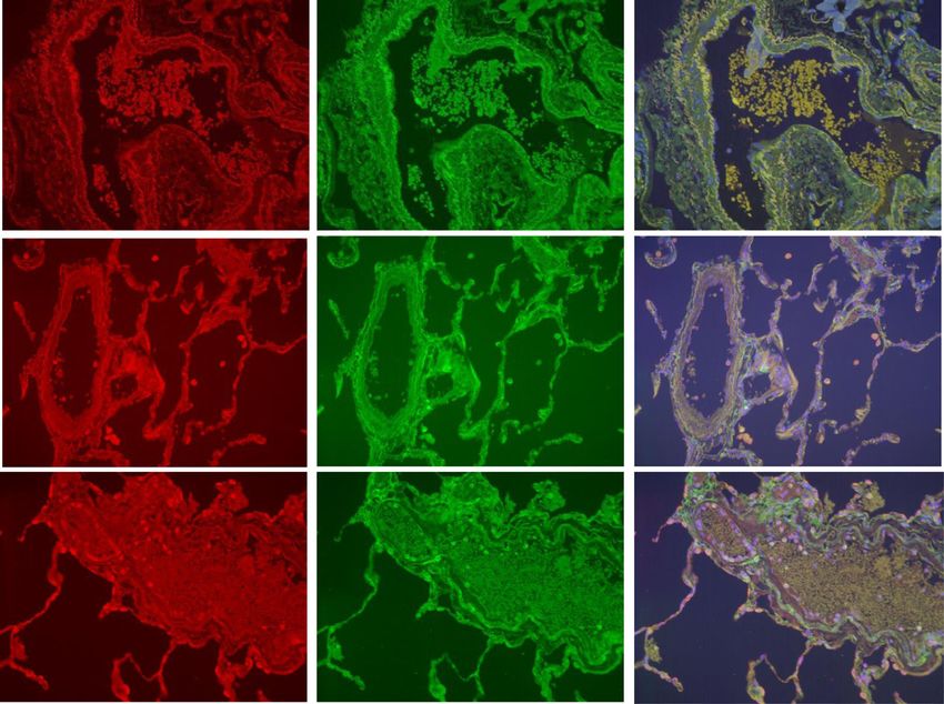
Ravi et al. Respiratory Research (2017) 18:90                                                                Page 2 of 12

interstitial lung disease [9], but whether increased          in COPD; namely increased replication and supressed
macrophage accumulation in COPD occurs by self-               apoptosis.
renewal is not understood.
  Costa et al reported increased migration of COPD per-       Methods
ipheral blood mononuclear cells towards C-X-C motif           Subjects
chemokine receptor 3 (CXCR3) and C-C motif chemokine          COPD patients (n = 91), smokers (S; n = 29) and healthy
receptor 5 (CCR5) ligands using single chemokines for         non-smokers (HNS; n = 37) were recruited for blood and
migration experiments [6]. Such experiments, however do       sputum sampling; S (with >10 pack-year smoking history)
not reflect the complex mixture of chemoattractants           and HNS had normal lung function. COPD patients had
present in the lungs [10–15]. Physiologically relevant com-   been diagnosed according to current guidelines [24] and
plex supernatants, such as those obtained from induced        were also seen during acute exacerbation (diagnosed
sputum could be used to further investigate the migratory     according to an increase in symptoms as described in
ability of COPD monocytes.                                    Additional file 1). COPD (n = 12), S (n = 9) and NS (non-
  CCR5 is the receptor for the monocyte chemoattract-         smokers; n = 6) undergoing surgical resection of suspected
ant C-C motif chemokine ligand 3 (CCL3) [16]. Studies         lung carcinoma were recruited. The research was ap-
using induced sputum and bronchoalveolar lavage have          proved by a local ethics committee; all participants pro-
shown that CCR5 ligand levels are increased in the lungs      vided written informed consent.
of COPD patients, suggesting a role for CCR5 signalling
in the recruitment of monocytes into COPD lungs               Clinical assessment
[12, 13, 16, 17]. Peripheral blood monocytes can be           Pre- and post- bronchodilator spirometry was performed
classified into 3 subtypes according to their expression of   as described in Additional file 1 [25]. COPD patients com-
CD14 (LPS receptor) and CD16 (FcγRIII receptor): CD14+        pleted the St George’s respiratory questionnaire (SGRQ)
+
 CD16- (‘Classical’), CD14+CD16+ (‘Intermediate’) and         and COPD Assessment Test (CAT).
CD14-CD16++ (‘Non-Classical’) [1]. Increased numbers of
pro-inflammatory CD14+CD16+ monocytes are found in            Sputum sampling
chronic inflammatory disease states such as rheumatoid        Sputum was induced and processed by the ‘two-step’
arthritis [18]. Furthermore, CD14+CD16+ cells have the        method [16]; PBS processed samples were used in the ex-
greatest surface expression of CCR5 [1, 19]. Monocyte         periments reported here.
subsets in COPD, and their expression of CCR5, have not
been previously reported.                                     Plasma cytokine measurement
  CCR5 expression is upregulated by interleukin-6 (IL-6)      Levels of plasma IL-6 and sIL-6R were measured using a
[20], a cytokine which trans-signals through a soluble re-    Meso Scale Discovery immunoassay (Gaithersburg (MD–
ceptor sIL-6R [21]. Plasma IL-6 levels are increased in a     USA) (details Additional file 1).
subset of stable COPD patients [22] and during COPD
exacerbations [23]. The systemic levels of sIL-6R have        Monocyte chemokine receptor expression
not been investigated in COPD; increased systemic IL-6/       Peripheral blood monocyte subtypes were identified by
sIL-6R signalling in COPD could upregulate blood              flow cytometry [1]. Proportional expression of CCR5 on
monocyte CCR5 expression, thereby promoting mono-             CD14++CD16-, CD14+CD16+ and CD14-CD16++ mono-
cyte recruitment into the lungs.                              cytes was determined (details Additional file 1).
  We have investigated COPD blood monocyte recruit-
ment with two major objectives in mind. Firstly, to char-     CCL3 and Sputum supernatant induced monocyte
acterise changes in the CCL3-CCR5 axis that could             chemotaxis
facilitate monocyte recruitment in COPD; we studied           CD14+ monocyte isolation and chemotaxis experiments
CCR5 expression on peripheral blood monocytes and             were performed as previously described (details Additional
plasma sIL-6R levels in COPD patients compared to             file 1) [16]. CD14+ monocytes did not undergo any form
controls. Secondly, to further investigate the hypothesis     of cytokine stimulation prior to the chemotaxis assay.
that monocyte recruitment from the blood is increased
in COPD; we studied COPD monocyte migration                   CD14+ monocyte cell culture
towards sputum supernatants and performed lung im-            CD14+ monocytes from 6 HNS were isolated and cul-
munohistochemistry studies to evaluate monocyte mi-           tured in the presence of recombinant human (rh) IL-6
gration from the pulmonary blood vessels of COPD              (20 ng/mL, R&D Systems, Abingdon UK), rh IL-6
patients compared to controls. We also performed im-          (20 ng/mL) + rh sIL-6R (40 ng/mL, R&D Systems) or
munohistochemistry studies to investigate an alternative      media alone (unstimulated control). Cell lysates were
mechanisms of pulmonary macrophage accumulation               harvested for gene expression analysis.
Ravi et al. Respiratory Research (2017) 18:90                                                                                                     Page 3 of 12

CD14+ monocyte CCR5 gene expression                                                Statistical analysis was performed using GraphPad
Quantitative polymerase chain reaction (QPCR) for de-                            Prism version 6 and IBM SPSS Statistics for Windows,
termination of CCR5 gene expression was performed as                             Version 23.0 (released 2015). Armonk, NY.
previously described (details Additional file 1) [26].
                                                                                 Results
                                                                                 The clinical characteristics of all study participants are
Immunohistochemistry and Immunofluorescence (IF)                                 stated in Table 1 (details of participants in individual
See Additional file 1 for details of tissue preparation, im-                     experiments are described in Additional file 3: Table S1).
aging and image analysis. The immunohistochemical tech-
nique employed for detection of CD34 (endothelial cell                           Plasma IL-6 and sIL-6R levels
surface glycoprotein [27]), neutrophil elastase (NE), Ki67                       Plasma IL-6 levels were significantly higher in COPD pa-
(cell-cycle maintaining protein) [28] and BCL-2 (B-cell                          tients, (n = 70; median 4.5 pg/mL) compared to HNS
CLL/lymphoma 2) (anti-apoptotic protein) (ADD REF) is                            (n = 15; median 0 pg/mL, p < 0.0001), with no other
detailed in Additional file 1.                                                   significant differences between groups (Fig. 1). Plasma
  Dual label immunofluorescence was performed to                                 sIL-6R levels were significantly higher in COPD patients
identify marginated ‘monocytic’ (cells apposed to the lu-                        compared to S (n = 15, medians; 5,338 pg/mL versus
minal endothelial surface) (CX3CR1+CD14+ and CX3CR1                              4,453 pg/mL respectively, p < 0.001), while the comparison
+
 CD16+) cells in the pulmonary microvasculature as de-                           of COPD vs. HNS (median 4,853 pg/mL) was not signifi-
scribed in Additional file 1.                                                    cant (p = 0.3).

                                                                                 Peripheral blood monocyte CCR5 expression
Statistical analysis                                                             The proportions of CD14++CD16-, CD14+CD16+ and
Normally distributed data were analysed using one way                            CD14-CD16++ monocytes were similar in COPD patients
ANOVA with application of Tukey’s post-test; unpaired                            (n = 15), S (n = 8) and HNS (n = 8) (Additional file 3:
t-tests were used where appropriate. Non-normally dis-                           Figure S1). A significantly greater proportion of CD14+
                                                                                 +
tributed data were analysed using the Kruskal-Wallis test                         CD16- monocytes from COPD patients expressed CCR5
with application of Dunn’s post-test. Mann-Whitney U                             compared to S or HNS; medians 5.2%, 0.9% and 0.8%, re-
tests were used as appropriate. Univariate correlation                           spectively (p < 0.05 for both comparisons; see Fig. 2). The
analysis was performed using the Spearman Rank test.                             proportion of CD14+CD16+ monocytes from COPD pa-
QPCR data was normally distributed and analysed using                            tients expressing CCR5 was significantly greater than S
one-way ANOVA with application of Dunnett’s multiple                             (medians 14.3% versus 4.9%; p < 0.05) but did not reach
comparisons test; details of a within subject analysis are                       statistical significance compared to HNS (median 2.7%,
described in Additional file 2.                                                  p = 0.09). There were no significant differences in the
Table 1 Demographic details of study participants
                                    COPD (n = 93)       S (n = 29)       HNS (n = 39)       COPD (n = 12) IHC/IF       S (n = 9) IHC/IF     NS (n = 6) IHC/IF
Age                                 66 (7)              54.4 (7)         41.6 (15.8)        66.3 (4.6)                 66.3 (6.6)           70 (5.7)
Gender (F:M)                        37:58               14:15            14:25              4:8                        4:5                  4:2
Current smoker (n)                  35                  26               0                  9                          9                    0
FEV1% predicted                     60.2 (18.9)         98.1 (14.1)      109.6 (14.4)       67.3 (25.6)                91 (11.9)            108 (7.2)
FEV1/FVC (%)                        52 (12.5)           75.5 (2.9)       82.5 (6.9)         67.3 (25.6)                91 (11.9)            108 (7.2)
GOLD I (%)                          18.3                -                -                  33.3                       -                    -
GOLD II (%)                         50.5                -                -                  25                         -                    -
GOLD III (%)                        26.9                -                -                  41.7                       -                    -
GOLD IV (%)                         4.3                 -                -                  0                          -                    -
CAT                                 17.1 (7.8)
SGRQ                                39.4 (20.3)
Smoking history (Pack Years)a       37 (13-122)         30.5 (12-67)     0                  42 (13-126)                50 (21-66)
ICS (%)                             65                  -                -
This table shows the grouped demographic details of subjects who participated MSD plasma cytokine analysis, flow cytometric (FACS) & qPCR, chemotaxis
(displayed on the left); lung immunohistochemistry/immunofluorescence (displayed on the right). Data is described by mean (SD). aData shown as median (range)
Abbreviations: FEV1 forced expiratory volume in 1 s, FVC forced vital capacity, CAT COPD Assessment Test and SGRQ St George’s Respiratory Questionnaire, ICS
inhaled corticosteroid, IHC Immunohistochemistry, IF Immunofluorescence, NS non smoker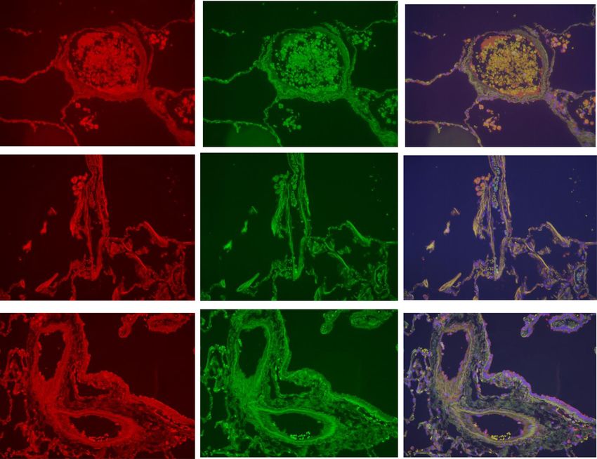
Ravi et al. Respiratory Research (2017) 18:90                                                                                                  Page 4 of 12

 Fig. 1 Plasma IL-6 and sIL-6R levels in COPD, S & HNS. The figure shows a plasma IL-6 and b plasma sIL-6R levels in COPD, S & HNS determined
 by MSD analysis. Each dot represents the value for an individual patient. The bar represents the median value. The statistical significance of differences
 observed was assessed by a The Kruskal-Wallis test with application of Dunn’s post-test and b ANOVA with application of Tukey’s post-test. The dashed
 line represents the lower limit of quantification (LLOQ)

proportion of CD14-CD16++ CCR5+ expressing cells be-                            curve observed; maximal chemotaxis levels were in the
tween COPD, S and HNS; medians 1%, 1.2% & 0.4%                                  concentration range 15.6 ng/ml to 62.5 ng/ml (Fig. 4).
respectively.
  There was no change in the proportion of monocyte
subsets or CCR5 expression at exacerbations compared                            CD14+ monocyte migration towards sputum supernatant
to stable state (n = 8 COPD patients; Additional file 3:                        The migration of HNS CD14+ monocytes towards sputum
Figure S2 and Table S2).                                                        supernatant from COPD patients and HNS was studied. A
                                                                                greater proportion of CD14+ monocytes migrated towards
Regulation of monocyte CCR5 gene expression                                     COPD sputum compared to HNS sputum; mean chemo-
CCR5 expression was significantly upregulated in CD14+                          taxis 14.3% versus 5.8%, respectively (p = 0.03) (Fig. 5).
monocytes cultured for four hours in the presence of                            Subsequent experiments used COPD sputum supernatant
both IL-6 and sIL-6R; the mean fold change in CCR5                              as the chemoattractant. HNS CD14+ monocytes (n = 8)
gene expression was 1.6 (p < 0.05). IL-6 stimulation                            demonstrated significantly greater chemotaxis compared
alone did not upregulate CCR5 gene expression. CD14+                            to COPD monocytes (n = 6); means 11.5% versus 4.4%
monocytes stimulated with IL-6 (solely or in conjunction                        (p < 0.05). There was a trend to significance when
with sIL-6R) for 19 h did not show alteration in CCR5                           comparing HNS to S, (n = 6; mean 5.6%, p = 0.07).
gene expression (Fig. 3 and within-subject analysis                               We performed subgroup analysis based on age; mono-
shown in Additional file 2).                                                    cytes of HNS >50 years of age (n = 3, mean age 57.7 years)
                                                                                with mean chemotaxis 16% demonstrated significantly
COPD CD14+ monocyte migration towards rhCCL3                                    greater migratory ability compared to younger HNS
COPD and HNS CD14+ monocytes showed similar migra-
Ravi et al. Respiratory Research (2017) 18:90 Page 5 of 12
The CX3CR1+ marginated cell population was com-
posed of CX3CR1+CD14+ and CX3CR1+CD16+ cells. The
absolute number and percentage of marginated CX3CR1
+
CD14+ cells per μm vessel circumference was signifi-
cantly reduced in COPD patients compared to both S and
NS (Table 2, Figs. 6 and 7). Similar results were obtained
for marginated CX3CR1+CD16+ cells in COPD patients
compared to S and NS (Table 2, Figs. 6 and 8). Negative
control immunofluorescence images are displayed in
Additional file 3: Figure S6.
Fig. 3 CCR5 gene expression by CD14+ monocytes. The figure
shows changes in gene expression by CD14+ monocytes stimulated Alveolar macrophage Ki67 and BCL2 expression
with IL-6 or IL-6 with sIL-6R compared with unstimulated CD14+
monocytes; cells were stimulated for either 4 or 19 h. The data is
COPD patients (n = 9) and S (n = 9) had significantly
shown as mean (SD). The statistical significance of differences ob- greater mean numbers of alveolar macrophages (59.2/
served was determined using ANOVA with application of Dunnett’s mm2 and 64.7/mm2 respectively) compared to NS (n = 6;
post-test (with basal gene expression being assigned as the ‘control’) 20.5/mm2, p < 0.05 and p < 0.01 respectively). The per-
centage of Ki67+ alveolar macrophages was low, and
greater in NS (mean 2%) compared to COPD patients
Marginated CX3CR1+ cells in the pulmonary
(mean 0.9%, p < 0.05 vs NS) (Fig. 9).
microvasculature
BCL2 was expressed by all COPD current smokers
Microvessels were identified by CD34 staining (Additional
(n = 9) and S (n = 9) alveolar macrophages, however BCL2
file 3: Figure S4). There were no significant differences in
was absent in the alveolar macrophages of NS (n = 6) and
the size of vessels or the absolute number of marginated
COPD former smokers (n = 3) (Fig. 10).
cells/ μm circumference observed between COPD patients
(n = 9), S (n = 9) and NS (n = 6) (Table 2). Intravascular
monocytic cells were identified by their expression of the Discussion
monocyte/macrophage surface marker CX3CR1. The me- We observed increased CCR5 expression on COPD
dian number of marginated CX3CR1+ cells per μm vessel blood monocytes. Increased plasma levels of sIL-6R may
circumference were significantly greater in NS (83.9 play a role in this observation, as IL-6 with sIL-6R up-
x10-4/μm) and S (31.5 x10⁻⁴/μm) compared to COPD regulated CCR5 gene expression in monocytes. However,
(1.5 × 10⁻⁴/μm;p < 0.01 and p < 0.05 respectively). The this increase in COPD monocyte CCR5 expression did
median percentage of marginated cells that expressed not confer a greater migratory ability; chemotaxis exper-
CX3CR1+ was significantly greater in HNS (83.1%) iments showed impairment of the migratory ability of
and S (46%) compared to COPD (9.3%; p < 0.001 and COPD peripheral blood monocytes. This impaired mi-
p < 0.05 respectively) (Table 2 & Fig. 5). A large num- gration was confirmed by examining monocyte margin-
ber of the CX3CR1- marginated cells in COPD pa- ation from blood vessels in the lungs. We have therefore
tients were neutrophils based on their appearance and found no evidence to support the theory that increased
nuclear morphology, which was confirmed by NE monocyte recruitment is responsible for increased lung
staining (Additional file 3: Figure S5). macrophage accumulation in COPD.
Fig. 4 Migration of COPD CD14+ monocytes towards rhCCL3. This figure shows the migration of CD14+ monocytes from a COPD (n = 3) and b
HNS (n = 3) towards a range of concentrations of rhCCL3 (250 – 2 ng/mL). Data is displayed as mean (SD)Ravi et al. Respiratory Research (2017) 18:90 Page 6 of 12
a b
p = 0.03 20 pRavi et al. Respiratory Research (2017) 18:90 Page 7 of 12
a b
c d
e f
Fig. 6 Marginated intravascular CX3CR1+, CX3CR1+CD14+ and CX3CR1+CD16+ cells. This figure shows the proportions of marginated CX3CR1+
(5a & 5b), CX3CR1+CD14+ (5c and 5d) and CX3CR1+CD16+ (5e & 5f) cells. The bar represents the median; each data point represents the
value for an individual patient. The statistical significance of differences observed was determined using Kruskal-Wallis test with application
of Dunn’s multiple comparisons test
of COPD CD14+ monocytes. We also observed increased compared to NS. The vascular endothelium of pulmonary
CCR5 expression on CD14++CD16- and CD14+CD16+ vessels in S and COPD expresses increased levels of adhe-
COPD blood monocytes, so it was perhaps surprising that sion molecules such as E-selectin, P selectin, ICAM-1,
COPD monocytes displayed impaired chemotaxis. It was ICAM-2 and VCAM-1 [34]. It is therefore unlikely that
therefore important that we evaluated monocyte migra- the reduced margination observed in COPD resulted from
tion into the lungs by a different method to further inves- attenuated endothelial adhesion molecule expression. The
tigate this observation. reduced margination was observed for both CX3CR1
+
C-X3-C motif chemokine receptor 1 (CX3CR1) is a CD14+ and CX3CR1+CD16+ cells in COPD patients.
widely used monocyte/macrophage marker [1, 33]. We Attenuated monocyte migration ability in COPD raises
found reduced margination of CX3CR1+ cells in COPD questions regarding the mechanisms of increased lungRavi et al. Respiratory Research (2017) 18:90 Page 8 of 12
a b c
COPD
d e f
S
g h i
NS
Fig. 7 CX3CR1 CD14 immunofluorescent staining of the pulmonary microvasculature of COPD, S and NS. These figures are representative images
for dual label immunofluorescent detection of CD14 by CX3CR1+ monocytes marginated within pulmonary microvessels in human lung tissue.
Representative images 6a-c) n = 9 COPD, 6d-f) n = 9 S and 6 g-i) n = 6 NS. Cell nuclei were counterstained with 4’,6-diamidino-2-phenylindole (blue).
CX3CR1+ cells were identified using an Alexa-568 conjugated donkey anti-rabbit secondary antibody (red 6a, 6d, 6 g). CD14+ cells were labelled with a
biotinylated rabbit anti-goat secondary antibody and detected using Streptavidin Dylight 488 (green 6b, 6e, 6 h). Composite images are shown (6c, 6f,
6i). Green/yellow autofluorescence is caused by intrinsically fluorescent tissue components such as elastic fibres and erythrocytes. Autofluorescence can
be distinguished from positive fluorescence by forming a composite image of the red, green and blue channels. Autofluorescence is visible in all three
channels and so appears as an amalgamation of the three colours. Positive fluorescence is visible in one channel only and thus appears as the pure
colour. Singly labelled cells appear in the composite image as either being red or green, dual labelled cells appear yellow. Examples of
immunoreactive cells are indicated by arrows (images taken using X20 objective lens). The white scale bar represents a length of 75 μm
macrophage numbers in COPD patients. Desch et al. re- have suggested that the turnover of these cells is indeed
cently described a “tissue monocyte” population in healthy very low whereas the turnover of interstitial lung macro-
human lungs that resembles monocytes, but expresses cell phages is high [39].
surface markers found in alveolar macrophages [3]. Popu- It has been reported that COPD lung macrophages ex-
lations described as “monocyte derived cells” were also press increased levels of a protein known as ‘Apoptosis in-
identified, which are thought to be monocytes that change hibitor of macrophage (AIM)’ which is associated with
phenotype and acquire new cell surface markers including delayed apoptosis [8]. A different study reported that alveo-
CD206 when recruited from the blood into the lungs, as lar macrophages from smokers displayed higher levels of
previously reported in mice [35]. Our results support the anti-apoptotic proteins including Bcl-xL [7]. These previous
concept that monocytes can be recruited into the lungs, as studies support the hypothesis that delayed apoptosis,
we observed marginated monocytes in control lung sam- caused by cigarette smoking, contributes to macrophage
ples. However, monocyte margination was reduced in accumulation in COPD. We observed that the anti-
COPD patients, indicating that increased lung macro- apoptotic protein BCL-2 was only present in current
phage numbers in COPD are not simply due to excessive smokers, with and without COPD. Our findings support a
blood monocyte recruitment. role for current smoking in prolonging alveolar macro-
Murine studies have shown that lung macrophages can phage lifespan. We speculate that the increased alveolar
be replenished by self-renewing, locally-derived progenitor macrophage numbers due to active smoking does not re-
cells [2, 36]. Human studies have also shown that resident turn to normal after smoking cessation, as the years of
lung mononuclear phagocyte populations can express the chronic cigarette smoking causing delayed apoptosis have
cell-cycle maintaining protein Ki67, suggesting that these permanently altered the homeostasis of lung macrophage
cells are engaged in self-renewal [37, 38]. We observed numbers. We also note that Kojima et al found increased
low levels of Ki67 expression amongst alveolar macro- AIM expression in COPD alveolar macrophages compared
phages, suggesting that a very limited proportion of these to both smoking and non-smoking controls, implicating
cells were actively undergoing self-renewal. Simian studies this particular anti-apoptotic protein in COPD specificRavi et al. Respiratory Research (2017) 18:90 Page 9 of 12
a b c
COPD
d e f
S
g h i
NS
Fig. 8 CX3CR1 CD16 Immunofluorescent staining of the pulmonary microvasculature of COPD, S and NS These figures are representative images
for dual label immunofluorescent detection of CD16 by CX3CR1+ monocytes marginated within pulmonary microvessels in human lung tissue.
Representative images 7a-c) 9 COPD, 7d-f) 9 S and 7 g-i) 6 NS. Cell nuclei were counterstained with 4’,6-diamidino-2-phenylindole (blue). CX3CR1+
cells were identified using an Alexa-568 conjugated donkey anti-rabbit secondary antibody red 7a, 7d, 7 g). CD16+ cells were labelled with a
biotinylated horse anti-mouse secondary antibody and detected using Streptavidin Dylight 488 (green 7b, 7e, 7 h). Composite images are shown
(7c, 7f, 7i). Autofluorescence is visible in all three channels and so appears as an amalgamation of the three colours. Positive fluorescence
is visible in one channel only and thus appears as the pure colour. Singly labelled cells appear in the composite image as either being
red or green, dual labelled cells appear yellow. Examples of immunoreactive cells are indicated by arrows (images taken using X20 objective lens). The
white scale bar represents a length of 75 μm
mechanisms [8]. Overall, these previous findings and our 6 signals through either membrane-bound IL-6R or sIL-6R
current observations indicate mechanisms of delayed apop- [21]. Increased sIL-6R levels may amplify the effects of IL-
tosis that can occur in macrophages from current smokers 6 [40]. CCR5 gene expression in microglial cells is upregu-
or COPD patients. lated following culture with IL-6 [20]; in the same study
In keeping with previously published studies, we ob- IL-6 stimulation caused a numerical, but not statistically
served increased plasma IL-6 levels in stable COPD pa- significant, increase in CCR5 gene expression in healthy
tients [22]. We also observed significantly increased blood monocytes [20]. We also observed a small increase
plasma sIL-6R levels in COPD patients compared to S. IL- in CCR5 expression with IL-6 alone, but significant
a b c
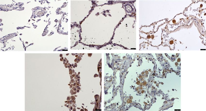
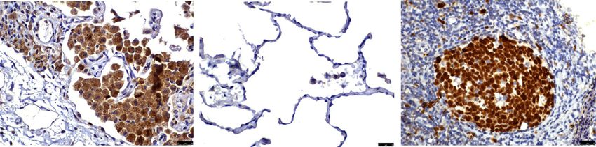
Fig. 9 Alveolar macrophage Ki67 expression. This figure shows immunohistochemical detection of Ki67 in lung resection specimens of COPD, S
and NS. Representative images of n = 9 COPD, n = 9 S and n = 6 NS. Positively labelling cells were visualised using DAB. Immunoreactive cells
(brown nuclei) are indicated with an arrow. 8a) Ki67 labelled lung resection specimen 8b) negative control 8c) tonsilar tissue (positive control).
Images taken using an X40 objective lensRavi et al. Respiratory Research (2017) 18:90 Page 10 of 12
a b c
d e
Fig. 10 BCL2 expression of alveolar macrophages. This figure shows immunohistochemical detection of BCL-2 in lung resection specimens of
COPD, S and NS. Representative images of n = 9 COPD, n = 3 COPD former smoker, n = 9 S and n = 6 NS. Positively labelling cells were visualised
using DAB (immunoreactive cells have brown cytoplasm and are indicated with an arrow). 9a) negative control, 9b) NS, 9c) S, 9d) COPD former
smoker and 9e) COPD current smoker. Images taken using an X 40 objective lens
induction was achieved when sIL-6R was also present, sug- of macrophages in sputum is increased in COPD patients
gesting an important role for IL-6 trans-signalling in the [14, 16]. Bronchoalveolar lavage supernatants are an alter-
regulation of CCR5 expression. An alternative mechanism native for chemotaxis experiments, representing a differ-
for CCR5 upregulation in monocytes is the effects of react- ent lung compartment where macrophages numbers are
ive oxygen species exposure, which can upregulate CCR5 increased. However, this is more invasive and the installa-
expression [41, 42]. tion of saline may cause excessive dilution.
Increased sIL-6R levels may promote inflammatory ac-
tivity in COPD by enhancing the effects of IL-6 through
trans-signalling. IL-6 is involved in the polarization of Conclusion
naïve CD4+ T lymphocytes towards the pro-inflammatory In conclusion, we have demonstrated that COPD mono-
Th17 effector phenotype [43]; furthermore, IL-6 sup- cytes show decreased migratory ability despite augmented
presses apoptosis of both innate and adaptive immune expression of CCR5. These findings indicate that in-
cells resulting in their persistence at foci of inflammation creased lung macrophage numbers in COPD lungs are
[44, 45]. not be due to excessive blood monocyte recruitment or al-
CD14+CD16+ monocytes are potent secretors of IL-1, veolar macrophage self-renewal; suppressed apoptosis
IL-6 and TNF-α [1], and expanded CD14+CD16+ mono- may be a factor leading to macrophage accumulation in
cyte populations are found in inflammatory disease states the lungs of COPD patients.
including atherosclerosis [46], obesity [47] and rheumatoid
arthritis [48]. We did not observe any change in monocyte Additional files
subsets in COPD patients compared to controls. Further-
more, there was no change in cellular subpopulations dur- Additional file 1: Details of methodology (pulmonary function, plasma
ing exacerbations, indicating no dysregulation of CD14 separation & cytokine measurement, flow cytometric monocyte
+ characterisation, CD14+ monocyte isolation, sputum supernatant induced
CD16+ cells in COPD. monocyte chemotaxis, CD14+ monocyte chemokine receptor gene
We elected to use sputum for chemotaxis experiments expression, Quantitative polymerase chain reaction, Immunohistochemistry
as there are increased levels of monocyte chemoattrac- and Immunofluorescence. (DOCX 34 kb)
tants in the sputum supernatants of COPD patients com- Additional file 2: Details of ‘Within-subject analysis’ (Generalised
Estimation Equation analysis) from CCR5 gene expression study (DOCX 15 kb)
pared to controls [14, 16]. Furthermore, the total numberRavi et al. Respiratory Research (2017) 18:90 Page 11 of 12
Additional file 3: Details of legends for additional Figures S1-S6. Table S1. Publisher’s Note
demographic details of patients from cytokine analysis, flow cytometry, Springer Nature remains neutral with regard to jurisdictional claims in published
chemotaxis and gene expression studies. Table S2. proportions of peripheral maps and institutional affiliations.
blood monocyte subtypes (CD14++CD16-, CD14+CD16+ & CD14-CD16++) in
stable and exacerbating COPD patients. Figure S1. proportions of monocyte Author details
1
sub-populations in the blood of COPD, S & HNS displayed as a graph. Figure NIHR Respiratory and Allergy Clinical Research Facility, Manchester
S2. changes in the CCR5 expression by monocyte sub populations during Academic Health Science Centre, University Hospital South Manchester NHS
COPD exacerbations displayed as a graph. Figure S3. Eeffect of age on Foundation Trust, University of Manchester, Manchester, UK. 2UCB, Slough,
CD14+ monocyte migration displayed as a graph. Figure S4. CD34 expres- Berkshire, UK. 3The Medicines Evaluation Unit, Wythenshawe Hospital, The
sion of pulmonary endothelial cells displayed as an immunohistochemistry Langley Building, Southmoor Road, Wythenshawe, Greater Manchester M23
image. Figure S5. neutrophils in the pulmonary microvasculature of COPD 9QZ, UK.
patients displayed as an immunohistochemistry image. Figure S6. Double
negative immunofluorescent image of tonsilar tissue stained using Received: 21 November 2016 Accepted: 1 May 2017
an immunofluorescence protocol with omission of CX3CR1 and
CD14 primary antibodies. (ZIP 2021 kb)
References
Abbreviations 1. Cros J, Cagnard N, Woollard K, Patey N, Zhang S-Y, Senechal B, Puel A,
AIM: Apoptosis inhibitor of macrophage; ANOVA: Analysis of variance; Biswas SK, Moshous D, Picard C, Jais J-P, D'Cruz D, Casanova J-L, Trouillet C,
BCL2: B-cell CLL/lymphoma 2; BCL-XL: B cell lymphoma extra large; Geissmann F. Human CD14dim Monocytes Patrol and Sense Nucleic acids
CAT: COPD assessment test; CCL: C-C motif chemokine ligand; CCR: C-C and viruses via TLR7 and TLR8 receptors. Immunity. 2010;33:375–86.
motif chemokine receptor; CD: Cluster of differentiation; COPD: Chronic 2. Yona S, Kim K-W, Wolf Y, Mildner A, Varol D, Breker M, Strauss-Ayali D,
obstructive pulmonary disease; C-X3-CR: CX3C chemokine receptor; CXCR: C- Viukov S, Guilliams M, Misharin A, Hume DA, Perlman H, Mailssen B, Zelzer
X-C motif chemokine receptor; HNS: Healthy non-smoker; ICAM: Intercellular E, Jung S. Fate mapping reveals origins and dynamics of monocytes and
adhesion molecule; IF: Immunofluorescence; IHC: Immunohistochemistry; tissue macrophages under homeostasis. Immunity. 2013;38:79–91.
IL: Interleukin; NE: Neutrophil elastase; NS: Non-smoker; PBMC: Peripheral 3. Desch AN, Gibbings SL, Goyal R, Kolde R, Bednarek J, Bruno T, Slanksy JE,
blood mononuclear cell; QPCR: Quantitative polymerase chain reaction; Jacobelli J, Mason R, Ito Y, Messier E, Randolph GJ, Prabagar M, Atif SM,
rh: Recombinant human; S: Smoker; SGRQ: St George’s Respiratory Questionnaire; Segura E, Xavier RJ, Bratton DL, Janssen WJ, Henson PM, Jakubzick C. Flow
sIL-6R: Soluble Interleukin-6 Receptor; TNF: Tumour necrosis factor; VCAM: Vascular cytometric analysis of mononuclear phagocytes in nondiseased human lung
cell adhesion molecule and lung-draining lymph nodes. Am J Respir Crit Care Med. 2016;193:614–26.
4. Hogg JC, Chu F, Utokaparch S, Woods R, Elliott WM, Buzatu L, Cherniak RM,
Rogers RM, Sciurba FC, Coxson HO, Pare PD. The Nature of Small Airway
Acknowledgements
Obstruction in Chronic Obstructive Pulmonary Disease. N Engl J Med. 2004;
We are grateful to Dr Louise Healy (UCB, Clough UK) for her assistance with
350:2645–53.
the MSD® analysis of plasma samples. The COPD Assessment Test was
5. Barnes PJ. Chronic obstructive pulmonary disease. N Engl J Med. 2000;343:
reproduced with the permission of GlaxoSmithKline (GSK, Brentford UK). GSK
269–80.
owns the Intellectual Property in the COPD Assessment Test. The SGRQ was
6. Costa C, Traves SL, Tudhope SJ, Fenwick PS, Belchamber KBR, Russel REK,
reproduced with the permission of St George’s University, London (UK). We
Barnes PJ, Donnelly LE. Enhanced monocyte migration to CXCR3 and CCR5
also wish to thank Ms Antonia Banyard for her generous technical assistance
chemokines in COPD. Eur Respir J. 2016;47:1093–102.
with regards the flow cytometric characterization of monocytes.
7. Tomita K, Caramori G, Lim S, Ito K, Hanazawa T, Oates T, Chiselita I, Jazrawi
E, Chung KF, Barnes PJ, Adcock IM. Increased p21CIP1/WAF1 and B Cell
Funding
Lymphoma Leukemia-XL expression and reduced apoptosis in alveolar
This study was in part funded by an unrestricted grant from UCB (Slough, UK).
macrophages from smokers. Am J Respir Crit Care Med. 2002;166:724–31.
8. Kojima J, Araya J, Hara H, Ito S, Takasaka N, Kobayashi K, Fujii S, Tsurushige
Availability of data and materials C, Numata T, Ishikawa T, Shimizu K, Kawaishi M, Saito K, Kamiya N, Hirano J,
All data generated or analysed during this study are included in this published Odaka M, Morikawa T, Hano H, Arai S, Miyazaki T, Kaneko Y, Nakayama K,
article [and its supplementary information files]. Kuwano K. Apoptosis inhibitor of macrophage (AIM) expression in alveolar
macrophages in COPD. Respir Res. 2013;14:30.
Authors’ contributions 9. Wahlström J, Berlin M, Sköld CM, Wigzell H, Eklund A, Grunewald J.
Conception: AKR, JP, JV, DS. Drafting the work or revising it: AKR, JP, RG, CSB, Phenotypic analysis of lymphocytes and monocytes/macrophages in
GB, MC, JV, DS. Final approval: AKR, JP, RG, CSB, GB, MC, JV, DS. Agreement to peripheral blood and bronchoalveolar lavage fluid from patients with
be accountable: AKR, JP, RG, CSB, GB, MC, JV, DS. All authors read and pulmonary sarcoidosis. Thorax. 1999;54:339–46.
approved the final manuscript. 10. Aaron SD, Vandemheen KL, Ramsay T, Zhang C, Avnur Z, Nikolcheva T,
Quinn A. Multi-analyte profiling and variability of inflammatory markers in
Competing interests blood and induced sputum in patients with stable COPD. Respir Res [serial
Dr Arjun Ravi, Dr Jonathan Plumb, Dr Sarah Mason, Ms Rosemary Gaskell, Dr online] 2010; vol. 11. Available from: https://respiratory-research.biomedcentral.
Caroline Broome and Mr George Booth have no conflicts of interest to com/articles/10.1186/1465-9921-11-41
declare. Dr Matthew Catley worked for UCB, Slough (UK). Professor Dave 11. Bafadhel M, McCormick M, Saha S, McKenna S, Shelley M, Hargadon B, Mistry V,
Singh has received lecture fees, research grants, consultancy fees and Reid C, Brightling CE. Profiling of sputum inflammatory mediators in asthma
support for conference attendance from various pharmaceutical companies and chronic obstructive pulmonary disease. Respiration. 2012;83:36–44.
including AstraZeneca, GlaxoSmithKline, Chiesi, Boehringer Ingelheim, Roche, 12. Capelli A, Di Stefano A, Gnemmi I, Balbo P, Cerutti CG, Balbi B, Lusuardi M,
Novartis, Cipla, Almirall and Merck. Donner CF. Increased MCP-1 and MIP1β in bronchoalveolar lavage fluid of
Professor Jørgen Vestbo has received lecture fees and consultancy fees from chronic bronchitics. Eur Respir J. 1999;14:160–5.
various pharmaceutical companies including GSK, Nycomed, Chiesi, Novartis, 13. Costa C, Rufino R, Traves SL, Silva JR LE, Barnes PJ, Donnelly LE. CXCR3 and
AstraZeneca and Boehringer Ingelheim. CCR5 chemokines in Induced Sputum from patients with COPD. Chest.
2008;133:26–33.
Consent for publication 14. Traves SL, Culpit SV, Russell REK, Barnes PJ, Donnelly LE. Increased levels of
Not applicable. the chemokines Gro-α and MCP1 in sputum samples from patients with
COPD. Thorax. 2002;57:590–5.
Ethics approval and consent to participate 15. Saetta M, Mariani M, Panina-Bordignon P, Turato G, Buonsanti C, Baraldo S,
The research was approved by the Greater Manchester (East) ethics committee: Bellettato CM, Papi A, Corbetta L, Zuin R, Sinigaglia F, Fabbri L. Increased
Approval numbers (10/H1003/108 & 05/Q140241)). expression of the chemokine receptor CXCR3 and its ligand CXCL10 inRavi et al. Respiratory Research (2017) 18:90 Page 12 of 12
peripheral airways of smokers with chronic obstructive pulmonary disease. Ma'ayan A, Riches DWH, Yokoyama WM, Ginhoux F, Henson PM, Randolph
Am J Respir Crit Care Med. 2002;165:1404–9. GJ. Minimal differentiation of classical monocytes as they survey steady-state
16. Ravi AK, Khurana S, Lemon J, Plumb J, Booth G, Healy L, Catley M, Vestbo J, tissues and transport antigen to lymph nodes. Immunity. 2013;39:599–610.
Singh D. Increased levels of soluble interleukin-6 receptor and CCL3 in COPD 36. Perdiguero E-G, Klapproth K, Schultz C, Busch K, Azzoni E, Crozet L, Garner
sputum. Respir Res. 2014;15:103. H, Trouillet C, de Bruijn M, Geissmann F, Rodewald H-R. Tissue-resident
17. Smyth LJC, Starkey C, Gordon FS, Vestbo J, Singh D. CD8 chemokine receptors macrophages originate from yolk-sac-derived erythro-myeloid progenitors.
in chronic obstructive pulmonary disease. Clin Exp Immunol. 2008;154:56–63. Nature. 2015;518:547–51.
18. Kawanaka N, Yamamura M, Aita T, Morita Y, Okamoto A, Kawashima M, 37. Brittan M, Barr LC, Anderson N, Conway Morris A, Duffin R, Marwick JA, Rossi
Iwashi M, Ueno A, Ohmoto Y, Makino H. CD14+, CD16+ blood monocytes F, Johnson S, Dhaliwal K, Hirani N, Rossi AG, Simpson AJ. Functional
and joint inflammation in rheumatoid arthritis. Arthritis Rheum. 2002;46: characterisation of human pulmonary monocyte-like cells in
2578–86. lipopolysaccharide-mediated acute lung inflammation. J Inflamm. 2014;11:9.
19. Weber C, Belge KU, von Hundelschausen P, Drauda G, Steppich B, Mack M, 38. Eguíluz-Gracia I, Schultz HHL, Sikkeland LIB, Danilova E, Holm AM, Pronk
Frankenberger M, Weber KSC, Ziegler-Heitbrock HW. Differential chemokine CJH, Agace WW, Iversen M, Andersen C, Jahnsen FL, Baekkevold ES. Long-
receptor expression and function in human monocyte subpopulations. J term persistence of human donor alveolar macrophages in lung transplant
Leukoc Biol. 2000;67:699–704. recipients. Thorax. 2016;71:1006–11.
20. Wang J, Crawford K, Yuan M, Wang H, Gorry PR, Gabuzda D. Regulation of 39. Cai Y, Sugimoto C, Arainga M, Alvarez X, Didier ES, Kuroda MJ. In vivo
CC chemokine receptor 5 and CD4 expression and human immunodeficiecy characterization of alveolar and interstitial lung macrophages in rhesus
virus Type 1 replication in human macrophages and microglia by T helper macaques: implications for understanding lung disease in humans.
Type 2 Cytokines. J Infect Dis. 2002;185:885–97. J Immunol. 2014;192:2821–9.
21. Fielding CA, McLoughlin RM, Mcleod L, Colmont CS, Najdovska M, Grail D, 40. Hurst SM, Wilkinson TS, McLoughlin RM, Jones S, Horiuchi S, Yamamoto N,
Ernst M, Jones SA, Topley N, Jenkins BJ. IL-6 regulates neutrophils trafficking Fuller GM, Topley N, Jones SA. IL-6 and its soluble receptor orchestrate a
during acute inflammation via STAT3. J Immunol. 2008;181:2189–95. temporal switch in the pattern of leucocyte recruitment seen during acute
22. Agusti A, Edwards LD, Rennard SI, Macnee W, Tal-Singer R, Miller BE, Vestbo inflammation. Immunity. 2001;14:705–14.
J, Lomas DA, Calverley PMA, Wouters E, Crim C, Yates JC, Silverman EK, 41. Saccani A, Saccani S, Orlando S, Sironi M, Ghezzi P, Mantovani A, Sica A.
Coxson HO, Bakke P, Mayer RJ, Celli B, for-the-Evaluation-of-COPD-Longitudinally- Redox regulation of chemokine receptor expression. Proc Natl Acad Sci.
to-Identify-Predictive-Surrogate-Endpoints-(ECLIPSE)-Investigators. Persistent 2000;97:2761–6.
Systemic Inflammation is Associated with Poor Clinical Outcomes in COPD: A 42. Lehoux G, Le Gouill C, Stankova J, Rola-Pleszcynski M. Upregulation of the
Novel Phenotype. PLoS One. 2012;7:e37483. expression of CCR5 by hydrogen peroxide in human monocytes. Med
23. Hurst JR, Perera WR, Wilkinson TMA, Donaldson GC, Wedzicha JA. Systemic Inflamm. 2003;12:29–35.
and upper and lower airway inflammation at exacerbation of chronic 43. Benwell R, Lee DR. Essential and synergistic Roles of IL-1 and IL-6 in human
obstructive pulmonary disease. Am J Respir Crit Care Med. 2006;173:71–8. Th17 differentiation directed by TLR ligand activated dendritic cells. Clin
24. Vogelmeier CF, Criner GJ, Martinez FJ, Anzueto A, Barnes PJ, Bourbeau J, Immunol. 2009;134:178–87.
Celli BR, Chen R, Decramer M, Fabbri LM, Frith PA, Halpin DMG, Victorina 44. Asensi V, Valle E, Meana A, Fierer J, Celada A, Alvarez V, Paz J, Coto E, Carton
López Varela M, Nishimura M, Roche N, Rodriguez-Rosin R, Sin DD, Singh D, JA, Maradona JA, Dieguez A, Sarasua J, Ocana M, Arribas JM. In vivo Interleukin-
Stockley RA, Vestbo J, Wedzicha JA, Agusti A. Global strategy for the 6 protects neutrophils from apoptosis in osteomyelitis. Infect Immun. 2004;72:
diagnosis, management and prevention of chronic obstructive lung disease 3823–38.
2017 Report - GOLD executive summary. Am J Respir Crit Care Med. 2017; 45. Atreya R, Finotto S, Mullberg J, Jostock T, Wirtz S, Schutz M, Bartsch B,
195:557–82. Holtmann M, Becker C, Strand D, Czaja J, Schlaak JF, Lehir HA, Autscbach F,
25. Wanger J, Clausen JL, Coates A, Pedersen O, Brusasco V, Burgos F, Casaburi Schurmann G, Nishimoto N, Yoshizaki K, Ito H, Kishimoto T, Galle PR, Rose-
R, Crapo R, Enright P, van der Grinten CPM, Gustafsson P, Hankinson J, John S, Neurath MF. Blockade of interleukin-6 trans-signaling suppresses T
Jensen R, Johnson D, MacIntyre N, McKay R, Miller MR, Navjas D, Pellegrino cell resistance against apoptosis in chronic intestinal inflammation: evidence
R, Viegi G. Standardization of the measurement of lung volumes. Eur Respir in Crohn's Disease and experimental colitis in-vivo. Nat Med. 2000;6:583–8.
J. 2005;26:511–22. 46. Rogacev KS, Cremers B, Zawada AM, Seiler S, Binder N, Ege P, Große-Dunker
26. Lea S, Plumb J, Metcalfe H, Spicer D, Woodman P, Fox JC, Singh D. The G, Heisel I, Hornof F, Jenken J, Rebling NM, Ulrich C, Scheller B, Böhm M,
effect of peroxisome proliferator-activated receptor-Ɣ ligands on in vitro Fliser D, Heine GH. CD14++CD16+ monocytes independently predict
and in vivo models of COPD. Eur Respir J. 2014;43:409–20. cardiovascular events: a cohort study of 951 patients referred for elective
27. Pusztazeri MP, Seelentag W, Bosman FT. Immunohistochemical expression coronary angiography. J Am Coll Cardiol. 2012;60:1512–20.
of endothelial markers CD31, CD34, von Willebrand Factor, and Fli-1 in 47. Rogacev KS, Ulrich C, Blömer L, Hornof F, Oster K, Ziegelin M, Cremers B,
normal human tissues. J Histochem Cytochem. 2005;54:385–95. Grenner Y, Geisel J, Schlitt A, Köhler H, Fliser D, Girndt M, Heine GH. Monocyte
heterogeneity in obesity and subclinical atherosclerosis. Eur Heart J. 2010;31:
28. Braun N, Papadopolous T, Müller-Hermelink H. Cell cycle dependent distribution
369–76.
of the proliferation-associated Ki-67 antigen in human embryonic lung cells.
48. Rossol M, Kraus S, Pierer M, Baerwald C, Wagner U. The CD14brightCD16+
Virchows Arch B, Cell pathol incl mol pathol. 1988;56:25–33.
Monocyte subset Is Expanded in Rheumatoid Arthritis and Promotes
29. Rahman I, Morrison D, Donaldson K, Macnee W. Systemic oxidative stress in
Expansion of the TH17 Cell Population. Arthritis Rheum. 2012;64:671–7.
asthma, COPD and smokers. Am J Respir Crit Care Med. 1996;154:1055–60.
30. Couillard A, Koechlin C, Cristol JP, Varray A, Prefaut C. Evidence of local
exercise induced systemic oxidative stress in chronic obstructive pulmonary
disease. Eur Respir J. 2002;20:1123–9.
31. Stadler N, Eggermann J, Vöö S, Kranz A, Waltenberger J. Smoking-induced Submit your next manuscript to BioMed Central
monocyte dysfunction is reversed by vitamin C supplementation in vivo. and we will help you at every step:
Arterioscler, Thromb Vasc Biol. 2007;27:120–6.
32. Wenisch C, Patruta S, Daxböck F, Krause R, Hörl W. Effect of age on human • We accept pre-submission inquiries
neutrophil function. J Leukoc Biol. 2000;67:40–5. • Our selector tool helps you to find the most relevant journal
33. McComb JG, Ranganathan M, Liu XH, Pilewski JM, Ray P, Watkins SC, Choi
• We provide round the clock customer support
AMK, Lee JS. CX3CL1 Up-regulation is associated with recruitment of
CX3CR1+ Mononuclear Phagocytes and T lymphocytes in the lungs during • Convenient online submission
cigarette smoke-induced emphysema. Am J Pathol. 2008;173:949–61. • Thorough peer review
34. González S, Hards J, van Eeden S, Hogg JC. The expression of adhesion
• Inclusion in PubMed and all major indexing services
molecules in cigarette smoke-induced airways obstruction. Eur Respir J.
1996;9:1995–2001. • Maximum visibility for your research
35. Jakubzick C, Gautier EL, Gibbings SL, Sojka DK, Schlitzer A, Johnson TE,
Ivanov S, Duan Q, Bala S, Condon T, van Rooijen A, Grainger JR, Belkaid Y, Submit your manuscript at
www.biomedcentral.com/submitYou can also read