Role of EXO1 nuclease activity in genome maintenance, the immune response and tumor suppression in Exo1D173A mice
←
→
Page content transcription
If your browser does not render page correctly, please read the page content below

Nucleic Acids Research, 2022 1
                                                                                                              https://doi.org/10.1093/nar/gkac616

Role of EXO1 nuclease activity in genome
maintenance, the immune response and tumor
suppression in Exo1D173A mice
Shanzhi Wang1,2 , Kyeryoung Lee1 , Stephen Gray 3,4 , Yongwei Zhang1 , Catherine Tang5 ,
Rikke B. Morrish6 , Elena Tosti1 , Johanna van Oers1 , Mohammad Ruhul Amin7 ,

                                                                                                                                                              Downloaded from https://academic.oup.com/nar/advance-article/doi/10.1093/nar/gkac616/6645635 by guest on 01 August 2022
Paula E. Cohen3 , Thomas MacCarthy5 , Sergio Roa 8,9,10,* , Matthew D. Scharff1,* ,
Winfried Edelmann1,* and Richard Chahwan 11,*
1
  Department of Cell Biology, Albert Einstein College of Medicine, 1300 Morris Park Avenue, NY 10461, USA,
2
  Current position: Department of Chemistry, University of Arkansas at Little Rock, Little Rock, AR 72204, USA,
3
  Department of Biomedical Sciences, Cornell University, NY 14853, USA, 4 Current position: School of Life Sciences,
University of Nottingham, Nottingham NG7 2UH, UK, 5 Department of Applied Mathematics and Statistics, Stony
Brook University, Stony Brook, NY, USA, 6 Current position: School of Physics and Astronomy, University of Exeter,
Exeter EX4 4QD, UK, 7 Department of Computer and Information Science, Fordham University, Bronx, NY, USA,
8
  Department of Biochemistry and Genetics, University of Navarra, 31008 Pamplona, Spain, 9 Centro de Investigacion
Biomedica en Red de Cancer (CIBERONC), Instituto de Salud Carlos III, Madrid, Spain, 10 Navarra Institute for Health
Research (IdiSNA), Pamplona, Spain and 11 Institute of Experimental Immunology, University of Zurich, Zurich 8057,
Switzerland

Received September 04, 2021; Revised May 30, 2022; Editorial Decision June 24, 2022; Accepted June 30, 2022

ABSTRACT                                                                        diate between Exo1+/ + and Exo1– /– mice, suggesting
DNA damage response pathways rely extensively on                                that both catalytic and scaffolding roles of EXO1 are
nuclease activity to process DNA intermediates. Ex-                             important for somatic hypermutation. Similarly, while
onuclease 1 (EXO1) is a pleiotropic evolutionary con-                           overall class switch recombination in Exo1DA/DA and
served DNA exonuclease involved in various DNA re-                              Exo1– /– mice was comparably defective, switch junc-
pair pathways, replication, antibody diversification,                           tion analysis suggests that EXO1 might fulfill an ad-
and meiosis. But, whether EXO1 facilitates these                                ditional scaffolding function downstream of class
DNA metabolic processes through its enzymatic or                                switching. In contrast to Exo1−/− mice that are infer-
scaffolding functions remains unclear. Here, we dis-                            tile, meiosis progressed normally in Exo1DA/DA and
sect the contribution of EXO1 enzymatic versus scaf-                            Exo1+/+ cohorts, indicating that a structural but not
folding activity by comparing Exo1DA/DA mice ex-                                the nuclease function of EXO1 is critical for meiosis.
pressing a proven nuclease-dead mutant form of                                  However, both Exo1DA/DA and Exo1– / – mice displayed
EXO1 to entirely EXO1-deficient Exo1−/− and EXO1                                similar mortality and cancer predisposition profiles.
wild type Exo1+/+ mice. We show that Exo1DA/DA and                              Taken together, these data demonstrate that EXO1
Exo1– /– mice are compromised in canonical DNA                                  has both scaffolding and enzymatic functions in dis-
repair processing, suggesting that the EXO1 enzy-                               tinct DNA repair processes and suggest a more com-
matic role is important for error-free DNA mismatch                             posite and intricate role for EXO1 in DNA metabolic
and double-strand break repair pathways. However,                               processes and disease.
in non-canonical repair pathways, EXO1 appears to
have a more nuanced function. Next-generation se-                               INTRODUCTION
quencing of heavy chain V region in B cells showed                              Exonuclease 1 (EXO1) is an evolutionarily conserved mem-
the mutation spectra of Exo1DA/DA mice to be interme-                           ber of the XPG/Rad2 family of metallonucleases. EXO1

* To
   whom correspondence should be addressed. Tel: +41 446353710; Email: richard.chahwan@uzh.ch
Correspondence may also be addressed to Winfried Edelmann. Tel: +1 718 678 1086; Email: winfried.edelmann@einsteinmed.org
Correspondence may also be addressed to Matthew D. Scharff. Tel: +1 718 430 3527; Email: matthew.scharff@einsteinmed.org
Correspondence may also be addressed to Sergio Roa. Tel: +34 948425600; Email: sroa@unav.es


C The Author(s) 2022. Published by Oxford University Press on behalf of Nucleic Acids Research.
This is an Open Access article distributed under the terms of the Creative Commons Attribution License (http://creativecommons.org/licenses/by/4.0/), which
permits unrestricted reuse, distribution, and reproduction in any medium, provided the original work is properly cited.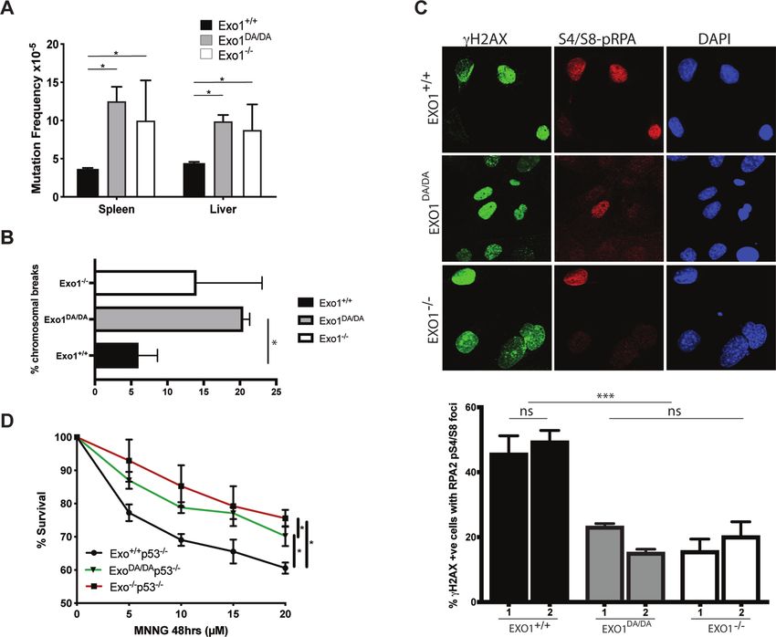
2 Nucleic Acids Research, 2022
and its biochemical nuclease activity was initially identi- tiation of homologous recombination (HR) mediated DNA
fied in extracts of fission yeast undergoing meiosis and has repair. Studies in yeast and mammals (22,23) suggest a two-
since been shown to fulfill broader biological functions in step model for DSB processing. MRE11-RAD50-NBS1
mammalian cells during DNA damage responses (DDR), (MRN) and CtIP initiate the ‘end-trimming’ of the DSB,
including DNA mismatch repair (MMR), DNA double- which is then followed by the generation of longer stretches
strand break repair (DSBR), and telomere maintenance of ssDNA by either EXO1 or the BLM/DNA2 helicase-
(1–3). In immune B cells, EXO1 can also mediate non- nuclease complex (22,24). Two physiological and beneficial
canonical forms of MMR that promote antibody diversi- subsets of DSBR occur during B cell maturation through
fication through somatic hypermutation (SHM) and class CSR and during meiosis in germ cells. Both processes are
switch recombination (CSR) (4). In germ cells, EXO1 me- dependent on adequate upstream DSBR signaling, but they
Downloaded from https://academic.oup.com/nar/advance-article/doi/10.1093/nar/gkac616/6645635 by guest on 01 August 2022
diates genetic diversity by promoting recombination dur- diverge considerably downstream. While meiosis relies on
ing meiosis (5). Catalytically, EXO1 has 5 to 3 exonucle- the error-free HR pathway for crossover formation, CSR
ase and 5 -flap endonuclease activities (6,7). Yet, these seem- relies on the error-prone pathway of non-homologous end
ingly distinct nuclease activities are now shown to be mech- joining (NHEJ) to generate intra-chromosomal switch re-
anistically integrated (8). gion translocations and facilitate isotype switching. As a
DNA mismatches are some of the most common forms result of this divergence, the dependency of these two pro-
of DNA damage, arising mainly during replication. MMR cesses on EXO1-mediated resection remains unclear.
is the main pathway that removes misincorporated nu- Deficiencies in MMR and DSBR associated processes
cleotides that result from erroneous replication. During can lead to chromosomal instability, infertility, neurode-
MMR, the coordination of mismatch recognition and exci- generation, tumorigenesis, and immune defects (25). Given
sion is facilitated by MutS homolog (MSH) and MutL ho- its pleiotropic roles in the DNA damage response, it is ex-
molog (MLH) complexes. Briefly, MSH2 forms a complex pected that defects in EXO1 could also lead to some of these
with MSH6 (MutS␣) to predominantly initiate the repair anomalies. Indeed, mice with an inactivating mutation in
of single-base mispairs and insertion/deletions (IDLs) and EXO1 had meiotic defects and sterility due to loss of chi-
with MSH3 (MutS) to predominantly initiate the repair of asmata during metaphase (26), impaired SHM and CSR,
larger IDLs of two to four bases. Subsequent to mismatch loss of mismatch repair activity, and a 30 fold increase of
recognition, MutS␣ and MutS interact with the MLH1- mutation frequency in mouse ES cells (4,5). EXO1 inacti-
PMS2 (MutL␣) complex in an ATP-dependent manner to vation also reduced overall survival in mice and caused an
initiate EXO1 mediated excision of the DNA strand carry- increased incidence of cancers, especially lymphomas (4,5).
ing the mismatched base(s). The repair process is completed In humans, germline mutations in EXO1 have been associ-
by DNA re-synthesis to fill the resulting single strand gap ated with hereditary nonpolyposis colorectal cancer/Lynch
and ligation. Thus far, EXO1 is the only known exonuclease syndrome (HNPCC/LS) (27).
that facilitates the removal of the mismatch-carrying strand To dissect the scaffolding function from the enzymatic
during MMR (9,10). MMR is shown to improve replica- function of EXO1, a mouse line with the cancer-associated
tive fidelity by over two orders of magnitude; hence, reduc- E109K mutation was previously generated and character-
ing genomic mutator phenotypes (9,11), and EXO1 plays ized (5). Residue E109 is a surface residue of the catalytic
an important part in that process. During MMR, EXO1 domain in the crystal structure (28) and, even though it does
directly interacts with MLH1, MSH2, and MSH3 both in not directly affect the catalytic site, it had been reported to
yeast (12,13) and mammalian cells (14,15). As a result, it be essential for the nuclease activity when the N-terminal
was speculated that EXO1 could fulfill both catalytic and fragment of human EXO1 was examined biochemically
structural roles during MMR but direct in vivo evidence (5,29). In vivo, homozygous mutant Exo1EK/EK knock-in
for this idea remained elusive (16). EXO1-independent exci- mice carrying the EXO1E109K showed defects in the DNA
sion in MMR has been described in yeast involving PCNA, damage response (DDR) and increased cancer predisposi-
Msh2/6 and Mlh1/Pms1 endonuclease (17,18). In addition, tion. Unexpectedly the Exo1EK/EK mice showed normal ac-
Exo1 independent MMR was proposed to involve replica- tivity in SHM, CSR, MMR, meiosis and were fertile, while
tion dependent strand displacement of the mismatch carry- Exo1−/− mice had defects in all of these processes. This was
ing strand in in vitro experiments (19). Interestingly, a non- interpreted to mean that EXO1 had an essential scaffolding
canonical error-prone subset of MMR occurs during B cell role, but its enzymatic activity could be replaced by some
maturation. During this process, initiated by AID-induced unknown factor during SHM, CSR, MMR, and meiosis
U:G mismatches in germinal center B cells, EXO1 is be- (5). However, this conclusion was challenged by two subse-
lieved to resect mismatches akin to its role during canonical quent independent biochemical observations, where for the
MMR. However, when repairing AID induced mismatches first time full-length non-tagged EXO1E109K was purified in
in antibody V regions, this process subsequently diverges vitro, and near WT activity was detected (30,31). In contrast
by the recruitment of error-prone polymerases, which were to the E109K mutation, these studies also confirmed that
shown to be recruited by ubiquitinated PCNA (20,21). the EXO1D173A mutation within the catalytic site, which
Another repair mechanism that relies on EXO1 activity had been previously reported to be nuclease-dead in yeast
is DSBR, which repairs some of the most toxic forms of and mammals (32–35), also caused the clear inactivation
DNA damage. DNA end resection of DSBs in the 5 to of EXO1 nuclease activity in vitro (6,30,31), offering a new
3 direction is a critical intermediate in this process. The alternative to reevaluate the nuclease and scaffold func-
ensuing single-stranded DNA (ssDNA) induces two inter- tions of EXO1 in vivo. To adequately separate the nucle-
dependent responses, namely checkpoint signaling and ini- ase and scaffold functions of EXO1, we herein generate a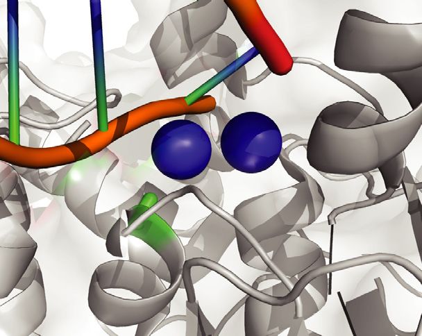
Nucleic Acids Research, 2022 3
new knock-in mouse line carrying the Exo1D173A mutation After washes in PBS + 0.1% Triton X-100, Alexa 488 goat
(Exo1DA ) and compare it to Exo1–/– and Exo1+/+ mice. anti-mouse/rabbit, and Alexa 598 goat anti-mouse/rabbit
Characterization of these animals confirms that EXO1 ex- (Molecular Probes) were used as secondary antibodies. Im-
hibits both scaffolding and catalytic functions, demonstrat- ages were acquired using a Bio-Rad Radiance 2100 (Nikon
ing that its exonuclease activity is essential in most DNA re- Eclipse E800) microscope using Lasersharp 2000 software
pair mechanisms (i.e. DDR, MMR, SHM and CSR) while (Zeiss).
it remains nonessential for meiosis, where EXO1 appears to
play a critical structural role. MNNG treatment
After crossing all models presented here to p53+/− mice,
Downloaded from https://academic.oup.com/nar/advance-article/doi/10.1093/nar/gkac616/6645635 by guest on 01 August 2022
MATERIALS AND METHODS
MEF cell lines were generated and pretreated with 20 M
Mice and MEF strains O-6-benzylguanine (O6BG, Sigma) before the addition of
The generation of Exo1−/− mice has been described MNNG to inhibit the repair of O6meG adducts by O-6-
previously (4). Exo1DA/DA mice were generated using methylguanine-DNA methyltransferase (MGMT, Sigma).
CRISPR/Cas9 technology (36–38). In brief, the mixture of After 48 h MNNG treatment, relative cell viability was de-
guide RNA targeting to the adjacent area of Exo1 D173 termined using thiazolyl blue tetrazolium bromide (MTT)-
coding region on exon-6 (CRISPR targeting sequence is conversion (Sigma) as per the manufacturer’s instructions.
gactctgacctcctcgcattTGG), Cas9 mRNA, and donor DNA The absorbance of converted dye was measured at a wave-
carrying the ∼1 kb homologous arms at each side and the length of 570 nm using a Perkin Elmer Victor X5 plate
surrounded desired DNA mutation were injected into zy- reader. Cell viability was calculated relative to DMSO-
gotes of C57BL/6J mice and then the injected fertilized eggs treated cells incubated in parallel.
were transferred into pseudopregnant CD1 female mice for
producing pups. The resulting offspring were genotyped Analysis of murine tumors and survival
by PCR and Sanger sequencing to identify the founders
carrying the desired Exo1 D173A mutation. The founders Mice were routinely observed until they became moribund
(F0) were mated with wild-type C57BL/6J mice, and then and/or signs of tumour development occurred and required
the resulting germline transmitted F1 heterozygotes were euthanasia. The Kaplan–Meier method was used to com-
bred to establish study cohorts. When indicated, Exo1+/+ , pare the overall survival curves of the mice, using the log-
Exo1+/DA and Exo1+/– mice were further crossed to Big Blue rank analysis to evaluate significance. All tumors were pro-
transgenic mice (58) or p53+/– mice (5). Mouse embryonic cessed for paraffin embedding, and sections were prepared
fibroblasts (MEFs) were isolated from embryos of pregnant for H&E staining and evaluated by a pathologist. Statisti-
mice at day 13.5 post-conception. MEF cell lines were pas- cal analysis of tumour incidence was performed using the
saged 4–6 times and cryopreserved. All protocols were per- Fisher’s exact test.
formed in accordance with the Animal Care and Use Com-
mittee of Albert Einstein College of Medicine (AECOM) Immunization and hypermutation analysis on VH186.2
under the direction of Public Health Service (PHS) Policy
on Humane Care and Use of Laboratory Animals. Age-matched mice (Exo1+/+ , Exo1−/− and Exo1DA/DA )
were immunized intraperitoneally with 200 g NP(33)-
CGG (BioSearch Technologies) on alum. For primary re-
Analysis of cII mutation frequencies sponse analysis, animals were sacrificed, and spleens were
Exo1+/+ , Exo1+/− and Exo1+/DA mice were crossed to Big removed 10 days after a single immunization. Subsequently,
Blue transgenic mice (56) to generate Exo1+/+ /BigBlue, RNA was extracted using Purelink RNA Mini Kit (Ther-
Exo1−/− /BigBlue, and Exo1DA/DA /BigBlue mice. Muta- moFisher) and cDNA was generated using AccuScript
tions in genomic DNA from the spleen and liver were de- High Fidelity 1st Strand cDNA Synthesis Kit (Stratagene).
tected using the Select-cII Mutation Detection System for The Mus musculus IGHV1-72*01 allele (IMGT accession
Big Blue Rodents (Stratagene) as we have done in the past number J00530:206–499), referred here as VH186.2, was
(58). amplified using a nested PCR as previously described (21),
and PCR products were gel purified and stored in 50 l
buffer containing 10 mM Tris (pH 8.0) until submission
CPT treatment and immunofluorescence
to TruSeq® DNA library preparation (Illumina) following
Primary MEFs were grown on coverslips and treated with manufacturer’s instructions. After quality control by Bio-
1 M CPT or DMSO (control). After 1 h, the drug was analyzer (Bio-Rad), libraries were pair-end sequenced on
removed and cells were pre-extracted for 5 min on ice in a MiSeq Sequencer (Illumina) at Einstein Epigenetics Fa-
10 mM Pipes buffer (pH 6.8) containing 300 mM sucrose, cility. Using an in-house computational pipeline based on
50 mM NaCl, 3 mM EDTA, 0.5% Triton X-100, and Pro- Kernel written in ultra-fast C++ and a GUI written in Java,
tease Inhibitor Mixture (EDTA-free; Roche) before fixation Illumina pair-reads were assembled and collapsed accord-
in 2% (wt/vol) paraformaldehyde for 15 min at 25◦ C. After ing to their unique molecular identifier (UMI) to recon-
fixation, cells were washed with PBS and then were blocked struct unique overlap VH186.2DJ regions. These sequences
with 5% (wt/vol) BSA and 0.1% Triton X-100 in PBS be- in FASTA format were then submitted to IMGT/HighV-
fore staining with mouse anti-␥ H2AX (Cell Signaling), rab- QUEST (39) for VH186.2-D-J usage and CDR3 clonal-
bit anti-RPA pS4/S8 (Bethyl), and DAPI (Vector) for 1 h. ity identification. Then, customized Python and R scripts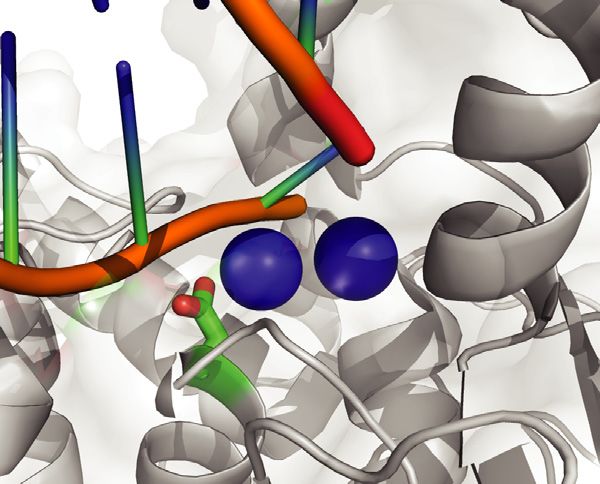
4 Nucleic Acids Research, 2022
were used for mutational analyses after randomly resam- antibodies conjugated to flurophores. After further wash-
pling during 1000 iterations one single sequence per CDR3 ing, slides were analyzed using an upright fluorescent mi-
clone, allowing us to increase our sensitivity for unique mu- croscope (Zeiss Axiophot with Zen 2.0 software).
tations, which are expected to derive from independent mu-
tational events, and avoid overestimating mutation frequen- Spermatocyte diakinesis spread preparations
cies by repeated counting of non-unique mutations inher-
ited within the clonal CDR3 lineage. For each group of Spread diakinesis preparations were made as described
Exo1+/+ , Exo1−/− and Exo1DA/DA iterations, frequencies of (44,45). Briefly, testis cells were liberated by manual dis-
mutation were normalized to the potential maximum num- section of tubules in 0.5% KCl. Multiple mixing and slow
ber of the corresponding mutation subtype as we have pre- centrifugation followed by fixation in 30% methanol: 10%
Downloaded from https://academic.oup.com/nar/advance-article/doi/10.1093/nar/gkac616/6645635 by guest on 01 August 2022
viously described (40), which corrects for sequence compo- acetic acid: 0.05% chloroform. Cells were finally fixed in
sition and size of CDR3 repertoire. 30% methanol: 10% acetic acid and then pipette onto heated
slides. Finally slides were stained in using Giemsa and im-
aged. Slides were imaged on a Zeiss Axiophot with Zen 2.0
Ex Vivo assay of class switching software.
Splenic cells were isolated from immunized animals
(Exo1+/+ , Exo1−/− and Exo1DA/DA ). Red blood cells RESULTS
and T cells were removed by ammonium chloride and
complement-dependent lysis with details described as previ- Generation of nuclease-dead Exo1D173A mice
ously (21). The rest of the cells were stimulated either using To create an inactivating nuclease-dead EXO1 mutation,
50 g/ml of LPS from Sigma-Aldrich (St. Louis, MO) or we generated a novel knock-in mouse line that carries the
using 50 g/ml of LPS plus 50 ng/ml of mouse IL-4 from EXO1D173A mutation (termed Exo1DA ) by CRISPR/Cas9
R&D Systems (Minneapolis, MN). After 4 days in com- technology in fertilized oocytes isolated from C57BL/6 fe-
plete RPMI media at 37◦ C, cells were stained against sur- male mice (Supplementary Figure S1A). The Exo1DA mu-
face IgM (ImmunoResearch) plus IgG1 (SouthernBiotech) tation was verified in the offspring by PCR and Sanger se-
or IgM plus IgG3 (SouthernBiotech), which was followed quencing (Supplementary Figure S1B). This mutant protein
by FACS analysis. was previously shown to have no detectable nuclease activ-
ity biochemically (30,31). Notably, the EXO1D173 residue is
Switch junction analysis evolutionarily conserved in both fission and budding yeast
as well as mouse and humans (Figure 1A, B). Furthermore,
After LPS stimulation for 4 days, splenic B cells were col- structural modeling of the resolved EXO1 crystal structure
lected for genomic DNA isolation using the DNeasy kit shows that residue EXO1D173 is embedded in the exonucle-
(Qiagen). S–S␥ 3 regions were amplified using nested PCR ase active site and is in direct contact with DNA moieties
with details and primers as previously described (41). PCR (8), which predicts a pronounced anomaly in the EXO1 nu-
products were purified with QIAquick PCR purification clease active site as a consequence of the EXO1D173A mu-
kit (Qiagen), ligated to Topo Blunt vector (Invitrogen) and tation (Figure 1C). However, such a perturbation is not ex-
transformed in E. coli chemical competent cells. Plasmids pected to affect protein folding and stability, DNA binding,
from individual E. coli colonies were purified and sequenced or MMR protein interactions as demonstrated by the dis-
for the inserted switch junctional regions. Switch junctions tinct domain dependencies (Figure 1B, C; Supplementary
were analyzed by alignment using BLAST2seq with the au- Figure S2A, B) and this is supported by studies in yeast and
tomatic low-complexity filter disabled and using consen- mammalian cells (30,31,33,34,46). Consistent with this, we
sus sequences from the NC 000078.6 GenBank record. The observed stable expression of mutant EXO1DA protein com-
nonparametric Kruskal-Wallis test followed by the Dunn’s parable to wild type EXO1 by Western-blot analysis, com-
post-tests were used to calculate P-values and interrogate pared to complete protein loss in previously characterized
whether the different groups showed identical distributions Exo1−/− mice (5) (Supplementary Figure S1C).
of microhomology lengths at the S–S␥ 3 junctions (com-
puted as 0 for blunt junctions, as positive values for short
The catalytic activity of EXO1 is predominant over scaffold-
and long microhomologies, and as negative values for in-
ing functions in canonical DDR
sertions). Frequency of insertions was calculated by divid-
ing the number of ocurrences by the total number of S– To measure the integrity of canonical error-free MMR func-
S␥ 3 junctions characterized in each group, and statistically tion in vivo, Exo1+/+ , Exo1DA/DA and Exo1–/- mice were
compared through Fisher’s exact tests. crossed to BigBlue mice (5,47). The accumulation of muta-
tions in the cII reporter gene was analyzed in genomic DNA
from the liver and spleen in all three cohorts. We observed
Meiosis
that mutation rates were consistently 2–3 times higher in
Analysis of Meiotic Prophase I was performed as previ- both Exo1−/− and Exo1DA/DA mutant mice compared to
ously described (42,43). After Chromosome spreading, the Exo1+/+ mice, thereby confirming that the catalytic role of
slides were washed, blocked, and then incubated with a EXO1 is essential for canonical error-free DNA mismatch
primary antibody against synaptonemal complex protein repair in mammalian cells in vivo (Figure 2A).
3 (SYCP3), MLH1, or CDK2. After subsequent washing In order to investigate the role of EXO1 in DSBR, we
and blocking, the slides were incubated with a secondary established primary mouse embryoninc fibroblasts (MEFs)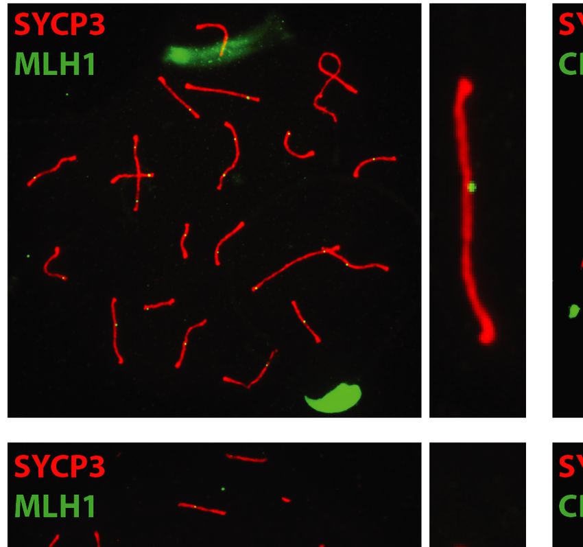
Nucleic Acids Research, 2022 5

                  A                                                E109                                        D173
                                                                                      ----
                                                                                      ----
                                                                                      ----
                                                                                      ----
                                                                                      ----
                                                                                      ----
                                                                                      ----

                  B

                                                                                                                                                                Downloaded from https://academic.oup.com/nar/advance-article/doi/10.1093/nar/gkac616/6645635 by guest on 01 August 2022
                  C
                         N-subdomain
                                             I-subdomain

                          WT                                                           D173A

Figure 1. Modeling EXO1 mutations in mice. (A) Jalview ClustalW alignment of EXO1 from the denoted species shows high evolutionary conservation of
the D173 residue. (B) Schematic structure depicting human EXO1 domains and binding partners MSH3, MSH2 and MLH1 which help mediate some of
EXO1 non-catalytic scaffolding functions. (C) Crystal structure of the N-terminal nuclease (enzymatic) domain (dark grey) of human EXO1 (left), with the
inset showing a 180◦ rotation of the active site; and D173A modeled structure (right), with the inset showing a 180◦ rotation of the active site. Active site
metals shown in blue. The RMSD of the D173A crystal structure and the pymol modeled D173A structure (based on WT) is 0.66. As a result, a modeled
D173A structure was presented to better demonstrate the active site metals (PDB: 3QEB). The insets highlights the difference between the proximity of
the metal ions (2 cyan circles) in the active site to the acidic D173 amino acid residue (green and red) compared to the neutral A173 amino acid residue
(green) mutation.

from Exo1+/+ , Exo1DA/DA and Exo1–/- mice and quantified                         nases of the PI3-family like ATM/ATR as a reaction at
chromomal breaks in metaphase spreads (Figure 2B). Com-                          DSBs (Figure 2C). This allowed us to score DNA end
pared to control Exo1+/+ , both Exo1−/− and Exo1DA/DA                            resection events during DSB repair, where both Exo1−/−
MEFs exhibited a similar increase in the percentage of cells                     and Exo1DA/DA MEFs showed significantly reduced co-
carrying chromosomal breaks, suggesting that EXO1 nu-                            localization of activated pRPA-S4/S8 and ␥ H2AX (Figure
clease is also essential for DSBR and chromosomal stabil-                        2C). These results indicate impaired DSB-resection in re-
ity. To examine whether the accumulation of these DSBs                           sponse to CPT treatment and suggest a critical role for the
responded to defects in EXO1 functions during end resec-                         catalytic activity of EXO1 during DSBR in mitotic cells.
tion to initiate DSBR, we exposed Exo1+/+ , Exo1DA/DA and                           MMR proteins also play a critical role in mediating ad-
Exo1–/- primary MEFs to camptothecin (CPT), which                                equate DDR signaling following genotoxic stress either di-
induces DSBs specifically in S-phase as a result of in-                          rectly by acting as DNA damage sensors to activate DDR
creased replication fork collapse intermediates (5). We then                     signaling networks (direct signaling model) or indirectly by
investigated the frequency and co-localization in foci of                        sequentially escalating a mutation in a ssDNA intermedi-
hyperphosphorylated RPA (S4/S8-pRPA) and ␥ H2AX,                                 ate to make a dsDNA break, which in turn triggers cell
which are targeted by DNA damage-activated protein ki-                           cycle arrest and cell death (futile cycle model) (48). To in-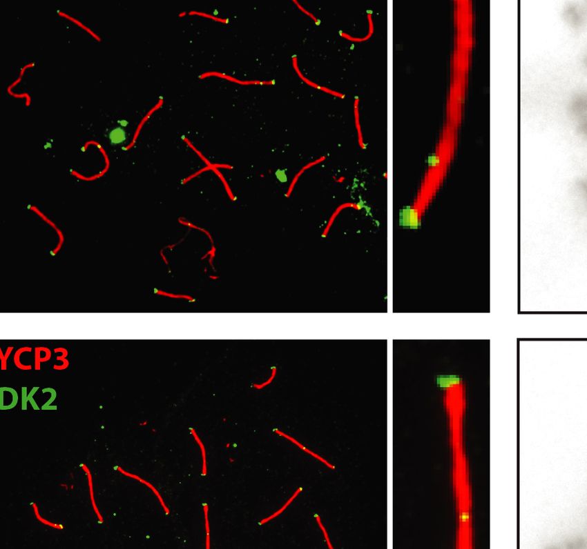
6 Nucleic Acids Research, 2022
Downloaded from https://academic.oup.com/nar/advance-article/doi/10.1093/nar/gkac616/6645635 by guest on 01 August 2022
Figure 2. In vivo nuclease activity of EXO1 is required for canonical DDR. (A) cll reporter gene mutation frequencies in spleen and liver of Exo1+/+ ,
Exo1DA/DA , and Exo1–/– mice (mice/genotype). Mutation frequency is increased by two- to threefold in EXO1−/− and EXO1DA/DA . (B) Quantification of
the number of chromomal breaks from metaphase spreads of the denoted Exo1 genotypes. At least two independent cell lines we used per genotype and
more than 100 cells where counted per cell line. Significance was established using t-test (*P < 0.01). (C) Representative photographs showing ␥ H2AX,
pS4/S8-RPA2, and DAPI stained MEFs after camptothecin treatment (2 h, 1 M) of the indicated genotypes. (Mag: 1000×; >80 cells were visualized per
genotype from three independent experiments). Histogram of the rates of ␥ H2AX-positive (DSB marker) cells with RPA2-pS4/S8 foci (ssDNA marker)
in MEFs of the indicated genotypes. ns: not significant; ***P < 0.001. (D) Cellular survival curves of MEFs from Exo1+/+ , Exo1DA/DA and Exo1–/- mice
in a p53-null background following 48h treatment with the denoted concentration of MNNG (x-axis). Error bars represent ± SD; ns: not significant;
*P-value < 0.01.
vestigate whether the EXO1D173A mutation could impair EXO1 catalytic function is important for survival and tumor
the eliciting of a G2-phase checkpoint arrest and apopto- suppression
sis in response to SN 1 DNA methylators such as MNNG DNA mismatches and DSBs are considered the most abun-
(49,50), we compared the survival of immortalized MEFs dant and harmful of all genotoxic lesions, respectively. Fail-
on a p53−/− background isolated from Exo1+/+ /p53−/− , ure to adequately repair these lesions can have severe ad-
Exo1DA/DA /p53−/− and Exo1−/− /p53−/− mice. We found verse effects on organisms, including genomic instability
that Exo1−/− and also Exo1DA/DA p53-deficient MEFs, al- and induction of tumorigenesis (51–53). To study the long-
though slightly less robustly in the Exo1DA/DA mutants, dis- term effects of EXO1 nuclease inactivation on survival and
played increased resistance to MNNG treatment compared cancer susceptibility, Exo1+/+ , Exo1−/− and Exo1DA/DA
to Exo1+/+ MEFs, suggesting inefficient DDR signaling in mouse cohorts were monitored for a period of up to 26
both EXO1 mutant backgrounds (Figure 2D). In conjunc- months. While Exo1+/+ mice exhibited the expected over-
tion with previous mechanistic studies showing the emer- all survival with 50% of mice still alive at 24 months of
gence of ssDNA and dsDNA breaks following MNNG ex- age, both Exo1−/− and Exo1DA/DA mice had a compara-
posure (30–32), these results indicate that the nuclease ac- ble decreased survival rate with 50% of mice dead at 16
tivity of EXO1 facilitates MMR-mediated DDR signaling.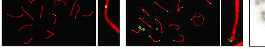
Nucleic Acids Research, 2022 7
months of age (Figure 3A). These accelerated death rates In B lymphocytes, generating and resolving DSB inter-
in Exo1−/− and Exo1DA/DA mice were due to increased can- mediates are critical to ensure diversity of antibody effec-
cer predisposition at a comparable incidence (Figure 3B). tor functions through CSR (53–56). To determine whether
The majority of Exo1DA/DA mice died of lymphoma (58%, the aforementioned lack of adequate ssDNA generation
n = 14/24) between six and 22 months of age (Figure 3C, observed in EXO1 nuclease-deficient cells (Figure 2) bears
D). In addition, three mice developed gastrointestinal ade- any physiological cellular phenotype in B cells, we con-
nomas (10%, n = 3/24) and two mice developed sarco- ducted ex vivo CSR experiments where DSBs at Ig switch
mas (8%, n = 2/24). In two mice, both lymphoma and sar- regions are obligate intermediates in the isotype recombi-
coma or lymphoma and fibroma were found. Five mice died nation process (53). To this end, B cell splenocytes were
before an autopsy could be performed (21%, n = 5/24). The isolated from Exo1+/+ (n = 8), Exo1−/− (n = 5) and
Downloaded from https://academic.oup.com/nar/advance-article/doi/10.1093/nar/gkac616/6645635 by guest on 01 August 2022
tumor spectrum on Exo1−/− was largely identical to that Exo1DA/DA (n = 8) mice and subjected to lipopolysaccha-
of Exo1DA/DA mice (Figure 3C,D). In addition, similar to ride (LPS) to induce IgM to IgG3 isotype switching or
Exo1−/− /p53−/− mice, the introduction of a p53 knockout LPS and interleukin-4 (IL-4) to induce IgM to IgG1 iso-
allele led to the rapid deaths of Exo1DA/DA /p53−/− mice be- type switching ex vivo (Figure 4C). Notably, both Exo1 mu-
tween two to three months of age due to the development tant cohorts exhibited a significant decrease in CSR effi-
of T cell lymphoma (100%, n = 17/17) ((5) and not shown). cacy towards both IgG3 and IgG1, compared to Exo1+/+
Taken together, these results indicate that an intact nuclease cohorts (Figure 4C and D), suggesting that EXO1 nucle-
activity is essential for EXO1 tumor suppressor functions. ase activity is critical for CSR. Next, to further explore
whether EXO1 nuclease was required not only to gener-
ate the switch region DSBs themselves but also to resolve
them, we cloned and sequenced S-S␥ 3 junctions from
Catalytic and structural functions of EXO1 are required for Exo1+/+ and Exo1−/− and Exo1DA/DA mutant splenic B
non-canonical DDR during antibody diverfication cells stimulated with LPS plus IL-4 for 72 h. Switch re-
To delineate the precise contribution of EXO1 structural gion DSBs are known to be primarily resolved by classical
and/or catalytic functions during SHM, we examined error- NHEJ (cNHEJ), though a significant subset is also repaired
prone repair of the VH186.2 heavy chain variable region through microhomology-mediated alternative-NHEJ (alt-
during primary immune responses in the Exo1+/+ (n = 7), EJ), where between 1 to 30 base pairs of ssDNA micro-
Exo1DA/DA (n = 8) and Exo1−/− (n = 3) cohorts (Figure homologies flanking the AID-induced DSB guide the aN-
4A and B). In the absence of either the EXO1 protein or HEJ repair process (57,58). We could observe that S-S␥ 3
its nuclease activity, a significant imbalance of mutations at junctions in Exo1DA/DA cells were different from those in
G:C and A:T pairs was revealed when frequencies of muta- Exo1+/+ or Exo1−/− cells (Figure 4E). While Exo1+/+ cells
tion were compared to those normally observed in Exo1+/+ showed the expected range of 2–20bp microhomology at
mice. While mutations at A:T pairs and error-prone Pol S–S␥ 3 junctions, Exo1−/− and Exo1DA/DA cells exhibited
hotspots were significantly reduced, the remaining muta- a slight decrease in long microhomologies. Furthermore,
tions were not only restricted but also increased at G:C Exo1DA/DA cells were further skewed towards the use of
pairs and AID hotspots in both Exo1–/– and Exo1DA/DA blunt joints, with a clear increase in the number and length
mice (Figure 4A). This pattern of nucleotide substitutions of insertions compared to the other two groups (Figure 4E,
strongly suggests that G:C mutations are due to replication inset). This resembles the phenotype previously observed
over the initial AID-induced U:G lesion without proper with the depletion of the DNA end-processing factor CtIP
EXO1-dependent resection of the surrounding area and, (59), although this notion has now been contested (60).
therefore, without the consequent induction of A:T muta- Taken together, these results appear to suggest that while
tions by the recruitment of error-prone DNA polymerases EXO1 nuclease activity contributes to the end resection
like Pol. Interestingly, the latter defect in error-prone re- mechanisms that promote microhomology-mediated aN-
pair at A:T sites was less dramatic in the presence of the mu- HEJ during CSR, a catalytically-dead protein may interfere
tant EXO1DA protein than in its complete absence (Figure with other factors that otherwise might participate and par-
4A), suggesting that some intermediate scaffolding func- tially compensate during aNHEJ in the complete absence
tions might still persist in Exo1DA/DA B cells. Consistently, of EXO1.
while a reduction in the spread of A:T mutations across the
length of the VH186.2 region, specially at Pol hotspots,
The structural function of EXO1, but not its catalytic activity,
could be observed in both EXO1-compromised models,
is critical for meiosis
Exo1–/- mice showed a more clear altered phenotype with
its few remaining A:T mutations distributed around CDR2 Like CSR, meiosis is another example of beneficial DSBs in
(Figure 4B). On the other hand, the overall distribution normal physiology. We had previously shown that Exo1−/−
and preference for mutations at G:C sites in the CDRs was mice of both sexes were sterile (5). Interestingly, Exo1DA/DA
maintained and even increased at non-hotspot G:C sites male and female mice are fertile, suggesting normal mei-
in Exo1–/– and Exo1+/+ mice (Figure 4B). Overall, analy- otic progression. To observe crossover events in pachynema,
sis of in vivo SHM at immunoglobulin genes suggests that we employed two well-characterized markers of these sites:
the structural and enzymatic functions of Exo1 work in tan- MLH1 and CDK2 (61–63), which colocalize on the paired
dem to provide adequate error-prone MMR around AID- chromosomes and the telomeres during meiotic crossing
induced U:G mismatches. over of mouse cells. In all our genotypic cohorts, MLH1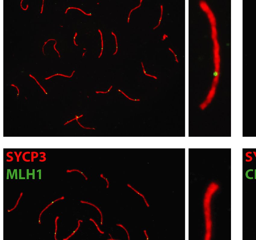
8 Nucleic Acids Research, 2022
Downloaded from https://academic.oup.com/nar/advance-article/doi/10.1093/nar/gkac616/6645635 by guest on 01 August 2022
Figure 3. The role of EXO1 nuclease-dead mutant on tumorigenesis. (A) Animal survival curve of the denoted genotypes showing faster death of
EXO1DA/DA (n = 24) and EXO1–/– (n = 15) mice compared to WT (n = 47). ***P-value < 0.0001. (B) Exo1−/− (n = 15) and EXO1DA/DA (n = 19),
which, as shown in (B), had a considerably higher tumor positive incidence compared to WT (n = 20). ***P-value < 0.001; ns: not significant. (C) repre-
sentative distribution of the tumor profiles of the samples quantified in (B). (D) representative H&E stains of the tumor samples denoted in (C). Bar is 50
m.
and CDK2 localized to crossover sites and CDK2 addi- DISCUSSION
tionally localized to telomeres (Figure 5). These crossover
As exemplified in this study, EXO1 fulfills many complex
markers remained associated with SYCP3, chromosome
even contradictory functions ranging from maintaining ge-
cores, and/or telomeres at a frequency and intensity that
nomic stability to promoting immune diversity as well as
is indistinguishable between Exo1+/+ and Exo1DA/+ and
meiotic recombination. We specifically were interested in
Exo1DA/DA mice (Figure 5), indicating normal meiosis pro-
dissecting the distinct contributions of the catalytic and
gression through prophase-I. In addition, by analyzing and
scaffolding functions of EXO1 in each of these genome
quantifying metaphase spreads, we could show that the
maintenance processes in vivo by comparing the phenotypes
number of chiasmata and bivalent chromosomes was also
of Exo1+/+ , Exo1DA/DA and Exo1–/– mice. The N-terminal
comparable in all groups (Figure 5). This is in contrast to
fragment of EXO1 encodes the nuclease domain bearing
our previous findings in Exo1−/− mice in which metaphase
the HNPCC-associated E109K mutation, which apparently
spreads predominantly displayed abnormal spindle struc-
compromised EXO1 nuclease activity in vitro when as-
tures with mostly achiasmatic univalent chromosomes (5).
sayed using tagged N-terminal fragments of both mouse
Altogether, these results suggest that EXO1 nuclease activ-
and human EXO1 (5,29). However, recent studies in which
ity, but not its structural function, is dispensable during mei-
the authors were finally able to isolate the full-length hu-
otic progression.
man EXO1E109K , without using any N-terminal tag, showed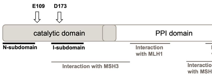
Nucleic Acids Research, 2022 9
A C Exo1-/-
Exo1+/+ Exo1DA/DA Exo1-/- Exo1+/+ Exo1DA/DA
(N=7) (N=8) (N=3) 7.76% 0.39% 0.63%
Total reads 1.47M 1.34M 0.59M
CDR3 clones 1080 681 497
IgG3
Mutations / clone 4.5 6.0 5.5
****
**** 0.4887 15
7.5
20.1% 7.21% 6.98%
Downloaded from https://academic.oup.com/nar/advance-article/doi/10.1093/nar/gkac616/6645635 by guest on 01 August 2022
G:C mutations 6.9 10.9 10.7
Mutation frequency (x10-3)
0
****
**** 0.0568 20
IgG1
AID hotspots 10
WRC/GYW 9.0 16.5 17.4 0
**** IgM
**** 4
****
2
D
175
A:T mutations LPS IL4+LPS
3.1 2.1 1.1
Relative switching efficiency (%)
0
**** 150
**** 5
**** **** IgG3
****
Polη hotspots 2.5
125 IgG1
WA/TW 4.7 2.1 0.9 0
100
75
ns ns
B
50
G:C mutations A:T mutations
non-hotspots AID hotspots non-hotspots Polη hotspots 25
CDR1 CDR2 CDR1 CDR2 CDR1 CDR2 CDR1 CDR2
1.00
0.75
0
+/
+
/D
A -/- +/
+
/D
A -/-
1 1
Exo1+/+
o 1 DA o o 1 DA o
Ex Ex
0.50
Ex o1 Ex o1
0.25 Ex Ex
0.00
Normalized mutation counts
1.00
E 0.4
insertions blunt microhomology (mh)
0.3
Frequency
0.75
Exo1DA/DA
0.2
short mh long mh
0.50
0.1
0.25 0.0
D /+
A
-/-
D
Exo1+/+
+
A/
0.00
1.00
0.75
Exo1DA/DA
Exo1-/-
0.50
0.25
Exo1-/-
0.00
30 60 90 120 150 180 210 240 270 300 30 60 90 120 150 180 210 240 270 300 30 60 90 120 150 180 210 240 270 300 30 60 90 120 150 180 210 240 270 300 -25 -20 -15 -10 -5 0 5 10 15 20 25
Nucleotide positions across VH186.2 region Sμ-S 3 junction length (bp)
Figure 4. Abrogation of EXO1 or its nuclease activity impairs antibody diversification. (A) Global analysis of in vivo SHM at the VH186.2 region during
primary response of NP-immunized mice. For comparison of average mutation frequencies of Exo1−/− and Exo1DA/DA to WT, contingency tables were
assigned to the mutation data and Pearson’s Chi-squared test with Yates’ continuity correction was applied. ****P-value < 0.0001. (B) Distribution of
unique mutations across the VH186.2 region and its CDRs as observed during the 1000 iterations that were performed for each genotype group and
normalized within each mutation subtype to maximum of 1. Mutations in G:C or A:T residues are split and are colored according to the hotspot definition
for AID (WRC/GYW) or poln (WA/TW). (C) Representative flow cytometry profiles analyzed by FloJo showing CSR efficiency in the three denoted
mouse cohorts. (D) Summary of ex vivo relative CSR efficiencies in WT (n = 8), Exo1DA/DA (n = 8) and Exo1−/− (n = 5) mice. As reference, mean
efficiency of switching in the WT group to IgG3 after stimulation with LPS or to IgG1 after stimulation with IL4 + LPS were defined as 100% in two
independent experiments. Relative percentages of switching to each isotype by animal and stimulation cocktail are depicted by dots, and bars represent
mean ± SD of the replicates. The background switching to IgG1 after LPS stimulation and to IgG3 after IL4 + LPS stimulation are also shown. ****P-
value < 0.0001; ns: not significant. (E) Distribution of blunt breakpoints (0 bp), microhomologies (from 1 to >10 bp in length) or insertions (represented
as negative integers) in S–S␥ 3 switch junctions cloned and sequenced in ex vivo LPS-stimulated splenic B cells from the different genotype groups (n > 22
per group). Dots represent individual S–S␥ 3 switch junctions, and red bars denote median length in the distribution of blunt junctions, microhomologies
and insertions within each group. The nonparametric Kruskal-Wallis test followed by the Dunn’s post-tests were used to explore these distributions. Long
microhomologies (>10 bp in length) in the wild-type group are highlighted in red, and increased frequency of insertions (as explored by Fisher’s exact
tests) in the Exo1DA/DA group is highlighted as an inset plot. ***P-value < 0.001.10 Nucleic Acids Research, 2022
A
Bivalents
+/+
Exo1
Downloaded from https://academic.oup.com/nar/advance-article/doi/10.1093/nar/gkac616/6645635 by guest on 01 August 2022
Bivalents
+/DA
Exo1
Bivalents
DA/DA
Exo1
B
30 25 25
Bivalents per cell
MLH1 foci per cell
CDK2 foci per cell
20 20
20
15 15
10 10
10
5 5
0 0 0
+/+ +/DA DA/DA +/+ +/DA DA/DA +/+ +/DA DA/DA
Exo1 Exo1 Exo1 Exo1 Exo1 Exo1 Exo1 Exo1 Exo1
Figure 5. EXO1 nuclease activity is dispensable for meiosis during spermatogenesis. (A) MLH1 and CDK2 (green) localize to the synaptonemal complex
SYCP3 (red) in WT (top), Exo1DA/+ (middle), and Exo1DA/DA (bottom) in pachytene spermatocytes. Left-hand panels show merged red and green channels,
while the right-hand inset shows representative merged channels of a sample chromosome showing MLH1 and CDK2 at crossover sites and additional
telomere-associated CDK2. Most right panels show the quantification of bivalent chromosome pairing during pachytene. (B) charts show the quantitation
of foci for each genotype at pachynema. Counts for WT, Exo1DA/+ and Exo1DA/DA are not statistically different from each other (by unpaired t-test with
Welch’s correction). The number of cells counted is depicted in grey (at least 45 cells per genotype). Values given are the number of foci per nucleus ± S.D.Nucleic Acids Research, 2022 11
retention of significant nuclease activity (30,31). A pos- other repair mechanisms such as post-replication repair but
sible explanation for these conflicting observations could we focus on the former two in this study (66,67). SHM and
be related to structural instability in the EXO1E109K mu- CSR are two immune diversification mechanisms in B cells
tant N-terminal fragment, together with the presence of that specifically require non-canonical error-prone MMR
a 6xHis-tag used for purification, which could both inter- and DSBR, respectively (68,69). What was unexpected,
fere with EXO1 enzymatic activity. Consistent with these though, is that during these two error-prone DNA repair
observations, canonical and non-canonical MMR (SHM processes, EXO1 seems to depend on both its catalytic and
and CSR in B cells) were not affected in Exo1EK/EK mice. non-catalytic scaffolding functions (tier-2). EXO1 seems to
Yet, Exo1EK/EK mice showed increased cancer predisposi- initiate both SHM and CSR processes through its nuclease
tion and displayed defective DDR and DSBR (5). This un- activity in the first instance. That is why our SHM analysis
Downloaded from https://academic.oup.com/nar/advance-article/doi/10.1093/nar/gkac616/6645635 by guest on 01 August 2022
expected limited phenotype might be related to the recently revealed a general increase in replication-fixed unrepaired
indentified overlap of the EXO1 nuclease domain with a C:G mutations and a decrease in error-prone introduced
PAR-interaction (PIN) domain, which has been proposed A:T mutations in both Exo1DA/DA and Exo1–/– models,
to facilitate recruitment and resection functions of EXO1 which is in line with other MMR factor deletions, includ-
during DSBR (64). Here, we bypassed those issues by cre- ing MSH2 or MSH6 depletion (68,69). Likewise, the overall
ating and characterizing a novel Exo1DA/DA knock-in mouse decrease of IgM to IgG1 and IgG3 switching was compa-
model carrying the nuclease-dead EXO1-D173A mutation rable in the two mutant Exo1 mouse models which in turn
(33,34) that represented an optimal in vivo system allowing are comparable to canonical MMR factors MSH2/MSH6
us to more precisely dissect potential structural and cat- double deletion (69), suggesting that the overall instigation
alytic functions of EXO1 in different mammalian genome of DSBs at switch junctions and their resolution by cN-
maintenance processes. Although a miniscule residual nu- HEJ highly depend on EXO1 nuclease activity. However,
clease activity was observed in purified human EXO1-DA upon further analysis of SHM mutation patterns, we could
in vitro (28,29), this does not seem to translate into in vivo observe that while the increase in G:C mutations fixed by
function in either yeast or humans (33–35). replication at AID-associated hotspot mutations were com-
Our results suggest that EXO1 may have a 3-tier func- parable between Exo1DA/DA and Exo1−/− , the recruitment
tion in the various DDR processes in which it participates. of error-prone mechanisms and Pol-mediated mutations at
This 3-tier approach depends on differential requirements A:T sites where only intermediary impaired in Exo1DA/DA
for the catalytic vs. scaffolding domains of EXO1: cat- compared to Exo1–/– B cells. These data suggest that dur-
alytic, scaffolding, or both. They also seem to suggest that ing complex error-prone MMR repair, additional scaffold-
the complexity of the DDR mechanism somehow directs ing functions of EXO1 are also needed. The rationale for
what activities of EXO1 are needed. In addition to the dif- how that could happen include: i) a passive reason, where
ferential role of EXO1 nuclease domain in various DDR nuclease-dead EXO1 could be recruited to sites of DNA
pathways, recent yeast studies have highlighted the impor- mismatches, but its lack of nuclease activity could act as
tant of EXO1’s unstructured C terminal domain for critical a dominant negative, thereby preventing subsequent down-
protein-protein interactions such as the binding with MSH2 stream MMR signaling. However, this rationale should also
and MLH1 (17,18,65). These studies also highlight the po- apply for canonical MMR as well, which we did not observe
tential of EXO1 independent resection via either Rad27 or in our assays. Another explanation could be the result of ii)
MLH1-PMS2 during MMR (17,18). Our poposed rationale an active scaffolding function of EXO1, which diversifies
for EXO1’s 3-tier function is in agreement with these studies its function. In this sense, PCNA was shown to bind and
and provide what is arguably the first demonstration of the increase EXO1 processivity at DSB sites (17,18,70,71), and
biological importance of EXO1 scaffold function. PCNA is required for A:T mutagenesis during error-prone
In tier-1, we show here that the role of EXO1 in canon- MMR through its recruitment of Pol (21). It is plausible
ical DDR pathways, such as error-free MMR and DSBR, that PCNA might still be recruited by the retained scaffold-
are primarily mediated by EXO1 nuclease activity. In our ing functions of the mutant EXO1DA protein and result in
assay measuring the MMR of spontaneous genomic mu- partial functionality, which would not otherwise occur in
tations, mutation frequencies were similar between EXO1 the complete absence of EXO1. Hence, such a scaffolding
depleted (Exo1−/− ) and EXO1 nuclease-dead (Exo1DA/DA ) function might contribute to the intermediate phenotype we
mouse models. Similarly, DSBR analyses through DNA end observed in Exo1DA/DA mice during the error-prone MMR
resection assays showed indistinguishable responses in these phase that introduces A:T mutations during SHM in B cells.
same mouse models. Furthermore, Exo1DA/DA and Exo1–/- Further supporting tier-2 in B cells, we also observed
mice exhibited similarly reduced survival due to the devel- independent contributions from the nuclease and scaf-
opment of lymphomas, sarcomas, and gastrointestinal ade- folding functions of EXO1 during CSR. Two variants of
nocarcinomas. These data suggest that error-free DNA re- error-prone DSBR, namely cNHEJ and microhomology-
pair through MMR and DSBR requires EXO1 resection mediated alt-EJ, are known to contribute during CSR,
activity and little or no distinct effects from its scaffold- which can be roughly dissected by studying antibody S-
ing function. Or that these two activities are completely S junctions (53). Our data highlight that Exo1DA/DA mice
epistatic or redundant in these particular repair mecha- show a clear preference for blunt-end NHEJ with an av-
nisms. erage of 0 bp S–S overlap compared to Exo1−/− B cells
That EXO1 is involved in two dichotomous DDR path- that show about 2–3 bp of S–S microhomology overlap.
ways, namely error-free and error-prone MMR and DSBR, Exo1DA/DA cells also have a decreased propensity for long
has been well documented. EXO1 can also be involved in microhomologies at the S–S junctions and at the same time12 Nucleic Acids Research, 2022
an increased propensity for insertions. This again could be CA248536 to W.E.]; UZH-URPP, BBSRC
explained by a (i) passive dominant-negative effect of the [BB/N017773/2]; SNF [CRSK-3 190550]; Vontobel [41309
Exo1DA/DA protein manifesting longer than usual residency to R.C.]; Agencia Estatal de Investigación [PID2020-
period on DSB sites. But it could also be argued that (ii) in 112994RB-I00/AEI/10.13039/501100011033]; Ministerio
complex scenarios such as during the competition between de Economı́a, Industria y Competitividad [SAF2017-
NHEJ and alt-EJ at S–S junctions, EXO1 can fulfill a more 82309-R/MINECO/AEI/FEDER, EU to S.R.]; M.D.S.
complex role than the basic nuclease activity primarily re- is supported by the Harry Eagle Chair, provided by the
quired for HR. This complexity could be mostly attributed National Women’s Division of the Albert Einstein Col-
to high selection for NHEJ and alt-EJ at S-S junctions lege of Medicine; S.R. was supported by Ministerio de
where EXO1 is not expected to fulfill a catalytic role but Economı́a y Competitividad through Programa Ramón
Downloaded from https://academic.oup.com/nar/advance-article/doi/10.1093/nar/gkac616/6645635 by guest on 01 August 2022
rather a scaffolding one. The recruitment of EXO1 by CtIP y Cajal [RYC-2014-16399/MEC]; R.C. was supported by
or DNA2 nucleases which are shown to bind, recruit, and AMS [SBF001\1005]; R.B.M. was supported by EPSRC
synergize with EXO1 nuclease activity, might contribute to [EP/M506527/1]. Funding for open access charge: NIH;
this S–S junction process through an EXO1 scaffolding role. SNF; BBSRC.
Finally, our data show that while EXO1 catalytic activ- Conflict of interest statement. SR receives research support
ity is completely dispensible for apparently normal mei- from Roche/Genentech (imCORE) and Gilead.
otic progression, it is critically dependent on the presence
of EXO1 protein. This tier-3 scaffolding EXO1 function
is surprising because meiosis is considered a specialized REFERENCES
and unique form of a hybrid between error-prone HR and 1. Szankasi,P. and Smith,G.R. (1995) A role for exonuclease I from S.
MMR which contributes to organismal diversity (72,73). pombe in mutation avoidance and mismatch correction. Science, 267,
It is not clear why EXO1 nuclease activity is not required 1166–1169.
2. Szankasi,P. and Smith,G.R. (1992) A single-stranded DNA
in meiosis, but it is consistent with budding yeast studies, exonuclease from Schizosaccharomyces pombe. Biochemistry, 31,
which confirm our findings and further suggest that the 6769–6773.
nickase activity of the MLH1–MLH3 complex is respon- 3. Käslin,E. and Heyer,W.D. (1994) A multifunctional exonuclease from
sible for crossover formation; albeit dependent on EXO1 vegetative schizosaccharomyces pombe cells exhibiting in vitro strand
protein but not on EXO1 nuclease activity (34). This could exchange activity. J. Biol. Chem., 269, 14094–14102.
4. Bardwell,P.D., Woo,C.J., Wei,K., Li,Z., Martin,A., Sack,S.Z.,
explain why both sexes of Exo1−/− mice are sterile (74). Parris,T., Edelmann,W. and Scharff,M.D. (2004) Altered somatic
Furthermore, recent studies in Exo1DA/DA mice showed that hypermutation and reduced class-switch recombination in
EXO1 is not an important contributor to 5 to 3 end resec- exonuclease 1-mutant mice. Nat. Immunol., 5, 224–229.
tion in murine meiosis or is substantially redundant with 5. Schaetzlein,S., Chahwan,R., Avdievich,E., Roa,S., Wei,K., Eoff,R.L.,
Sellers,R.S., Clark,A.B., Kunkelf,T.A., Scharff,M.D. et al. (2013)
other resection activities (74). In fact, recent studies suggest Mammalian Exo1 encodes both structural and catalytic functions
that EXO1 facilitates the MLH1-MLH3 nuclease activity in that play distinct roles in essential biological processes. Proc. Natl.
meiosis (33,75) and EXO1 protein but not its nuclease activ- Acad. Sci. U.S.A., 110, E2470–E2479.
ity might mediate the DNA nicks protection during meiotic 6. Lee,B.I. and Wilson,D.M. (1999) The RAD2 domain of human
crossover (76). exonuclease 1 exhibits 5 to 3 exonuclease and flap structure-specific
endonuclease activities. J. Biol. Chem., 274, 37763–37769.
In conclusion, we show that EXO1 fulfills 3-tier activity 7. Qiu,J., Qian,Y., Chen,V., Guan,M.X. and Shen,B. (1999) Human
in genome maintenance depending on the process it is in- exonuclease 1 functionally complements its yeast homologues in
volved in and the complexity of that DNA repair process. DNA recombination, RNA primer removal, and mutation avoidance.
One tier is predominantly dependent on the nuclease func- J. Biol. Chem., 274, 17893–17900.
8. Orans,J., McSweeney,E.A., Iyer,R.R., Hast,M.A., Hellinga,H.W.,
tion of EXO1 such as canonical MMR and DSBR. Another Modrich,P. and Beese,L.S. (2011) Structures of human exonuclease 1
tier depends primarily on the scaffolding function, such as DNA complexes suggest a unified mechanism for nuclease family.
meiosis, and a final tier depends on the concerted act of nu- Cell, 145, 212–223.
clease and scaffolding functions during non-canonical re- 9. Sertic,S., Quadri,R., Lazzaro,F. and Muzi-Falconi,M. (2020) EXO1:
pair processes in B cells for antibody diversification. a tightly regulated nuclease. DNA Repair (Amst)., 93, 102929.
10. Mimitou,E.P. and Symington,L.S. (2009) DNA end resection: many
nucleases make light work. DNA Repair (Amst)., 8, 983–995.
11. Keelagher,R.E., Cotton,V.E., Goldman,A.S.H. and Borts,R.H.
DATA AVAILABILITY (2011) Separable roles for Exonuclease I in meiotic DNA
Sequencing data are available through GEO acces- double-strand break repair. DNA Repair (Amst)., 10, 126–137.
12. Tishkoff,D.X., Boerger,A.L., Bertrand,P., Filosi,N., Gaida,G.M.,
sion GSE183344. Mutation analysis scrpits are on Kane,M.F. and Kolodner,R.D. (1997) Identification and
Github: https://gitlab.com/maccarthyslab/SHMPrep and characterization of Saccharomyces cerevisiae EXO1, a gene encoding
https://gitlab.com/maccarthyslab/SHMServer. an exonuclease that interacts with MSH2. Proc. Natl. Acad. Sci.
U.S.A., 94, 7487–7492.
13. Tran,P.T., Simon,J.A. and Liskay,R.M. (2001) Interactions of Exo1p
SUPPLEMENTARY DATA with components of MutL␣ in Saccharomyces cerevisiae. Proc. Natl.
Acad. Sci. U.S.A., 98, 9760–9765.
Supplementary Data are available at NAR Online. 14. Tishkoff,D.X., Amin,N.S., Viars,C.S., Arden,K.C. and
Kolodner,R.D. (1998) Identification of a human gene encoding a
homologue of Saccharomyces cerevisiae EXO1, an exonuclease
FUNDING implicated in mismatch repair and recombination. Cancer Res., 58,
5027–5031.
NIH [CA72649, CA102705 to M.D.S., 1R01AI132507- 15. Schmutte,C., Marinescu,R.C., Sadoff,M.M., Guerrette,S.,
01A1 to M.D.S. and T.M., CA76329, CA222358, Overhauser,J. and Fishel,R. (1998) Human exonuclease I interactsYou can also read


