RNA-Targeting Splicing Modifiers: Drug Development and Screening Assays - MDPI

Page created by Frederick Hodges
 
CONTINUE READING
RNA-Targeting Splicing Modifiers: Drug Development and Screening Assays - MDPI
molecules
Review
RNA-Targeting Splicing Modifiers: Drug Development and
Screening Assays
Zhichao Tang, Junxing Zhao                  , Zach J. Pearson, Zarko V. Boskovic             and Jingxin Wang *

                                          Department of Medicinal Chemistry, University of Kansas, Lawrence, KS 66047, USA;
                                          zhichao.tang@ku.edu (Z.T.); zhao.junxing@ku.edu (J.Z.); zjp@ku.edu (Z.J.P.); zarko@ku.edu (Z.V.B.)
                                          * Correspondence: wang.jingxin@ku.edu

                                          Abstract: RNA splicing is an essential step in producing mature messenger RNA (mRNA) and
                                          other RNA species. Harnessing RNA splicing modifiers as a new pharmacological modality is
                                          promising for the treatment of diseases caused by aberrant splicing. This drug modality can be used
                                          for infectious diseases by disrupting the splicing of essential pathogenic genes. Several antisense
                                          oligonucleotide splicing modifiers were approved by the U.S. Food and Drug Administration (FDA)
                                          for the treatment of spinal muscular atrophy (SMA) and Duchenne muscular dystrophy (DMD).
                                          Recently, a small-molecule splicing modifier, risdiplam, was also approved for the treatment of SMA,
                                          highlighting small molecules as important warheads in the arsenal for regulating RNA splicing.
                                          The cellular targets of these approved drugs are all mRNA precursors (pre-mRNAs) in human
                                          cells. The development of novel RNA-targeting splicing modifiers can not only expand the scope
                                of drug targets to include many previously considered “undruggable” genes but also enrich the
         
                                          chemical-genetic toolbox for basic biomedical research. In this review, we summarized known
Citation: Tang, Z.; Zhao, J.; Pearson,    splicing modifiers, screening methods for novel splicing modifiers, and the chemical space occupied
Z.J.; Boskovic, Z.V.; Wang, J.            by the small-molecule splicing modifiers.
RNA-Targeting Splicing Modifiers:
Drug Development and Screening
                                          Keywords: alternative splicing; high-throughput screening; antisense oligonucleotide; small molecule;
Assays. Molecules 2021, 26, 2263.
                                          splicing modifier; RNA-targeting
https://doi.org/10.3390/
molecules26082263

Academic Editors:
Maria Chatzopoulou, Angela Russell
                                          1. Introduction
and Simona Rapposelli                          The splicing of messenger RNA precursors (pre-mRNAs) by the spliceosome is an
                                          essential processing step occurring before the translation of most nuclear encoded eu-
Received: 26 February 2021                karyote genes. Some genes encoded in eukaryotic organelles (e.g., mitochondria [1])
Accepted: 9 April 2021                    and in prokaryotic cells [2] must undergo spliceosome-independent RNA splicing before
Published: 14 April 2021                  translation. In the past two decades, gene- or exon-specific splicing modifiers have been
                                          developed for the treatment of several human disease states, including spinal muscular
Publisher’s Note: MDPI stays neutral      atrophy (SMA) [3,4], Duchenne muscular dystrophy (DMD) [5–7], and influenza virus
with regard to jurisdictional claims in   infection [8], as well as pre-clinical drug development for diseases such as frontotemporal
published maps and institutional affil-   dementia and parkinsonism linked to chromosome 17 (FTDP-17) [9], yeast infection [10],
iations.                                  and familial dysautonomia [11]. Instead of a traditional protein-targeting drug modality,
                                          these compounds act through direct binding to the pre-mRNAs.

                                          1.1. Chemistry in RNA Splicing Reactions
Copyright: © 2021 by the authors.              There are two general types of splicing reactions to process messenger RNAs (mRNAs)
Licensee MDPI, Basel, Switzerland.        —the distinction being made based on the mechanisms of breaking the phosphate group in
This article is an open access article    the 50 splice sites (i.e., the exon/intron junction at the 50 of the intron to be spliced). Both
distributed under the terms and           mechanisms start with a nucleophilic attack, but what distinguishes them is the identity
conditions of the Creative Commons
                                          of the nucleophiles. These can either be (a) an adenosine in the intron, namely the branch
Attribution (CC BY) license (https://
                                          point, or (b) an exogenous guanosine cofactor that non-covalently binds to the intron
creativecommons.org/licenses/by/
                                          aptamer [12] (Figure 1). The first type of splicing reaction occurs in the maturation of all the
4.0/).

Molecules 2021, 26, 2263. https://doi.org/10.3390/molecules26082263                                        https://www.mdpi.com/journal/molecules
RNA-Targeting Splicing Modifiers: Drug Development and Screening Assays - MDPI
reviews) and a family of intron sequences, namely group II introns, in bacteria, plants,
                                and yeast [2]. This type of splicing reaction removes the intron as a cyclized fragment,
                                intron lariat, in two steps (Figure 1a). First, the 2′-OH of the branching adenosine initiates
                                nucleophilic attacks on the 5′ splice site and frees the 5′ exon (exon 1). Second, the 3′-OH
Molecules 2021, 26, 2263        of the 5′ exon attacks the 3′ phosphate group of the intron (3′ splice site) to rejoin the2 two                    of 27
                                exons with a simultaneous release of the intron lariat (Figure 1a). In animal cells, the intron
                                lariats usually do not encode proteins and are commonly linearized and destroyed within
                                minutes     [15]. However,
                                nuclear encoded          mRNAs   some  of the intron lariats
                                                                   by spliceosomes        (see refcan  remain
                                                                                                    [13,14]   for cyclic
                                                                                                                   recentand    be exported
                                                                                                                            reviews)            to the
                                                                                                                                       and a family
                                cytoplasm       to  regulate    cellular  functions      [15].  These    cytosolic     stable
                                of intron sequences, namely group II introns, in bacteria, plants, and yeast [2]. This type     intron   lariats   are,
                                therefore,    a  source    of circular RNAs      (circRNAs),     whose     regulatory     role
                                of splicing reaction removes the intron as a cyclized fragment, intron lariat, in two steps     is not fully  under-
                                stood
                                (Figure  [16].
                                           1a). First, the 20 -OH of the branching adenosine initiates nucleophilic attacks on
                                      The
                                       0
                                the 5 splice second      typefrees
                                                  site and     of splicing   involves
                                                                   the 50 exon    (exon a1).family
                                                                                               Second,of the
                                                                                                          intron    sequences:
                                                                                                               30 -OH              groupattacks
                                                                                                                        of the 50 exon      I introns
                                                                                                                                                    the
                                  0
                                [12].  In this type
                                3 phosphate        group of splicing
                                                            of the intron     0
                                                                      reaction,    a segment
                                                                           (3 splice    site) to of  the the
                                                                                                  rejoin  intron
                                                                                                               twofolds
                                                                                                                      exonsand   forms
                                                                                                                              with        a binding-
                                                                                                                                    a simultaneous
                                pocket
                                releasefor     exogenous
                                           of the              guanosine
                                                     intron lariat   (Figure(Figure
                                                                                1a). In1b)animal
                                                                                            [12]. The   3′-OH
                                                                                                    cells,         of the G
                                                                                                            the intron        then usually
                                                                                                                           lariats  serves asdo  a nu-
                                                                                                                                                   not
                                cleophile    and attacks
                                encode proteins        and arethecommonly
                                                                  5′ splice site.  After the
                                                                               linearized    and5′ destroyed
                                                                                                   splice site within
                                                                                                                 breaksminutes
                                                                                                                           and is guanylated,
                                                                                                                                     [15]. However, the
                                nucleophilic       G  is  displaced   by a  G   at the  3′  splice  site in  the   binding
                                some of the intron lariats can remain cyclic and be exported to the cytoplasm to regulate      pocket.  The   free   3′-
                                OH    of  the  5′  exon    subsequently    attacks    the   3′ splice  site  similarly    to  finalize
                                cellular functions [15]. These cytosolic stable intron lariats are, therefore, a source of circular     the  splicing
                                reaction.    In group Iwhose
                                RNAs (circRNAs),            intron regulatory
                                                                    splicing, the    intron
                                                                                  role  is notproduct    remains linear
                                                                                                fully understood        [16]. (Figure 1b).

      Figure 1. Splicing reaction with (a) adenosine as the branch point, which occurs in spliceosome-dependent
                                                                                          spliceosome-dependent splicing and
      group II introns, and (b) exogenous guanosine binding in the group
                                                                    group II introns.
                                                                             introns.

                                      The
                                      In   second type
                                         eukaryotes,      of splicing
                                                        most            involves a family
                                                               of the pre-mRNAs              of intron
                                                                                       are spliced   by sequences:
                                                                                                         spliceosomes.group
                                                                                                                          ForI this
                                                                                                                               introns [12].
                                                                                                                                    reason,
                                In this
                                we  cantype
                                         alsoof  splicing
                                              group    thereaction,
                                                           splicing areactions
                                                                        segment into
                                                                                   of thetwo
                                                                                          intron  folds
                                                                                              major      and formsnamely
                                                                                                      categories,    a binding-pocket
                                                                                                                            spliceosome-for
                                exogenous and
                                dependent    guanosine    (Figure 1b)
                                                   -independent          [12]. The
                                                                     splicing.      30 -OH
                                                                                These   two of the G
                                                                                            types   of then  serves
                                                                                                        splicing    as a nucleophile
                                                                                                                 distribute            and
                                                                                                                             distinctively
                                attacks       0 splice site. After the 50 splice site breaks and is guanylated, the nucleophilic G
                                         the 5kingdoms
                                in different               of life. Spliceosome, as alluded to already, only exists in eukaryotic
                                is displaced   by a G atinthe    0 splice site in the binding pocket. The free 30 -OH of the 50 exon
                                cells and is enclosed       the3 cell  nucleus. On the other hand, the spliceosome-independent
                                subsequently                    0
                                group   I and IIattacks
                                                  intronstheare3 never
                                                                   spliceobserved
                                                                          site similarly  to finalize
                                                                                     in animal   cellsthebutsplicing
                                                                                                             they arereaction.
                                                                                                                        broadlyInfound
                                                                                                                                    groupinI
                                intron splicing, the intron product remains linear (Figure 1b).
                                      In eukaryotes, most of the pre-mRNAs are spliced by spliceosomes. For this reason,
                                we can also group the splicing reactions into two major categories, namely spliceosome-
                                dependent and -independent splicing. These two types of splicing distribute distinctively in
                                different kingdoms of life. Spliceosome, as alluded to already, only exists in eukaryotic cells
                                and is enclosed in the cell nucleus. On the other hand, the spliceosome-independent group
                                I and II introns are never observed in animal cells but they are broadly found in prokaryotic
                                cells. Group I and II introns also exist in eukaryotic organelles, such as plant chloroplasts
RNA-Targeting Splicing Modifiers: Drug Development and Screening Assays - MDPI
Molecules 2021, 26, x FOR PEER REVIEW                                                                                                               3 of 28

Molecules 2021, 26, 2263                                                                                                                             3 of 27
                                    prokaryotic cells. Group I and II introns also exist in eukaryotic organelles, such as plant
                                    chloroplasts and yeast mitochondria. Other than messenger RNA (mRNA), transfer RNA
                                    (tRNA) [17] and ribosomal RNA (rRNA) [12] can also be spliced in a spliceosome-inde-
                                    and yeast mitochondria. Other than messenger RNA (mRNA), transfer RNA (tRNA) [17]
                                    pendent manner.
                                    and ribosomal RNA (rRNA) [12] can also be spliced in a spliceosome-independent manner.
                                    1.2.
                                    1.2. Spliceosome-Dependent
                                          Spliceosome-Dependent Alternative
                                                                        Alternative Splicing
                                                                                      Splicing
                                          For
                                           For nuclear
                                                nuclear encoded
                                                           encoded eukaryote
                                                                        eukaryote genes,
                                                                                     genes, aa single
                                                                                               single pre-mRNA          sequence may
                                                                                                        pre-mRNA sequence             may have
                                                                                                                                             have more
                                                                                                                                                    more
                                    than   one    splice   pattern,   leading   to  different   protein    isoforms     from   the
                                    than one splice pattern, leading to different protein isoforms from the same gene. The mech-      same    gene.   The
                                    mechanism        that  generates    these  splice  variants   is termed     alternative
                                    anism that generates these splice variants is termed alternative splicing. The alternativesplicing.    The  alterna-
                                    tive  splicing
                                    splicing          of a particular
                                                of a particular           pre-mRNA
                                                                   pre-mRNA             may happen
                                                                                  may happen              in a single
                                                                                                   in a single    normal normal     cell, different
                                                                                                                            cell, different   tissuestis-
                                                                                                                                                        or
                                    sues   or  development        stages,  and   some   disease    states  (e.g.,  when
                                    development stages, and some disease states (e.g., when a gene mutation is present).   a gene    mutation     is pre-
                                                                                                                                                      It is
                                    sent).  It is estimated
                                    estimated     that 90–95%  thatof90–95    % of the
                                                                      the human         human pre-mRNAs
                                                                                    pre-mRNAs        can undergo   can  undergo alternative
                                                                                                                      alternative    splicing [18] splic-
                                                                                                                                                      (for
                                    ing  [18] (for
                                    reviews,     see reviews,    see ref [19–21]).
                                                      ref [19–21]).
                                          The
                                           The process
                                                 process forfor choosing
                                                                choosing the the spliced
                                                                                 spliced sites
                                                                                           sites is
                                                                                                 is highly
                                                                                                     highly regulated
                                                                                                               regulated and
                                                                                                                           and is is considered
                                                                                                                                     considered to  to be
                                                                                                                                                        be
                                    more complicated
                                            complicated       than   the  recognition    of the  primary      intron   sequences.      Apart   from    the
                                    terminal and and branch
                                                       branchpoint
                                                                 pointsequences
                                                                         sequenceswithin
                                                                                       withinthethe   introns,
                                                                                                   introns,       other
                                                                                                               other     short
                                                                                                                     short   RNA RNA      sequences
                                                                                                                                     sequences          in
                                                                                                                                                   in the
                                    the  pre-mRNA
                                    pre-mRNA              are also
                                                     are also  crucialcrucial  in determining
                                                                         in determining             the splice
                                                                                             the splice           sitespatterns.
                                                                                                           sites and     and patterns.      In general,
                                                                                                                                     In general,    there
                                    there
                                    are fourareclasses
                                                 four classes
                                                          of suchof such
                                                                     shortshort   sequences,
                                                                            sequences,          namely
                                                                                           namely     exonicexonic   splicing
                                                                                                                splicing        enhancer
                                                                                                                           enhancer           (ESE),
                                                                                                                                         (ESE),  exonicex-
                                    onic  splicing
                                    splicing          silencer
                                                silencer   (ESS),(ESS),  intronic
                                                                   intronic         splicing
                                                                              splicing        enhancer
                                                                                        enhancer     (ISE),(ISE),   and intronic
                                                                                                             and intronic     splicingsplicing  silencer
                                                                                                                                          silencer   (ISS)
                                    (ISS)
                                    (Figure(Figure     2). These
                                               2). These     RNA RNA        sequences
                                                                     sequences            arereferred
                                                                                   are also    also referred      to as cis-acting
                                                                                                         to as cis-acting     regulatoryregulatory
                                                                                                                                              sequence se-
                                    quence
                                    elements.  elements.     The cis-acting
                                                   The cis-acting      elementselements    exertregulatory
                                                                                   exert their    their regulatory
                                                                                                                 effect effect  of splicing
                                                                                                                         of splicing           by bind-
                                                                                                                                        by binding      to
                                    ing  to different
                                    different             RNA-binding
                                                 RNA-binding        proteins  proteins  or noncoding
                                                                               or noncoding      (nc) RNAs  (nc)[22],
                                                                                                                  RNAs     [22],function
                                                                                                                       which       which function
                                                                                                                                             as splicingas
                                    activators
                                    splicing       or repressors.
                                                activators             These proteins
                                                              or repressors.              and ncRNAs
                                                                                These proteins              are alsoare
                                                                                                   and ncRNAs          referred    to as trans-acting
                                                                                                                          also referred     to as trans-
                                    regulatory
                                    acting          elements,
                                             regulatory          whichwhich
                                                            elements,      directly  or indirectly
                                                                                  directly            interact
                                                                                            or indirectly        with with
                                                                                                             interact   the spliceosome
                                                                                                                              the spliceosome  subunits
                                                                                                                                                     sub-
                                    (Figure
                                    units      2). 2).
                                            (Figure

                 Regulatory mechanism for spliceosome-dependent
      Figure 2. Regulatory                    spliceosome-dependent splicing.
                                                                         splicing. ESE
                                                                                   ESE == exonic splicing enhancer,
                                                                                                           enhancer, ESS
                                                                                                                     ESS = exonic splicing
      silencer, ISE
      silencer, ISE == intronic splicing enhancer,
                                         enhancer, ISS
                                                    ISS == intronic splicing silencer.                           the 55′0 splice was shown
                                                                             silencer. A stem-loop structure at the
      as an
      as an illustration
            illustrationof offunctional
                              functionalstructural
                                         structuralelements
                                                     elementsininregulating
                                                                   regulatingRNARNAsplicing. TheThe
                                                                                      splicing.   figure is modified
                                                                                                     figure           fromfrom
                                                                                                             is modified     ViralZone, SIB
                                                                                                                                 ViralZone,
      Swiss  Institute  of Bioinformatics.
      SIB Swiss Institute of Bioinformatics.

                                           There are  aretwotwolarge
                                                                  largeprotein
                                                                          proteinfamilies
                                                                                    families of of
                                                                                                trans-acting
                                                                                                    trans-actingelements
                                                                                                                    elements in eukaryotic
                                                                                                                                  in eukaryotic cells:cells:
                                                                                                                                                        het-
                                    erogeneous
                                    heterogeneous     nuclear
                                                          nuclear(hn)(hn)
                                                                       RNP RNP[23][23]
                                                                                    andand
                                                                                         serine-arginine
                                                                                             serine-arginine  (SR) proteins
                                                                                                                 (SR)  proteins [24].  The
                                                                                                                                   [24].     members
                                                                                                                                           The  members   of
                                    hnRNP
                                    of hnRNP    family    cancan
                                                     family    be both   splicing
                                                                    be both         activators
                                                                               splicing          and repressors
                                                                                          activators                [23], although
                                                                                                        and repressors                  the most
                                                                                                                            [23], although       theabun-
                                                                                                                                                      most
                                    dant
                                    abundantproteins    in this
                                                    proteins   infamily,   hnRNP
                                                                   this family,  hnRNPA1 and    A2, usually
                                                                                            A1 and              act as
                                                                                                       A2, usually   actsplicing   repressors
                                                                                                                         as splicing     repressors[23].[23].
                                                                                                                                                         SR
                                    proteins,
                                    SR proteins,   on on
                                                       thetheother
                                                               otherhand,
                                                                       hand,primarily
                                                                               primarily function
                                                                                           functionasassplicing
                                                                                                           splicingactivators
                                                                                                                    activators[24].
                                                                                                                                  [24].Some
                                                                                                                                         Someproteins,
                                                                                                                                                  proteins,
                                    such as as far
                                                 farupstream
                                                      upstreamelement-binding
                                                                    element-bindingprotein protein1 (FUBP1),
                                                                                                      1 (FUBP1),  can
                                                                                                                    canexist
                                                                                                                          existas as
                                                                                                                                  either   a splicing
                                                                                                                                      either   a splicingac-
                                    tivator
                                    activator  or aorrepressor
                                                       a repressor [25–27].  The regulatory
                                                                        [25–27].    The regulatoryrole ofrole
                                                                                                           suchofproteins    depends
                                                                                                                  such proteins           on the context
                                                                                                                                       depends      on the
                                    context
                                    of          of theorgenes
                                        the genes                 or the presence
                                                           the presence               of the
                                                                            of the drug       drug treatment
                                                                                           treatment     [28]. [28].
                                           Although pre-mRNAs are single-stranded (ss), stable secondary or tertiary structures
                                    form by by intramolecular
                                                  intramolecularRNA    RNAfolding.
                                                                              folding.Some
                                                                                         Some  of of
                                                                                                  these   RNA
                                                                                                      these     structural
                                                                                                              RNA   structuralelements
                                                                                                                                   elementsare functional
                                                                                                                                                are func-
                                    in regulating
                                    tional               splicing
                                              in regulating          (see Sections
                                                                splicing              2.2 and
                                                                           (see Sections         2.5 for
                                                                                             2.2 and   2.5examples).
                                                                                                            for examples).It isItworth
                                                                                                                                 is worth  noting
                                                                                                                                              noting that
                                                                                                                                                       thatit
                                    is  still  debated     whether     RNA    secondary     structures     are
                                    it is still debated whether RNA secondary structures are commonly used to  commonly        used   to  regulate    RNA
                                    splicing. In an in vitro splicing system, it was demonstrated that only stem-loops with
                                    ~50 bp perfect match at the junction of the splices site can induce exon skipping [29].
RNA-Targeting Splicing Modifiers: Drug Development and Screening Assays - MDPI
Molecules 2021, 26, 2263                                                                                                                        4 of 27

                                       1.3. Spliceosome-Independent Splicing
                                             To date, three major types of spliceosome-independent splicing have been discovered
                                       for group I [30,31], group II [32,33], and tRNA introns [34,35]. Besides the three major types,
                                       the name “group III intron” was suggested for a rare splicing mechanism in mRNA genes
                                       of chloroplasts in euglenid protists [36,37], which has not been found elsewhere. The three
                                       major types of spliceosome-independent splicing are found in all three domains of life, but
                                       only tRNA splicing is observed in vertebrates [38]. The group I introns are self-cleaving
                                       ribozymes [39] and usually do not require a protein, although some RNA maturase can
                                       facilitate intron folding and promote splicing [40]. Figure 1b illustrates the mechanism of
                                       group I intron splicing. The group II intron splicing mechanism is similar to the spliceosome-
                                       dependent splicing (e.g., using an intronic A as a branch point, Figure 1a). The group II
                                       intron splicing machinery as a possible origin of the spliceosome in evolution has been
                                       suggested [41]. Both groups I and II introns are mostly found in bacteria, archaea, and
                                       chloroplast or mitochondria in plants and yeast [32]. They form complicated, functional,
                                       tertiary structures in RNA splicing [30,33], which can serve as druggable targets [10].
                                             In plant chloroplasts and bacteria, the tRNA genes sometimes contain a group I or II
                                       intron and are spliced accordingly [34,42]. However, tRNAs are spliced via a completely
                                       different mechanism in eukaryotes and archaea [34,43]. This mechanism involves an
                                       endonuclease and a ligase to remove the introns and re-join the exons, respectively [38].
                                       In mammalian cells, there are multiple copies of tRNA in the genome (e.g., more than
                                       500 copies in human [44]), and 6% of the encoded tRNAs must be processed by tRNA
                                       splicing for their function [45]. The key differences between spliceosome-dependent and
                                       major types of spliceosome-independent splicing mechanisms are summarized in Table 1.

                 Table 1. Key differences between spliceosome-dependent, group I, group II intron, and tRNA splicing.

                                        Spliceosome                 Group I Intron               Group II Intron                     tRNA 1
                                                                   Bacteria, archaea,
                                                                                               Fungi, algal plastids,
         Organisms                       Eukaryotes             bacteriophages, plants,                                      Eukaryotes, archaea
                                                                                                bacteria, archaea
                                                                         fungi
                                                                  Bacterial/archaeal,          Bacterial/archaeal,        vertebrate/plant nucleus,
          Location                        Nucleus
                                                               mitochondria, chloroplast    mitochondria, chloroplast       yeast cytosol, archaea
        Branch point                        A                   Exogenous G cofactor                    A                      (not applicable)
                                    Dynamic, ~6.5 kb on
         Intron size                                                250–500 nt [39]                 2–3 kb [47]                 6–133 nt [48]
                                       average [46]
       Intron format                      Lariat                        Linear                       Lariat                      Linear
    Protein involvement                Spliceosome                   Not necessary            Reverse transcriptase         Endonuclease, ligase
                   1   tRNAs in bacteria are sometimes spliced by the group I intron mechanism and are not included in this table.

                                       2. Known RNA-Targeting Splicing Modifiers for the Treatment of Human Diseases
                                             Pharmacological modulation of RNA splicing may be used in two situations as a
                                       therapeutic modality for human diseases: (1) for diseases caused by mutations that induce
                                       aberrant RNA splicing, pre-mature stop codons, or reading frameshift. For the treatment of
                                       these diseases, correcting the mis-splicing or restoring the disrupted open reading frame
                                       by modulating splicing would be a promising strategy. Examples in this group include
                                       rare genetic diseases, such as DMD, SMA, and FTDP-17 (see Sections 2.1, 2.2 and 2.5);
                                       (2) for diseases that are not caused by aberrant splicing, nonsense, or frameshift mutations,
                                       however, modulating splicing might lead to a reduced expression level of certain gene
                                       isoforms and ultimately mitigate the disease state. This category includes different infectious
                                       diseases (Sections 2.3 and 2.4). The pharmacological approach targeting cis-acting regulatory
                                       elements in the pre-mRNAs has recently been validated as a strategy to alter the splice
                                       pattern in a gene-specific manner in cells, animal models, and patients.
                                             There are two major classes of clinically validated splicing modifiers: (1) antisense
                                       oligonucleotides (ASOs) and (2) small molecules. ASOs are the current gold standard
                                       for modulating RNA splicing. In contrast to RNA interference (RNAi) that uses double-
                                       stranded (ds) RNAs to degrade the target pre-mRNAs through the RNA-induced silencing
RNA-Targeting Splicing Modifiers: Drug Development and Screening Assays - MDPI
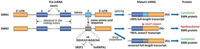
Molecules 2021, 26, 2263                                                                                                                       5 of 27

                                     complex (RISC) [49], ASOs are ssDNA/RNAs that act by directly binding to the target
                                     pre-mRNAs through Watson-Crick base-pairing. Such stoichiometric binding to the cis-
                                     acting regulatory element prevents the pre-mRNA sequence from recruiting the splicing
                                     regulatory proteins or alters the equilibrium of the splicing-regulatory RNA structures.
                                     Therefore, the ASO binding to the splicing enhancer sequences (ESEs or ISEs) results
                                     in splicing inhibition, whereas ASO binding to the splicing silencer sequences (ESSs or
                                     ISSs) leads to splicing activation. Besides splicing modifiers, ASOs can also be used as a
                                     chemical-genetic tool to induce RNase H-dependent RNA degradation [50,51] or as a steric
                                     block [52–54] against the ribosome assembly.
                                          Generally, the cellular uptake for ASOs is poor, which is the intrinsic limitation for
                                     their application [55]. Of particular interest is the inability of ASOs to cross the blood-brain
                                     barrier [55]. Illustrating this point is the FDA-approved ASO for the treatment of SMA,
                                     nusinersen, which must be administered by intrathecal injection into the cerebrospinal fluid
                                     to act on motor neurons in the central nervous system [3]. In addition, the immunogenicity
                                     of long ASOs is a liability for therapeutic uses and should be closely monitored in drug
                                     development [56]. On the other hand, cell-permeable RNA-targeting small molecules are
                                     an emerging pharmacological modality to modulate splicing. Risdiplam is the first small-
                                     molecule splicing modifier approved by the FDA for the treatment of SMA. Risdiplam
                                     readily crosses the blood-brain barrier and is formulated as an oral drug [4]. Risdiplam is
                                     also the first approved drug acting through binding to a non-ribosomal RNA. There are a
                                     number of excellent reviews on the topic of RNA-targeting small molecules in drug devel-
                                     opment (for recent reviews, see ref [57–60]). In this review, we only focused on the drug
                                     development of RNA-targeting splicing modifiers, including ASOs and small molecules
                                     (Table 2). RNA editing with CRISPR associated proteins (Cas) is another promising strategy
                                     that could potentially be used for modulating splicing. However, this review does not
                                     cover RNA editing due to lack of clinical validation and limitation of the scope (for recent
                                     reviews on RNA editing, see [61,62]).

                                                  Table 2. Disease-related splicing modifiers.

          Disease             Target Gene                      Name                              Category 1                 Development Stage
                                                           Nusinersen [3]                       ASO (MOE)                        Approved
                                                          E1v1.11 -ASO [63]                     ASO (PMO)                          Phase I
                                                          SMN-PNA [64]                          ASO (PNA)                         Cellular
                                                           Risdiplam [65]                      small molecule                    Approved
           SMA                   SMN2
                                                          Branaplam [66]                       small molecule            Phase II/III (discontinued)
                                                            PK4C9 [67]                         small molecule                    Preclinical
                                                           LDN-2014 [68]                       small molecule                    Preclinical
                                                          HSMNEx7D [69]                         ASO (PMO)                        Preclinical
                            Dystrophin Ex51                Eteplirsen [70]                      ASO (PMO)                        Approved
                            Dystrophin Ex51               Drisapersen [71]                     ASO (20 -OMe)                    Discontinued
                            Dystrophin Ex53               Golodirsen [72]                       ASO (PMO)                        Approved
           DMD              Dystrophin Ex53                Viltolarsen [5]                      ASO (PMO)                        Approved
                            Dystrophin Ex45               Casimersen [73]                       ASO (PMO)                         Phase III
                            Dystrophin Ex45                 DS-5141 [74]                       ASO (ENA 2 )                      Phase I/II
     Yeast infection         group II intron              Intronistat B [10]                   small molecule                      Cellular
                                                          E1.4, E5.3 [75]                      ASO (PMO)                         Preclinical
                                                         E10α, E10β [76]                      ASO (20 -OMe)                       Cellular
                                                       bipartite ASO [77]                      ASO (PNA)                          Cellular
          FTDP-17                MAPT            Mitoxantrone– bipartite ASO [78]      small molecule-ASO conjugate               Cellular
                                                         compound 9 [9]                       small molecule                      Cellular
                                                        compound 2 [79]                       small molecule                      Cellular
                                                             7-26S [11]                         ASO (MOE)                       Preclinical
                                                             SSO1 [80]                          ASO (OMe)                         Cellular
       Familial
                                IKBKAP                     ExSpeU1 [81]                             ASO                         Preclinical
     Dysautonomia
                                                            Kinetin [82]                       small molecule              Phase I (discontinued)
                                                           RECTAS [83]                         small molecule                     Cellular
    Influenza A virus
                            RNA segment 7                  Radavirsen [8]                       ASO (PMO)                          Phase I
         infection
      1See Section 4.1 for abbreviations of ASO categories. 2 ENA = 20 -O, 40 -C-ethylene-bridged nucleic acid [84], an analog of locked nucleic
      acid (LNA).
RNA-Targeting Splicing Modifiers: Drug Development and Screening Assays - MDPI
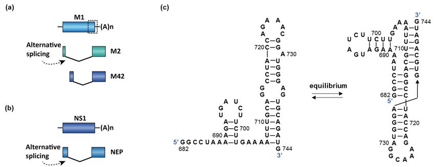
pression of dystrophin protein, which is an essential component connecting the cytoskel-
                             eton of a muscle fiber to the surrounding extracellular matrix through the cell membrane
                             [85]. DMD patients demonstrate muscle weakness from a young age and have a mean life
                             expectancy of 19.0 years [86]. About 70 % of patients with deletion mutations are amenable
Molecules 2021, 26, 2263     to partial dystrophin restoration by single exon skipping. For example, 14% of the DMD                               6 of 27
                             patients have a deletion in the genomic DNA sequence containing exons 49 and 50 [87].
                             Importantly, the nucleotide number in an exon is not necessarily an integer multiple of 3
                             for keeping a complete set of codons. The Δexon 49–50 transcript happens to cause a read-
                              2.1. Duchenne Muscular Dystrophy (DMD)
                             ing frameshift and creates a premature stop codon on exon 51. This frameshift and some
                             other DMD
                                     nonsenseis anmutations
                                                    X-linked recessive       genetic disease.
                                                                 in DMD patients         can bothCertain
                                                                                                     create agene    mutations
                                                                                                               premature      stop affect
                                                                                                                                     codon,thelead-
                                                                                                                                                expres-
                              sion
                             ing  toof  dystrophin protein,
                                      nonsense-mediated              which
                                                                 decay        is an
                                                                          of the     essential[88,89].
                                                                                   transcript     component       connecting
                                                                                                           A clinically           the cytoskeleton
                                                                                                                          validated    strategy is
                              ofskip
                             to  a muscle
                                       the stopfiber  to the surrounding
                                                   codon-containing        exons  extracellular     matrix
                                                                                    in the transcript        through
                                                                                                          (exon   51) andtherestore
                                                                                                                              cell membrane
                                                                                                                                      the reading   [85].
                             frame (Figure 3) [90]. Eteplirsen is the first FDA-approved ASOs for DMD. Eteplirsenlife
                              DMD      patients     demonstrate        muscle     weakness      from    a  young     age   and   have   a   mean
                              expectancy
                             binds   to the ofESE 19.0
                                                    of years
                                                        exon 51 [86].
                                                                    andAbout
                                                                         induces 70%exon
                                                                                       of patients
                                                                                            skipping, with   deletion
                                                                                                         which          mutations
                                                                                                                  further   leads toare     amenable
                                                                                                                                       correction
                              to partial    dystrophin      restoration     by   single   exon   skipping.     For
                             of the reading frame. Although the dystrophin is shorter than the wild-type protein,    example,     14%   of  the DMD
                                                                                                                                                  the
                              patients
                             Δexon        have
                                      49–51       a deletionisinstill
                                               dystrophin            thepartially
                                                                         genomicfunctional
                                                                                       DNA sequence[91]. containing exons 49 and 50 [87].
                              Importantly,
                                   Apart from   thethe
                                                     nucleotide
                                                         Δexon 49–51number     in an exon
                                                                           genotype,      manyis not  necessarily
                                                                                                 deletions    clusteranbetween
                                                                                                                         integer multiple
                                                                                                                                    exons 44of     3 for
                                                                                                                                                 and
                              keeping
                             55,          a complete
                                 and therefore,      thisset  of codons.
                                                           region    has alsoThebeen∆exon    49–50 transcript
                                                                                       recognized                  happens
                                                                                                      as a “hotspot”     targettoforcause   a reading
                                                                                                                                      ASO-based
                              frameshift
                             drugs.   Similarandtocreates
                                                    exon 51, a premature
                                                                 exclusion of  stop   codon
                                                                                  exons        on exon
                                                                                           53 and         51. This
                                                                                                    45 would     alsoframeshift     and some
                                                                                                                       result in partially        other
                                                                                                                                               func-
                             tional dystrophin in 8% and 8% of the DMD patients, respectively [92]. Two ASOs, go- to
                              nonsense      mutations      in DMD      patients     can  both   create  a  premature      stop   codon,    leading
                              nonsense-mediated
                             lodirsen    and viltolarsen decay    of the
                                                               were        transcript
                                                                      discovered         [88,89].
                                                                                       and  approvedA clinically
                                                                                                          for the validated
                                                                                                                    treatment strategy
                                                                                                                                  of DMDisintopa-  skip
                              the  stop   codon-containing          exons   in  the  transcript    (exon   51)  and
                             tients with a confirmed deletion of the dystrophin gene that is amenable to exon 53 skip-restore   the  reading     frame
                              (Figure
                             ping        3) [90].
                                    [7,93].         Eteplirsen
                                             Casimersen            is the
                                                              is the       firstapproved
                                                                       fourth     FDA-approved ASO drugASOs  forfor
                                                                                                                  DMDDMD.     Eteplirsen
                                                                                                                          patients    who havebindsa to
                              the ESE ofmutation
                             confirmed        exon 51 of  andtheinduces
                                                                  dystrophinexongeneskipping,     which further
                                                                                         that is amenable      to exonleads  to correction
                                                                                                                          45 skipping     [94]. of
                                                                                                                                                 DS-the
                              reading
                             5141         frame.ASO
                                    is another      Although      the dystrophin
                                                          that induces                  is shorterand
                                                                           exon 45 skipping          thanhasthe  wild-typethe
                                                                                                              completed        protein,
                                                                                                                                  Phase the     ∆exon
                                                                                                                                           I/II clin-
                              49–51
                             ical trialdystrophin
                                         (NCT02667483) is still[74].
                                                                partially functional [91].

                                      Therapeuticstrategy
                           Figure3.3.Therapeutic
                           Figure                 strategyofofEteplirsen
                                                               Eteplirsenfor
                                                                           forthe
                                                                                thetreatment
                                                                                     treatmentofofDMD.
                                                                                                   DMD.

                                   Apart from the ∆exon 49–51 genotype, many deletions cluster between exons 44 and
                             55, and therefore, this region has also been recognized as a “hotspot” target for ASO-based
                             drugs. Similar to exon 51, exclusion of exons 53 and 45 would also result in partially
                             functional dystrophin in 8% and 8% of the DMD patients, respectively [92]. Two ASOs,
                             golodirsen and viltolarsen were discovered and approved for the treatment of DMD in
                             patients with a confirmed deletion of the dystrophin gene that is amenable to exon 53
                             skipping [7,93]. Casimersen is the fourth approved ASO drug for DMD patients who have
                             a confirmed mutation of the dystrophin gene that is amenable to exon 45 skipping [94].
                             DS-5141 is another ASO that induces exon 45 skipping and has completed the Phase I/II
                             clinical trial (NCT02667483) [74].

                              2.2. Spinal Muscular Atrophy
                                  Spinal muscular atrophy (SMA) is one of the most common lethal genetic diseases in
                             newborns [95]. The cause of SMA in the most severe type (type I) is a recessive homozygous
                             deletion within the survival of motor neuron 1 (SMN1) gene in chromosome 5 [95]. Humans
                             have two nearly identical genes, SMN1 and SMN2. However, the protein produced by
                             SMN2 cannot fully compensate for the loss of SMN1 in type I SMA patients. It was
                             demonstrated that a single nucleotide difference (C-to-T change at exon 7 at +6) in exon
                             7 of SMN2 causes ~85% of the exon 7 skipping [96], leading to an inactive SMN isoform
                             (∆exon 7, Figure 4). The SMN2 exon 7 contains a regulatory RNA secondary structure at
                             the 30 -end, namely terminal stem-loop 2 (TSL2) (Figure 5) [97]. Reverse genetic studies
RNA-Targeting Splicing Modifiers: Drug Development and Screening Assays - MDPI
ture at the 3′-end, namely terminal stem-loop 2 (TSL2) (Figure 5) [97]. Reverse genetic
                                             studies showed that TSL2 is inhibitory [97]. Destabilizing TSL2 by point-mutations leads
                                             to exon 7 inclusion. On the contrary, strengthen TSL2 causes exon 7 skipping even in
                                             SMN1 [97], probably because the formation of TSL2 partially competes with the binding
                                             of U1 snRNP.
         Molecules 2021, 26, 2263                                                                                                          7 of 27
                                                   The exact mechanism of SMN in motor neuron maintenance and survival is not fully
                                             elucidated. To date, there are a collection of drugs used in clinics or in the development
                                             pipelines for the treatment of SMA (for recent reviews, see refs [98,99]). Homozygous de-
                                             letion
                                             showed of that
                                                        bothTSL2
                                                             SMN1     and SMN2
                                                                  is inhibitory     is embryonic
                                                                                 [97].             lethal.
                                                                                        Destabilizing TSL2Therefore,    type I patients
                                                                                                             by point-mutations   leads usually
                                                                                                                                        to exon
                                             7 inclusion.
                                             have          On the
                                                   a wildtype  SMN2contrary,  strengthen
                                                                       +/+ gene. One of theTSL2   causes exon
                                                                                             therapeutic        7 skipping
                                                                                                           strategies       even
                                                                                                                      to cure SMAinisSMN1   [97],
                                                                                                                                      to restore
                                             probably because
                                             full-length splicingthe
                                                                   offormation
                                                                      SMN2 with   of aTSL2 partially competes
                                                                                       pharmacological          with the
                                                                                                         intervention     binding
                                                                                                                        (Figure 4).of U1 snRNP.

                  Figure 4. Differential splicing patterns for SMN1 and SMN2. ~85% ~85% of the SMN2 transcript has exon 7 skipped due to the
                  C-to-T
                  C-to-T transition, which mediates the loss of interaction of SRSF1 and
                          transition, which  mediates  the loss of interaction of SRSF1 and gain  of the
                                                                                             gain of the interaction
                                                                                                         interaction of
                                                                                                                      of hnRNPA1.
                                                                                                                         hnRNPA1. Most
                                                                                                                                     Most type
                                                                                                                                           type II
ules 2021, 26, x FOR
                  SMA PEER  REVIEW
                        patients  have deleted  SMN1   but wildtype  SMN2.   Nusinersen, RG-7916,  and  LMI-070   are all        9 of 28 therapy
                                                                                                                          interventional
                  SMA patients have deleted SMN1 but wildtype SMN2. Nusinersen, RG-7916, and LMI-070 are all interventional therapy to
                  to restore exon 7 inclusion in SMN2.
                  restore exon 7 inclusion in SMN2.

                                                  The current FDA-approved ASO, nusinersen, was obtained by chemical modification
                                             of ASO-10-27 (masking intron 7 +10 to +27, which covers the ISS-N1 region). ISS-N1 is a
                                             15 nucleotide (intron 7 +10 to +24) inhibitory cis-element located downstream of the 5′
                                             splicing site of exon 7 and was found to play a dominant role in inducing exon 7 skipping
                                             [100]. Several splicing factors were identified to bind ISS-N1, including hnRNPA1/A2 and
                                             SRSF10 [101,102]. It is likely the effect of nusinersen comes from sequestration of these
                                             factors from binding to ISS-N1. Following intrathecal injections, nusinersen significantly
                                             improves motor function in these SMA patients and restores the SMN level in the central
                                             nervous system and peripheral tissues, leading to a significant improvement of the sur-
                                             vival rate [103]. Besides ISS-N1 in the intron 7, an ASO targeting the ISS in the intron 6, E1
                                             region, is also efficacious in human cells and mouse models (Figure 5) [104]. Similar to
                                             nusinersen, ASO E1v1.11 prevents E1 region from recruiting splicing repressor proteins and
                                             thus rescues exon 7 inclusion [104]. RG-7800 is the first-in-class small-molecule splicing
                                             modifier (PTC/ Roche) tested in a clinical trial for the treatment of SMA. RG-7800 increases
                                             the production of full-length SMN2 mRNA upon oral administration in mouse models
                                             [105]. The clinical trial was stopped as a precautionary measurement for a retinal toxicity
                                             issue observed in cynomolgus monkeys after chronic daily oral dosing for 39 weeks [105].
                                             Risdiplam (RG-7916) is a close analog of RG-7800 and is a second-generation molecule

               Figure 5. (a) Binding
                             Figure 5.sites of the existing
                                        (a) Binding sites ofdrugs for thedrugs
                                                            the existing  treatment  of treatment
                                                                               for the  SMA. Risdiplam
                                                                                                  of SMA.has  two binding
                                                                                                           Risdiplam       sitesbind-
                                                                                                                      has two    on the SMN2
                             ing sites
               pre-mRNA exon7.     (b) on
                                       Thethe  SMN2 pre-mRNA
                                            structures            exon7. (b) The structures
                                                       of known RNA-targeting                of known
                                                                                 small-molecule        RNA-targeting
                                                                                                  splicing modifiers forsmall-mole-
                                                                                                                         SMN2 exon 7.
                             cule splicing modifiers for SMN2 exon 7.
                                                  The exact mechanism of SMN in motor neuron maintenance and survival is not fully
                                             elucidated.
                              2.3. Virus Infection       To date, there are a collection of drugs used in clinics or in the development
                                    Viruses pipelines
                                             can adoptfor  the treatment
                                                        different strategiesoftoSMA  (for recent
                                                                                 maximize        reviews,
                                                                                            the use of theirsee
                                                                                                             DNArefsor[98,99]).
                                                                                                                        RNA ge-Homozygous
                                             deletion of both SMN1    and  SMN2   is embryonic    lethal. Therefore,
                              nomes and encode more than one peptide with a single nucleic acid sequence. Such strat-  type I patients usually
                              egies include RNA editing (in Ebola virus [111]), programmed frameshift (in coronavirus
                              [112]), and alternative splicing (in influenza A virus [113]). Viruses hijack the host spliceo-
                              some to process its transcript, and this process may be assisted by some viral proteins.
                                   Influenza A virus (IAV), a highly infectious and unpredictable respiratory pathogen,
RNA-Targeting Splicing Modifiers: Drug Development and Screening Assays - MDPI
Molecules 2021, 26, 2263                                                                                            8 of 27

                           have a wildtype SMN2+/+ gene. One of the therapeutic strategies to cure SMA is to restore
                           full-length splicing of SMN2 with a pharmacological intervention (Figure 4).
                                 The current FDA-approved ASO, nusinersen, was obtained by chemical modification
                           of ASO-10-27 (masking intron 7 +10 to +27, which covers the ISS-N1 region). ISS-N1 is a 15
                           nucleotide (intron 7 +10 to +24) inhibitory cis-element located downstream of the 50 splicing
                           site of exon 7 and was found to play a dominant role in inducing exon 7 skipping [100].
                           Several splicing factors were identified to bind ISS-N1, including hnRNPA1/A2 and
                           SRSF10 [101,102]. It is likely the effect of nusinersen comes from sequestration of these
                           factors from binding to ISS-N1. Following intrathecal injections, nusinersen significantly
                           improves motor function in these SMA patients and restores the SMN level in the central
                           nervous system and peripheral tissues, leading to a significant improvement of the survival
                           rate [103]. Besides ISS-N1 in the intron 7, an ASO targeting the ISS in the intron 6, E1
                           region, is also efficacious in human cells and mouse models (Figure 5) [104]. Similar to
                           nusinersen, ASO E1v1.11 prevents E1 region from recruiting splicing repressor proteins and
                           thus rescues exon 7 inclusion [104]. RG-7800 is the first-in-class small-molecule splicing
                           modifier (PTC/ Roche) tested in a clinical trial for the treatment of SMA. RG-7800 increases
                           the production of full-length SMN2 mRNA upon oral administration in mouse models [105].
                           The clinical trial was stopped as a precautionary measurement for a retinal toxicity issue
                           observed in cynomolgus monkeys after chronic daily oral dosing for 39 weeks [105].
                           Risdiplam (RG-7916) is a close analog of RG-7800 and is a second-generation molecule with
                           enhanced potency and improved pharmacokinetics and safety properties [65]. Risdiplam is
                           approved by the FDA for the treatment of patients in all stages of SMA in August 2020 [106].
                           Branaplam (Novartis, Cambridge, MA, USA) is another small-molecule splicing modifier
                           and was tested in a Phase 2/3 clinical trial for the treatment of SMA. In the SMN∆7 mouse
                           model, branaplam treatment increased full-length SMN mRNA and protein levels, and
                           extended survival [66]. The clinical trial was terminated after risdiplam was developed to
                           a more advanced stage.
                                 Risdiplam and branaplam have partially overlapping mechanism of action. They
                           act by stabilizing the transient double-strand RNA structure formed by the SMN2 pre-
                           mRNA and U1 snRNP complex [107,108]. NMR studies demonstrated that a risdiplam
                           analog, SMN-C5, binds to a bulged A between the 50 splice site and U1 snRNA and
                           enhances the spliceosome recognition (Figure 5) [107]. However, the chromatography
                           study demonstrated that the in vitro binding between branaplam and 50 splice site requires
                           holo U1 snRNP, rather than U1 snRNA alone [108]. The exact binding nucleotides for
                           branaplam have not been elucidated. Compared to branaplam, risdiplam analogs not only
                           bind to the interface between 50 splice site and U1 snRNA, but also to a secondary binding
                           site near the 30 of the exon. RNA immunoprecipitation demonstrated that risdiplam
                           analogs preferentially bind to a GA-rich sequence that matched the sequence of exon 7 +22
                           to +30 [28]. The analysis of other risdiplam-sensitive genes in splicing demonstrated that
                           the GA-rich sequence was slightly enriched at the 30 splice site but does not have a strong
                           consensus in all sequences [109,110]. PK4C9 is another small molecule reported as an
                           SMN2 splicing modifier (Figure 5b). It was identified by a target-based screening against
                           TSL2 in exon 7. In SMA patient fibroblast cells, PK4C9 increased exon 7 inclusion by
                           40% at 40 µM, coupled with a 1.5-fold increase in SMN protein level. Reverse genetics
                           demonstrated that the 50 splice site (GAGUAAGU) of exon 7 is likely to be the target of
                           PK4C9. NMR and molecular dynamics studies indicate that PK4C9 may improve the
                           accessibility of the 50 splice site via binding to TSL2 and stabilizing a tri-loop structure of
                           TSL2 [67].

                           2.3. Virus Infection
                                Viruses can adopt different strategies to maximize the use of their DNA or RNA
                           genomes and encode more than one peptide with a single nucleic acid sequence. Such
                           strategies include RNA editing (in Ebola virus [111]), programmed frameshift (in coron-
RNA-Targeting Splicing Modifiers: Drug Development and Screening Assays - MDPI
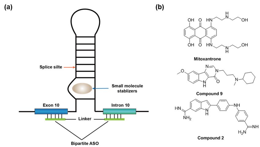
Molecules 2021, 26, 2263                                                                                                                     9 of 27

                                   avirus [112]), and alternative splicing (in influenza A virus [113]). Viruses hijack the host
                                   spliceosome to process its transcript, and this process may be assisted by some viral proteins.
                                         Influenza A virus (IAV), a highly infectious and unpredictable respiratory pathogen,
                                   represents a substantial threat to public health [114]. The outbreak of Spanish flu (H1N1) in
                                   1918 killed 21–50 million people globally [115], one of the deadliest pandemics in recorded
                                   human history. According to the United States Centers for Disease Control and Prevention
                                   (CDC), there have been 9.3–45 million cases of seasonal flu every year in the U.S. from
                                   2010 to 2020, with 12,000–61,000 flu-associated deaths annually [116] (access date 11 April
                                   2021). Two dominant influenza A strains (H1N1, H3N2) are currently circulating in the
                                   United States and globally [117] (access date 11 April 2021). Both influenza A and B are
                                   ssRNA(–) viruses (“–“ indicates the RNA sequence is reversely complemented to the coding
                                   sequence), and their genome contains eight RNA segments.
                                         RNA splicing occurs in segments 7 and 8, encoding matrix protein (M) and nonstruc-
                                   tural protein (NS1), respectively (Figure 6a,b) [118]. The splicing of M is essential because
                                   the splice variants M1 (matrix protein) and M2/M42 (ion channel) are both required
Molecules 2021, 26, x FOR PEER REVIEW
                                                                                                                                            for
                                                                                                                                       10 of 28
                                   viral replication. The M1 matrix protein assembles to form virus-like particles, which are a
                                   prerequisite for the budding process. M2 forms an ion channel on the nucleocapsid envelop
                                   and is essential for viral entry. Mutually exclusive secondary structures at the 30 splice
                                   splice
                                   site ofsite
                                            the of
                                                 Mthe  M are
                                                    gene  geneproposed
                                                                are proposed     to control
                                                                           to control         the splice
                                                                                        the splice        pattern
                                                                                                     pattern       [115,119].
                                                                                                              [115,119].         Specifically,
                                                                                                                            Specifically,   the
                                   the hairpin
                                   hairpin        conformation
                                              conformation        promotes
                                                             promotes          splicing
                                                                          splicing  of MofandM and    production
                                                                                                 production    of M2 of (Figure
                                                                                                                         M2 (Figure     6c).
                                                                                                                                  6c). In    In
                                                                                                                                           con-
                                   contrast,   the pseudoknot    conformation     disrupts    binding   of the  trans-acting
                                   trast, the pseudoknot conformation disrupts binding of the trans-acting factor SRSF1 to an   factor  SRSF1
                                   to an exonic
                                   exonic          splice enhancer
                                            splice enhancer   (ESE) and(ESE)  andthe
                                                                          blocks    blocks
                                                                                       splicethe  splice
                                                                                               site      site6c),
                                                                                                    (Figure   (Figure    6c), andfavors
                                                                                                                  and therefore     therefore
                                                                                                                                            the
                                   favors   the production   of M1   [115,120].  However,     the hairpin  or the  pseudoknot
                                   production of M1 [115,120]. However, the hairpin or the pseudoknot structures were not          structures
                                   were   not discovered
                                   discovered               by whole-genome
                                                 by whole-genome        chemical chemical      probing in
                                                                                  probing analyses       analyses   in virus-infected
                                                                                                           virus-infected                 cells
                                                                                                                              cells [121,122].
                                   [121,122].   A recent
                                   A recent study         study demonstrated
                                                     demonstrated                   that IAV
                                                                       that IAV strains    that strains  that are
                                                                                                 are adapted       adapted
                                                                                                               in avian    andin   avian host
                                                                                                                                 human     and
                                   human     host cells
                                   cells contain        contain
                                                   different      different
                                                             splicing        splicingRNA
                                                                        regulatory      regulatory
                                                                                            structuralRNA   structural
                                                                                                         elements    at the  30 splice
                                                                                                                          elements   atsite
                                                                                                                                         the of
                                                                                                                                              3′
                                   splice
                                   M [113].siteThe
                                                of M [113]. The
                                                   inhibition  ofinhibition   of NS1 also
                                                                  NS1 also reduces          reduces
                                                                                       the viral      the viralinreplication
                                                                                                  replication     vitro [123].inInfluenza
                                                                                                                                  vitro [123].A
                                   Influenza
                                   virus hijacksA virus hijacks
                                                   the host      the host spliceosome
                                                            spliceosome                    by thewith
                                                                            by the interaction      interaction
                                                                                                         U2 andwith     U2 andthrough
                                                                                                                  U6 snRNA        U6 snRNA  the
                                   through    the viral
                                   viral protein   NS1 protein
                                                        [118]. NS1 [118].

      Figure 6. (a,b)
                 (a,b) Two
                        Two genes
                              genes in
                                     ininfluenza
                                         influenzaAAundergo
                                                        undergosplicing:
                                                                  splicing:MMand
                                                                               andNS1.  The
                                                                                     NS1.  Thedotted
                                                                                                dottedboxbox
                                                                                                          in M
                                                                                                             in is
                                                                                                                M the   proposed
                                                                                                                    is the         region
                                                                                                                            proposed       re-
                                                                                                                                      region
      sponsible for
      responsible  forsplicing
                        splicingswitch
                                  switch(enlarged
                                          (enlargedinin(c)).
                                                        (c)).The
                                                              Thespliced
                                                                  splicedisoform
                                                                          isoformofofNS1
                                                                                      NS1isisnamed
                                                                                              namednuclear
                                                                                                      nuclear export
                                                                                                              export protein
                                                                                                                       protein (NEP).
                                                                                                                                (NEP). (c) An
                                                                                                                                       (c) An
      equilibrium between
      equilibrium   between aa hairpin
                                 hairpin and
                                          and aa pseudoknot
                                                 pseudoknot structure
                                                               structure was
                                                                         was proposed
                                                                              proposed to
                                                                                        to control
                                                                                            control the
                                                                                                    the splicing
                                                                                                        splicing of
                                                                                                                 of MM in
                                                                                                                        in influenza
                                                                                                                           influenza A
                                                                                                                                     A [115].
                                                                                                                                        [115].

                                          ASOs have been developed to target the       the splicing
                                                                                           splicing of
                                                                                                    of M1/M2.
                                                                                                       M1/M2. Radavirsen
                                                                                                                Radavirsen (AVI-7100)
                                                                                                                              (AVI-7100)
                                    is the
                                        themost
                                            mostadvanced
                                                   advanceddrug  drugcandidate  that
                                                                        candidate     showed
                                                                                   that  showed good safety
                                                                                                  good      andand
                                                                                                        safety  tolerability profile
                                                                                                                    tolerability       in a
                                                                                                                                   profile
                                    Phase   I clinical
                                    in a Phase          trial trial
                                                  I clinical  [8]. Radavirsen   is a 20-mer
                                                                    [8]. Radavirsen            ASOASO
                                                                                      is a 20-mer   targeting a conserved
                                                                                                         targeting           region
                                                                                                                   a conserved        that
                                                                                                                                   region
                                    controls   M1/M2
                                    that controls   M1/M2splicing.   The sequence
                                                               splicing.             of radavirsen
                                                                          The sequence               andand
                                                                                          of radavirsen   experimental  datadata
                                                                                                             experimental      for for
                                                                                                                                    thethe
                                                                                                                                         in
                                    in vitro
                                    vitro     drug
                                           drug      effect
                                                 effect  hashas
                                                              notnot  been
                                                                   been    published.
                                                                         published.
                                          Another notable example of viral splicing is in human immunodeficiency virus 1
                                    (HIV-1). HIV-1 is an ssRNA(+) retrovirus. Unlike IAV, all the viral genes are encoded on
                                    a single strand of RNA. HIV is the cause of the acquired immunodeficiency syndrome
                                    (AIDS). In 2018, 37.9 million people were living with the virus in the world, and 770,000
                                    people died from AIDS-related disease [124]. The ~9,000 nucleotide HIV-1 genome tran-
RNA-Targeting Splicing Modifiers: Drug Development and Screening Assays - MDPI
Molecules 2021, 26, 2263                                                                                                                 10 of 27

                                         Another notable example of viral splicing is in human immunodeficiency virus 1
                                   (HIV-1). HIV-1 is an ssRNA(+) retrovirus. Unlike IAV, all the viral genes are encoded on a
                                   single strand of RNA. HIV is the cause of the acquired immunodeficiency syndrome (AIDS).
                                   In 2018, 37.9 million people were living with the virus in the world, and 770,000 people
                                   died from AIDS-related disease [124]. The ~9000 nucleotide HIV-1 genome transcribes over
                                   50 functional mRNAs by using alternative splicing [125], including several essential genes,
                                   such as tat and rev [126]. There are two introns to be spliced in tat and rev (Figure 7), and
                                   the splicing of the 50 intron is a prerequisite to 30 intron splicing [126]. The pre-mRNA is
                                   differentially spliced to produce distinct mRNAs coding either for the tat or rev proteins
                                   (Figure 7). The protein Rev controls the export of unspliced or partially spliced transcript
                                   from the nucleus, which is crucial for viral replication and packaging [127,128]. Although
Molecules 2021, 26, x FOR PEER REVIEW                                                                                    11 of 28
                                   earlier work demonstrated that splicing-switching ASOs reduced the release of HIV virions
                                   in cells [129], to our knowledge, no splicing modifier has shown promising in vivo activities
                                   for the treatment of HIV infection.

                                Figure 7. RNA splicing in HIV-1 genes tat and rev. The figure represents only two sets of the potential
                                Figure  7. RNA
                                splice sites      splicing
                                             out of        in HIV-1 genes
                                                    four alternative        tatsites
                                                                     50 splice  andand
                                                                                     rev.eight
                                                                                          The figure represents
                                                                                               alternative        only
                                                                                                           30 splice    two
                                                                                                                     sites   sets The
                                                                                                                           [125]. of the po- is
                                                                                                                                      figure
                                tential splice sites out of four alternative 5′ splice sites and eight alternative 3′ splice sites [125]. The
                                modified from ViralZone, SIB Swiss Institute of Bioinformatics.
                                figure is modified from ViralZone, SIB Swiss Institute of Bioinformatics.
                                      Apart from IAV and HIV, RNA splicing is also essential for some other viruses, such
                                Table 3. Selected
                                as human          human pathogenic
                                            T-lymphotropic          viruses withB19,
                                                            virus, parvovirus    essential RNA splicing
                                                                                     and human          processes. 1. The genes
                                                                                                  papillomavirus
                                that undergo RNA
                                           Virus  splicing for these viruses
                                                 Name                        are summarized
                                                                        Essential Genes     inViral
                                                                                               TableRegulatory
                                                                                                     3.        Factors
                                            Influenza A virus                   M1, M2/M42                      NS1 [118]
                                Table 3. Selected
                                  human            human pathogenic
                                           immunodeficiency   virus 1 viruses with
                                                                                tat,essential
                                                                                     rev; env RNA splicing processes.
                                                                                                                rev [128]
                                     Human T-lymphotropic virus                 env, tax, rex                 rex [130,131]
                                              Virus Name                    Essential Genes             Viral Regulatory Factors
                                             Parvovirus B19                      VP1, VP2
                                          Influenza
                                       Human         A virus
                                                 Papillomavirus 1             M1,
                                                                               E1, M2/M42
                                                                                    E2, L1, L2                  NS1  [118]
                                                                                                                 E2 [132]
                                 human immunodeficiency virus 1                tat, rev; env                    rev [128]
                                    Human T-lymphotropic virus                 env, tax, rex                  rex [130,131]
                                2.4. Yeast Infection
                                            Parvovirus B19                      VP1, VP2
                                      Antifungal    drugs against
                                      Human Papillomavirus    1    fungal infections
                                                                              E1, E2, L1,especially
                                                                                           L2       against invasive
                                                                                                                E2 [132]fungal infec-
                                tions remain an unmet clinical need because of the unsatisfactory treatment outcomes
                                with   currently
                                2.4. Yeast          available antifungal drugs. One key fundamental challenge in new anti-
                                             Infection
                                fungal   drug discovery
                                      Antifungal     drugs is     that fungi
                                                              against    fungaland   yeast cells
                                                                                  infections       have similar
                                                                                               especially          biochemical
                                                                                                             against                pathways
                                                                                                                       invasive fungal          to
                                                                                                                                            infec-
                                humans      [133].  Group    II  introns   are  large   autocatalytic    RNA    motifs
                                tions remain an unmet clinical need because of the unsatisfactory treatment outcomes with that   adopt   complex
                                tertiary
                                currently  structures
                                              availableand   catalyzedrugs.
                                                         antifungal       RNA splicing
                                                                                  One key[134].     They are found
                                                                                             fundamental      challengein plants,
                                                                                                                            in newfungi,    yeast,
                                                                                                                                      antifungal
                                and  various    lower   eukaryotes,      but  are not  present   in  mammals      [135].
                                drug discovery is that fungi and yeast cells have similar biochemical pathways to hu-     Due   to their  absence
                                in
                                mansmammals      and essential
                                       [133]. Group     II intronsroles    in fungal
                                                                     are large         metabolism,
                                                                                 autocatalytic   RNAgroupmotifs II introns
                                                                                                                that  adoptmay      servetertiary
                                                                                                                              complex      as po-
                                tential  targets
                                structures     andfor  highlyRNA
                                                    catalyze    specific    antifungal
                                                                        splicing   [134].drug
                                                                                           They discovery.
                                                                                                   are found in plants, fungi, yeast, and
                                      Recently,    the  Pyle  group    reported     a series of
                                various lower eukaryotes, but are not present in mammals [135].  antifungal   agents Duewith
                                                                                                                           to novel    structures
                                                                                                                               their absence   in
                                that
                                mammals and essential roles in fungal metabolism, group II introns may serve asidentified
                                      target   yeast  group    II  introns   by inhibiting   its  ribozyme    activity   [10].  They    potential
                                two  compounds
                                targets   for highlytermed
                                                       specificintronistat
                                                                   antifungalAdrug
                                                                                 and intronistat
                                                                                       discovery. B which were shown to inhibit group
                                II intron   splicing  in S. cerevisiae   in  vivo
                                      Recently, the Pyle group reported a series  and   consequently     cause
                                                                                             of antifungal       its growth
                                                                                                              agents           inhibition.
                                                                                                                        with novel           They
                                                                                                                                      structures
                                were   also shown
                                that target           to selectively
                                               yeast group    II intronsbindbygroup    II intron
                                                                                inhibiting         tertiary structure
                                                                                             its ribozyme     activity without
                                                                                                                         [10]. They affecting the
                                                                                                                                       identified
                                other two known splicing systems (intron I and spliceosome). Importantly, intronistat A
                                and intronistat B showed little toxicity in human cells, indicating specificity for fungi.

                                2.5. Frontotemporal Dementia and Parkinsonism Linked to Chromosome 17 (FTDP-17)
Molecules 2021, 26, 2263                                                                                           11 of 27

                           two compounds termed intronistat A and intronistat B which were shown to inhibit group
                           II intron splicing in S. cerevisiae in vivo and consequently cause its growth inhibition. They
                           were also shown to selectively bind group II intron tertiary structure without affecting the
                           other two known splicing systems (intron I and spliceosome). Importantly, intronistat A
                           and intronistat B showed little toxicity in human cells, indicating specificity for fungi.

                           2.5. Frontotemporal Dementia and Parkinsonism Linked to Chromosome 17 (FTDP-17)
                                FTDP-17 is an autosomal dominant neurodegenerative disorder with symptoms of
                           behavioral changes and cognitive and motor impairment [136]. The disease is primarily
                           caused by mutations in the microtubule-associated protein tau (MAPT) gene [137]. The tau
                           protein stabilizes neuronal microtubules under normal conditions. More than 38 mutations
                           were identified in FTDP-17 patients, and the majority of the patients have a distorted
                           proportion of tau isoforms [136]. Some mutations (e.g., c.892A > G [138], intron 10 +16C >
                           T [139]) promote an exon 10 inclusion splice isoform, namely 4-repeat (4R), which leads
                           to tau hyperphosphorylation and aggregation, and ultimately pathogenic neurofibrillary
                           tangles [140,141].
                                Molecules that inhibit tau phosphorylation and aggregation, anti-tau antibodies, and
                           ASOs targeting MAPT splicing and expression have been developed for the treatment of
                           FTDP-17 (for a recent review, see ref [142]). It was shown that some ASOs that induce
                           exon skipping also causes the reduction of expression level of the pathogenic tau, probably
                           through nonsense-mediated decay pathway [75].
                                Besides ASO-based tau expression inhibitors, drug molecules that induce the exon
                           10 skipping in the mutant MAPT would also be of potential therapeutic interest for restoring
                           the aberrant splice pattern for FTDP-17 [9]. ASOs bound to either the 50 or 30 splice site of
                           exon 10 were demonstrated to induce exon 10 skipping [76]. An RNA stem-loop structure
                           in intron 10 near the 50 splice site acts as a splicing silencer (Figure 8a). This stem-loop
                           structure can be destabilized via pathogenic mutations in the 50 splice sites of exon 10 [143].
                           Therefore, stabilizing the stem-loop structure is a strategy to induce exon 10 skipping
                           and restore the non-pathogenic MAPT isoform. An anticancer drug, mitoxantrone, was
                           identified in a high-throughput screening campaign as the stem-loop structure ligand
                           and inhibitor of exon 10 splicing (Figure 8b) [144]. NMR experiments demonstrated
                           that mitoxantrone stabilizes the stem-loop structure through intercalating into the stem
                           base [145]. Several other small-molecule splicing modifiers were identified by the Disney
                           group via structure-based design [9,79] or phenotypic screening (e.g., “compound 9” and
                           “compound 2” from the original report [9] (Figure 8b). Compound 9 was shown to stabilize
                           an internal bulge of the stem-loop structure and reduce exon 10 splicing [9]. The Wolfe
                           group also demonstrated that a bipartite ASO that is complementary to the flanking arms
                           of the stem-loop can destabilize the structure and, thereby, inhibit the exon 10 splicing
                           (Figure 8a) [77]. Importantly, a conjugate of this bipartite ASO and mitoxantrone is more
                           potent than the ASO or mitoxantrone alone in the in vitro splicing reactions [78]. To our
                           knowledge, this small molecule–ASO conjugate is the first small molecule–ASO chimera to
                           modulate RNA splicing.
lize an internal bulge of the stem-loop structure and reduce exon 10 splicing [9]. The Wolfe
                   group also demonstrated that a bipartite ASO that is complementary to the flanking arms
                   of the stem-loop can destabilize the structure and, thereby, inhibit the exon 10 splicing
                   (Figure 8a) [77]. Importantly, a conjugate of this bipartite ASO and mitoxantrone is more
                   potent than the ASO or mitoxantrone alone in the in vitro splicing reactions [78]. To our
Molecules 2021, 26, 2263                                                                                                         12 of 27
                   knowledge, this small molecule–ASO conjugate is the first small molecule–ASO chimera
                   to modulate RNA splicing.

                Figure
     Figure 8. (a)      8. (a) A stem-loop
                   A stem-loop              controls
                                  controls the  exon the exon 10 splicing
                                                      10 splicing         in the gene
                                                                  in the MAPT    MAPT   gene
                                                                                      and theand  the binding
                                                                                              binding   site of site
                                                                                                                the of
                                                                                                                     known splicing
                the The
     modifiers. (b) known    splicingofmodifiers.
                        structures                (b) The structures
                                        known RNA-targeting          of known RNA-targeting
                                                                small-molecule                small-molecule
                                                                               splicing modifiers for MAPT exon splicing
                                                                                                                     10.
                modifiers for MAPT exon 10.
                                2.6. Familial Dysautonomia
                2.6. Familial Dysautonomia
                                      Familial dysautonomia (FD) is an inherited autosomal recessive disease which occurs
                      Familial dysautonomia    (FD) isamong
                                almost exclusively     an inherited autosomalJewish
                                                               the Ashkenazi    recessive  disease
                                                                                        with        which
                                                                                              a carrier    occurs of about 1 in
                                                                                                        frequency
                almost exclusively
                                30 [146]. Features of this disease include loss of pain and temperaturein
                                     among   the Ashkenazi    Jewish with  a carrier frequency   of about  1   30
                                                                                                             sensation, gastroin-
                [146]. Features of this disease include  loss of pain and  temperature   sensation,  gastrointes-
                                testinal dysfunction, respiratory abnormality, autonomic crises, progressive optic atrophy
                tinal dysfunction,
                                andrespiratory    abnormality,
                                     gait ataxia [147].          autonomic
                                                        FD was caused        crises, progressive
                                                                        by mutations   in the geneoptic  atrophy
                                                                                                    IKBKAP   which encodes IKK
                                complex-associated protein (IKAP)/elongator protein 1 (ELP1) [148,149]. More than 99%
                                cases were found to be related with a T to C mutation at the donor splice site of intron 20
                                (intron 20 +6T > C). This mutation causes inefficient use of intron 20 donor splice site and
                                results in skipping of exon 20 in the mature IKBKAP mRNA. Skipping of exon 20 generates
                                a frameshift and consequently introduces a stop codon in the reading frame of exon 21,
                                eventually generates truncated IKAP protein [147].
                                      There is currently no targeted therapy for FD, but efforts have been made to correct
                                exon 20 skipping and restore the full length IKAP protein expression. Based on their
                                experience in discovery of ASO treatments for SMA, the Krainer group used a two-step
                                screening approach to identify cis-elements in IKBKAP pre-mRNA and ASOs that could
                                restore exon 20 splicing in FD patient fibroblasts [11]. Two splicing silencer elements were
                                identified separately in intron 20 and intron 19. One ASO that masks intron 20 +7 to
                                +26 nucleotides was found to be most effective and induced completely inclusion of exon
                                20 in IKBKAP transcript and statistically significant increase in IKAP protein levels in FD
                                patient fibroblast cells [11]. In vivo effect was also validated in a mouse model with ASO
                                7-26S which contains a more stable phosphorothioate backbone in structure. Similarly, the
                                Andresen group also identified a splicing silencer in intron 20 which acts as the binding
                                site of hnRNP A1, one splicing repressor [80]. Their most active ASO candidate, SSO1
                                which masks intron 20 +11 to +35 nucleotides was able to induce completely restoration
                                of IKBKAP exon 20 inclusion and dramatic increase in IKAP protein levels in FD patient
                                fibroblast cells at low nanomolar concentration.
                                      The Pagani group described a novel strategy to correct exon 20 skipping by using
                                exon-specific U1 snRNAs (ExSpeU1s) [81]. ExSpeU1s are snRNA like particles that comple-
                                mentarily bind to intronic regions downstream of the 50 splice site of skipped exons and
                                facilitate exon recognition by recruiting the spliceosomal components, thus increase exon
                                inclusion during splicing. Lentiviral transduction of FD fibroblasts with the most potent
                                ExSpeU1s increased full length IKBKAP mRNA by three-fold and restores IKAP protein
Molecules 2021, 26, 2263                                                                                                              13 of 27

                                   level to ~80% of the normal fibroblasts. In a TgFD9 transgenic mouse model, intraperi-
                                   toneal delivery of ExSpeU1s adeno-associated virus particles successfully increased the
                                   production of full-length human IKBKAP transcript and protein [81].
                                        Small molecule splicing modifiers were also reported to be able to correct exon 20 skip-
                                   ping and induce full length IKAP protein expression. Kinetin, a plant cytokinin used
                                   as anti-aging skin care agent, was found to dramatically increase exon 20 inclusion and
                                   consequent wild type IKBKAP mRNA amount as well as IKAP protein in FD cell lines [150].
                                   Mechanism study revealed that this effect is independent of FD mutation while a motif
                                   containing CCA element at the joint of 30 end of exon 20 and 50 splice site was determined
                                   to be necessary for kinetin’s activity [151]. Kinetin was the first small molecule splicing
                                   modifier entered clinical trial. However, the trial was discontinued because of withdrawal
                                   of participants [82]. A kinetin analog, termed RECTAS, was identified as a more potent
                                   splicing modifier than kinetin in promoting exon 20 inclusion and IKAP expression in FD
                                   patient cells [83].

                                   3. Screening Methods for RNA Splicing Modifiers
                                        The deciding process for splice site selection can be influenced by many regulatory
                                   factors. For example, more than 46 different proteins were discovered as regulatory factors
                                   for the exon 7 inclusion/skipping in the SMN2 gene [152]. Due to this complexity, pheno-
                                   typic screening is a widely used approach to identify novel splicing modifiers. as Among
                                   these, “ASO walking” for ASOs [101,153] and cell-based phenotypic assays for small
Molecules 2021, 26, x FOR PEER REVIEW
                                   molecules [10,89,154] find broadest use. Both clinically tested small molecules, branaplam 14 of 28
                                   and risdiplam, were originally uncovered by phenotypic screening campaigns [108,155].
                                  Target-based rational design for small-molecule splicing modifiers is also emerging and
                                  were   elicited
                                   stem-loop      fora MAPT
                                                and           splicing
                                                       collection      via docking the
                                                                  of RNA-binding        MAPT [9].
                                                                                    molecules  exonIn10 regulatory
                                                                                                      this          stem-loop
                                                                                                           review, we           andon
                                                                                                                        will focus   a
                                   collection  of RNA-binding     molecules  [9]. In this review, we  will
                                   the approaches for the discovery of RNA-targeting splicing modifiers.   focus on  the approaches
                                   for the discovery of RNA-targeting splicing modifiers.
                                    3.1. “ASO Walking” Uncovers Cis-Acting Factors
                                   3.1. “ASO Walking” Uncovers Cis-Acting Factors
                                          ASOs act through antagonizing the binding between the pre-mRNA and trans-acting
                                          ASOs act through antagonizing the binding between the pre-mRNA and trans-acting
                                    regulatory RNA elements or the formation of functional structures. ASO binding se-
                                   regulatory RNA elements or the formation of functional structures. ASO binding sequences
                                    quences are usually determined by an empirical method, “ASO walking”. Figure 9 illus-
                                   are usually determined by an empirical method, “ASO walking”. Figure 9 illustrates two
                                    trates two ASO walkings in the SMN2 gene within exon 7 [153] and intron 7 [101], respec-
                                   ASO walkings in the SMN2 gene within exon 7 [153] and intron 7 [101], respectively. 15-mer
                                    tively.were
                                   ASOs     15-mer
                                                usedASOs   werethe
                                                      to target   used   to target
                                                                     region  wherethe    region where
                                                                                      neighboring    ASOs neighboring
                                                                                                            overlappedASOs      overlapped
                                                                                                                          by 10 nucleotides
                                    by 10 nucleotides
                                   (exon                (exon
                                           7 +16 to intron     7 +16
                                                            7 +40     to intron
                                                                    region        7 +40 region
                                                                            is shown).    In an is
                                                                                                 inshown).   In an in
                                                                                                    vitro splicing     vitroASOs
                                                                                                                    assay,   splicing  assay,
                                                                                                                                   masking
                                    ASOs   masking   exon   7 +36  to +50  and   intron  7 +11  to +25  strongly   induces
                                   exon 7 +36 to +50 and intron 7 +11 to +25 strongly induces the splicing of exon 7 [153].  the splicing  of
                                    exon  7 [153]. Further   optimization    uncovered     two   ESS/ISS   elements   that
                                   Further optimization uncovered two ESS/ISS elements that can be targeted by ASOs: exon   can be  targeted
                                   7by+34
                                        ASOs:  exon
                                           to +48,    7 +34 toTSL2
                                                   targeting    +48, (Figure
                                                                       targeting9)TSL2    (Figure
                                                                                    [97], and       9) [97],
                                                                                               intron   7 +11and  intron
                                                                                                               to +24,  an7hnRNP
                                                                                                                             +11 to +24,  an
                                                                                                                                      A2/B1
                                    hnRNP    A2/B1  binding
                                   binding site (Figure 9).   site (Figure   9).

      Figure 9. Binding sites for the ASOs used in the SMN2 exon 7 and intron 7 walk. The position of complementarity of each
      Figure 9. Binding sites for the ASOs used in the SMN2 exon 7 and intron 7 walk. The position of complementarity of each
      ASO along the sequence of interest is indicated by a horizontal line. +: promotion of the exon 7 inclusion, −: inhibition
      ASO along the sequence of interest is indicated by a horizontal line. +: promotion of the exon 7 inclusion, −: inhibition of
      of
      thethe
          exonexon   7 inclusion,
                 7 inclusion,      *: no
                               *: no      effect
                                      effect     on alternative
                                             on alternative     splicing.
                                                            splicing. The The figure
                                                                          figure     is modified
                                                                                 is modified fromfrom  refs [101,153]
                                                                                                  refs [101,153]      (copyright
                                                                                                                 (copyright      © 2021
                                                                                                                             © 2021 Hua
      Hua   et al. [153]; © 2021  The  American    Society of Human    Genetics [101]). The “ASO walking”   experiment   determined
      et al. [153]; © 2021 The American Society of Human Genetics [101]). The “ASO walking” experiment determined the splic-         the
      splicing  silencer
      ing silencer        elements
                     elements  (red (red   underline).
                                      underline).

                                   3.2. High-Throughput Screening (HTS) Assays for Small-Molecule Splicing Modifiers
                                         The ASO-based splicing modifiers have some unfavorable pharmacokinetic proper-
                                   ties. For example, they cannot cross the blood-brain barrier and have a low distribution in
                                   the bladder and stomach [55]. Small-molecule splicing modifiers have the propensity to
                                   overcome these problems. In addition, the identification of small-molecule splicing mod-
You can also read