"REHABILITATION PROCESS USING ELECTROMYOGRAPHY AND BIOFEEDBACK " - Biomedical Engineering Degree Final Degree Project
←
→
Page content transcription
If your browser does not render page correctly, please read the page content below
Final Degree Project
 Biomedical Engineering Degree

“REHABILITATION PROCESS USING
 ELECTROMYOGRAPHY AND
 BIOFEEDBACK “

 Barcelona, June 2021
 Author: Francisco José Domingo Gil
 Director/s: José Luis Parreño Catalan
 Tutor: Manel Puig Vidal
ACKNOWLEDGEMENTS
I would like to express my gratitude to both my director José Luis Parreño, for trusting me and
giving me the opportunity of developing the project with him, and my tutor Manel Puig for helping
me, teaching me, and supporting me during the development of the project.

Moreover, to my family and friends for giving me the necessary support to carry on the project.

Thank you.

 I
Abstract
A good rehabilitation routine is essential for the best possible recovery after an injury or to increase
the quality of life of those who suffer from neuromusculoskeletal diseases. It is of particular
relevance to maintain the motivation throughout all the process, for which videogames may play an
essential role.

Biofeedback is a process that provides real time information from psychophysiological recordings
about the levels at which physiological systems are functioning. In this project the biofeedback
system has been implemented by means of a low-cost EMG system created using Arduino.

The EMG system has been developed using an Olimexino-328 microcontroller and an EMG-Shield,
both from Olimex. The program was developed using Arduino IDE. To assess the quality of the
signal of the prototype, it was compared to a professional EMG device, the DataLog from Biometrics
Ltd. The comparison showed promising results although it could be improved by means of post-
processing algorithms.

An audio-visual Biofeedback system targeting maximum strength and explosiveness of the muscles
was created using Python. Flappy Bird game commands were changed to control them with the
EMG low-cost prototype. The flap logic of the game is guided by a threshold fixed automatically by
the software at the 60% of the maximum signal obtained by the EMG system.

The system was tested to optimize its performance and fix possible flaws. Although it is simple and
further investigation may be needed, having in mind the fewer resources used, the system
performance is encouraging, and a clinical trial should be performed to assess its real behaviour,
usefulness, and efficiency for rehabilitation purposes.

Keywords
Rehabilitation – Arduino – EMG – Low-cost – Audio-visual Biofeedback

 II
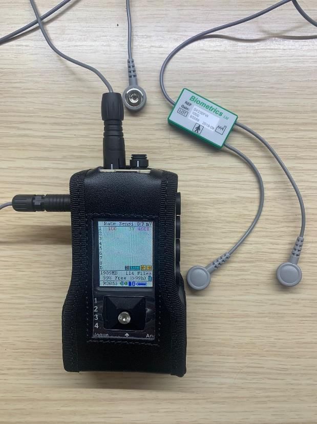
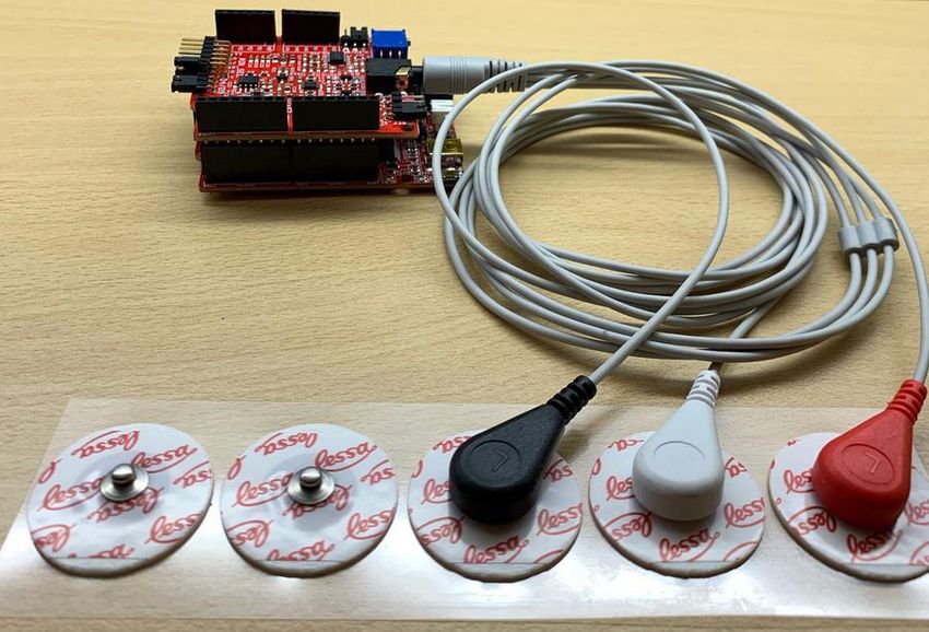
List of figures
Figure 1.1 Signal transmission schematic. Extracted from [1] ....................................................... 1
Figure 1.2 sEMG Biofeedback loop. Extracted from [5] ................................................................ 2
Figure 2.1 FREEEMG system. The sensors can be seen. Extracted from [8] ............................... 6
Figure 2.2 New sEMG device........................................................................................................ 6
Figure 2.3 Myo ArmBand. The bracelet includes the sensors Extracted from [15] ........................ 8
Figure 2.4 Mobile robot controlled with the MyoArmBand Extracted from [15] .............................. 8
Figure 2.5 Predefined gestures that the armband detects. Extracted from [15] ............................ 8
Figure 2.6 Lokomat RABWSTT System. Extracted from [18] ...................................................... 10
Figure 2.7 Elbow extension target activity Extracted from [19] .................................................... 10
Figure 2.8 EMG-Biofeedback activity Extracted from [19] ........................................................... 11
Figure 3.1 Trigno Research+ EMG System Extracted from [24] ................................................ 13
Figure 3.2 Noraxon Dynamometer Extracted from [25] ............................................................... 13
Figure 3.3 Sierra Summit EMG System. Extracted from [26] ...................................................... 13
Figure 3.4 Myontec EMG belt. It includes an shcematic of how it works. Extracted from [27] ..... 14
Figure 3.5 Cometa waterproof EMG System. Extracted from [28] .............................................. 14
Figure 3.6 ThoughtTech Biofeedback software Extracted from [29] ............................................ 14
Figure 3.7 Zukor Interactive Biofeedback game. Extracted from [30] .......................................... 15
Figure 3.8 Zukor Interactive Balance platform. Extracted from [30] ............................................ 15
Figure 3.9 Kangaroo activity, designed to target strength, duration, and progressive resistance.
Extracted from [31]....................................................................................................................... 15
Figure 5.1 Olimex EMG Shield. Personal source ........................................................................ 20
Figure 5.2 Olimexino-328 board. Personal source ...................................................................... 20
Figure 5.3 Cable electrodes in the upper part and electrodes in the lower part. Personal source21
Figure 5.4 Assembled EMG System. Personal source ............................................................... 21
Figure 5.5 Raw EMG signal in Arduino IDE. Personal source..................................................... 22
Figure 5.6 Electrodes placed for Arduino and DataLog signal recording at the same time. Personal
source .......................................................................................................................................... 22
Figure 5.7 DataLog EMG Device from Biometrics Ltd. Personal source ..................................... 23
Figure 5.8 DataLog EMG signal seen in its own software. Personal source ............................... 23
Figure 5.9 Arduino EMG signal histogram before normalizing intensity. Personal source .......... 24
Figure 5.10 DataLog EMG signal histogram before normalizing intensity. Personal source ....... 24
Figure 5.11 DataLog vs Arduino EMG normalized amplitude histogram. Personal source ......... 24
Figure 5.12 Arduino vs DataLog signal. High amplitude corresponds to bíceps voluntary
contraction. Personal source ....................................................................................................... 25
Figure 5.13 Empty Display Screen. Personal source .................................................................. 26
Figure 5.14 Diagram which shows origin and direction of increasing values in pygame surfaces.
Personal source ........................................................................................................................... 27
Figure 5.15 Screen with background and bird implemented. Personal source ............................ 28
Figure 5.16 Screen with background, bird and pipes implemented. Personal source ................. 28
Figure 5.17 Game Over screen. Personal source ....................................................................... 29
Figure 5.18 Pop-up box for fixing the threshold. Personal source ............................................... 30
Figure 7.1 WBS of the project. It has 3 main parts. Personal source .......................................... 32

 III
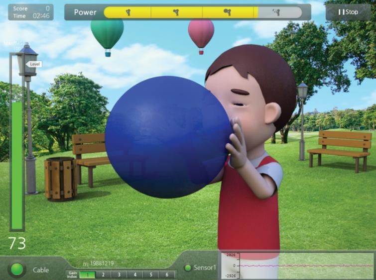
Figure 7.2 The GANTT chart gives an idea of the start and end dates of each task and is important
for monitoring specific tasks. The red bars indicate the activities from the critical path. Personal
source .......................................................................................................................................... 36
Figure 7.3 PERT diagram of the project. Each node includes the number, the Early and Last times.
The red lines indicate the critical path.. Personal source ............................................................. 37

 IV
List of tables
Table 4.1 Studied solutions for the hardware, programming environment, type of biofeedback
system, and biofeedback development method. .......................................................................... 16
Table 7.1 Dictionaries for each of the activities carried out in the project and references in the WBS.
..................................................................................................................................................... 34
Table 7.2 Used to calculate the final time for each activity defined in the WBS........................... 35
Table 7.3 The first column corresponds to the number associated to each WBS activity, in the
second column the letter associated to the activity, followed by the previous activities and then the
consequent ones. In the last 3 columns we find the duration of each task, its start time and its
ending time. ................................................................................................................................. 36
Table 8.1 SWOT matrix of the project. ........................................................................................ 38
Table 9.1 Cost analysis of the project.......................................................................................... 41

 V
INDEX
ACKNOWLEDGEMENTS ............................................................................................................... I
Abstract.......................................................................................................................................... II
 List of figures ............................................................................................................................ III
 List of tables .............................................................................................................................. V
1. Introduction ........................................................................................................................... 1
 1.1. Motivation ...................................................................................................................... 2
 1.2. Objectives ...................................................................................................................... 3
 1.3. Methodology .................................................................................................................. 3
 1.4. SCOPE and Limitations ................................................................................................. 3
 1.5. Location of the project .................................................................................................... 4
2. State of the art....................................................................................................................... 5
 2.1. EMG............................................................................................................................... 5
 2.1.1. EMG applications ................................................................................................... 7
 2.2. Biofeedback ................................................................................................................... 9
3. Analysis of the market ......................................................................................................... 13
 3.1. EMG............................................................................................................................. 13
 3.2. Biofeedback ................................................................................................................. 14
4. Conception engineering ...................................................................................................... 16
 4.1. Study of solutions......................................................................................................... 16
 4.1.1. EMG Hardware .................................................................................................... 16
 4.1.2. Arduino vs DataLog signal acquisition possibilities .............................................. 17
 4.1.3. Type of Biofeedback system ................................................................................ 17
 4.1.4. Biofeedback programming environment............................................................... 17
 4.1.5. Biofeedback development method ....................................................................... 18
 4.2. Proposed solution ........................................................................................................ 18
5. Detailed engineering ........................................................................................................... 20
 5.1. EMG system ................................................................................................................ 20
 5.2. Arduino signal and DataLog signal comparison ........................................................... 22
 5.3. Biofeedback system ..................................................................................................... 25
6. Experimental validation ....................................................................................................... 31
7. Project execution schedule ................................................................................................. 32

 VI
7.1. Work Breakdown Structure .......................................................................................... 32
 7.1.1. WBS Dictionary .................................................................................................... 32
 7.2. Task sequence matrix .................................................................................................. 34
 7.2.1. GANTT ................................................................................................................. 36
 7.2.2. PERT ................................................................................................................... 36
8. Technical viability ................................................................................................................ 38
 8.1. Strengths ..................................................................................................................... 38
 8.2. Weaknesses ................................................................................................................ 39
 8.3. Opportunities................................................................................................................ 39
 8.4. Threats ......................................................................................................................... 39
9. Economic viability ................................................................................................................ 41
10. Normative and legal aspects ............................................................................................ 42
11. Conclusions and future steps ........................................................................................... 43
12. References ...................................................................................................................... 45
Appendixes .................................................................................................................................. 49
 Appendix 1- EMG Arduino Code ............................................................................................. 49
 Appendix 2- Save Arduino signal in a txt file............................................................................ 49
 Appendix 3- Arduino signal and DataLog signal comparison................................................... 49
 #Import needed libraries .................................................................................. 49
 Appendix 4- Flappy Bird EMG-Biofeedback ............................................................................ 51

 VII
1. Introduction
As you know skeletal muscles are the motors that allow humans to move. The mechanism of action
of muscles is a very complex procedure which incorporate signal transmission along nerve fibres
and across neuromuscular junctions, electrical activation of the muscle fibres, which are organized
in elementary units known as motor units, which finally produce forces that act on the tendons of
the muscles and allows bones movement. ElectroMyoGraphy is a technique which evaluates the
health condition of all this procedure. It translates the signal into graphs or numbers which can be
used for many applications such as diagnosis or treatment. [1]

Understanding EMG signals implies the understanding of muscles and the way they generate
bioelectrical signals. It implies understanding how specific mechanisms and phenomena influence
the signals, as well as the inverse problem, which is even more difficult, and consists of
understanding how signals reflect certain mechanism and phenomena and allow their identification
and description [1].

The electric signal is produced in the cortex, and
travels all along the spinal cord until it arrives to the
motoneuron, which transmits the signal to the
skeletal muscle. Figure 1.1 shows a simplified
schematic of the signal transmission [1].

There are two types of EMG: neurologic EMG and
kinesiologic EMG. Each one has some advantages
and disadvantages shown in Table 1.1.

 Figure 1.1 Signal transmission schematic. Extracted from [1]

 Neurologic EMG (nEMG) Kinesiologic EMG (sEMG)
 Position Directly in the muscle In the surface
 Invasiveness Invasive, painful. Use of Non-Invasive. Use of
 needles electrodes
 Complexity Highly complex, need of high Simple, lower knowledge
 anatomy knowledge needed
 Precision High precision, detects signal Low precision, limited to
 from small muscles, individual surface and big muscles.
 action potentials Difficult to get signal from an
 isolated muscle
 Table 1.1 Neurologic vs Kinesiologic EMG

EMG applications have been growing since the development of this technology. Numerous
applications for this technology have been developed in clinical practice, such as diagnosing of
neuromuscular diseases analysing and determining abnormalities or for treatment purposes which
could be improving ergonomics or muscular rehabilitation, what is called biofeedback. Biofeedback
is defined by the Association for Applied Psychophysiology and Biofeedback (AAPB) as a process
that provides real time information from psychophysiological recordings about the levels at which
physiological systems are functioning. This means that by means of sEMG, biofeedback measures

 1
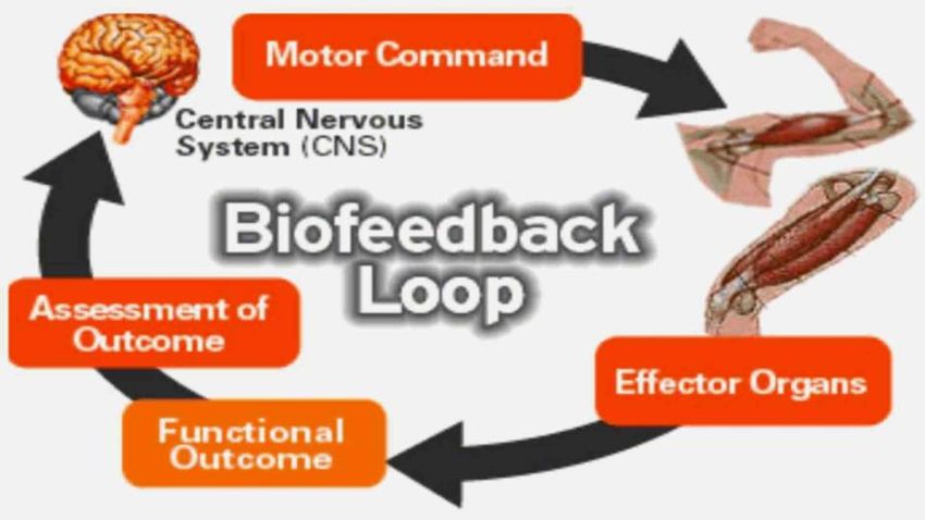
and transforms the physiological information from muscles into visual and/or audio signals [2, 3, 4,
5].

When an injury occurs, not only bones, ligaments and muscles are hurt, but also special nerve
receptors. These receptors, found in muscles, send great amount of information back to the brain.
If during rehabilitation the receptors are not retrained, injury is more likely to reoccur. Is in this
situation where biofeedback becomes important. It helps the patient to retrain the muscles and
receptors thank to the outcome that they receive. The loop of biofeedback is shown in Figure 1.2.
The brain produces a command which arrives to the effector organs, the muscles. Thanks to sEMG,
the signal can be monitored, and an outcome is obtained. This raw outcome is a graph, which does
not make any sense to the patient. Therefore, it must be processed and transformed in another
outcome more recognizable to the patient, so the signal is transformed into an acoustic or visual
feedback [5].

 Figure 1.2 sEMG Biofeedback loop. Extracted from [5]

 1.1. Motivation
Since very young I have suffered from different injuries myself such as elbow luxation, collarbone
fracture or knee injury, therefore I have had to attend rehabilitation several times and during
different stages of my life, therefore I have been able to experience myself the evolution of the
technology for rehabilitation.

In addition, I have always had a great interest on technology, and my own experience showed me
how technology can improve people’s quality of life, but also that many times rehabilitation
processes are tough and boring, which can lead to the abandonment of the process. When I saw
the opportunity of developing a project in that field, I thought that it could be a great opportunity to
apply my own experience and to gain knowledge.

Initially the project was focused on shoulder rehabilitation and acoustic biofeedback, but during the
development of it, the director and me decided that it could be better to generalize to the
rehabilitation process since biofeedback could be applied to many muscles of the body. Also, the
biofeedback system was changed to a more complex type, which would be much more interesting
both for me as a student and the challenge that it would suppose completing it, and for the project

 2
itself since it would make more motivating the rehabilitation process, fact that as it will be explained
later, is a very important point.

 1.2. Objectives
This work was based on the belief that rehabilitation procedures could be improved to make them
more friendly and effective. A study in Argentina shows that exists a highly abandonment ratio of
rehabilitation procedures [6]. I believe that a more entertaining rehabilitation procedure would
reduce this abandonment percentage and biofeedback could have a fundamental work on it.

The main goals of this project were the following:

 - Assess the use of biofeedback and EMG in rehabilitation.
 - Compare the signal obtained in the low-cost EMG systems with a professional EMG
 system.
 - Develop an EMG and biofeedback low-cost system.
 - Clinical trial of the project in Institut de Biomecànica Clínica.

 1.3. Methodology
The project was performed with the help of José Luis Parreño and it consisted of the following
stages:

 • Stage 1: Bibliographic research of both technologies was performed to get familiarized with
 the topic. It was focused on the applications that EMG and biofeedback can be used.
 • Stage 2: the development of the EMG and biofeedback system was performed. This phase
 consisted of getting the needed material for the project and the development of the
 software needed for both obtaining the EMG signal and the development of the
 Biofeedback interface.
 • Stage 3: Comparing the EMG signal of my prototype with a DataLog from Biometrics Ltd.
 A professional EMG system.
 • Stage 4: This last phase consisted mainly in writing the present report. It consists of an
 introduction of both technologies, then a description of the possibilities we had for the EMG
 and biofeedback development and the final solution. Next the whole process of developing
 the system. Then the technical and economic viability, and the timeline. Finally, a
 conclusion of the project.

 1.4. SCOPE and Limitations
As this was a final degree project, time has been an important limitation. It has been carried out
from February 2021 to June 2021. The main limitation where time affected is that it would have
been interested to apply this project for patients to perform the efficacy of the system, but
biofeedback rehabilitation procedures need a much larger time window, which made impossible to
complete this objective.

Covid-19 pandemics has been another large limitation, which forced to perform from distance this
project as much as it was possible. Probably if this pandemic did not happen, we could have started
the project in April 2020 and the clinical study could have been performed. This also caused that
the meetings with the director had to be online and I could not attend the biomechanics laboratory

 3
where the project was intended to be performed, what would have helped me on understanding
better the applications of Biofeedback.

Considering all these limitations the Scope of this project included:

 • Bibliographic research of EMG, Biofeedback applications and types of biofeedback
 currently used.
 • Development of an EMG prototype using Arduino
 • Development of a Biofeedback software
 • Comparing EMG signal of my prototype with the DataLog system.
 • Discussion of the differences between the signals

 1.5. Location of the project
The project described has been performed in collaboration with José Luis Parreño, director of the
Institut de Biomecànica Clínica.

 4
2. State of the art
To develop an EMG system and an effective biofeedback software, it has been necessary to
understand at which point is the current technology and some biofeedback systems to get an idea
of the software I could develop.

 2.1. EMG
To correctly perform a sEMG analysis, several points are needed to be took in account. There are
different variables that will lead to wrong results if they are neglected.

First of all, the electrode shape. It is defined as the shape of the conductive area of the SEMG
electrodes. In literature both square and circular electrodes are reported to be used for SEMG
recordings. As SENIAM project (Surface ElectroMyoGraphy for the Non-Invasive Assessment of
Muscles) states, not much difference in performance and pick up can be expected. As long as the
total surface are for both electrodes is the same, the skin impedance of both electrodes will almost
be equal, so no influence can be expected. However, the European inventor showed that circular
electrodes are preferred [7].

Second, the electrode size. It is defined as the size of the conductive area of a SEMG electrode.
This parameter is important since an increase of the size, is expected that the view of the electrodes
increases. It can be shown that has an integrative effect on the SEMG signal, increasing the
detected amplitude and decreasing the high frequency contents. In general, it is important that the
size of electrodes is large enough to be able to record a reasonable pool of motor units, but small
enough to avoid crosstalk from other muscles. Both the SENIAM and the European inventor
recommend electrodes of 10mm of diameter maximum [7].

Third, the inter electrode distance. It is defined as the centre-to-centre distance between the
conductive areas of 2 bipolar electrodes. The influence of the inter electrode distance on the
recorded area and crosstalk is a relevant item. SENIAM recommend an inter electrode distance of
20mm. If the SEMG is performed in small muscles, the distance should not exceed ¼ of the muscle
fibre length. This will avoid unstable recording due to tendon and motor endplate effects [7].

The preparation of the skin is a very important factor to get a good electrode-skin contact. It will
help to obtain better SEMG signal and lower noise and artifacts. There are different techniques and
combinations in this field, but SENIAM recommends shaving the patient, and then clean it with
alcohol and allow it to vaporise so the skin is dry before electrodes placement [7].

Surface EMG devices have been developing since 1950. In that time the devices used analogic
systems. From that point the systems have been continuously evolving, developing digital systems
to the actuality, which most of the are portable devices with wireless sensors. One of the most
advanced EMG devices is FREEMG. It is a SEMG device which uses 4G technology. It
distinguishes from its competitors for the high signal accuracy, the absence of wires, the lightness
and reduced size of the probe and many other properties which allows to perform analysis of any
type of movement, for any muscle altering the minimum possible the movement of the subject. It is
connected to the PC through a USB receiver, which can manage up to 20 sensors simultaneously.
Even though, the sensors have an internal memory to ensure that the signal is recorded in case

 5
that a temporary disconnection occurs, and to allow recording in wide places where the subject can
be far from the USB receiver [8].

 Figure 2.1 FREEEMG system. The sensors can be seen. Extracted from [8]

Some of the problems of this type of cutting-edge technologies are that they are expensive and
difficult to operate, which make impossible to use by non-specialists. Hence a new sEMG device,
which is defined as simple to use and highly accessible, has been developed for people who are
not familiar with EMG. But before using it in professional activities, the validity and reliability must
be stablished. To do so, this new sEMG system, showed in Figure 2.2, was compared to the
aforementioned FREEEMG. The signal was recorded in participants which performed three
maximal voluntary isometric contractions for 5 seconds each with a 5-minute rest between each
one. The electrodes were placed following the SENIAM protocols, and the signal was recorded at
the same time. After analysing the recorded data, it was confirmed that the new system valid
enough to monitor muscle activity during daily life exercise [9].

 Figure 2.2 New sEMG device.

During or after the acquisition process, different signal filtering and processing procedures are
used. First, a denoising step is performed. It can be denoising by hardware or by software. The
hardware denoising improves the performance of the device using physical filters. The software
denoising is done using filters or wavelet transforms. The wavelet transform is an extension to
traditional Fourier transform. Its coefficients have different characteristics at each scale of noise

 6
and signal, so the idea is to remove these components generated by noise at each scale. Then the
inverse transform is used to reconstruct the original signal [10].

If a comparison between analog and digital filters is started, it will not end fast. In one hand, analog
filters are faster which reduces the delay to 0, but they need of several components which can
reduce its accuracy due to temperatures or difference in manufacturing. Analog filters then,
increase power consumption and cost. In the other hand, digital filters may be slower, but since in
EMG acquisition devices, the sampling frequency is at the range of kHz, the delay is negligible. In
addition, they have other advantages such as, they act exactly equal, no matter the circumstances,
and allow changing the notch frequency from 50Hz in Europe to 60Hz in North America easily by
changing only one parameter. Furthermore, it allows the implementation of real-time algorithm such
as artifact suppression or other devices control [11].

The 50hz signal is one of the main signals that must be removed from any acquired data. It can be
done using analog or digital filter. In this case, by the reasons explained before, a comb digital filter
was used. This comb filter was designed as a high pass filter and fixing a sampling frequency of
50hz, that Is the frequency to be filtered. A Butterworth and Chebyshev filter was used. The filter
was designed using Matlab FDA filter tool. First, the filter zeros, poles and gain were determined
as if it was an analog lowpass filter. Then, by bilinear transformation, the analog filter was
transformed into a digital filter, and then to a zero-pole-gain form again. It gives as result the filter
coefficients as floating-point numbers [11].

In EMG recordings other types of noise appear such as artifacts. Artifacts in EMG are different
errors in the signal that can come from different sources such as cable movements or relative
movements of the muscle to the EMG sensor, for example at the beginning and the end of a
contraction. This error must be removed to obtain the clearest possible signal, an option is using
digital high pass filters, since artifacts are in the range of 0-20Hz, and a cut-off frequency at that
range can be fixed. After an evaluation of the filter using different cut-off frequencies, the optimal
signal was obtained with fc=60Hz with a Chebyshev filter. In addition, two lowpass filters were
applied. One of them removes the high frequency noise after the high pass, and the other performs
a smoothing to get a DC output signal. The fc was fixed at 531Hz for removing high frequency, and
3.1Hz for the smoothing step [11].

 2.1.1. EMG applications
EMG applications have been growing during last years as consequence of the development of new
technologies such as machine learning and artificial intelligence. Many of the new uses of EMG are
related to gesture recognition. The increase in computing power has brought the presence of many
computing devices in the daily life of human beings. Hand Gesture Recognition, from now on HGR,
models are human-computer systems that determine which gesture was performed. HGR models
need acquiring big amount of data using different sensors such as inertial units, gloves, and SEMG
sensors. All of them have limitations, for examples gloves cannot be used by amputees, and SEMG
generate noisy data. Even though, all these sensors collect date related to a real movement, EMG
also extracts the intention of the movement, therefore it might be useful with amputees who cannot
execute the movements. As it has been explained, EMG record the electrical activity of skeletal
muscles which have two types of contractions: static and dynamic. Each of them can be modelled

 7
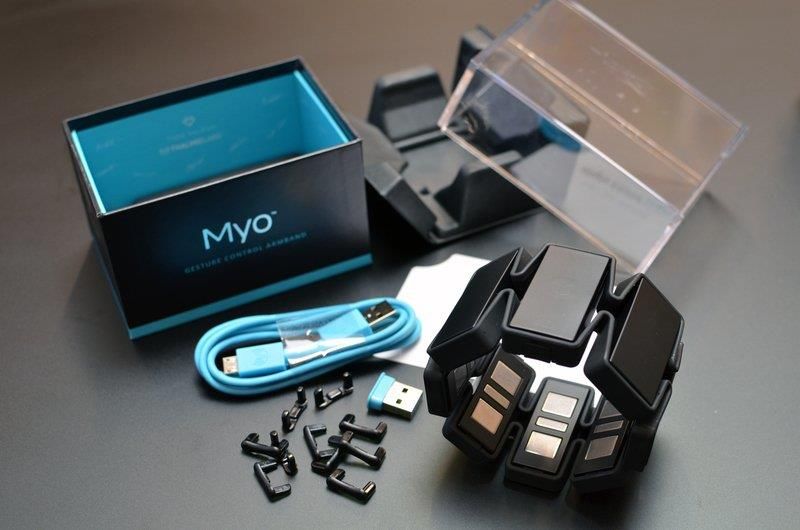
using mathematical models, and the combination of the two of them results on an EMG model.
However, the mathematical models are not used in HGR as consequence of the parameter
estimation difficulty [12,13,14].

Therefore, machine learning is used. It can obtain a solution using different techniques. By applying
ML, a wide range of possibilities for myoelectric control opens, and it allows using EMG signals to
control prostheses or drones. In conventional amplitude-based control, one EMG channel us used
for controlling only one function of the device, and when it exceeds a predefines threshold, this
function is activated. For many applications HGR models are required to work in real time so one
the user performs an action, the system gives him a response, but it is important that this response
is fast enough to be perceived as instantaneous [12].

An example of this application is Myo Armband. It is a method for gesture control which is equipped
with EMG and Inertial sensors. The inertial sensor uses an accelerometer, a gyroscope, and a
magnetometer to obtain the maximum precision. With the combination of these two different
sensors, a mobile robot can be controlled [15]. The following figures show the connection between
the two devices:

 Figure 2.3 Myo ArmBand. The bracelet includes the sensors Figure 2.4 Mobile robot controlled with the MyoArmBand
 Extracted from [15] Extracted from [15]

 .

The user receives feedback from the device withExtracted from indicators:
 two different [5] LEDs and a haptic
feedback vibration. When a movement is produced the armband detects the electrical signal, and
comparing the signal detected by the different sensors, it can be classified in 5 predefined gestures
which are shown in Figure 2.5 [15].

 Figure 2.5 Predefined gestures that the armband detects. Extracted from [15]

Each of these gestures will produce a movement to the robot, which can be go forward, backwards
turn left, turn right, or stop. In this article it is shown that the robot control successfully exhibited
real-time human-robot interaction through hand gesture using a low-cost SEMG device. However,

 8
it also has some drawbacks. For example, it cannot be used for medical applications such
controlling robotic prostheses since these need six degrees of freedom axes, and the Myo has a
smaller number of degrees. In addition, the accuracy and precision could differ between users or
in cases where the muscles have impaired development [15].

Apart from these complex applications, EMG can be used for many rehabilitation purposes to
perform and study and evaluation of the process. Incidence of surgery in shoulder lesions affecting
the rotator cuff have increased a 238% from 1995 to 2009 in the United States. Many reports
indicate failure after surgery, in most cases within the first 3 to 6 months post-surgery. Different
factors affect the outcome, such as the size of the injury, the tissue quality, or the location, but
postoperative rehabilitation protocol is one of the most important factors. After a rotator cuff surgery,
it is important to improve range of motion in the early stages, but without overloading surgical repair
which could lead to failure. The gold-standard for evaluating muscle activation and therefore,
loading in limb, is EMG. It is important to categorize the activation into different levels, to control
when it could be harmful to the rotator cuff. Different reports indicate that an activation greater than
15% could be harmful, therefore it is important to control muscle activation at any moment of the
rehabilitation process. To do so, a control group was used which performed different exercises and
the professionals assessed the percentage of activation that every exercise caused. Then, the
exercises which had a lower activation than 15% were in the users. There were four types of
exercises: actives, passives, active-assisted with the non-affected limb, active- assisted with bars.
The three first cases showed activation below the 15% of the maximum, but the last one, showed
higher. If EMG were not used, the exercise would still be used in the rehabilitation process, which
could lead to a bad recovery [16].

 2.2. Biofeedback
Since biofeedback can be used to train a specific function of the body and improve its voluntary
control, several medical applications have appeared. It is often used to train neuromotor control
during rehabilitation processes. Neuromotor control consists of selecting the right fiber types and
activate them with precise timing. For such purposes, different biofeedback systems can be used.
One of the simplest systems is drawing a graphic line. Initially the line is horizontal and is asked to
the patients to raise the line without altering the pace or direction of the studied movement. After
the trial, the data recorded using sEMG was processed and it was shown that with an appropriate
training paradigm, most skeletal muscle groups that are voluntarily activated could be trained to
increase their coherence [17].

Based on the above, biofeedback can be used for simple purposes such as muscular rehabilitation
after an injury, to more complex such as training the walking ability on people with nerval damage.
Robotic-assisted body weight sported treadmill training (RABWSTT) has been used in gait
rehabilitation for people with neurological conditions, It reduces fatigue of both the patient and the
clinician what enables prolonged walking training. In addition, it provides guidance in the lower
limbs movement, and this type of extensive exposure to a task-specific repetitive training helps
promote reorganization of the primary motor cortex and functional outcomes can be improved in
patients with neurological conditions like spinal cord injuries. In this case a Lokomat system was
used for RABWSTT, which is a system used for gait training and rehabilitation, and it was combined
with an EMG-biofeedback system. It is important that during the training, the patient has an active

 9
participation, because in the contrary, it will not improve as much as he should. That is why
biofeedback is used. In this case, an audio feedback was generated if the muscle activation was
less than 30% of maximal recruitment to encourage active participation during the stance phase of
the gait cycle. In most neurological diseases high repetition of task-specific training with proper
sensory feedback are essential elements for neuroplasticity after spinal cord injury. EMG-
biofeedback systems enhance muscle contraction, and it has been proven that visual and audio
feedback can promote muscle recruitment,
increase muscle performance, and promote
better improvement in lower limbs
performance in people with neurological
conditions. It was also used to monitor
muscle contraction and enhance modulation
in central nervous system without any
adverse effect, what indicates that it is a
feasible and safe way to promote
participation of subjects during training. So,
the use of EMG-biofeedback during
RABWSTT is a valid treatment for promoting
independent walking ability what can help
improve independence in daily activities in Figure 2.6 Lokomat RABWSTT System. Extracted from [18]
people with SCI and enables them to enhance
their walking endurance that promotes social re-integration [18].

Biofeedback has been used in children with spastic cerebral palsy (CP) which can suffer from
muscle imbalance in the elbow joint and associated reaching movement incoordination. Different
treatments such as neurodevelopmental treatment, strengthening or resistance training have been
sued, but outcome results have been variable. These methodologies do not provide accurate or
quantified biofeedback about muscle activation imbalance as it would in EMG biofeedback
trainings, and in addition, undesirable compensatory movement cannot be controlled. Therefore, a
hybrid model of EMG-VR system has
been recently developed to provide
accurate biofeedback and motivation to
restore muscle imbalance between the
triceps and biceps during elbow
reaching movements in children with
CP. This system is designed to provide
a real-time visual feedback about
triceps and biceps muscle activation
patterns, during functional VR games.
These games were designed to
 Figure 2.7 Elbow extension target activity Extracted from [19]
improve self-motivation, so they were
enjoyable and improve functional
strength [19].

 10
This system provides real-time audio-visual biofeedback information about muscle activation which
enabled the patient to acquire the motor task more consciously at the cortical motor control level,
which is essential for biofeedback training. The game consisted of a blowing balloon VR game
which was provided for triceps activation. The VR system provides a fun experience and improves
motivation for children to perform elbow extension more implicitly minimising undesirable
compensatory movement patterns. This game was used for increasing triceps muscle contraction
while inhibiting biceps activity. It was compared the immediate effects of EMG biofeedback and
EMG-VR feedback, and the latest methodology showed superior improvements [19].

 Figure 2.8 EMG-Biofeedback activity Extracted from [19]

Biofeedback has been used for other type of diseases such as disorder that affect the pelvic floor.
In such cases, biofeedback is used along with EMG sensors. This therapy is designed to improve
abdominal push effort and its coordination during defecation, anal sphincter tone at rest and
voluntary contraction of it. In this case a visual biofeedback display is used, seeing a visual color-
coded graphic of anal canal and rectal pressure, patients’ objective is to squeeze the anal sphincter
muscles, without co-contracting the brace muscles and maintaining chest breathing. Using this
therapy, patients which have increased rectal sensation can be trained to tolerate larger rectal
volumes and patients with hyposensation can recognize lower rectal volumes and contract the
pelvic floor muscles in response to rectal distension [20].

Another very common disease is low back pain. It can have different origins such as heavy lifting
or a bad posture at your workplace. Therefore, health professionals often prescribe postural training
as preventative measure to reduce forces on the lumbar spine when lifting. This therapy may lead
to short-term changes in behaviour, but the long -term effects are uncertain. In this case
biofeedback was provided by two inertial sensors attached to the lumbar spine. These inertial
sensors measured the range of motion of the lumbar spine, and when the 80% of range of motion
was achieved, an audio biofeedback was produced by a software. The 80% threshold was set
because when it is exceeded, passive loading on the lumbar spine significantly increased. When
the results were compared with a non-biofeedback group, a positive outcome was determined. The
differences of flexion in the same task at the end of a 20 minute session the non-biofeedback group
performed almost full flexion for lifting tasks, while the biofeedback group only 64% [21].

 11
Biofeedback devices can be used for postural training to those that hold a static posture during
prolonged computer work and can develop neck pain. The device used for the detection of the
posture was Lumo Lift which is attached to the skin below the mid-clavicle. The subjects performed
the same task with and without the biofeedback system, and after analysing the results, it is shown
that once more, biofeedback task giver better results, however, long-term effects are not
investigated [22].

Biofeedback-based videogames can be also used on improving balance challenges in autism
spectrum disorder which a relatively high number of individuals struggle with postural stability. A
visual-biofeedback system was implemented through a videogame, to perform a balance training
that improves postural stability. Commercial videogames may not provide sufficient biofeedback to
improve balance performance in autism, hence a prototype was developed with a simple interface
and the possibility of hardening the difficulty to personalize the game depending on the capabilities
and improvements of the subject. Existing devices such as Microsoft Kinect and Nintendo Wii
balance board were used since these allow monitoring of balance and posture. To improve
motivation, a training with the designed videogame and genuine Nintendo games was developed.
After a six-week period training, participants improved balance times in the prototypes game. On
average, participants almost doubled the amount of time that they were capable of standing at one
foot. As conclusion, these findings suggest that visual-based biofeedback training improves
balance in autism [23].

 12
3. Analysis of the market
 3.1. EMG
There are several EMG devices in the market, each of them has different properties and functions,
hence it is important to evaluate the application that it will be destined for and then decide which
fits better.

Delsys is a company which has different sEMG systems depending on the need of each client. It
is mainly focused on research groups and hospitals. They offer systems with EMG and inertial
sensors such as Trigno Research+,
which can detect signal from up to 32
wireless sensors simultaneously and
allows integration with third-party
software plugins. There are different
sensor depending on the application,
for example, for gait analysis, fingers,
face, motor control or dynamic
movements. Each of them has a
different sampling frequency, for
example the gait analysis has a
maximum sampling frequency of Figure 3.1 Trigno Research+ EMG System Extracted from [24]
4370Hz, and the face sensor up to
2222Hz [24].

Another system which also uses inertial sensors is Ultium EMG from
Noraxon and also has up to 32 EMG channels available, but in this
case the maximum sampling frequency is 4000Hz. However, the
software allows to select high pass or low pass filters with
predefined cut-off frequency. It allows connecting other sensors
such as a hand grip dynamometer which measures grip strength.
The combination of EMG recording with the dynamometer allows a
better understanding of the injury. Noraxon’s software is one of the Figure 3.2 Noraxon Dynamometer
most popular part of the system since it is extremely user-friendly Extracted from [25]

and offers extensive reporting options [25].

A system which offers a very different point of view is Sierra Summit
from Cadwell. It can be used with surface electrodes or needles. This
system is the first EMG system which offers a fully integrated
ultrasound system, what improves safety margins for the needles’
injection. It also allows deep nerves stimulation. Compared to the
Noraxon and Delsys systems, it offers only 12 EMG channels and
does not include inertial sensors. It is highly customizable, fact that
allows building the system that covers the needs that the therapist
 Figure 3.3 Sierra Summit
needs [26]. EMG System. Extracted from
 [26]

 13
A totally different approach is made by Myontec. This company designed textile products which
incorporate EMG sensors. Its products are destined to athletes in therapy or in training session with
a professional. They have different products such as a shirt or shorts, which have an 8 channel
EMG device. Its latest product is a belt which is used for measuring low back EMG. This belt was
developed for identifying factors behind low back pain, therefore can be used by athletes but also
by workers who do repetitive lifting tasks [27].

 Figure 3.4 Myontec EMG belt. It includes an shcematic of how it works. Extracted from [27]

Even though lately many EMG wireless sensors have appeared,
they were not designed for aquatic sports. Cometa systems offers
the first waterproof EMG sensors. This approach is really interesting
for assessing possible injury risk in swimmers. It also has an inertial
sensor on board and up to 16 EMG channels with accelerometers.
Its sampling frequency can be fixed up to 2000Hz. In addition, the
EMG systems from Cometa have been developed to have a fixed
delay from the acquisition of the signal to the analysis output. This
delay can be removable when used with other EMG systems and it
 Figure 3.5 Cometa waterproof
is so short that does not affect in any way real time monitoring of EMG System. Extracted from [28]
the signals [28].

 3.2. Biofeedback
One of the biggest companies in the biofeedback
systems field is ThoughtTech. They offer
measure instrumentation but what it is more
important the biofeedback systems. They offer
different hardware and software packages
depending on the needs of the customer. Its EMG
biofeedback systems use up to 3 EMG sensors
which are connected to a computer. The signal
received by the computer is used for controlling
the games that they developed [3, 29]. Figure 3.6 ThoughtTech Biofeedback software Extracted
 from [29]

 14
They have partnered with Zukor interactive, which have different videogames depending on the
muscle rehabilitation purpose: strength, resistance, explosiveness, and other muscular capabilities.
They offer games from different fields such as sports, cars or animals what helps them to increase
the public and maintain the motivation among their patients. In the actuality they are developing
some VR games, which will improve the immersion and the training even more. In its offer there is
a tool which is used for balance analysis and training [30].

 Figure 3.8 Zukor Interactive Balance platform.
 Figure 3.7 Zukor Interactive Extracted from [30]
 Biofeedback game. Extracted from
 [30]

 Accelerated Care Plus designed a sEMG tool which is
used in the treatment of dysphagia. The tool is named Synchrony and enables using VR activities
to visualize therapeutic swallowing activity. The different activities included in the tool are work-rest
cycles, the diver, designed to target tongue base retraction, endurance, and specificity of lingual
movement; the Kangaroo designed to target strength, duration, and progressive resistance; and
the bow and arrow which is designed to target skill-based training and timing. This tool is
continuously improving thanks to the feedback received from final users and dysphagia researchers
[31].

 Figure 3.9 Kangaroo activity, designed to target strength, duration, and progressive resistance. Extracted from [31]

 15
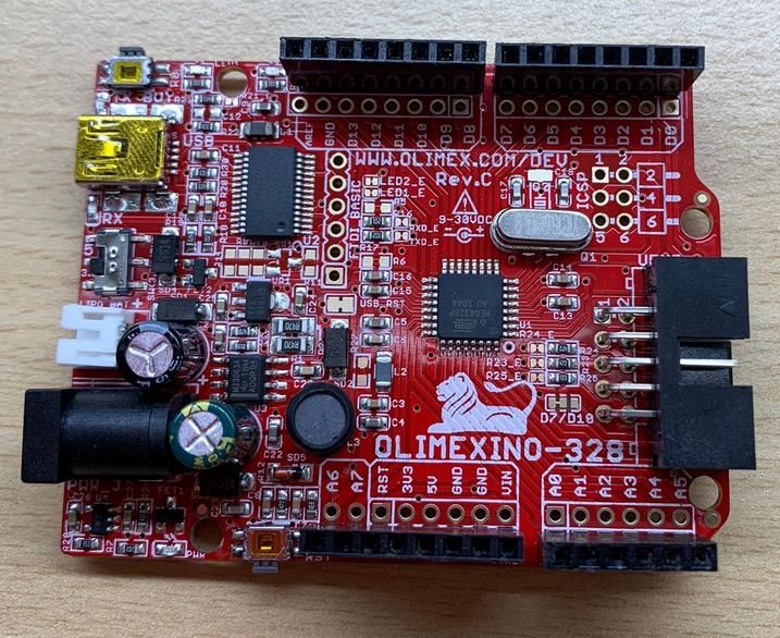
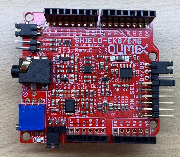
4. Conception engineering
In the following section, the possible hardware used, the possible programming environment, the
type of biofeedback system and the method for developing the system is discussed. The different
options are shown in Table 3.1. The final solution is described into further detail in the detailed
engineering section.

 Studied solutions
 EMG Hardware - Individual components, using
 resistances, capacitors, and
 operation amplifiers.
 - Integrated circuits and
 microcontrollers.
 Arduino vs DataLog - Record signal at different times.
 - Record signal at the same time.
 Type of Biofeedback system - Visual (LED).
 - Acoustic (Sound).
 - Complex (Game).
 Biofeedback programming environment - Python.
 - C
 Biofeedback development method - Integral development of the game
 - Downloading a game and adding
 biofeedback needed features
Table 4.1 Studied solutions for the hardware, programming environment, type of biofeedback system, and biofeedback
development method.
 4.1. Study of solutions
 4.1.1. EMG Hardware
For the EMG system there were two options, make us of protoboard and individual components or
using integrated circuits and microcontrollers such as Arduino.

The use of a protoboard and individual components is a more complex solution. It would have been
needed a highly study of all the properties of an electronic circuit, such as the gain to get a signal,
the frequencies in which the EMG signal works, and therefore a complex study of all the
components needed to perform the different filters needed. An advantage of this solution is that it
has the option of including an Isolation phase, what would give the prototype the possibility of
passing official tests and laws such as EN-60601, to be certified as a medical device.

In general, it would have made the project much larger, and taking in consideration the limitations
explained before, it would have difficult the development of the biofeedback system.

To test all these components and the correct implementation of the circuit it would have been
needed many other instruments such as oscilloscopes and waveforms generators, that would have
made more difficult the task due to covid limitations, or buying a all-in-one instrument such as
Analog Discovery 2, which would have make a lot more expensive the project.

Arduino is an Open-source electronic prototyping platform which enables users to create interactive
electronic objects. In this case I was needed of a basic microcontroller board and a board which
integrated the EMG circuit. This solution provided me the possibility of dedicating more time to the

 16
biofeedback system, which is more important in the project. Once I received the two components
needed, I only had to develop the software, which was not difficult thanks to Arduino software that
provides an easy environment to control this microcontroller.

 4.1.2. Arduino vs DataLog signal acquisition possibilities
Comparing the two different signals is difficult, since it is important to record the same amount of
data, with the same sampling frequency, and the acquisition points as near as possible. I had two
options. Recording the signal at different times allows to use the electrodes exactly at the same
point, but other problems appear. It is impossible to reproduce the same exact signal two different
times, and it is impossible to superpose the two signals because the contraction would take place
at different times.

Recording the signal at the same time has the opposite advantages and disadvantages. The
electrodes cannot be at the same point, but the signal is the same and it allows me to superpose
them in order to compare the signals from the different devices.

 4.1.3. Type of Biofeedback system
The biofeedback system could have been implemented using mainly a visual, an acoustic or a
more complex system.

Visual biofeedback can be applied using binary systems such as a simple LED, that is powered-on
or powered-off, a digital system showing numbers depending on the activation level or a graph that
shows continuously the signal. It has the disadvantage that the patient needs to be in front of a
screen, therefore when the biofeedback is applied to rehabilitation procedures in which the patient
must move along the room, it makes difficult to perform it [32].

Acoustic biofeedback is based on an individual sound or multiple sounds that could be used for
playing a song. It can also be binary, which indicates if the activation threshold has been reached.
An advantage of this modality is that is can be used in daily activity, allowing to give feedback to
the patient only when is needed, to make sure that he is focused on his daily tasks. It also solves
the problem of visual biofeedback because the sound can be listened even though the patient is
far from the feedback source [32].

Complex biofeedback makes use of both previous modalities. This system is generally
implemented in different types of games, fact that makes the rehabilitation procedure more
entertaining and motivating. The main disadvantage is that it is more complex to carry out since a
game must be programmed [32].

 4.1.4. Biofeedback programming environment
Python has large library of built-in functions library which facilitates programming. For my purpose,
it has pygame library which facilitates programming a game and its controls. In general, it is a
simple language and environment, and it is easy to learn and write and read.

C is a more complex language, with a limited number of built-in functions. The syntax is harder;
therefore, it is more difficult to learn, write and read.

 17
You can also read