Refracture of the cemented vertebrae after percutaneous vertebroplasty: risk factors and imaging findings
←
→
Page content transcription
If your browser does not render page correctly, please read the page content below
Xiong et al. BMC Musculoskeletal Disorders         (2021) 22:459
https://doi.org/10.1186/s12891-021-04355-w

 RESEARCH ARTICLE                                                                                                                                  Open Access

Refracture of the cemented vertebrae after
percutaneous vertebroplasty: risk factors
and imaging findings
Yu-chao Xiong1†, Wei Guo2†, Fan Xu1, Ci-ci Zhang1, Zhi-ping Liang1, Li Wu1, Song Chen1 and Xu-wen Zeng1*

  Abstract
  Background: To determine the related imaging findings and risk factors to refracture of the cemented vertebrae
  after percutaneous vertebroplasty (PVP) treatment.
  Methods: Patients who were treated with PVP for single vertebral compression fractures (VCFs) and met this study’s
  inclusion criteria were retrospectively reviewed from January 2012 to January 2019. The follow-up period was at
  least 2 years. Forty-eight patients with refracture of the cemented vertebrae and 45 non-refractured patients were
  included. The following variates were reviewed: age, sex, fracture location, bone mineral density (BMD),
  intravertebral cleft (IVC), kyphotic angle (KA), wedge angle, endplate cortical disruption, cement volume, surgical
  approach, non-PMMA-endplate-contact (NPEC), cement leakage, other vertebral fractures, reduction rate (RR), and
  reduction angle (RA). Multiple logistic regression modeling was used to identify the independent risk factors of
  refracture.
  Results: Refracture was found in 48 (51.6%) patients. Four risk factors, including IVC (P = 0.005), endplate cortical
  disruption (P = 0.037), larger RR (P = 0.007), and NPEC (P = 0.006) were found to be significant independent risk
  factors for refracture.
  Conclusions: Patients with IVC or larger RR, NPEC, or endplate cortical disruption have a high risk of refracture in
  the cemented vertebrae after PVP.
  Keywords: Vertebroplasty, Risk factors, Magnetic resonance imaging, Spinal fractures, Bone cements

Background                                                                            embolism, fractures in the adjacent vertebrae, and
Percutaneous vertebroplasty (PVP) is a minimally inva-                                refracture of previously treated vertebrae have been re-
sive technique for the treatment of vertebral compres-                                ported [6–10]. However, recompression in cemented
sion fractures (VCFs). Most clinical studies [1–5] have                               vertebrae may lead to aggravation of the kyphotic de-
reported that this treatment can provide immediate pain                               formity, vertebral height loss, and even compression of
relief and biomechanical stability, and restore partial ver-                          the spinal cord by vertebral body fracture, which usually
tebral height. Despite these excellent clinical results,                              requires further treatment [8, 9]. Some researchers [8,
complications such as cement leakage, infection,                                      11, 12] believe that cement distribution patterns may be
                                                                                      an important predisposing factor to refracture. Kim [13]
* Correspondence: gzshszhyyfsk@163.com                                                reported that the intravertebral cleft (IVC) might be a
†
 Yu-chao Xiong and Wei Guo both contributed equally to this work as co-               significant risk factor. Although research has highlighted
first author.
1
 Department of Radiology, Guangzhou Red Cross Hospital, Jinan University,
                                                                                      many risk factors, refracture of the cemented vertebrae
396 Tongfu Road, Guangzhou 510220, Guangdong Province, China                          remains a controversial topic.
Full list of author information is available at the end of the article

                                       © The Author(s). 2021 Open Access This article is licensed under a Creative Commons Attribution 4.0 International License,
                                       which permits use, sharing, adaptation, distribution and reproduction in any medium or format, as long as you give
                                       appropriate credit to the original author(s) and the source, provide a link to the Creative Commons licence, and indicate if
                                       changes were made. The images or other third party material in this article are included in the article's Creative Commons
                                       licence, unless indicated otherwise in a credit line to the material. If material is not included in the article's Creative Commons
                                       licence and your intended use is not permitted by statutory regulation or exceeds the permitted use, you will need to obtain
                                       permission directly from the copyright holder. To view a copy of this licence, visit http://creativecommons.org/licenses/by/4.0/.
                                       The Creative Commons Public Domain Dedication waiver (http://creativecommons.org/publicdomain/zero/1.0/) applies to the
                                       data made available in this article, unless otherwise stated in a credit line to the data.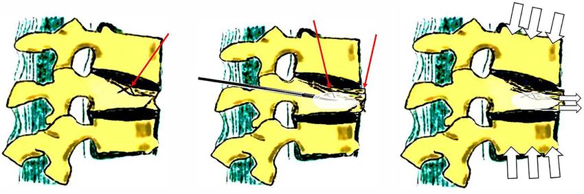
Xiong et al. BMC Musculoskeletal Disorders   (2021) 22:459                                                    Page 2 of 9

  The treatment of refracture in cemented vertebrae re-      parallel to the superior and inferior edges of the pedicle.
mains challenging. The treatment strategy for vertebral      The needle was positioned in the optimal position as
fractures need to be changed when the risks of PVP out-      confirmed by C-arm, that is, the tip reached the anterior
weighs the efficacy. Thus, the purpose of this study was     third of the vertebral body and the middle height of the
to assess the related imaging findings and risk factors of   midline. After the stylet was removed from the trocar, a
patients who experienced refracture of the cemented          formulated polymethylmethacrylate (PMMA) mixture
vertebrae after PVP.                                         was instilled, filling the fractured bone. The cement in-
                                                             jection process was performed slowly, and strictly moni-
Methods                                                      tored under C-arm fluoroscopy in the lateral plane to
Patient selection                                            avoid cement leakage. The bone cement filled the frac-
This retrospective cohort study was conducted from           tured vertebrae in the anterior third of the vertebral
January 1, 2012 to January 1, 2019 in the spine surgery      body as much as possible to form an effective mechan-
department of our hospital. The research program was         ical column. The injection was immediately stopped
approved by Institutional Review Board of Guangzhou          when cement leakage was seen in the segmental vein,
Red Cross Hospital, and all procedures were performed        adjacent intervertebral disk, epidural space, or epidural
according to the Declaration of Helsinki. All patients re-   vein. After PVP, all patients were allowed to ambulate
ceived written informed consent before operation.            the day after surgery.
  A total of 1303 patients who were diagnosed with VCF
(T4-L5) receiving single level PVP were enrolled in this     Imaging examinations
study. Patients who met the following criteria were          Prior to PVP and at least 24 months of follow-up, all pa-
excluded:                                                    tients underwent spinal MR examinations of the spine
                                                             supine position. The MR examinations were performed
  (i) pathological vertebral fractures secondary to tumor,   with a 1.5-T (Siemens Avanto) imager with the following
       severe inflammation, or long-term corticosteroid      sequences: a sagittal T1- weighted spin-echo sequence
       use;                                                  (TR, 535 ms; TE, 11 ms), a sagittal T2-weighted spin-
  (ii) patients without available radiographs or magnetic    echo sequence (TR, 3500 ms; TE, 90 ms), and a spectral
       resonance imaging (MRI);                              attenuated inversion recovery (SPAIR) sequence (TR,
  (iii)No history of PVP surgery;                            3500 ms; TE, 90 ms). Prior to PVP and within 2 weeks of
  (iv) patients with neurologic deficits;                    PVP and following underwent anteroposterior and lat-
  (v) follow-up time less than 24 months;                    eral radiographs of the spine supine position.
  (vi) patients with hyperparathyroidism,
       hyperthyroidism, or other bone metabolic diseases     Radiological assessment
                                                             All images were analyzed during a time span of 2 weeks.
  The inclusion criteria were as follows:                    Images were randomly evaluated by two experienced
                                                             musculoskeletal radiologists in a random order, each
  (i) patients who had plain films preoperatively,           blinded to the clinical information. In our study, the in-
       immediately after surgery, and at the final follow-   ter observer correlation coefficient (ICC) of all radiology
       up;                                                   parameters was excellent (ICC > 0.85). A consensus was
  (ii) patients who underwent MRI preoperatively and at      reached when two observers disagreed on the first
       last follow-up;                                       reading. Face-to-face training was conducted before the
  (iii)follow-up period of at least 2 years;                 study.
  (iv) patients with a bone density scan before the PVP;
  (v) single-level symptomatic VCF treated with PVP;         Anatomical locations of the involved vertebrae
                                                             The anatomical locations of the involved vertebrae
  Based on these criteria, a total of 93 patients were en-   were divided into two groups: vertebrae outside the
rolled in our study (75 women, 18 men).                      thoracolumbar junction (from T4 to T9 or L3 to L5)
                                                             and vertebrae at the thoracolumbar junction (from
Operative procedure                                          T10 to L2) [15].
All patients received bilateral or unilateral PVP in the
prone position under the guidance of C-arm fluoroscopy       Intravertebral cleft (IVC)
after local anesthesia (1% lidocaine). According to          The IVC was detected as an area of signal loss (gas-
Jensen’s technique [14], under C-arm fluoroscopic con-       containing space) or showing marked hyperintensity
trol, 11-gauge or 13-gauge bone biopsy needles were en-      (fluid collection) on the preoperative sagittal T2-
tered the pedicle in a slightly descending manner or         weighted images [15, 16].
Xiong et al. BMC Musculoskeletal Disorders (2021) 22:459 Page 3 of 9
Endplate cortical disruption contact with the upper and lower endplates [20]. The
Endplate cortical disruption was determined as evident patterns of NPEC were classified as NPEC on the
discontinuation in the cortical endplate as seen on the upper endplate, NPEC on the lower endplate, NPEC
preoperative sagittal T2/T1-weighted images [17]. on the upper and lower endplates, and no NPEC on
anteroposterior and lateral radiography of the treated
Kyphotic angle (KA) (Cobb’s angle) and reduction angle vertebra [21].
(RA)
KA was defined as the angle between the upper endplate Clinical data analysis
of the upper vertebra and the lower endplate of the lower The medical records were retrospectively analyzed to
vertebra. At L5, the KA was defined as the angle between collect the use of anti-osteoporosis drug. Medicament
the upper endplate of L4 and the upper endplate of the sa- for the treatment of osteoporosis include zoledronic
cral vertebra. RA was calculated as the difference between acid, calcium and vitamin D supplements. Effective anti-
preoperative and immediate postoperative KA [18]. osteoporosis therapy needs to meet the minimum drug
ownership rate of 80% within 6 months [22]. Patient
Wedge angle (WA) demographics, including gender, age, interval (the period
WA was defined as the angle between the upper end- between the start of new back pain related to MRI-
plate line and the lower endplate of the fractured confirmed fracture and the time of PVP), other vertebral
vertebra. fractures, surgical approach, bone mineral density
(BMD), and cement volume were also analyzed.
Vertebral compression rate (CR), reduction rate (RR) and
compression rate increase (CRI) (Fig. 1) Statistical analysis
CR refers to the ratio of vertebral height of the fractured All statistical analyses were performed using statistics
vertebrae to the average vertebral height of the upper software (SPSS, Chicago, IL, USA). P < 0.05 indicated a
and lower vertebrae at the same site [18]. At L5, CR was statistically significant difference. Logistic regression uni-
the ratio of L4 vertebral height to L5 vertebral height at variate and multivariate analyses were used to assess the
the same site. RR was calculated as the difference be- risk factors for refracture of the cemented vertebrae after
tween preoperative and immediate postoperative CR PVP. The possible risk factors with P value less than or
[18].CRI was defined as the difference in CR between equal to 0.10 in univariate analysis were input into the
immediately after surgery and the last follow-up. final multivariate logistic regression model. After adjust-
ing other risk factors, the significance of each risk factor
Cement leakage on refracture was tested.
Cement leakage was defined as any cement present in
the space beyond the cortical margin [19]. Results
In total, 93 patients (refracture group, n = 48; non-
Non-PMMA-endplate-contact (NPEC) refracture group, n = 45) were reviewed. Patients in the
NPEC was defined as postoperative plain radiographs refracture group were followed for 1.2–25.9 months (mean,
showing that the injected PMMA did not come into 9.3 months; median, 11.2 months). In the refracture group,
Fig. 1 The reduction rate (RR) and compression rate increase (CRI) were calculated as aboveXiong et al. BMC Musculoskeletal Disorders (2021) 22:459 Page 4 of 9
the CRI was 15.1–40.2% (mean, 23.87%; standard deviation (P < 0.001), NPEC (P < 0.001), and kyphotic angle (P =
(SD), 7.89%). Patients in the non-refracture group were 0.014) were significant factors for refracture of the
followed for 24.8–46.6 months (mean, 33.2 months; me- cemented vertebrae after PVP (Table 1). On multivariate
dian, 38.4 months). analysis, however, IVC (P = 0.005; odds ratio, 27.12; 95%
Univariate analysis revealed that IVC (P < 0.001), confidence interval [CI]: 2.67, 275.38), endplate cortical
endplate cortical disruption (P = 0.026), reduction rate disruption (P = 0.037; odds ratio, 3.23; 95% confidence
Table 1 Univariate analysis: clinical factors and imaging finds in the refracture and non-refracture groups
clinical factors and imaging finds Refracture(n = 48) Non-refracture(n = 45) P value
Age (years) 79.65 ± 7.90 78.29 ± 7.21 0.303
Grender
Men 8 10
Women 40 35 0.498
Fracture location
Thoracolumbar 33 36
Non-thoracolumnar 15 9 0.215
BMD(g/cm2) 0.664 ± 0.15 0.73 ± 0.16 0.686
o
kyphotic angle ( ) 17.56 ± 9.51 12.58 ± 9.64 0.014
Wage angle (o) 10.88 ± 6.28 10.20 ± 5.01 0.204
IVC
Present 18 1
Absent 30 44Xiong et al. BMC Musculoskeletal Disorders (2021) 22:459 Page 5 of 9
Table 2 Outcome of multivariate logistic regression analysis misunderstood cement block cracked [12]. In the
OR (95% CI) P value present study, there was vertebral bone marrow edema,
Endplate cortical disruption 3.23 (1.07–9.75) 0.037 and the loss of vertebral height only in the bony verte-
IVC 27.12 (2.67–275.38) 0.005
bra, not in the cement mass. For these reasons, we rec-
ommend using the term ‘refracture’ to describe this
RR (%) 2.94 (1.33–6.47) 0.007
condition. In previous studies, recompression or refrac-
NPEC 1.99 (1.23–3.24) 0.006 ture of augmented vertebrae were defined as a height re-
Data were analyzed with logistic regression. Multivariable analysis adjusted for duction of 1 mm or 4 mm on follow-up radiographs [12,
Endplate cortical disruption, IVC, RR, and NPEC
CI Confidence interval, OR Odds ratio 15, 20, 24–27]. Due to the magnification ratio on radio-
graphs, the measurement of height loss can easily lead to
interval [CI]: 1.07, 9.75), larger RR (P = 0.007; odds ratio, incorrect evaluation. In addition, several long-term stud-
2.94; 95% confidence interval [CI]: 1.33, 6.47), and NPEC ies of patients who were post-vertebral augmentation
(P = 0.006; odds ratio, 1.99; 95% confidence interval [CI]: have showed that in up to 30% of patients have a gradual
1.23, 3.24) showed significance after adjustment for decrease in vertebral body height of 10 to 15% after PVP
other variables (Table 2). between 12 and 24 months [28]. Thus, in this study, the
Analysis of the relationship between endplate cortical criterion of 15% decrease in height [18] and presence of
disruption and the displacement of the anterior edge of vertebral bone marrow edema was adopted.
the vertebral body found that the anterior movement of Although the risk factors for refracture in cemented ver-
the vertebral body was 3.12 ± 2.62 mm for with endplate tebrae after PVP have been previously reported [8, 11], to
cortical disruption, and 1.67 ± 2.18 mm for without end- our knowledge this is the first study to report the risk fac-
plate cortical disruption (P < 0.05). tors and imaging findings of refracture based on bone
marrow edema as a diagnostic basis.
Discussion The incidence of refracture in cemented vertebrae
Researchers have not uniformly described the loss of was 3.68%(48/1303) in this study, which was approxi-
vertebral height and the criteria for unified diagnosis of mately consistent with the findings in previous studies,
height loss in cemented vertebrae after PVP. He [12] which ranged from 0.56 to 27.63% [8, 9, 18, 24, 29].
and Kim [20] described ‘recompression’ of previously However, the author infers that the true incidence of
treated vertebrae. The term ‘recompression’ might be refractures should be higher than the expected data in
confused with additional loss of vertebral height, includ- the present study. This is because some patients with
ing osteoporosis [23]. Heo [9] and Yu [18] reported refracture did not seeking medical services and the
using recollapse to describe the loss of the same verte- present study used rigorous inclusion and exclusion cri-
brae after PVP. The term ‘recollapse’ might be teria and grouping criteria.
Fig. 2 An 82-year-old woman with refracture after PVP. MRI and lateral plain X-ray of an 82-year-old woman with a T11 compression fracture. a
Sagittal SPAIR showed a T11 compression fracture with bone marrow edema, IVC (black arrow), and endplate cortical disruption (white arrow). b
Postoperative X-ray showed the cemented vertebrae with NPEC on the lower endplate (black arrow) and displacement of the anterior edge of
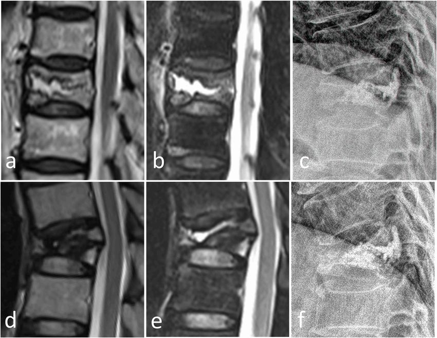
the vertebral body (white arrow). c-d MRI and lateral X-ray at 5 months after PVP showed loss of height of T11 and bone marrow edemaXiong et al. BMC Musculoskeletal Disorders (2021) 22:459 Page 6 of 9 We evaluated the risk factors for refracture in cemen- form NPEC (Fig. 2). In our study, the incidence of ted vertebrae. IVC showed a statistically significant rela- NPEC was significantly higher in patients with IVC tionship to refracture (Table 2), which corresponds with (94.74%) than without IVC (78.38%). Further analysis many previous studies [8, 9, 11, 18, 30, 31]. IVC is a risk also indicated that NPEC was a risk factor associated factor for refracture in cemented vertebrae and can be with refracture (Table 2). Zhang [24] found that pa- explained by two factors; namely, the IVC factor and tients without NPEC had a lower risk of recompression secondary changes caused by IVC. compared with patients with NPEC on the upper and With regard to the IVC factor, IVC provides radio- lower endplates. Hou [25] found that the smaller the logical evidence of osteonecrosis [29, 32, 33]. Osteo- distance between PMMA and the endplate, the lower necrosis of the involved vertebrae would progress after the incidence of recompression. Heo [20] found that PVP, which would eventually weaken the structural recompression occurred in unsupported areas of rigidity of the vertebral body and result in refracture of PMMA. Our findings are consistent with previous re- the remainder of the vertebral body (Figs. 2 and 3) [9]. search. Bone cement is in contact with both the upper In addition, the fractured vertebrae with IVC was usually and lower endplates, so it can provide a better support a solid lump cased volumetric pressure effect that may in the vertical direction since the load is transmitted aggravate the process of osteonecrosis [9]. Heo et al. [9] through both the upper and lower endplates, which are also reported that the timing of PVP is of great import- harder in nature. When bone cement had NPEC, the ance for patients with IVC, given that it is likely unfavor- load did not transmitted through the cementless area, able during the early phase of osteonecrosis. Premature resulting in a stress shielding effect, so the bone cement PVP may cause collateral vessels in necrotic bone to fail may serve to concentrate stress on the surrounding fra- to form. gile bones and lead to refracture. With regard to secondary changes caused by the IVC In our study, compared with the VCFs without IVC, factor, IVC was related to cement distribution pattern the height of the vertebral body with IVC increased from [9, 12, 18] and vertebral height restoration [34]. 7.19 to 12.63%. Michael [34] also reported that vertebro- When bone cement is injected, it often enters the plasty increased the height of the fractured vertebrae, low-pressure zone (IVC zone), which causes the bone and these effects were most remarkable in fractured ver- cement to form solid lump cement, making it easier to tebrae with IVC. Further analysis indicated that RR was Fig. 3 A 59-year-old man with refracture after PVP. Preoperative T2W and SPAIR images (a, b) showed a T10 compression fracture with IVC. Postoperative lateral radiographs (c) after 2 days showed NPEC on the lower and upper endplates. The T2WI, SPAIR image, and lateral radiograph 5 months after PVP (d-f) showed refracture of the cemented T10 vertebra with loss of height and bone marrow edema
Xiong et al. BMC Musculoskeletal Disorders     (2021) 22:459                                                                          Page 7 of 9

a risk factor associated with refracture (Table 2). Lin                   anterior edge of the vertebral body can offset the force
[11] also found that cemented vertebrae with significant                  of the bone cement to diffuse into the trabecular bone.
vertebral height restoration after PVP were prone to                      As a result, the cemented vertebrae with endplate cor-
refracture. Too much recovery of the vertebral body may                   tical disruption is more vulnerable to refracture.
lead to increased tension of the paravertebral soft tissue,                  Previous research found that BMD was a risk factor
which may lead to increased mechanical load on the en-                    for refracture after surgery [31, 35–37]. However, in our
larged vertebrae or more unstable fractures. Conse-                       study BMD was not a risk factor for refracture. We
quently, the risk of refracture of involved vertebrae                     speculate that this is due to the fact that this study used
increased with a greater degree of height restoration.                    bone marrow edema as a diagnostic criterion for refrac-
   Overall, IVC affected RR and NPEC, but RR and IVC                      ture. Villarraga [37] found that the loss of height in
were not all dependent on the impact of IVC. The                          cemented vertebrae was the natural development of
NPEC, IVC, and RR were independent risk factors for                       osteoporosis concluded from finite element model ana-
refracture in cemented vertebrae after PVP.                               lysis, so the loss of height in cemented vertebrae caused
   In our study, endplate cortical disruption was also an                 by osteoporosis may not cause bone marrow edema.
independent risk factor for refracture in cemented verte-                    Although some studies [1–5] reported that PVP could
brae after PVP (Table 2) (Fig. 4). We explained the oc-                   provide significantly pain relief in patients with VCFs,
currence of refracture after PVP as a biomechanical                       more and more studies [38, 39] does not support signifi-
model (Fig. 5). When there is endplate cortical disrup-                   cant clinically benefits from PVP comparaed with pla-
tion, the anterior edge of the vertebral body will move                   cebo. Refarcture in cemented vertebrae may be one of
forward in the process of bone cement injection (Fig. 2).                 the reasons why pain relief is not better than placebo. In
Anterior vertebral displacement causes two conse-                         our opinion, looking for strategies for poor clinical ef-
quences. On one hand, the axial force of the vertebral                    fects of PVP provides clinicians with a pragmatic
body will be partially dispersed laterally, and the verte-                method of how to best treat patients.
bral body is weak against lateral pressure. On the other                     This study had several limitations. First, it was a retro-
hand, the fractured vertebral body cannot be sufficiently                 spective study with a single center and relatively small
filled with bone cement, because the displacement of the                  sample size, and a prospective, multi-center studies with

 Fig. 4 A, 85-year-old woman with refracture after PVP. Preoperative lateral radiographs, SPAIR, and T2W images (a-c) showed a T12 compression
 fracture with endplate cortical disruption (black arrow). Postoperative lateral radiographs (d) after 1 day showed NPEC on the lower and upper
 endplates. The SPAIR image and lateral radiograph 6 months after PVP (e, f) showed refracture of the cemented T12 vertebra with loss of height
 and bone marrow edema
Xiong et al. BMC Musculoskeletal Disorders            (2021) 22:459                                                                                Page 8 of 9

  Fig. 5 Schematic diagram of refracture caused by endplate cortical disruption. Shows the presence of endplate cortical disruption (a). When the
  bone cement is injected, the displacement of the anterior edge of the vertebral body offsets the force of the bone cement to diffuse into the
  trabecular bone and causes NPEC (b). When subjected to axial pressure, part of the force will migrate to the horizontal direction, and the
  vertebral body is weak against the horizontal direction, resulting in refracture (c)

a larger sample size are required to ensure the universal-                      performed preliminary data preparations. All authors read and approved the
ity of our conclusions. Second, two-dimensional X-ray                           final manuscript.

was used to determine the NPEC. In these images,
                                                                                Funding
NPEC may be underestimated. To accurately assess                                No funding was obtained for this study.
NPEC, three-dimensional CT scans will help. Finally,
although there is no significant difference in effective                        Availability of data and materials
                                                                                The datasets used and analysed during the current study are available from
anti-osteoporosis therapy between the refracture group
                                                                                the corresponding author on reasonable request.
and the non-refracture group, a large proportion of pa-
tients receiving effective anti-osteoporosis treatment in                       Declarations
this study may lead to bias.
                                                                                Ethics approval and consent to participate
                                                                                The study was approved by Ethics Committee of Guangzhou Red Cross
Conclusions                                                                     Hospital (reference number 2020/125/02). In this retrospective study, written
Four independent risk factors were significantly associ-                        informed consent was obtained from all patients included in this study.

ated with refracture of the cemented vertebrae after
                                                                                Consent for publication
PVP, including intravertebral cleft, non-PMMA-                                  Not applicable.
endplate-contact, increased reduction rate, and endplate
cortical disruption. Therefore, the current surgical                            Competing interests
                                                                                All authors declare that they have no competing interests.
methods and treatment strategies may need to be
adjusted on the basis of the risk factors of patients.                          Author details
                                                                                1
                                                                                 Department of Radiology, Guangzhou Red Cross Hospital, Jinan University,
Abbreviations                                                                   396 Tongfu Road, Guangzhou 510220, Guangdong Province, China.
                                                                                2
PVP: Percutaneous vertebroplasty; VCFs: Vertebral compression fractures;         Department of Radiology, Wuhan Third Hospital, Tongren Hospital of
BMD: Bone mineral density; IVC: Intravertebral cleft; KA: Kyphotic angle;       Wuhan University, 241 Liuyang Road, Wuhan 430063, Hubei Province, China.
NPEC: Non-PMMA-endplate-contact; RR: Reduction rate; RA: Reduction angle;
PMMA: Polymethylmethacrylate; MRI: Magnetic resonance imaging;                  Received: 27 December 2020 Accepted: 11 May 2021
WA: Wedge angle; CR: Compression rate; CRI: Compression rate increase;
CI: Confidence interval
                                                                                References
Acknowledgments                                                                 1. Anderson PA, Froyshteter AB, Tontz WL Jr. Meta-analysis of vertebral
We thank Peter Mittwede, MD, PhD, from Liwen Bianji, Edanz Editing China            augmentation compared with conservative treatment for osteoporotic
(www.liwenbianji.cn/ac), for editing the English text of a draft of this            spinal fractures. J Bone Miner Res. 2013;28(2):372–82. https://doi.org/10.1
manuscript. We thank Dr. Yahui Liu for helping with the drawing of this             002/jbmr.1762.
article.                                                                        2. Clark W, Bird P, Gonski P, Diamond TH, Smerdely P, McNeil HP, et al. Safety
                                                                                    and efficacy of vertebroplasty for acute painful osteoporotic fractures
Authors’ contributions                                                              (VAPOUR): a multicentre, randomised, double-blind, placebo-controlled trial.
X.W.Z, Y.C.X and W. G were responsible for the study concept design,                Lancet. 2016;388(10052):1408–16. https://doi.org/10.1016/S0140-6736(1
analysis and interpretation of data, drafting of the manuscript, and critical       6)31341-1.
revision of the manuscript for intellectual content. X.W.Z, Y.C.X and F. X      3. De Leacy R. New, high-quality evidence for Vertebroplasty in the
conducted data analyses and all the authors contributed to the                      Management of Painful Recent Compression Fractures: review of the
interpretation of data. Z.P.L and S. C were responsible for the analysis and        VAPOUR trial. World Neurosurg. 2016;96:596–8. https://doi.org/10.1016/j.
interpretation of data. L. W and C.C.Z conducted data collection and                wneu.2016.09.043.
Xiong et al. BMC Musculoskeletal Disorders (2021) 22:459 Page 9 of 9
4. Klazen CA, Lohle PN, de Vries J, Jansen FH, Tielbeek AV, Blonk MC, et al. 23. Liu JT, Li CS, Chang CS, Liao WJ. Long-term follow-up study of osteoporotic
Vertebroplasty versus conservative treatment in acute osteoporotic vertebral vertebral compression fracture treated using balloon kyphoplasty and
compression fractures (Vertos II): an open-label randomised trial. Lancet. vertebroplasty. J Neurosurg Spine. 2015;23(1):94–8. https://doi.org/10.3171/2
2010;376(9746):1085–92. https://doi.org/10.1016/S0140-6736(10)60954-3. 014.11.SPINE14579.
5. Masala S, Mastrangeli R, Petrella MC, Massari F, Ursone A, Simonetti G. 24. Zhang L, Wang Q, Wang L, Shen J, Zhang Q, Sun C. Bone cement
Percutaneous vertebroplasty in 1,253 levels: results and long-term distribution in the vertebral body affects chances of recompression after
effectiveness in a single Centre. Eur Radiol. 2009;19(1):165–71. https://doi. percutaneous vertebroplasty treatment in elderly patients with osteoporotic
org/10.1007/s00330-008-1133-4. vertebral compression fractures. Clin Interv Aging. 2017;12:431–6. https://doi.
6. Kim YJ, Lee JW, Park KW, Yeom JS, Jeong HS, Park JM, et al. Pulmonary org/10.2147/CIA.S113240.
cement embolism after percutaneous vertebroplasty in osteoporotic 25. Hou Y, Yao Q, Zhang G, Ding L, Huang H. Polymethylmethacrylate
vertebral compression fractures: incidence, characteristics, and risk factors. distribution is associated with recompression after vertebroplasty or
Radiology. 2009;251(1):250–9. https://doi.org/10.1148/radiol.2511080854. kyphoplasty for osteoporotic vertebral compression fractures: a
7. Schmid KE, Boszczyk BM, Bierschneider M, Zarfl A, Robert B, Jaksche H. retrospective study. PLoS One. 2018;13(6):e0198407. https://doi.org/10.1371/
Spondylitis following vertebroplasty: a case report. Eur Spine J. 2005;14(9): journal.pone.0198407.
895–9. https://doi.org/10.1007/s00586-005-0905-7. 26. McKiernan F, Faciszewski T, Jensen R. Reporting height restoration in
8. Kang SK, Lee CW, Park NK, Kang TW, Lim JW, Cha KY, et al. Predictive risk vertebral compression fractures. Spine (Phila Pa 1976). 2003;28(22):2517–21
factors for refracture after percutaneous vertebroplasty. Ann Rehabil Med. disucssion 2513.
2011;35(6):844–51. https://doi.org/10.5535/arm.2011.35.6.844. 27. Black DM, Cummings SR, Karpf DB, Cauley JA, Thompson DE, Nevitt MC,
9. Heo DH, Chin DK, Yoon YS, Kuh SU. Recollapse of previous vertebral et al. Randomised trial of effect of alendronate on risk of fracture in women
compression fracture after percutaneous vertebroplasty. Osteoporosis Int. with existing vertebral fractures. Fracture intervention trial research group.
2009;20(3):473–80. https://doi.org/10.1007/s00198-008-0682-3. Lancet. 1996;348(9041):1535–41.
10. Suzuki N, Ogikubo O, Hansson T. Previous vertebral compression fractures 28. Jacobson RE, Palea O, Granville M. Progression of vertebral compression
add to the deterioration of the disability and quality of life after an acute fractures after previous vertebral augmentation: technical reasons for
compression fracture. Eur Spine J. 2010;19(4):567–74. https://doi.org/10.1 recurrent fractures in a previously treated vertebra. Cureus. 2017;9(10):e1776.
007/s00586-009-1162-y. https://doi.org/10.7759/cureus.1776.
11. Lin WC, Lee YC, Lee CH, Kuo YL, Cheng YF, Lui CC, et al. Refractures in 29. Chen LH, Hsieh MK, Liao JC, Lai PL, Niu CC, Fu TS, et al. Repeated
cemented vertebrae after percutaneous vertebroplasty: a retrospective analysis. percutaneous vertebroplasty for refracture of cemented vertebrae. Arch
Eur Spine J. 2008;17(4):592–9. https://doi.org/10.1007/s00586-007-0564-y. Orthop Trauma Surg. 2011;131(7):927–33. https://doi.org/10.1007/s00402-01
12. He D, Lou C, Yu W, Zhu K, Wu Z, Liu F, et al. Cement distribution patterns 0-1236-7.
are associated with recompression in cemented vertebrae after 30. Summa A, Crisi G, Cerasti D, Ventura E, Menozzi R, Ormitti F. Refractures in
percutaneous Vertebroplasty: a retrospective study. World Neurosurg. 2018; cemented vertebrae after percutaneous vertebroplasty and pain relief after
120:e1–7. https://doi.org/10.1016/j.wneu.2018.06.113. a second procedure: a retrospective analysis. Neuroradiol J. 2009;22(2):239–
13. Kim YJ, Lee JW, Kim KJ, Chung SK, Kim HJ, Park JM, et al. Percutaneous 43. https://doi.org/10.1177/197140090902200216.
vertebroplasty for intravertebral cleft: analysis of therapeutic effects and 31. Li YX, Guo DQ, Zhang SC, Liang YK, Mo GY, Li DX, et al. Risk factor analysis
outcome predictors. Skelet Radiol. 2010;39(8):757–66. https://doi.org/10.1 for re-collapse of cemented vertebrae after percutaneous vertebroplasty
007/s00256-009-0866-8. (PVP) or percutaneous kyphoplasty (PKP). Int Orthop. 2018;42(9):2131–9.
14. Jensen ME, Evans AJ, Mathis JM, Kallmes DF, Cloft HJ, Dion JE. Percutaneous https://doi.org/10.1007/s00264-018-3838-6.
polymethylmethacrylate vertebroplasty in the treatment of osteoporotic 32. Dupuy DE, Palmer WE, Rosenthal DI. Vertebral fluid collection associated
vertebral body compression fractures: technical aspects. AJNR Am J with vertebral collapse. AJR Am J Roentgenol. 1996;167(6):1535–8. https://
Neuroradiol. 1997;18(10):1897–904. doi.org/10.2214/ajr.167.6.8956592.
15. Lin WC, Lu CH, Chen HL, Wang HC, Yu CY, Wu RW, et al. The impact of 33. Naul LG, Peet GJ, Maupin WB. Avascular necrosis of the vertebral body: MR
preoperative magnetic resonance images on outcome of cemented imaging. Radiology. 1989;172(1):219–22. https://doi.org/10.1148/radiology.1
vertebrae. Eur Spine J. 2010;19(11):1899–906. https://doi.org/10.1007/s00586- 72.1.2740507.
010-1434-6. 34. Teng MM, Wei CJ, Wei LC, Luo CB, Lirng JF, Chang FC, et al. Kyphosis
16. Wu AM, Chi YL, Ni WF. Vertebral compression fracture with intravertebral correction and height restoration effects of percutaneous vertebroplasty.
vacuum cleft sign: pathogenesis, image, and surgical intervention. Asian AJNR Am J Neuroradiol. 2003;24(9):1893–900.
Spine J. 2013;7(2):148–55. https://doi.org/10.4184/asj.2013.7.2.148. 35. Liebschner MA, Rosenberg WS, Keaveny TM. Effects of bone cement volume
and distribution on vertebral stiffness after vertebroplasty. Spine (Phila Pa
17. Hong SJ, Lee S, Yoon JS, Kim JH, Park YK. Analysis of intradiscal cement
1976). 2001;26(14):1547–54. https://doi.org/10.1097/00007632-200107150-
leakage during percutaneous vertebroplasty: multivariate study of risk
00009.
factors emphasizing preoperative MR findings. J Neuroradiol J de
36. Klazen CA, Venmans A, de Vries J, van Rooij WJ, Jansen FH, Blonk MC, et al.
Neuroradiologie. 2014;41(3):195–201. https://doi.org/10.1016/j.neurad.2013.
Percutaneous vertebroplasty is not a risk factor for new osteoporotic
07.004.
compression fractures: results from VERTOS II. AJNR Am J Neuroradiol. 2010;
18. Yu WB, Jiang XB, Liang D, Xu WX, Ye LQ, Wang J. Risk factors and score for
31(8):1447–50. https://doi.org/10.3174/ajnr.A2148.
recollapse of the augmented vertebrae after percutaneous vertebroplasty in
37. Villarraga ML, Bellezza AJ, Harrigan TP, Cripton PA, Kurtz SM, Edidin AA. The
osteoporotic vertebral compression fractures. Osteoporosis Int. 2019;30(2):
biomechanical effects of kyphoplasty on treated and adjacent nontreated
423–30. https://doi.org/10.1007/s00198-018-4754-8.
vertebral bodies. J Spinal Disord Tech. 2005;18(1):84–91. https://doi.org/10.1
19. Gao C, Zong M, Wang WT, Xu L, Cao D, Zou YF. Analysis of risk factors
097/01.bsd.0000138694.56012.ce.
causing short-term cement leakages and long-term complications after
38. Buchbinder R, Johnston RV, Rischin KJ, Homik J, Jones CA, Golmohammadi
percutaneous kyphoplasty for osteoporotic vertebral compression fractures.
K, et al. Percutaneous vertebroplasty for osteoporotic vertebral compression
Acta Radiologica (Stockholm, Sweden: 1987). 2018;59(5):577–85.
fracture. Cochrane Database Syst Rev. 2018;4:CD006349.
20. Kim YY, Rhyu KW. Recompression of vertebral body after balloon
39. Firanescu CE, de Vries J, Lodder P, Venmans A, Schoemaker MC, Smeets AJ,
kyphoplasty for osteoporotic vertebral compression fracture. Eur Spine J.
et al. Vertebroplasty versus sham procedure for painful acute osteoporotic
2010;19(11):1907–12. https://doi.org/10.1007/s00586-010-1479-6.
vertebral compression fractures (VERTOS IV): randomised sham controlled
21. Oka M, Matsusako M, Kobayashi N, Uemura A, Numaguchi Y. Intravertebral
clinical trial. BMJ. 2018;361:k1551.
cleft sign on fat-suppressed contrast-enhanced MR: correlation with cement
distribution pattern on percutaneous vertebroplasty. Acad Radiol. 2005;12(8):
992–9. https://doi.org/10.1016/j.acra.2005.05.003. Publisher’s Note
22. Bawa HS, Weick J, Dirschl DR. Anti-osteoporotic therapy after fragility Springer Nature remains neutral with regard to jurisdictional claims in
fracture lowers rate of subsequent fracture: analysis of a large population published maps and institutional affiliations.
sample. J Bone Joint Surg Am. 2015;97(19):1555–62. https://doi.org/10.2106/
JBJS.N.01275.You can also read