Refining the treatment of spinal cord lesions: experience from 500 cases
←
→
Page content transcription
If your browser does not render page correctly, please read the page content below
NEUROSURGICAL
           FOCUS                                                                                                            Neurosurg Focus 50 (5):E22, 2021

                          Refining the treatment of spinal cord lesions: experience
                          from 500 cases
                          *Manfred Westphal, MD, Klaus C. Mende, MD, and Sven O. Eicker, MD
                          Department of Neurosurgery, University Medical Center Hamburg-Eppendorf, Hamburg, Germany

                          OBJECTIVE Tumorous lesions of the spinal cord, as well as some vascular lesions like cavernous hemangiomas, de-
                          mand careful consideration as to the indication and approach for surgery. As these lesions are rare in any departmental
                          series, refinement of treatment strategies evolves over long periods. In this context, the authors evaluated a series of
                          500 intramedullary lesions for approach, technique, outcome, complications, and follow-up.
                          METHODS Five hundred intramedullary lesions in 460 patients were treated with a continuously evolving departmental
                          strategy between 1985 and 2020. No lesions of the cauda equina or filum terminale were included. The focus of the
                          evaluation was on the adaptation of exposure, resective methodology, sequelae, imaging, and rate of recurrence. Thirty-
                          seven patients were children at the time of diagnosis.
                          RESULTS Among the 348 neoplastic lesions, the largest subtype was ependymoma (n = 192, 55.2%), followed by as-
                          trocytoma (n = 89, 25.6%). As a trend, metastases (n = 21) have become more frequent and more apparent only in the
                          past 15 years. Reoperations for recurrent or progressive cases or referrals after incomplete resection were performed
                          in 56 cases, mostly for progressive diffuse or pilocytic astrocytomas. Among the vascular lesions, 68 (54.8%) were
                          hemangioblastomas, followed by 56 (45.2%) cavernous hemangiomas. All intramedullary tumors were approached
                          through a midline myelotomy, refining an en bloc resection technique for endophytic tumors to increase the rate of radi-
                          cal resection. Cavernous hemangiomas reaching the surface and hemangioblastomas were approached directly. Com-
                          plete removal was possible in 77.2% of endophytic tumors but in only 41.7% of diffuse tumors. All WHO grade II diffuse
                          astrocytomas, WHO grade III tumors, and glioblastoma progressed despite treatment according to standard regimens.
                          Vascular lesions were regularly removed completely, with only 1 recurrence of a large hemorrhagic thoracic cavernous
                          hemangioma. The major sequelae were sensory deficits and neuropathic pain. Stabilizing instrumentation was placed in
                          5 cases of spinal deformity, mostly when more than 4 levels were affected, and in the pediatric population.
                          CONCLUSIONS In a large series of intramedullary surgeries, refinement of treatment strategies related to exposure,
                          implementation of intraoperative adjuncts such as ultrasound, intraoperative neuromonitoring, resective strategies, and
                          reconstruction were evaluated. The authors found that for almost any defined, endophytic medullary lesion, a safe and
                          complete removal can be offered.
                          https://thejns.org/doi/abs/10.3171/2021.2.FOCUS201107
                          KEYWORDS spinal cord lesion; intramedullary; surgery; cavernoma; angioblastoma; ependymoma; astrocytoma

I
     ntrinsic tumors of the spinal cord represent 2% to                           on space-occupying intramedullary lesions, including in-
     10% of all neoplasms of the CNS,1–7 in reality being at                      trinsic or metastatic tumors, but also hemangioblastomas
     the lower end of the range. They are more common in                          (HAs) and cavernous hemangiomas.
young adults4 and have a different histological spectrum                             Because intramedullary spinal cord lesions (IMSCLs)
than in the adult group. Surgery of these complex and                             are uncommon and often present with subtle symptoms,
rare lesions is challenging, as the cord is a densely func-                       diagnosis is often delayed;8 the primary counseling about
tional and vulnerable structure. Evolving microsurgical                           the nature of the lesion and presumably dismal prognosis
strategies, expanding imaging technologies, and a grow-                           often generate unwarranted anxiety in patients. Treatment
ing number of technological adjuncts have led to constant                         inevitably involves surgery, in most cases for complete
evolution and refinement of spinal cord surgery, which we                         or maximally safe resection, but also for biopsy. Biopsy
revisited here in a large institutional series that focuses                       should be limited to select cases, as the findings oftentimes

ABBREVIATIONS CEG = Cooper-Epstein grade; HA = hemangioblastoma; IMSCL = intramedullary spinal cord lesion; IONM = intraoperative neuromonitoring; MPS =
methylprednisolone; NASCIS II = National Acute Spinal Cord Injury Study 2; MEP = motor evoked potential; SSEP = somatosensory evoked potential.
SUBMITTED January 1, 2021. ACCEPTED February 17, 2021.
INCLUDE WHEN CITING DOI: 10.3171/2021.2.FOCUS201107.
* M.W. and K.C.M. contributed equally to this work.

©AANS 2021, except where prohibited by US copyright law                                                      Neurosurg Focus Volume 50 • May 2021          1

                                                                                                             Unauthenticated | Downloaded 10/26/21 09:26 PM UTC
Westphal et al.
TABLE 1. Spectrum of spinal cord lesions in adults and infants from 1985 to 2020
No. of Lesions (%)
In Adults In Children Total
Ependymoma 184 (36.8) 8 (1.6) 192 (38.4)
Astrocytoma 70 (14.0) 19 (3.8) 89 (17.8)
Glioblastoma 6 (1.2) 2 (0.4) 8 (1.6)
Oligodendroglioma 4 (0.8) 0 (0.0) 4 (0.8)
Melanocytoma 4 (0.8) 0 (0.0) 4 (0.8)
Ganglioglioma/-cytoma 9 (1.8) 4 (0.8) 13 (2.6)
Lipoma 12 (2.4) 1 (0.2) 13 (2.6)
Metastasis 21 (4.2) 0 (0.0) 21 (4.2)
Inflammatory 6 (1.2) 1 (0.2) 7 (1.4)
Cystic lesion 9 (1.8) 1 (0.2) 10 (2.0)
Teratoma 1 (0.2) 3 (0.6) 4 (0.8)
Inconclusive biopsy 10 (2.0) 1 (0.2) 11 (2.2)
Cavernoma 53 (10.6) 3 (0.6) 56 (11.2)
Angioblastoma 66 (13.2) 2 (0.4) 68 (13.6)
Total 455 (91.0) 45 (9.0) 500 (100)
are inconclusive; the procedure should be reserved for a terial blood pressure of approximately 80 mm Hg) was
priori unresectable cases or histological confirmation for maintained. Intraoperative neuromonitoring (IONM) is a
implementation of other treatment modalities,9 especially standard of spinal cord surgery, and motor evoked poten-
in cases of lymphoma.2,4 Notwithstanding the poor expec- tial (MEP), somatosensory evoked potential (SSEP), and
tations, advancements in and defined limits of the surgical D-wave monitoring have routinely been employed during
procedures allow for safe treatment of most lesions. There tumor resection since 1996. A methylprednisolone (MPS)
are numerous reports containing rather small series.10–14 regimen based on the National Acute Spinal Cord Injury
Acknowledging that different but equivalent approaches Study 2 (NASCIS II) findings for traumatic spinal cord
may evolve in different centers, we present our series of injuries was initiated before the dura mater was opened.
500 cases not in an attempt to prescribe definitive stan- Data analysis was performed using IBM SPSS Statis-
dards, but to contribute to the advancements in intramed- tics version 26 (IBM Corp.) and Microsoft Excel 2019.
ullary lesion surgery in general. In this study, a variety of Cross tables and chi-square test analyses were performed
sizable defined subgroup lesions were treated surgically in for nominal and ordinal data. Parametric data were ana-
the same center, with techniques and strategies evolving lyzed using the Student t-test. A logistic regression analy-
as the series grew and as constant adaptations were made sis was performed for the impact of preoperative clinical
from earlier reports.15–17 status on clinical outcome. All results were deemed sig-
nificant at p < 0.05.
Methods
Records from 500 surgeries in 460 patients for IMSCLs Results
were either retrieved from archives or documented in a Between 1985 and 2020, 500 intramedullary lesions
prospective database. Histology was based on the respec- were surgically treated. Of these lesions, 348 were tumors,
tive WHO classification used at the time, and no reevalu- with ependymomas being the most prominent (n = 192;
ation was performed according to the new WHO 2016 Fig. 1), followed by astrocytomas of all grades (n = 89;
classification for cases from earlier decades (Table 1). Fig. 2). There were 124 vascular pathologies (Fig. 3). The
Symptoms and neurological deficits were scored using the remaining 28 pathologies were intramedullary cysts (n =
Cooper-Epstein grade (CEG),18 which allowed for a stan- 10), diffuse inflammation (n = 7), or had no conclusive pa-
dardized evaluation of clinical change in the perioperative thology (n = 11; Fig. 4, Table 1). The mean patient age was
and follow-up periods. 41 ± 17 years, with statistically significant differences be-
All patients underwent Gd-enhanced whole-spine MRI tween subgroups and growth patterns: endophytic, 44 ± 15
at the time of diagnosis to assess possible multiple lesions years; diffuse, 34 ± 18 years; vascular, 41 ± 14 years; and
and enable level counting from below. The extent of resec- metastases, 54 ± 19 years (ANOVA, p < 0.001; differences
tion was based on Gd-enhanced postoperative MRI per- between diffuse and all other groups were significant at p
formed within 6 weeks postoperatively at the latest and < 0.01 [Bonferroni], as was the difference between vas-
preferably within 48 hours for contrast-enhancing lesions. cular and metastatic lesions). The mean time from initial
Total intravenous anesthesia was routinely used, and an symptoms to diagnosis was 93 ± 114 weeks, which was not
adequate perfusion pressure (a maximum of a mean ar- significantly different between the subgroups.
2 Neurosurg Focus Volume 50 • May 2021
Unauthenticated | Downloaded 10/26/21 09:26 PM UTCWestphal et al.
FIG. 1. Sagittal MR images of representative spinal cord ependymomas. A: Contrast-enhanced T1-weighted image showing the
intramedullary tumor from T10 to T12. B: Image showing different parts with partially solid and cystic proportions. C: Contrast-en-
hanced T1-weighted image showing a second case with extended spinal cord ependymoma from C0 to C3. D: Image demonstrat-
ing the corresponding syringomyelia. E: Image showing the postoperative result after complete tumor removal.
Clinical Status or upper-extremity function (chi-square test, p < 0.001).
In 19% (74/390) of patients, the preoperative CEG was Deficits progressed in 84.0% of endophytic lesions, 87.7%
0 for both upper and lower extremities, and the lesions of diffuse lesions, 86.4% of vascular lesions, and 92.3% of
were discovered incidentally. Of the remaining patients, metastatic lesions.
58.5% presented with very mild deficits (CEG 0–1), 25.6% At discharge, upper- and lower-extremity CEGs were
with mild deficits (CEG 2), and 11.5% with moderate to improved or equal to those at the first presentation in
high deficits (CEG 3); 4.4% presented with major impair- 69.5% and 60.6% of patients, respectively (chi-square test,
ment (CEG 4 or 5) and severe deficits concerning lower- p < 0.001). On short-term follow-up (6 months), in 40.2%
FIG. 2. Exemplary presentation of a pilocytic astrocytoma. A: Sagittal T2-weighted image of the thoracolumbar spine. B and
C: Postoperative kyphotic deformity despite reconstruction of the dorsal elements of the spinal column (B) requiring spinal
stabilization (C; with carbon fiber–reinforced PEEK screws to reduce susceptibility artifacts on postoperative imaging). D: Sagittal
T2-weighted MR image of the cervical spine, showing diffuse astrocytoma (IDH mutated). E: Sagittal T1-weighted MR image of the
cervical spine showing anaplastic astrocytoma.
Neurosurg Focus Volume 50 • May 2021 3
Unauthenticated | Downloaded 10/26/21 09:26 PM UTCWestphal et al.
FIG. 3. Vascular lesions. A and B: Sagittal (A) and axial (B) T2-weighted images showing a small eccentrically located cavernous
hemangioma. Hemosiderin (black) is clearly visible in panel B. C and D: Sagittal (C) and axial (D) T2-weighted MR images illus-
trating the dimensions that a cavernous hemangioma can achieve in the cervical spine. E and F: Sagittal (E) and axial (F) images
showing a highly vascularized hemangioblastoma of the thoracic spinal cord. G: Sagittal image of a small angioblastoma with a
corresponding cyst in the cervical spine.
of patients upper-extremity neurological function further ties (Table 2) and 58.5% of patients for the lower extremi-
increased, as it did in 24% for the lower extremities. In to- ties (Table 3).
tal, stabilization or improvement at the 6-month follow-up Improvement in lower-extremity function at 6 months
was recorded in 88.4% of patients for the upper extremi- after surgery was more likely if the preoperative neuro-
FIG. 4. A and B: Sagittal T2- (A) and T1-weighted (B) MR images of the cervical spine, showing a rare case of unclear extended
neoplastic tethering of the spinal cord, with cystic and solid tumor parts and corresponding syringomyelia. C: Axial image of the T2
vertebra. The etiology remains unclear even after biopsy.
4 Neurosurg Focus Volume 50 • May 2021
Unauthenticated | Downloaded 10/26/21 09:26 PM UTCWestphal et al.
TABLE 2. Lower-extremity CEGs at 6 months
Preop CEG
Postop CEG 0 1 2 3 4 5 Overall
0 8.0% 11.5% 4.0% 1.5% 1.5% 0.0% 26.5%
1 3.0% 23.0% 8.5% 1.0% 1.5% 0.0% 37.0%
2 0.0% 3.5% 10.0% 7.0% 1.0% 0.0% 21.5%
3 0.5% 0.5% 2.5% 4.5% 2.5% 1.0% 11.5%
4 0.0% 0.5% 0.5% 1.0% 1.0% 0.5% 3.5%
5 0.0% 0.0% 0.0% 0.0% 0.0% 0.0% 0.0%
Overall 11.5% 39.0% 25.5% 15.0% 7.5% 1.5%
The values across the diagonal (shown in shading and/or bold) represent the stable patients. The values under the diagonal represent patients
who declined, and the values above the diagonal represent patients who experienced neurological improvement. Improvement was seen in
12%, 46.5% remained stable, and 41.5% had a decline.
logical impairment was minor (OR 1.704/CEG for a stable the dura mater is still closed (Fig. 5). In cases in which
or better outcome, 95% CI 1.27–2.29; logistic regression, the arachnoid space is obliterated and the dura mater is
p < 0.001). opened, the cord may rise out of the opening. To avoid in-
jury in such cases of swelling, it is mandatory to drain ce-
Recurrent Lesions rebrospinal fluid from the ventral space above the lesion.
Fifty-six patients in our cohort had recurrent lesions. The main aspect of this series was the use of a CO2 laser
Of all endophytic tumors, 21 were surgeries for recurrent in an attempt at in toto removal of endophytic tumors, as
tumor (15 were previous operations at our institution and illustrated in Fig. 6B and C. By dislodging the tumor from
6 were external referrals). In diffuse tumors, 26 were re- its bed, thereby alternating from pole to pole, severing ad-
current lesions (19 of these were previous operations at hesions with 1.5 W of CO2 laser power, and uplifting the
our institutions and 7 were external), and for metastases, increasingly mobile tumor, the dissection plane is opened
only 1 recurrent case was treated. Eight surgeries were with minimal traction. This allows for stripping the tracts
performed on a recurring vascular lesion. Because of the from the tumor surface, resulting in radical removal. With
technical advances made over the course of this study, clear visibility of the ventral vessels entering and branch-
recurrence rates were significantly reduced over time; ing at the surface of the tumor, avoidance of injury to the
patients operated on before the year 2000 had an overall anterior spinal artery was possible in all cases.
recurrence rate of 19.8% versus 7.2% for patients operated In diffuse tumors, in which the midline approach does
on after that year (chi-square test, p = 0.001). not open a clear dorsal tumor surface and dissection plane
on which the tumor can be followed in its circumference
Surgery (Fig. 6), color and texture may allow for an internal decom-
All intrinsic tumors are approached through the mid- pression and decreasing intramedullary pressure through-
line, so the exposure is by laminotomy. Care is taken to out debulking. This results in the appearance of a transition
gain adequate access to the poles of the tumors, so that zone that may be carefully “thinned out,” considering that
there is a straight view to the entrance of the ventral dis- fiber tracts will be embedded in the outer layer. For pilocyt-
section plane. After exposure of the dura mater, adequate ic astrocytoma or diffuse astrocytoma grade II, this may
exposure is verified by ultrasound so that the edges of the still lead to long-term control. Glioblastoma and metasta-
lamina above or below can be removed if necessary while ses are also only debulked inside the margins of change of
TABLE 3. Upper-extremity CEGs at 6 months
Preop CEG
Postop CEG 0 1 2 3 4 Overall
0 41.7% 3.5% 2.5% 0.0% 0.0% 47.7%
1 11.1% 15.6% 3.0% 0.5% 0.0% 30.2%
2 4.0% 7.0% 5.0% 2.0% 0.0% 18.0%
3 0.5% 0.5% 1.0% 1.0% 0.0% 3.0%
4 0.0% 0.0% 0.5% 0.5% 0.0% 1.0%
Overall 57.3% 26.6% 12.0% 4.0% 0.0%
The values across the diagonal (shown in shading and/or bold) represent the stable patients. The values under the diagonal represent patients
who declined, and the values above the diagonal represent patients who experienced neurological improvement. Improvement was seen in
25.1%, 63.3% remained stable, and 11.5% experienced decline.
Neurosurg Focus Volume 50 • May 2021 5
Unauthenticated | Downloaded 10/26/21 09:26 PM UTCWestphal et al.
FIG. 5. Intraoperative ultrasound verification of the tumor extent and localization before opening the dura mater. A: Sagittal
contrast-enhanced T1-weighted MR image from C1 to T3 with an ependymoma at C5–7. A solid tumor is in the dashed circle
(cranial syringomyelia is also displayed). The solid circle shows the tumor cyst. B: The solid tumor is perfectly visualized before
opening the dura mater. C: The cystic part of the tumor is shown.
texture quality, followed by standard radiation therapy and of differential vulnerability due to asymmetrical tumor
chemotherapy. With metastases becoming notably more growth and indicated the side of more adhesiveness or sur-
frequent in the past years and with extended survival of face assimilation to fiber tracts or invasion. Sudden changes
cancer patients, the need for interdisciplinary consultation in SSEPs were seen when too much pressure was exerted
for chemotherapeutic and especially for radiosurgical op- when the dissection plane was opened on one side. In re-
tions, and adequate sequencing, is emerging.19 sponse, we let that area rest and continued on the contra-
Gross-total resection was possible in 77.2% (95/123) of lateral side. Slowly diminishing SSEPs and MEPs were
our endophytic tumor cases, but only in 41.7% (30/72) of observed when duration of surgery was long and were dis-
the diffuse tumors and 72.7% of the metastases (chi-square regarded when no surgical maneuver could be associated
test, p < 0.001). The CO2 laser was used in 92.9% of all ep- with the decreases. These monitoring changes usually oc-
endymoma resections. A macroscopically complete resec- curred later in the procedure, and the resection was always
tion was achieved in 82.0% of all cases in which a CO2 completed to the extent anticipated for the respective lesion.
laser was employed. Consequently, the recurrence rate of
ependymomas throughout the whole series, including the Complications and Neurological Sequelae
“learning curve,” was 17.5% (28/160), 14.0% (n = 13) for CSF leaks requiring surgical intervention were detect-
those that underwent total resection and 31.8% (n = 7) for ed in 8 cases. Revision surgery due to postoperative hema-
subtotal resection (chi-square test, p = 0.11). toma was necessary for 4 cases. There were no surgical
Vascular lesions such as HA and cavernous hemangio- site infections.
ma have clear margins, mostly delineated by a clear plane As for sensory findings, nearly all patients who had
of gliosis (Fig. 6). Cavernoma can be gradually shrunk by their tumor accessed through a midline approach reported
draining its cyst so that it is dissected away from the glio- dysesthesia of the lower body and diminished propriocep-
sis into the cavity resulting from evacuation. HAs, on the tion in the immediate postoperative period; however, these
contrary, are left intact and are carefully devascularized in symptoms did not impair their walking if there was no
their pial embedding, arteries before veins, and then after preexisting severe motor deficit. The sensory deficits were
opening the pia circumferentially, slowly shrunk with the always more pronounced on the side where the tumor was
bipolar away from the gliotic bed, and finally removed in more adherent to the inner surface of the tumor cavity re-
toto to prevent recurrence. Vascular lesions were totally flecting the area of origin, which especially in endophytic
removed in 93.4% of cases. astrocytoma may be an area of unilateral infiltration op-
posite a clean dissection plane contralaterally. A small
Electrophysiological Findings proportion of patients developed neuropathic pain as glio-
SSEPs were found to frequently decrease by 30% uni- sis became part of the postoperative consolidation of the
laterally with the midline incision. This was indicative resection cavity.
6 Neurosurg Focus Volume 50 • May 2021
Unauthenticated | Downloaded 10/26/21 09:26 PM UTCWestphal et al.
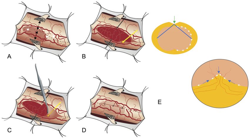
FIG. 6. A: Spinal cord alignment altered by an IMSCL with hypervascularization, midline shift (black arrows), and bulging of the
spinal cord. Midline identification can be achieved by perforating vessels (white arrows), nerve root symmetry, and visualization of
the dorsal median sulcus (dashed gray line). B: Pial stay sutures (black lines) are advocated by some but are met with skepticism
by others. The CO2 laser (yellow line) may be helpful in lateral tumor dissection. C: After lateral and pole dissection, the tumor may
be lifted, and the plane of dissection followed with a CO2 laser to cut even small adhesions. D: Using pial sutures to close the cord
may prevent complications through adhesions. E: The midline or dorsal sulcus offers an approach from which the lateral aspects
can be reached with near equidistance (blue lines). The direction of dissection along the surface (white arrows), approaching the
midline from both sides (blue arrows), and localization of feeding vessels in the ventral part of the spinal cord.
Spinal Deformity For exposure of nonexophytic, completely intramedul-
Laminotomies have been used for exposure in all pri- lary tumors, mostly ependymoma and astrocytoma, we
mary cases of this series since 1990. In recurrent cases exclusively used the midline approach to the cord, as have
in which lesion removal became difficult because of ex- many others.22–26 With the tumor mass mostly located in
tensive fibrosis, stepwise laminectomy was performed to the central cord, the midline or dorsal sulcus offers an ap-
protect the dural sac. Even with laminotomy, extensive proach from which the lateral aspects can be reached with
cervical swan neck deformity developed in 4 patients and almost equidistance without unnecessarily injuring fiber
progressive thoracic kyphosis in 1 patient (Fig. 2A–C); all tracts (Fig. 6E). Thus, no asymmetrical strain is put on the
were children. This developed over a course of 1 to 5 years tumor surroundings as in the dorsolateral approach, which
and led to spinal instrumentation. In adults, mild swan is proposed as an alternative.27 Identifying the midline for
neck deformity that did not cause neurological symptoms a longitudinal incision of the pia mater can be difficult due
progressed in some patients for 3 to 5 years and then sta- to the altered anatomy and torsion in the presence of an
bilized, with patients refusing offered corrections after the underlying tumor with asymmetrical cord distension (Fig.
deformity became fixed and no surgery was required if 6A), but vessels perforating at the midline into the depth of
there was no recurrence. Spinal deformity was more likely the cord serve as a landmark, as may the dorsal root entry
to occur in multilevel laminotomies of 4 or more segments. zones. In contrast, exophytic lesions, often cavernoma and
HA, are approached from their exposed surface, and for
Discussion ventrolateral lesions, a longer exposure and rotation of the
We present a large and complex series of institution- cord by detaching and gently pulling on the dental liga-
al experience with intramedullary surgery, mostly for tu- ment allows access.
mors, which is the continuous experience of a very small Likewise, the method of tumor removal has to be indi-
team (see Acknowledgments) over a specific period.16–21 vidually adapted, but we advocate that solid intramedul-
Our main finding was that there are some general prin- lary tumors should be left intact as described above. This
ciples that should be standardized and followed in every en bloc removal appears to be crucial for obtaining high
surgery, but there are also entity-specific strategies that rates of complete resections for well-circumscribed intra-
need to be adopted. medullary tumors, as is also reported by other authors.28
Neurosurg Focus Volume 50 • May 2021 7
Unauthenticated | Downloaded 10/26/21 09:26 PM UTCWestphal et al.
By outward traction or pushing of the tumor starting at the small and nonreactive, these tumors should just be ob-
poles, the dissection plane to be targeted opens up. Thus, served with regular MRI follow-up at yearly intervals un-
slowly approaching the equator from the cranial as well as less symptoms develop. Slow growth, changed contrast
the caudal pole in order to raise only half the tumor at the characteristics, and cyst formation may lead to a discus-
time, the traction enables a safe severance of the fiber tracts sion of preemptive surgery with the patient. This is rel-
surrounding the tumor surface. Working along the clearly evant in HA. Most of our HA cases were spontaneous and
visible tumor/cord interface, it has even been claimed that therefore single, but patients who have von Hippel–Lindau
IONM may not be necessary in cases resected with this syndrome frequently present with multiple intramedullary
technique.28 This approach may have to be modified in HAs. One study proposed early spinal surgery in these
long tumors that extend over 3 or 4 segments, so that a cases, even for asymptomatic lesions, since symptoms,
completely freed segment is cut off and a new starting pole once present, were often found to be irreversible (71.4%
is created to continue along the same methodology. In our did not improve after surgery).30 We propose a more con-
series, what has greatly facilitated this resective technique servative approach, as there has to be a trade-off between
is use of the CO2 laser, which is used to open up the dis- the preventive aspect and the acceptable side effects, which
section plane mostly at the lateral aspects where adhesions can be disproportionate for small, completely asymptom-
and vascular bridges resist the microdissector used to shell atic lesions. Since it is hard to predict future developments
the tumor out of its bed. Applying a short CO2 laser burst in patients’ lives, leaving as much of the spine intact as
through which the thermal energy dissipates in the act possible is our foremost concern.
of evaporation of the adhesive spot on the tumor surface As for sequelae, spinal deformity may have severe re-
ensures maximal safety for the cord tissue (Fig. 6B and constructive consequences. Undisputedly, optimal expo-
C). To keep a long, multilevel myelotomy and enlarging sure for a centrally located intramedullary lesion should
resection cavity open during the length of the procedure allow the entire circumference of the tumor to be reached
is also a matter of controversy—pial 6-0 stay sutures are at the equator, with equal bilateral exposure, which is best
advocated by some23 but are met with skepticism by others achieved through a midline approach. Thus, the generally
(R. Spetzler, personal communication)—so we tend to use favored keyhole approaches31 are inadequate in this situ-
them (modified to 8 × 0) only in long exposures with wide ation, except for lateral surface lesions like cavernomas.
opening over the exposed tumor in the midline so that they Multilevel laminotomies in different techniques are now
more or less “hold” rather than “pull” the well-detached standard; laminectomy has become inappropriate because
dorsal columns. of frequently observed deformities, especially in the cervi-
While en bloc resection is a decisive aspect for radical cal spine and in young patients, causing a paradigm shift in
removal and has a low recurrence rate for circumscribed the 1990s.25 In a long-term cohort of 55 pediatric patients,
tumors, this does not hold true for diffuse or invasive tu- new-onset spinal deformity after IMSCL surgery was re-
mors. In these cases, debulking is the first step, possibly ported to be as high as 16%, with stabilization required
opening boundaries at which a transition zone to normal in 55% of patients.32 The risk for deformity is reduced
cord tissue becomes apparent. Guidance can be obtained with smaller exposures,33 and even further with lamino-
from working with experienced specialists to interpret plasty,34,35 but it is still a risk that cannot be completely
monitoring or intraoperative stimulation findings,4,29 as avoided and should be discussed with the patient prior to
well as fluorescence in some lesions.4,22 However, this is obtaining their consent for surgery (Fig. 2). Laminoplasty
not broadly validated and depends very much on histology, also offers advantages should recurrence or progression
individual texture characteristics, and, foremost, personal necessitate another exposure.
experience. In diffuse astrocytomas, where the complete While patients usually adapt to proprioceptive or senso-
cord is one homogeneous tumor-infiltrated entity, biopsy is ry deficits, the development of neuropathic pain,25,36 which
advocated only to obtain proper diagnosis to guide thera- reaches up to 60% in some series,22 can be a dramatic se-
peutic decisions. Also, neuroglial tumors like ganglioglio- quela. This can occur during gliotic scar consolidation of
mas or gangliocytomas, reflecting the essential matrix of the resection cavity and may fluctuate within a period of 2
the cord, lack a clearly defined border. This only allows a years postoperatively, but thereafter it becomes a burden to
conservative wedge resection guided by consistency and the patient as it may even increase over time. Adhesions of
color, with careful attention to the electrophysiology. Dur- the pial opening to the dorsal dura mater incision can re-
ing such debulking, the CO2 laser has also been found to sult in tethering causing microtrauma and can worsen over
be helpful. time, so untethering may be necessary, but may still recur.
Emergency situations are rare for intramedullary le- Pain management is difficult and may require multimodal
sions. Intratumoral hemorrhages may occur in ependy- interventions. Limiting its occurrence, pial closure of the
moma and HA and are the typical presenting sign for resection cavity with microsutures is attempted wherever
cavernous hemangiomas. This is usually accompanied by possible. To allow the resection cavity to collapse as much
extensive edema, so that high-dose MPS or dexametha- as possible, the arachnoid space should be opened because,
sone is recommended to alleviate symptoms. If the level of due to long-term pressure in slowly expanding tumors, it
paraparesis is not higher than grade II, waiting for several may be broadly adherent to the dura mater and completely
days or even weeks to let the cord recover from the impact obliterated.
results in a much less vulnerable perilesional environment. In our treatment regimen for IMSCL, we adopted a
There is also the issue of how to deal with tumors that routine MPS scheme based on the NASCIS II findings
are found incidentally and produce no symptoms. When that investigated traumatic spinal cord injuries.37–39 In
8 Neurosurg Focus Volume 50 • May 2021
Unauthenticated | Downloaded 10/26/21 09:26 PM UTCWestphal et al.
NASCIS II, 24 hours of early MPS treatment (< 8 hours 2. Grimm S, Chamberlain MC. Adult primary spinal cord tu-
after injury) showed an improvement in sensory func- mors. Expert Rev Neurother. 2009;9(10):1487–1495.
tion at the 6-month follow-up and in motor function at 3. Hirano K, Imagama S, Sato K, et al. Primary spinal cord
tumors:review of 678 surgically treated patients in Japan. A
the 1-year follow-up.37,40,41 An initial bolus of 30 mg/kg multicenter study. Eur Spine J. 2012;21(10):2019–2026.
was applied before myelotomy, followed by a continuous 4. Juthani RG, Bilsky MH, Vogelbaum MA. Current manage-
application of 5.4 mg/kg/hr for 24 hours. While there is ment and treatment modalities for intramedullary spinal cord
criticism concerning the NASCIS II scheme, it may not tumors. Curr Treat Options Oncol. 2015;16(8):39.
apply to its use in resection of IMSCL. The controversy 5. Karikari IO, Nimjee SM, Hodges TR, et al. Impact of tumor
concerning the timing of MPS application is irrelevant to histology on resectability and neurological outcome in primary
the elective surgical situation since it coincides with the intramedullary spinal cord tumors:a single-center experience
timing of the trauma and exactly mimics the situation of with 102 patients. Neurosurgery. 2011;68(1):188–197.
6. Schellinger KA, Propp JM, Villano JL, McCarthy BJ. De-
an experimental spinal cord trauma in a laboratory situa- scriptive epidemiology of primary spinal cord tumors. J
tion, and therefore is different from an accident situation, Neurooncol. 2008;87(2):173–179.
where the exact mechanism of the trauma remains unclear. 7. Tobin MK, Geraghty JR, Engelhard HH, et al. Intramedul-
Also, the complications associated with high-dose steroids lary spinal cord tumors:a review of current and future treat-
were found in a collective of patients who sustained severe ment strategies. Neurosurg Focus. 2015;39(2):E14.
trauma, often with significant comorbidities. 8. Samartzis D, Gillis CC, Shih P, et al. Intramedullary spinal
With the illustrated evolution of departmental standards, cord tumors:part I—epidemiology, pathophysiology, and
diagnosis. Global Spine J. 2015;5(5):425–435.
it appears that a wide spectrum of even formidable lesions 9. Kakareka M, Moncman R, Georges J, et al. Pediatric spinal
can be safely approached. Establishing rigid schemes for cord biopsy:a case series from a high-volume referral center.
this kind of surgery over the last 35 years has led to short- J Clin Neurosci. 2019;65:34–40.
ened hospital stays, shortened procedures, and the elimi- 10. Richards O, Goacher E, Pal D, et al. Intramedullary spinal
nation of many obstacles inhibiting a smooth workflow. cord tumours—a single centre, 10-year review of clinical and
Key elements in this regard are optimal neuroimaging, pathological outcomes. Br J Neurosurg. 2021;35(2):125–128.
monitoring, MPS, standardized laminotomy, transdural 11. Samuel N, Tetreault L, Santaguida C, et al. Clinical and
ultrasound, the availability of experienced neuropathology pathological outcomes after resection of intramedullary spi-
nal cord tumors:a single-institution case series. Neurosurg
for frozen section, the awareness of pitfalls (e.g., trapped Focus. 2016;41(2):E8.
CSF, monitoring artifacts, unexpected histology), and the 12. Schebesch KM, Mueller S, Wendl C, et al. Recurrence rates
presence of a small, dedicated team to build a learning and functional outcome after resection of intrinsic intramed-
experience. The use of a CO2 laser may be departmentally ullary tumors. Clin Neurol Neurosurg. 2015;134:60–66.
specific, as others use different microsurgical technology. 13. Seki T, Hida K, Yano S, et al. Surgical outcomes of high-
grade spinal cord gliomas. Asian Spine J. 2015;9(6):935–941.
14. Svoboda N, Bradac O, de Lacy P, Benes V. Intramedullary
Conclusions ependymoma:long-term outcome after surgery. Acta Neuro-
Our series offers evidence that for rare lesions, which chir (Wien). 2018;160(3):439–447.
can have a broad variety of individual presentation, suffi- 15. Cristante L, Hermann HD. Radical excision of intramedul-
lary cavernous angiomas. Neurosurgery. 1998;43(3):424–431.
cient frequency of a procedure, and limitation to a small 16. Cristante L, Herrmann HD. Surgical management of intra-
team with infrequent changes of personnel, allows for in- medullary hemangioblastoma of the spinal cord. Acta Neuro-
cremental refinements so that even a rare procedure can chir (Wien). 1999;141(4):333–340.
become standardized. This also ensures that optimal tech- 17. Cristante L, Herrmann HD. Surgical management of in-
niques will be passed on to trainees, maintaining a constant tramedullary spinal cord tumors:functional outcome and
high quality for the outcome of the patients. Whatever these sources of morbidity. Neurosurgery. 1994;35(1):69–76.
refinements are in specialized centers, the main emerging 18. Cooper PR, Epstein F. Radical resection of intramedullary
aspect is the elimination of a defeatist view in patients and spinal cord tumors in adults. Recent experience in 29 pa-
tients. J Neurosurg. 1985;63(4):492–499.
referring physicians toward intramedullary lesions, to gain 19. Payer S, Mende KC, Westphal M, Eicker SO. Intramedullary
confidence from series like this that most patients will be spinal cord metastases:an increasingly common diagnosis.
able to walk home and continue with their normal lives, Neurosurg Focus. 2015;39(2):E15.
and even be cured in cases of low-grade endophytic tumors. 20. Eicker SO, Floeth FW, Kamp M, et al. The impact of fluo-
rescence guidance on spinal intradural tumour surgery. Eur
Spine J. 2013;22(6):1394–1401.
Acknowledgments 21. Westphal M. Intramedullary tumors. In:Tonn JC, Reardon
We thank Luca Papavero and Eric Fritzsche, who were a long- DA, Rutka JT, Westphal M, eds. Oncology of CNS Tumors.
term part of the team, which began with Hans-Dietrich Herrmann 3rd ed. Springer;2019:633–657.
and Loris Cristante. We also thank Tammam Abboud, Cindy 22. Aghakhani N, Messerer M, David P, et al. Intramedullary
Schwarz, and Romy See for intraoperative monitoring. We thank ependymomas:a French retrospective multicenter study of
Sabine Wuttke for the excellent graphical support in this paper. 221 cases. Article in French. Neurochirurgie. 2017;63(5):
391–397.
23. Brotchi J. Intramedullary astrocytomas surgery in adult pa-
References tients:the rationale for cautious surgery. World Neurosurg.
1. Boström A, Kanther NC, Grote A, Boström J. Management 2013;80(5):e139–e140.
and outcome in adult intramedullary spinal cord tumours:a 24. Brotchi J, Noterman J, Baleriaux D. Surgery of intramedul-
20-year single institution experience. BMC Res Notes. 2014;7: lary spinal cord tumours. Acta Neurochir (Wien). 1992;116(2-
908. 4):176–178.
Neurosurg Focus Volume 50 • May 2021 9
Unauthenticated | Downloaded 10/26/21 09:26 PM UTCWestphal et al.
25. Klekamp J. Treatment of intramedullary tumors:analysis of 36. Nakamura M, Tsuji O, Iwanami A, et al. Central neuropathic
surgical morbidity and long-term results. J Neurosurg Spine. pain after surgical resection in patients with spinal intramed-
2013;19(1):12–26. ullary tumor. J Orthop Sci. 2012;17(4):352–357.
26. Kucia EJ, Bambakidis NC, Chang SW, Spetzler RF. Surgical 37. Bracken MB, Shepard MJ, Collins WF, et al. A randomized,
technique and outcomes in the treatment of spinal cord epen- controlled trial of methylprednisolone or naloxone in the
dymomas, part 1:intramedullary ependymomas. Neurosur- treatment of acute spinal-cord injury. Results of the Second
gery. 2011;68(1)(Suppl Operative):57–63. National Acute Spinal Cord Injury Study. N Engl J Med.
27. Katsigiannis S, Carolus AE, Schmieder K, Brenke C. Pos- 1990;322(20):1405–1411.
terolateral myelotomy for intramedullary spinal cord tumors: 38. Tsutsumi S, Ueta T, Shiba K, et al. Effects of the Second
the other way to do it? Acta Neurochir (Wien). 2020;162(1): National Acute Spinal Cord Injury Study of high-dose meth-
101–107. ylprednisolone therapy on acute cervical spinal cord injury—
28. Sweeney KJ, Reynolds M, Farrell M, Bolger C. Gross total results in spinal injuries center. Spine (Phila Pa 1976). 2006;
resection rates of grade II/III intramedullary ependymomas 31(26):2992–2997.
using the surgical strategy of en-bloc resection without intra- 39. Young W. NASCIS. National Acute Spinal Cord Injury
operative neurophysiological monitoring. Br J Neurosurg. Study. J Neurotrauma. 1990;7(3):113–114.
2017;31(3):364–368. 40. Bracken MB. Steroids for acute spinal cord injury. Cochrane
29. Jin SH, Chung CK, Kim CH, et al. Multimodal intraoperative Database Syst Rev. 2012;1(1):CD001046.
monitoring during intramedullary spinal cord tumor surgery. 41. Bracken MB, Shepard MJ, Collins WF Jr, et al. Methylpred-
Acta Neurochir (Wien). 2015;157(12):2149–2155. nisolone or naloxone treatment after acute spinal cord injury:
30. Van Velthoven V, Reinacher PC, Klisch J, et al. Treatment of 1-year follow-up data. Results of the second National Acute
intramedullary hemangioblastomas, with special attention Spinal Cord Injury Study. J Neurosurg. 1992;76(1):23–31.
to von Hippel-Lindau disease. Neurosurgery. 2003;53(6):
1306–1314.
31. Mende KC, Krätzig T, Mohme M, et al. Keyhole approaches Disclosures
to intradural pathologies. Neurosurg Focus. 2017;43(2):E5. Dr. Eicker: honoraria from Stryker and Spineart.
32. Ahmed R, Menezes AH, Awe OO, et al. Long-term incidence
and risk factors for development of spinal deformity follow- Author Contributions
ing resection of pediatric intramedullary spinal cord tumors.
J Neurosurg Pediatr. 2014;13(6):613–621. Conception and design: Westphal, Eicker. Acquisition of data:
33. Knafo S, Court C, Parker F. Predicting sagittal deformity Mende, Eicker. Analysis and interpretation of data: Mende. Draft-
after surgery for intramedullary tumors. J Neurosurg Spine. ing the article: Westphal, Eicker. Critically revising the article:
2014;21(3):342–347. Westphal. Approved the final version of the manuscript on behalf
34. McGirt MJ, Garcés-Ambrossi GL, Parker SL, et al. Short- of all authors: Westphal. Statistical analysis: Mende.
term progressive spinal deformity following laminoplasty
versus laminectomy for resection of intradural spinal tumors: Correspondence
analysis of 238 patients. Neurosurgery. 2010;66(5):1005– Manfred Westphal: University Medical Center Hamburg-Eppen-
1012. dorf, Hamburg, Germany. westphal@uke.de.
35. Montano N, Trevisi G, Cioni B, et al. The role of laminoplas-
ty in preventing spinal deformity in adult patients submitted
to resection of an intradural spinal tumor. Case series and
literature review. Clin Neurol Neurosurg. 2014;125:69–74.
10 Neurosurg Focus Volume 50 • May 2021
Unauthenticated | Downloaded 10/26/21 09:26 PM UTCYou can also read