Rare variants in Toll-like receptor 7 results in functional impairment and downregulation of cytokine-mediated signaling in COVID-19 patients
←
→
Page content transcription
If your browser does not render page correctly, please read the page content below
Genes & Immunity                                                                                                                                     www.nature.com/gene

                  BRIEF COMMUNICATION                                    OPEN

                  Rare variants in Toll-like receptor 7 results in functional
                  impairment and downregulation of cytokine-mediated
                  signaling in COVID-19 patients
                  Stefania Mantovani 1,92, Sergio Daga 2,3,92, Chiara Fallerini 2,3, Margherita Baldassarri 2,3, Elisa Benetti 3, Nicola Picchiotti 4,5,
                  Francesca Fava 2,3,6, Anna Gallì 7, Silvia Zibellini 7, Mirella Bruttini2,3,6, Maria Palmieri 2,3, Susanna Croci 2,3, Sara Amitrano6,
                  Diana Alaverdian2,3, Katia Capitani 2,3,8, Simone Furini 3, Francesca Mari 2,3,6, Ilaria Meloni 2,3, GEN-COVID Multicenter Study*,
                                                                  ✉                                  ✉
                  Elisa Frullanti 2,3,93, Mario U. Mondelli 1,9,93 and Alessandra Renieri 2,3,6,93

                  © The Author(s) 2021

                      Toll-like receptors (TLR) are crucial components in the initiation of innate immune responses to a variety of pathogens, triggering
                      the production of pro-inflammatory cytokines and type I and II interferons, which are responsible for innate antiviral responses.
                      Among the different TLRs, TLR7 recognizes several single-stranded RNA viruses including SARS-CoV-2. We and others identified rare
                      loss-of-function variants in X-chromosomal TLR7 in young men with severe COVID-19 and with no prior history of major chronic
1234567890();,:

                      diseases, that were associated with impaired TLR7 signaling as well as type I and II IFN responses. Here, we performed RNA
                      sequencing to investigate transcriptome variations following imiquimod stimulation of peripheral blood mononuclear cells isolated
                      from patients carrying previously identified hypomorphic, hypofunctional, and loss-of-function TLR7 variants. Our investigation
                      revealed a profound impairment of the TLR7 pathway in patients carrying loss-of-function variants. Of note, a failure in IFNγ
                      upregulation following stimulation was also observed in cells harboring the hypofunctional and hypomorphic variants. We also
                      identified new TLR7 variants in severely affected male patients for which a functional characterization of the TLR7 pathway was
                      performed demonstrating a decrease in mRNA levels in the IFNα, IFNγ, RSAD2, ACOD1, IFIT2, and CXCL10 genes.

                      Genes & Immunity; https://doi.org/10.1038/s41435-021-00157-1

                  INTRODUCTION                                                                               identified rare loss-of-function (LOF) variants in X-chromosomal
                  Coronavirus disease 2019 (COVID-19), caused by the severe acute                            TLR7 that were associated with impaired TLR7 signaling as well as
                  respiratory syndrome coronavirus 2 (SARS-CoV-2) [1], has rapidly                           type I and II IFN responses [4, 5]. Another study revealed that at
                  developed into a global pandemic of enormous consequences.                                 least 3.5% of patients with life-threatening COVID-19 pneumonia
                  COVID-19 is characterized by a broad spectrum of clinical                                  had genetic mutations at candidate loci known to be involved in
                  manifestations in humans, ranging from asymptomatic to mild                                TLR3- and IRF7-dependent induction and amplification of IFN-I [7].
                  symptomatic to severe pneumonia accompanied by multiorgan                                     Interferons are rapidly produced following viral infection and
                  failure [2]. Older age, male sex, hypertension, diabetes, and obesity                      induce potent first-line defense mechanisms against viruses that
                  are all indicators identified as risk factors predisposing to severe                        are key in host–virus standoff [8]. The initial sensing of pathogens
                  disease [2]. In addition, and perhaps underlying some of these                             is mediated by innate pattern recognition receptors that include
                  indicators, specific genetic factors may more precisely explain the                         Toll-like receptors (TLRs). The intracellular signaling cascades
                  predisposition of some individuals to develop severe disease                               triggered by TLRs lead to the transcriptional expression of
                  requiring hospitalization and even admission to intensive care                             inflammatory mediators that coordinate the elimination of
                  units [2]. Increasing evidence suggests that defects in responsive-                        pathogens and infected cells. Interestingly, among the different
                  ness to type I interferons (IFN-I) are of prime importance. Indeed,                        TLRs, TLR7 binds to single-stranded RNA viruses, such as influenza
                  genetic variants that decrease IFN-I production and the develop-                           A virus, HIV-1, hepatitis C virus, HBV RNA intermediates, and SARS-
                  ment of anti-IFN-I autoantibodies have been associated with more                           CoV-2 as well as binding to synthetic guanine-rich RNA sequence
                  severe COVID-19 [3–7]. Recently, two studies in young men with                             analogs such as imiquimod (IMQ) [9–13]. Upon virus infection or
                  severe COVID-19 and no history of major chronic diseases                                   agonist stimulation, TLR7 dimerizes in the endosome to initiate

                  1
                   Division of Clinical Immunology and Infectious Diseases, Department of Medicine, Fondazione IRCCS Policlinico San Matteo, Pavia, Italy. 2Medical Genetics, University of Siena,
                  Siena, Italy. 3Med Biotech Hub and Competence Center, Department of Medical Biotechnologies, University of Siena, Siena, Italy. 4Department of Mathematics, University of Pavia,
                  Pavia, Italy. 5University of Siena, DIISM-SAILAB, Siena, Italy. 6Genetica Medica, Azienda Ospedaliero-Universitaria Senese, Siena, Italy. 7Department of Hematology Oncology,
                  IRCCS Fondazione Policlinico San Matteo, Pavia, Italy. 8Core Research Laboratory, ISPRO, Florence, Italy. 9Department of Internal Medicine and Therapeutics, University of Pavia,
                  Pavia, Italy. 92These authors contributed equally: Stefania Mantovani, Sergio Daga. 93These authors jointly supervised this work: Elisa Frullanti, Mario U. Mondelli, Alessandra
                  Renieri. *A list of authors and their affiliations appears at the end of the paper. ✉email: m.mondelli@smatteo.pv.it; alessandra.renieri@unisi.it

                  Received: 10 September 2021 Revised: 11 November 2021 Accepted: 22 November 2021
S. Mantovani et al.
2
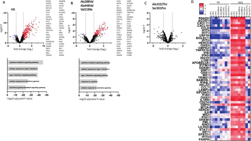
Fig. 1 DEGs in PBMC from HDs and patients carrying TLR7 variants stimulated with IMQ. A–C Volcano plots showing DEGs. Red dots show
upregulated genes (log2FC ≥ 1.5 with adjusted p value ≤ 0.05) and blue dots represent downregulated genes (log2FC ≤ −1,5; adjusted p
value ≤ 0.05). The DEGs with the top 50 absolute FC are reported. A Three healthy donors. B Patients (n = 3) carrying the Ala288Val, Ala488Val,
and Val219 Ile variants. C Patients carrying Ala1032Thr (n = 1) and Ser301Pro (n = 1) variants. Gene Ontology biological process terms
significantly overrepresented among the genes increased by IMQ are shown in the lower panel in (A, B, D). Heatmap of logCPM values for the
top DEGs in HDs and patients carrying TLR7 variants after IMQ stimulation. IMQ imiquimod, NS non stimulated.
TLR7-mediated MyD88 signal transduction, resulting in the unstimulated PBMC. The genes displaying the top 50 absolute
activation of mitogen-activated protein kinase cascades and NF- FC are listed in Fig. 1A. We used the Gene Ontology (GO) database
κB [14]. Signaling in human immune cells by TLR7 has been to perform GO-biological process enrichment analysis of DEGs.
documented to trigger production of pro-inflammatory cytokines, Cytokine-mediated signaling and cellular response to interferons
including tumor necrosis factor α (TNF-α), interleukin (IL)-6, IL-1β, were upregulated pathways in HDs (Fig. 1A, lower panel). Patients
and IL-12 as well as IFN-I. IFN-I regulates a range of immune carrying hypomorphic or hypofunctional variants displayed 108
responses through the IFN-I receptor, resulting in the transcription upregulated genes (log2FC ≥ 1.5; adjusted p value ≤ 0.05) and 5
of hundreds of IFN-stimulated genes (ISGs) whose joint action downregulated genes (log2FC ≤ −1.5; adjusted p value ≤ 0.05),
leads to the generation of an “antiviral state” [8, 14]. most of which were the same observed in HDs (Fig. 1B). The GO-
To gain insight into TLR7-linked mechanisms of severe COVID- biological process enrichment analysis identified the same
19, we performed RNA sequencing (RNA-Seq) to carefully upregulated pathways (Fig. 1B, lower panel). Interestingly, RNA-
characterize transcriptome variations following IMQ stimulation seq analysis in patients carrying LOF variants showed that none
of peripheral blood mononuclear cells (PBMC) isolated from had genes with an adjusted p value ≤ 0.05 (Fig. 1C), suggesting a
patients carrying previously identified LOF TLR7 variants [5]. In profound impairment of the TLR7 pathway. As shown in the heat
addition, we found new TLR7 variants in severely affected males map (Fig. 1D), for most of the 50 genes with the highest FC in HDs
for which functional characterization of the pathway was also after IMQ stimulation we noticed a significant upregulation in
performed. patients carrying hypomorphic and hypofunctional variants but
not in patients with LOF variants. A notable exception was IFNγ for
which a failure to induce upregulation following stimulation was
RESULTS AND DISCUSSION also observed in cells harboring the hypofunctional and hypo-
To study more deeply the functional effects of TLR7 variants, after morphic variants. It has been shown that at around day 10 in
TLR7 stimulation with IMQ in comparison with unstimulated cells, subjects with COVID-19, IFN-I decreased while IFNγ remained
we performed RNA-Seq experiments on PBMC from healthy stable [15], promoting the resolution of lung inflammation.
donors (HDs) and from patients carrying the functionally Therefore, administration of IFN-I might be considered a
hypomorphic variants Ala288Val and Ala448Val, the hypofunc- therapeutic option for TLR7 mutated patients. The efficacy of
tional variant Val219Ile, and the LOF variants Ala1032Thr and IFN-I therapy would depend on whether it is administered early in
Ser301Pro. As shown in Fig. 1A, B, we observed several the course of the disease. Patients with a severe course of COVID-
differentially expressed genes (DEGs) in the HDs as well as in 19 are usually admitted to the hospital after a few days at home
the patients carrying hypomorphic and hypofunctional variants. In making it difficult to identify those in need of IFN-I treatment.
contrast, when LOF variants were analyzed, no DEGs were found Indeed, inappropriate administration of IFN-I to the wrong
(Fig. 1C). Specifically, TLR7 stimulation induced a strong response patients or at the wrong time point could be counterproductive
in HDs with 211 genes significantly upregulated (log2 fold change by triggering the cytokine storm. A more attractive therapeutic
(FC) ≥ 1.5; adjusted p value ≤ 0.05) and 19 downregulated genes option would be IFNγ, which is not only useful in patients with
(log2FC ≤ −1.5; adjusted p value ≤ 0.05) compared with hypomorphic mutations but, in addition, can stabilize the
Genes & ImmunityS. Mantovani et al.
3
Fig. 2 Gene expression analysis in PBMC of patients carrying TLR7 variants stimulated with IMQ. A Pedigree of P6 (A upper panel) and P10
(A lower panel) shows the segregation of the variant within the family. Squares represent male family members; circles, females. Black symbols
indicate individuals harboring the TLR7 variant. Individuals infected by SARS-CoV-2 are indicated by a virus symbol () close to the individual
symbol. B–G PBMC from COVID-19 patients and four unaffected male controls (HDs) were stimulated with IMQ at 5 μg/mL or cell culture
medium. A quantitative PCR assay was performed and 2−ΔΔCt was calculated using HPRT1 as the housekeeping gene. Fold change in mRNA
expression of genes induced by IMQ with respect to cell culture medium was calculated: *p < 0.05; **p < 0.01; ***p < 0.001.
inflammatory response and does not require timely carrying the hypomorphic variant Ala288Val showed a reduced,
administration. though not statistically significant, production of IFNα protein
As shown in Fig. 1, in HDs and in patients carrying hypomorphic after TLR7 engagement (Supplementary Fig. 2). Overall, the
and hypofunctional variants, but not in patients carrying LOF transcriptomic profile of cells harboring LOF TLR7 variants showed
variants, TLR7 engagement triggered an antiviral response a wide deficiency of ISGs while both hypomorphic and LOF
upregulating typical antiviral ISGs (RSAD2, APOBEC3A, HERC5, mutations displayed a reduction of IFNγ transcription.
OASs, MXs, IFITs, and IFITMs family genes) as well as proinflamma- We next extended the analysis to two additional rare TLR7
tory cytokine and chemokine genes (IL6, CXCL10, CXCL11, CCL8, variants: the already reported Arg920Lys variant (P6) [5], and the
CCL2, CCL7) [8]. Interestingly, IL6 and CXCL10 were found to be new Asp41Glu (P10) variant predicted to be deleterious from in
involved in the mechanisms sustaining the cytokine-storm, a silico analysis (Fig. 2A). The two variants were identified in two
peculiar aspect of SARS-COV-2 infection which, at least in severe severely affected male patients aged 49 and 79 years, one in each.
cases, is responsible for diffuse alveolar damage and multi-organ In order to functionally characterize the TLR7 pathway, we
failure [16]. Furthermore, IFITMs inhibit cellular entry of SARS-CoV performed a gene expression profile analysis of PBMC from
and MERS-CoV [17]. We also observed upregulation of CCL2 and patient P6 and from two relatives of patient P10 following
CCL7, two pivotal chemokines for monocyte recruitment, both of stimulation with IMQ. We found a statistically significant decrease
which were found enriched in bronchoalveolar fluid (BALF) from in mRNA levels for IFNα and IFNγ genes in P6, P10-II-I, and P10-II-III
patients with severe COVID-19 [18, 19]. Moreover, the TNFSF10 compared with HDs (Fig. 2B, C). We further analyzed some of the
gene (TRAIL), an apoptosis-related gene that was previously found genes showing the highest FC in the HDs transcriptomic profile
to be upregulated in BALF and PBMC from COVID-19 patients [20], and observed a significant decrease of mRNAs encoding for
was also upregulated after IMQ stimulation in HD and in patients RSAD2, ACOD1, and IFIT2 genes in P6, demonstrating a profound
carrying hypomorphic and hypofunctional variants. Meanwhile, impairment of the TLR7 signaling pathway in response to
we observed a marked induction of negative regulators (such as TLR7 stimulation (Fig. 2D–F). Of note, it was reported that RSAD2,
USP18, IL1RN, and ACOD1), suggesting stimulation of negative in addition to the role of direct suppressor of viral replication,
feedback loops. The functional status of the cells was evaluated by promotes TLR7- and TLR9-mediated production of IFNα. [21].
stimulating PBMC from patients and HDs with the TLR4 agonist Moreover, we observed that CXCL10 mRNA was markedly reduced
lipopolysaccharide (LPS). Intracellular production of IL6 was in P6, P10-II-I, and P10-II-III compared with HDs (Fig. 2G). Overall,
evaluated in monocytes (as shown in Supplementary Fig. 1). The our data expand previous findings on the TLR7 role in rare
frequencies of CD3−CD14+IL6+ cells were comparable in patients Mendelian forms of COVID-19 and provide further insights into the
and HDs, indicating that cells from patients harboring TLR7 altered pathways that might contribute to disease severity.
variants were functionally active. IFNα and IFNγ protein produc-
tion was evaluated in the supernatant of PBMC from HDs and from
a small number of patients after TLR7 engagement. The data MATERIALS AND METHODS
showed a trend toward a lower production of IFNα as well as IFNγ Male COVID-19 patients were selected from the Italian GEN-COVID cohort
proteins in patients carrying LOF TLR7 variants. The patient [5]. Cases were selected according to the following inclusion criteria: (i)
male gender; (ii) young age (S. Mantovani et al.
4
in TLR7. Exclusion criteria were: (i) SARS-CoV-2 infection not confirmed by 16. Coperchini F, Chiovato L, Croce L, Magri F, Rotondi M. The cytokine storm in
PCR. Relatives of patients 6 and 10 were contacted to obtain a blood COVID-19: an overview of the involvement of the chemokine/chemokine-
sample. Segregation analysis of the variants was performed with Sanger receptor system. Cytokine Growth Factor Rev. 2020;53:25–32.
sequencing on an ABI3130 Genetic Analyzer. PBMC isolation, IMQ 17. Huang IC, Bailey CC, Weyer JL, Radoshitzky SR, Becker MM, Chiang JJ, et al.
stimulation, and qPCR were performed as previously described [5]. The Distinct patterns of IFITM-mediated restriction of filoviruses, SARS coronavirus,
primers used are listed in Supplementary Table 1. Supernatants of PBMCs and influenza A virus. PLoS Pathog. 2011;7:e1001258.
stimulated with IMQ, LPS, or medium alone were measured for IFNα 18. Shi C, Pamer EG. Monocyte recruitment during infection and inflammation. Nat
(Invitrogen) and IFNγ production (Bio-Techne) according to the manufac- Rev Immunol. 2011;11:762–74.
turer’s instructions. PBMC were stimulated in vitro with LPS at 1 µg/ml for 19. Zhou Z, Ren L, Zhang L, Zhong J, Xiao Y, Jia Z, et al. Heightened innate immune
4 h, then IL6 production was examined in CD3− CD14+ cells by flow responses in the respiratory tract of COVID-19 patients. Cell Host Microbe.
cytometry. Briefly, 3 × 10⁵ PBMC were stained with anti-CD3 BV605 and 2020;27:883–890.e2.
anti-CD14 BB700 mAbs, fixed and permeabilized with the BD Cytofix/ 20. Xiong Y, Liu Y, Cao L, Wang D, Guo M, Jiang A, et al. Transcriptomic characteristics
Cytoperm kit in the presence of anti-IL6 BV421 (Becton Dickinson) of bronchoalveolar lavage fluid and peripheral blood mononuclear cells in
according to the manufacturer’s instructions. RNA quality was assessed COVID-19 patients. Emerg Microbes Infect. 2020;9:761–70.
by Fragment Bioanalyzer (Agilent); all samples exhibited RNA quality 21. Saitoh T, Satoh T, Yamamoto N, Uematsu S, Takeuchi O, Kawai T, et al. Antiviral
numbers greater than 8. The libraries for RNA-seq were performed protein Viperin promotes Toll-like receptor 7- and Toll-like receptor 9-mediated
according to the Illumina TruSeq Stranded mRNA Library preparation type I interferon production in plasmacytoid dendritic cells. Immunity.
protocol and sequenced in multiplex with HiSeq 2500 platform (Illumina) 2011;34:352–63.
in 50 nucleotides, paired-end read configuration. Sequencing data were 22. Torre D, Lachmann A, Ma'ayan A. BioJupies: automated generation of interactive
analyzed using the BioJupies platform [22]. notebooks for RNA-seq data analysis in the cloud. Cell Syst. 2018;7:556–561.
WES and Genotype (GWAS) data were generated within the GEN-COVID
Data Repository (GCGDR). In order to be able to store and analyze the
massive amount of genomic data generated with the analysis of the entire
cohort of samples populating the biobank, we relied on the NIG. External ACKNOWLEDGEMENTS
users can upload and analyze data using the NIG pipeline by registering This study is part of the GEN-COVID Multicenter Study, https://sites.google.com/dbm.
and creating a specific project. A section dedicated to COVID-19 samples unisi.it/gen-covid, the Italian multicenter study aimed at identifying the COVID-19
has been created within the NIG database (http://nigdb.cineca.it/) that host genetic bases. Specimens were provided by the COVID-19 Biobank of Siena,
provides variant frequencies as a free tool for both clinicians and which is part of the Genetic Biobank of Siena, member of BBMRI-IT, of Telethon
researchers. Network of Genetic Biobanks (project No. GTB18001), of EuroBioBank, and of RD-
Connect. We thank the CINECA consortium for providing computational resources
and the Network for Italian Genomes (NIG; http://www.nig.cineca.it) for its support.
REFERENCES We thank private donors for the support provided to AR (Department of Medical
1. Zhu N, Zhang D, Wang W, Li X, Yang B, Song J, et al. China Novel Coronavirus Biotechnologies, University of Siena) for the COVID-19 host genetics research project
Investigating and Research Team. A Novel Coronavirus from Patients with (D.L n.18 of March 17, 2020). We also thank the COVID-19 Host Genetics Initiative
Pneumonia in China, 2019. N Engl J Med. 2020;382:727–33. (https://www.covid19hg.org/), MIUR project “Dipartimenti di Eccellenza 2018–2020”
2. Richardson S, Hirsch JS, Narasimhan M, Crawford JM, McGinn T, Davidson KW, to the Department of Medical Biotechnologies University of Siena, Italy, and “Bando
et al. Presenting characteristics, comorbidities, and outcomes among 5700 Ricerca COVID-19 Toscana” project to Azienda Ospedaliero-Universitaria Senese. We
patients hospitalized with COVID-19 in the New York City Area. J Am Med Assoc. thank Intesa San Paolo for the 2020 charity fund dedicated to the project N B/2020/
2020;323:2052–9. 0119 “Identificazione delle basi genetiche determinanti la variabilità clinica della
3. Park A, Iwasaki A. Type I and Type III interferons—induction, signaling, evasion, risposta a COVID-19 nella popolazione italiana”. Generous support was also received
and application to combat COVID-19. Cell Host Microbe. 2020;27:870–8. from private donations by Mrs. Maurizio Traglio, Enzo Cattaneo, and Alberto Borella.
4. van der Made CI, Simons A, Schuurs-Hoeijmakers J, van den Heuvel G, Mantere T, We thank Francis P. Crawley for revising the paper. We thank the CINECA consortium
Kersten S, et al. Presence of genetic variants among young men with severe for providing computational resources and the Network for Italian Genomes (NIG)
COVID-19. J Am Med Assoc. 2020;324:1–11. http://www.nig.cineca.it for its support. We thank private donors for the support
5. Fallerini C, Daga S, Mantovani S, Benetti E, Picchiotti N, Francisci D, et al. Asso- provided to A.R. (Department of Medical Biotechnologies, University of Siena) for the
ciation of Toll-like receptor 7 variants with life-threatening COVID-19 disease in COVID-19 host genetics research project (D.L n.18 of March 17, 2020). We also thank
males: findings from a nested case-control study. Elife. 2021;10:e67569. the COVID-19 Host Genetics Initiative (https://www.covid19hg.org/); MIUR project
6. Bastard P, Rosen LB, Zhang Q, Michailidis E, Hoffmann HH, Zhang Y, et al. “Dipartimenti di Eccellenza 2018–2020” to the Department of Medical Biotechnol-
Autoantibodies against type I IFNs in patients with life-threatening COVID-19. ogies University of Siena, Italy. We also thank Intesa San Paolo for the 2020 charity
Science. 2020;370:eabd4585. fund dedicated to the project N. B/2020/0119 “Identificazione delle basi genetiche
7. Zhang Q, Bastard P, Liu Z, Le Pen J, Moncada-Velez M, Chen J, et al. Inborn errors determinanti la variabilità clinica della risposta a COVID-19 nella popolazione italiana”
of type I IFN immunity in patients with life-threatening COVID-19. Science. and the Tuscany Region for funding within “Bando Ricerca COVID-19 Toscana”
2020;370:eabd4570. project supporting research at the Azienda Ospedaliero-Universitaria Senese. We also
8. Grandvaux N, tenOever B. R, Servant M. J, Hiscott J. The interferon antiviral thank Mrs. Maurizio Traglio, Enzo Cattaneo and Alberto Borella for private donations.
response: from viral invasion to evasion. Curr Opin Infect Dis. 2002;15
(Jun):259–67.
9. Lee J, Wu CC, Lee KJ, Chuang TH, Katakura K, Liu YT, et al. Activation of anti-
hepatitis C virus responses via Toll-like receptor 7. Proc Natl Acad Sci USA. AUTHOR CONTRIBUTIONS
2006;103:1828–33. Conceptualization: SM, SD, CF, IM, MUM, and AR. Data curation: SM, SD, CF, IM, FM,
10. Heil F, Hemmi H, Hochrein H, Ampenberger F, Kirschning C, Akira S, et al. Species- AR, MUM, and GEN‐COVID Multicenter Study. Formal analysis: SM, SD, CF, EB, NP, and
specific recognition of single-stranded RNA via toll-like receptor 7 and 8. Science. SF. Funding acquisition: AR. Investigation: MB, SA, IM, EF, FM, and AR. Methodology:
2004;303:1526–9. MB, FF, AG, SZ, MB, MP, SC, SA, DA, and KC. Resources: AR and MUM; Software: SM,
11. Isogawa M, Robek MD, Furuichi Y, Chisari FV. Toll-like receptor signaling inhibits SD, EB, NP, and SF; Supervision: EF, IM, FM, AR, and MUM. Validation: IM, AR, and
hepatitis B virus replication in vivo. J Virol. 2005;79:7269–72. MUM. Visualization: CG, CF, MB, FF, CF, AR, and RM. Writing-original draft: SM, SD, CF,
12. Poulas K, Farsalinos K, Zanidis C. Activation of TLR7 and innate immunity as an EF, IM, AR, and MUM. All authors have reviewed and approved the paper.
efficient method against COVID-19 pandemic: imiquimod as a potential therapy.
Front Immunol. 2020;11:1373.
13. Mele D, Mantovani S, Oliviero B, Grossi G, Ludovisi S, Mondelli MU, et al. Hepatitis
C virus inhibits CD4 T cell function via binding to Toll-like receptor 7. Antiviral ETHICS
Res. 2017;137:108–11. The GEN-COVID study was performed in accordance with the relevant international,
14. Petes C, Odoardi N, Gee K. The Toll for trafficking: Toll-like receptor 7 delivery to EU, national, and institutional guidelines and approved in advance by the University
the endosome. Front Immunol. 2017;8:1075. Hospital (Azienda Ospedaliero-Universitaria Senese) Ethical Review Board, Siena, Italy
15. Galani IE, Rovina N, Lampropoulou V, Triantafyllia V, Manioudaki M, Pavlos E, et al. (Prot n. 16917, dated March 16, 2020). Clinical trial registration NCT04549831. Patients
Untuned antiviral immunity in COVID-19 revealed by temporal type I/III interferon or legally authorized representatives provided informed consent for participating in
patterns and flu comparison. Nat Immunol. 2021 Jan;22:32–40. the study.
Genes & ImmunityS. Mantovani et al.
5
COMPETING INTERESTS Open Access This article is licensed under a Creative Commons
The authors declare no competing interests. Attribution 4.0 International License, which permits use, sharing,
adaptation, distribution and reproduction in any medium or format, as long as you give
appropriate credit to the original author(s) and the source, provide a link to the Creative
ADDITIONAL INFORMATION Commons license, and indicate if changes were made. The images or other third party
Supplementary information The online version contains supplementary material material in this article are included in the article’s Creative Commons license, unless
available at https://doi.org/10.1038/s41435-021-00157-1. indicated otherwise in a credit line to the material. If material is not included in the
article’s Creative Commons license and your intended use is not permitted by statutory
Correspondence and requests for materials should be addressed to Mario U. regulation or exceeds the permitted use, you will need to obtain permission directly
Mondelli or Alessandra Renieri. from the copyright holder. To view a copy of this license, visit http://creativecommons.
org/licenses/by/4.0/.
Reprints and permission information is available at http://www.nature.com/
reprints
© The Author(s) 2021
Publisher’s note Springer Nature remains neutral with regard to jurisdictional claims
in published maps and institutional affiliations.
GEN-COVID MULTICENTER STUDY
Francesca Montagnani3,10, Mario Tumbarello3,10, Ilaria Rancan3,10, Massimiliano Fabbiani10, Barbara Rossetti10, Laura Bergantini11,
Miriana D’Alessandro11, Paolo Cameli11, David Bennett11, Federico Anedda12, Simona Marcantonio12, Sabino Scolletta12,
Federico Franchi12, Maria Antonietta Mazzei13, Susanna Guerrini13, Edoardo Conticini14, Luca Cantarini14, Bruno Frediani14,
Danilo Tacconi15, Chiara Spertilli Raffaelli15, Marco Feri16, Alice Donati16, Raffaele Scala17, Luca Guidelli17, Genni Spargi18, Marta Corridi18,
Cesira Nencioni19, Leonardo Croci19, Gian Piero Caldarelli20, Davide Romani21, Paolo Piacentini21, Maria Bandini21, Elena Desanctis21,
Silvia Cappelli21, Anna Canaccini22, Agnese Verzuri22, Valentina Anemoli22, Manola Pisani22, Agostino Ognibene23,
Alessandro Pancrazzi23, Maria Lorubbio23, Massimo Vaghi24, Antonella D’Arminio Monforte25, Federica Gaia Miraglia25,
Raffaele Bruno26,27, Marco Vecchia26, Massimo Girardis28, Sophie Venturelli28, Stefano Busani28, Andrea Cossarizza29, Andrea Antinori30,
Alessandra Vergori30, Arianna Emiliozzi30, Stefano Rusconi31,32, Matteo Siano32, Arianna Gabrieli31, Agostino Riva30,31,
Daniela Francisci33,34, Elisabetta Schiaroli33, Francesco Paciosi33, Andrea Tommasi33, Pier Giorgio Scotton35, Francesca Andretta35,
Sandro Panese36, Stefano Baratti36, Renzo Scaggiante37, Francesca Gatti37, Saverio Giuseppe Parisi38, Francesco Castelli39,
Eugenia Quiros-Roldan39, Melania Degli Antoni39, Isabella Zanella40,41, Matteo Della Monica42, Carmelo Piscopo42, Mario Capasso43,44,45,
Roberta Russo43,44, Immacolata Andolfo43,44, Achille Iolascon43,44, Giuseppe Fiorentino46, Massimo Carella47, Marco Castori47,
Filippo Aucella48, Pamela Raggi49, Rita Perna49, Matteo Bassetti50,51, Antonio Di Biagio50,51, Maurizio Sanguinetti52,53, Luca Masucci52,53,
Alessandra Guarnaccia52, Serafina Valente54, Oreste De Vivo54, Gabriella Doddato2,3, Mirjam Lista2,3, Giada Beligni2,3,
Floriana Valentino2,3, Kristina Zguro3, Rossella Tita6, Annarita Giliberti2,3, Maria Antonietta Mencarelli6, Caterina Lo Rizzo6,
Anna Maria Pinto6, Francesca Ariani2,3,6, Laura Di Sarno2,3, Elena Bargagli11, Marco Mandalà55, Alessia Giorli55, Lorenzo Salerni55,
Patrizia Zucchi56, Pierpaolo Parravicini56, Elisabetta Menatti57, Tullio Trotta58, Ferdinando Giannattasio58, Gabriella Coiro58, Fabio Lena59,
Gianluca Lacerenza59, Cristina Mussini60, Enrico Martinelli61, Luisa Tavecchia62, Mary Ann Belli62, Lia Crotti63,64,65,66,67,
Gianfranco Parati63,64, Maurizio Sanarico68, Francesco Raimondi69, Filippo Biscarini70, Alessandra Stella70, Tiziana Bachetti71,
Maria Teresa La Rovere72, Serena Ludovisi73, Maurizio Bussotti74, Simona Dei75, Sabrina Ravaglia76, Rosangela Artuso77,
Elena Andreucci77, Giulia Gori77, Angelica Pagliazzi77, Erika Fiorentini77, Antonio Perrella78, Francesco Bianchi3,78, Paola Bergomi79,
Emanuele Catena79, Riccardo Colombo79, Sauro Luchi80, Giovanna Morelli80, Paola Petrocelli80, Sarah Iacopini80, Sara Modica80,
Silvia Baroni81, Francesco Vladimiro Segala82, Marco Falcone83, Giusy Tiseo83, Chiara Barbieri83, Tommaso Matucci83, Davide Grassi84,
Claudio Ferri84, Franco Marinangeli85, Francesco Brancati86, Antonella Vincenti87, Valentina Borgo87, Stefania Lombardi87, Mirco Lenzi87,
Massimo Antonio Di Pietro88, Francesca Vichi88, Benedetta Romanin88, Letizia Attala88, Cecilia Costa88, Andrea Gabbuti88,
Roberto Menè63,64, Marta Colaneri26, Patrizia Casprini89, Giuseppe Merla43,90, Gabriella Maria Squeo90 and Marcello Maffezzoni91
10
Department of Medical Sciences, Infectious and Tropical Diseases Unit, Azienda Ospedaliera Universitaria Senese, Siena, Italy. 11Unit of Respiratory Diseases and Lung
Transplantation, Department of Internal and Specialist Medicine, University of Siena, Siena, Italy. 12Department of Emergency and Urgency, Medicine, Surgery and Neurosciences,
Unit of Intensive Care Medicine, Siena University Hospital, Siena, Italy. 13Department of Medical, Surgical and Neuro Sciences and Radiological Sciences, Unit of Diagnostic
Imaging, University of Siena, Siena, Italy. 14Rheumatology Unit, Department of Medicine, Surgery and Neurosciences, University of Siena, Policlinico Le Scotte, Siena, Italy.
15
Department of Specialized and Internal Medicine, Infectious Diseases Unit, San Donato Hospital Arezzo, Arezzo, Italy. 16Department of Emergency, Anesthesia Unit, San Donato
Hospital, Arezzo, Italy. 17Cardio-Thoracic and Neurologic Department, Pneumology Unit and Respiratory Intesive Care Unit, San Donato Hospital, Arezzo, Italy. 18Department of
Emergency, Anesthesia Unit, Misericordia Hospital, Grosseto, Italy. 19Department of Specialized and Internal Medicine, Infectious Diseases Unit, Misericordia Hospital, Grosseto,
Italy. 20Clinical Chemical Analysis Laboratory, Misericordia Hospital, Grosseto, Italy. 21Dipartimento di Prevenzione, Azienda USL Toscana Sud Est, Arezzo, Italy. 22Dipartimento
Tecnico-Scientifico Territoriale, Azienda USL Toscana Sud Est, Arezzo, Italy. 23Clinical Chemical Analysis Laboratory, San Donato Hospital, Arezzo, Italy. 24Chirurgia Vascolare,
Ospedale Maggiore di Crema, Crema, Italy. 25Department of Health Sciences, Clinic of Infectious Diseases, ASST Santi Paolo e Carlo, University of Milan, Milano, Italy. 26Division of
Infectious Diseases I, Fondazione IRCCS Policlinico San Matteo, Pavia, Italy. 27Department of Clinical, Surgical, Diagnostic, and Pediatric Sciences, University of Pavia, Pavia, Italy.
28
Department of Anesthesia and Intensive Care, University of Modena and Reggio Emilia, Modena, Italy. 29Department of Medical and Surgical Sciences for Children and Adults,
University of Modena and Reggio Emilia, Modena, Italy. 30HIV/AIDS Department, National Institute for Infectious Diseases, IRCCS, Lazzaro Spallanzani, Rome, Italy. 31III Infectious
Diseases Unit, ASST-FBF-Sacco, Milan, Italy. 32Department of Biomedical and Clinical Sciences Luigi Sacco, University of Milan, Milan, Italy. 33Infectious Diseases Clinic, “Santa
Maria” Hospital, University of Perugia, Perugia, Italy. 34Infectious Diseases Clinic, Department of Medicine 2, Azienda Ospedaliera di Perugia and University of Perugia, Santa Maria
Hospital, Perugia, Italy. 35Department of Infectious Diseases, Treviso Hospital, Local Health Unit 2 Marca Trevigiana, Treviso, Italy. 36Clinical Infectious Diseases, Mestre Hospital,
Venezia, Italy. 37Infectious Diseases Clinic, ULSS1 Belluno, Italy. 38Department of Molecular Medicine, University of Padova, Padova, Italy. 39Department of Infectious and Tropical
Diseases, University of Brescia and ASST Spedali Civili Hospital, Brescia, Italy. 40Department of Molecular and Translational Medicine, University of Brescia, Brescia, Italy
Genes & ImmunityS. Mantovani et al.
6
41
Clinical Chemistry Laboratory, Cytogenetics and Molecular Genetics Section, Diagnostic Department, ASST Spedali Civili di Brescia, Brescia, Italy. 42Medical Genetics and
Laboratory of Medical Genetics Unit, A.O.R.N. “Antonio Cardarelli”, Naples, Italy. 43Department of Molecular Medicine and Medical Biotechnology, University of Naples Federico II,
Naples, Italy. 44CEINGE Biotecnologie Avanzate, Naples, Italy. 45IRCCS SDN, Naples, Italy. 46Unit of Respiratory Physiopathology, AORN dei Colli, Monaldi Hospital, Naples, Italy.
47
Division of Medical Genetics, Fondazione IRCCS Casa Sollievo della Sofferenza Hospital, San Giovanni Rotondo, Italy. 48Department of Medical Sciences, Fondazione IRCCS Casa
Sollievo della Sofferenza Hospital, San Giovanni Rotondo, Italy. 49Clinical Trial Office, Fondazione IRCCS Casa Sollievo della Sofferenza Hospital, San Giovanni Rotondo, Italy.
50
Department of Health Sciences, University of Genova, Genova, Italy. 51Infectious Diseases Clinic, Policlinico San Martino Hospital, IRCCS for Cancer Research Genova, Genova,
Italy. 52Microbiology, Fondazione Policlinico Universitario Agostino Gemelli IRCCS, Catholic University of Medicine, Rome, Italy. 53Department of Laboratory Sciences and
Infectious Diseases, Fondazione Policlinico Universitario A. Gemelli IRCCS, Rome, Italy. 54Department of Cardiovascular Diseases, University of Siena, Siena, Italy. 55Otolaryngology
Unit, University of Siena, Siena, Italy. 56Department of Internal Medicine, ASST Valtellina e Alto Lario, Sondrio, Italy. 57Study Coordinator Oncologia Medica e Ufficio Flussi Sondrio,
Sondrio, Italy. 58First Aid Department, Luigi Curto Hospital, Polla, Salerno, Italy. 59Department of Pharmaceutical Medicine, Misericordia Hospital, Grosseto, Italy. 60Infectious
Diseases Clinics, University of Modena and Reggio Emilia, Modena, Italy. 61Department of Respiratory Diseases, Azienda Ospedaliera di Cremona, Cremona, Italy. 62U.O.C.
Medicina, ASST Nord Milano, Ospedale Bassini, Cinisello Balsamo (MI), Balsamo, Italy. 63Istituto Auxologico Italiano, IRCCS, Department of Cardiovascular, Neural and Metabolic
Sciences, San Luca Hospital, Milan, Italy. 64Department of Medicine and Surgery, University of Milano-Bicocca, Milan, Italy. 65Istituto Auxologico Italiano, IRCCS, Center for Cardiac
Arrhythmias of Genetic Origin, Milan, Italy. 66Istituto Auxologico Italiano, IRCCS, Laboratory of Cardiovascular Genetics, Milan, Italy. 67Member of the European Reference Network
for Rare, Low Prevalence and Complex Diseases of the Heart-ERN GUARD-Heart, Rome, Italy. 68Independent Data Scientist, Milan, Italy. 69Scuola Normale Superiore, Pisa, Italy.
70
CNR-Consiglio Nazionale delle Ricerche, Istituto di Biologia e Biotecnologia Agraria (IBBA), Milano, Italy. 71Direzione Scientifica, Istituti Clinici Scientifici Maugeri IRCCS, Pavia,
Italy. 72Istituti Clinici Scientifici Maugeri IRCCS, Department of Cardiology, Institute of Montescano, Pavia, Italy. 73Fondazione IRCCS Ca’ Granda Ospedale Maggiore Policlinico,
Milan, Italy. 74Istituti Clinici Scientifici Maugeri IRCCS, Department of Cardiology, Institute of Milan, Milan, Italy. 75Health Management, Azienda USL Toscana Sudest, Tuscany, Italy.
76
IRCCS C. Mondino Foundation, Pavia, Italy. 77Medical Genetics Unit, Meyer Children’s University Hospital, Florence, Italy. 78Department of Medicine, Pneumology Unit,
Misericordia Hospital, Grosseto, Italy. 79Department of Anesthesia and Intensive Care Unit, ASST Fatebenefratelli Sacco, Luigi Sacco Hospital, Polo Universitario, University of
Milan, Milano, Italy. 80Infectious Disease Unit, Hospital of Lucca, Lucca, Italy. 81Department of Diagnostic and Laboratory Medicine, Institute of Biochemistry and Clinical
Biochemistry, Fondazione Policlinico Universitario A. Gemelli IRCCS, Catholic University of the Sacred Heart, Rome, Italy. 82Clinic of Infectious Diseases, Catholic University of the
Sacred Heart, Rome, Italy. 83Department of Clinical and Experimental Medicine, Infectious Diseases Unit, University of Pisa, Pisa, Italy. 84Department of Clinical Medicine, Public
Health, Life and Environment Sciences, University of L’Aquila, L’Aquila, Italy. 85Anesthesiology and Intensive Care, University of L’Aquila, L’Aquila, Italy. 86Medical Genetics Unit,
Department of Life, Health and Environmental Sciences, University of L’Aquila, L’Aquila, Italy. 87Infectious Disease Unit, Hospital of Massa, Massa, Italy. 88Infectious Diseases Unit,
Santa Maria Annunziata Hospital, USL Centro, Florence, Italy. 89Laboratory of Clinical Pathology and Immunoallergy, Florence-Prato, Italy. 90Laboratory of Regulatory and
Functional Genomics, Fondazione IRCCS Casa Sollievo della Sofferenza, San Giovanni Rotondo (Foggia), Foggia, Italy. 91University of Pavia, Pavia, Italy.
Genes & ImmunityYou can also read