PREVENTION AND TREATMENT OF 'TENNIS ELBOW'
←
→
Page content transcription
If your browser does not render page correctly, please read the page content below
Gudelj, J. and Kosinac, Z.: Prevention and treatment of ‘tennis elbow’                Sport Science 6 (2013) 1: 113‐117

                          PREVENTION AND TREATMENT OF ‘TENNIS ELBOW’
                                      Jelena Gudelj and Zdenko Kosinac
                                                      Split, Croatia

                                                                                                        Review paper

Abstract
‘Tennis elbow’ is overuse injury caused by frequent repeated contraction of hand and fingers extensors
resulting chronic stress muscles and tendons. The main symptom is pain that occurs on the outside of the
elbow, but can be felt in the upper arm or the outer side of the forearm. The diagnosis of tennis elbow using
computed tomography (CT), ultrasound, arthroscopy, magnetic resonance imaging (MRI) and clinical
diagnosis, which is the basis and most important diagnostic method. Depending on the stage of disease and
treatment is different. Great importance is given to kinesiotherapy, especially stretching exercises. Except
stretching exercises, kinesiotherapy also include isotonic and isometric exercises. Indications for operative
treatment is the existence of symptoms for more than six months, despite adequate treatment conducted
non-operatively.

Key words: tennis elbow, prevention, non-operatively, operative treatment

Introduction                                                      Etiopathogenesis of lateral epicondylitis

Tennis is played by millions of people of all ages                The disease is one degenerative process resulting
around the world. It is a sport that is very                      from     fatigue  due     to hamstring    injuries,
interesting, exciting and usually do not cause                    weaknesses and likely changes due to poor blood
serious medical problems. In the elbow, however,                  circulation. Regardless of the type of tissue
problems arise. Inflammation and degenerative                     response to injury is inflammation, which includes
changes in the elbow resulting in the medial and                  a number of changes in the final core networks,
lateral humeral epikondil. Epicondylitis humeri is                blood and connective tissue. Inflammatory
entenzitis that appears on the same starting point                reaction is a very complex reaction in which
extensor hands and fingers on the lateral epikondil               involved different types of cells, a number of
humerus, which is called the lateral (radial)                     enzymes, many physiologically active substances
epicondylitis (Grisogono, 1989). Inflammation of                  and other. Changes nerve causes severe pain in
the lateral epicondyle, or more often referred to as              the area of the damaged tendon or her handle the
a "tennis elbow" is one of the injuries of which                  often subtle morphological changes. There comes
suffer most sports and recreation (tennis) and                    to perineuritisa flange nerve sheath that
pitchers (baseball) and cricket players, javelin                  subsequently compressed nerve.
throwers, handball players, etc. (dactilografi,
shoemakers, dentists). Despite the name "tennis
elbow", professional tennis players are an absolute
minority, only 5 % of the total number of people
with inflammation of the lateral epicondyle. Lateral
epicondylitis (tennis elbow) occurs 7-10 times
more than the median, is more common in men,
usually occurs in middle aged between 30-50
years. The first description of symptoms
suggestive of lateral epicondylitis gave back
1873rd the German doctor Runge & Morris (1882).
Describing the symptoms of tennis introduces
name tennis arm (Engl. lawn tennis arm), while a                  Figure 1. Changes that occur in the tendons due to
year later Major changed the name for tennis                                       lateral epicondylitis
elbow, which is held to this day. Lateral                           (1. Ulna, 2. Humerus; m. extensor carpi radialis
epicondylitis is one of the most popular and also                    brevis; 4. m. extensor digitorum communis)
the most common overuse injuries of the
locomotor system in humans, manifested by pain                    Changes in the tendon can be seen in edema and
in the outer part of the elbow. Appears at the                    necrosis of the tendon, and after only a short time
starting point caput commune extensor hands and                   in the damaged area accumulate inflammatory
fingers on the lateral epikondil. Damage of                       cells (Figure 1). The inflammatory phase of the
mioentensial apparatus is caused by repeated                      regeneration of damaged tendons supplements
muscle contractions. The causes of pain are                       with the growing binder, in the beginning of
microscopic rupture tendon insertions. The pain                   cellular which eventually becomes richer collagen
occurs in the elbow and forearm. Because of                       fibers. Blood vessels multiply, but over time their
injuries is reduced vascularization in the affected               number is decreasing. The scar is different than
tendon perch and nervous endings are irritated                    normal tendon irregularity layout and structure of
and     resulting  inflammation.     Repetition   of              bundles of collagen fibers, and it is often found
movement can lead to a complete tendon rupture.                   more blood capillaries.

                                                                                                                  113
Gudelj, J. and Kosinac, Z.: Prevention and treatment of ‘tennis elbow’ Sport Science 6 (2013) 1: 113‐117
Changes tendon insertions can be seen in edema According to Peterson (2002), while playing tennis
and hemorrhage, sometimes there is a tear of the should pay attention to the following items: 1)
tendon or bone, connective scar bronchial often good footwork, so that the player is properly set
degenerates, so most of the tendons, and often up to the ball, 2) the ball should be in the right
the periosteum, becoming almost acellular and place at the right time to hit the racket, 3) on
imbued with a homogeneous mass of hyaline. impact should be applied throughout the shoulder
These scars often spread to surrounding tissue and body as when hitting the shuttle on the racket
structures by changing their appearance. Changes would not be disrupted paths, 4) it is very
nerves causes severe pain in the area of the important foundation courses, which should be
damaged tendon or her handle the often subtle slow to reduce the speed of the balls, 5) the ball
morphological changes. There comes to should be light; wet balls or balls with too little air
perineuritisa flange nerve sheath that pressure becomes severe, and 6) the right
subsequently compressed nerve (Renström & equipment - the racket should be selected
Peterson, 2002). Risk factors for lateral individually, taking into account the technique of
epicondylitis can be divided into two groups: the playing, 7) Great so. “swet spot” (center of the
use of inadequate equipment and the performance racket hitting the surface area on which generated
impact improper technique. Causal role play the smallest torsion -twisting on impact balls). Hit
racket and his posture, tension mat on it, the size balls out of the surface increases the vibrations.
and weight of the racket, the width of the handle,
the weight of the balls and hitting her mid Treatment of 'tennis elbow'
racquet, type of terrain, the intensity of impact Treatment can be nonsurgical and surgical.
players. Performing impact improper technique
(backhand shot) leads in connection with the Nonsurgical treatment
development of lateral epicondylitis. Concentric The objectives of non-operative treatment are: 1)
contraction that occurs when the shot incorrectly Relieve pain and inflammation control mioentesial
performed shortens the muscles in order to appliances; 2) Enhancing healing of mioentesial
maintain the tension needed to stabilize the wrist. appliances; 3) Control further action; 4) Increase
As a result of shortness muscle maximum load muscle strength and endurance of the muscles; 5)
occurs at the ends of the muscle or tendon. This Restoring the proper range of motion; and 6)
creates a certain force that is transmitted along Restore maximum functional ability, and 7
them to their starting point at the lateral Prevention of repeated movements on mioentesial
epikondilus humerus. Such repeated contractions apparatus (Kosinac, 2008; Grisogono, 1989).
resulting in chronic stress of mioentesial Treatment is important to start as early as
apparatus, which therefore reduce vascularization possible, at the appearance of the first symptoms.
and irritated nerve endings and the resulting Here are the most common and mistaken,
aseptic inflammation. because the first symptoms are usually not paid
sufficient attention and continues with the activity
Aim unchanged intensity. Treatment is divided into
three phases: 1) The first stage is the most
The main objective of this paper is to highlight the important holiday of the working and sporting
benefits and importance of preventive measures in activities. The patient should avoid movements
the development of "tennis elbow". Likewise, the that cause pain, but it has to perform active
goal of this paper is to describe the current movements of the rest of the limb to avoid
methods of treatment of tennis elbow due to the stiffness and other complications. At this stage in
stage of disease and therapeutic procedures in the the account comes Cryotherapy several times
treatment of tennis elbow. during the day. Cryotherapy reduces the pain
lowering conductivity sensory nerves, reduces
Prevention of 'tennis elbow'
inflammation and the island of vasoconstriction
Prevention of sports injuries, generally the primary and lowers levels of a chemical reaction. If the
task of a sports coach, a sports doctor and the present endemic, it is necessary to raise the grip
athlete. More than half of the injuries can be of your hand. It is recommended that you wear a
avoided by proper dosing load to prevent fatigue brace for the wrist that held his fist in the forward
and muscle fatigue in athletes. To ensure and position of the 200, which tendon release
implement prevention, ie to reduce the risk of excessive tension, and allows full mobility of the
overuse injuries, it is essential to know the risk fingers and elbow, 2) The second phase is
factors and their possible effect and negative characterized by the absence of pain at rest, or
effect on the development of overuse injuries. the appearance of increased pain when performing
From a theoretical point of view can be movements due to the increased load. At this
distinguished: general preventive measures and stage continues cryotherapy and starts with the
specific preventive measures. General preventive individual program of physical training. Of great
measures include: stretching as a leader in the importance are stretching exercises for increasing
prevention and treatment and strengthening - the length mioentezijskog apparatus at rest can be
strengthening the muscles of the forearm to reduced by stretching and during the execution of
stabilize the wrist. Preventive measures for certain movements. In addition to stretching
playing tennis are properly playing (technique) exercises, at this stage, complementing the
and avoiding asymmetric training techniques. isotonic and isometric exercises.
114Gudelj, J. and Kosinac, Z.: Prevention and treatment of ‘tennis elbow’ Sport Science 6 (2013) 1: 113‐117
From electrotherapy procedures used high-voltage
galvanic stimulation current that creates the
piezoelectric effect which helps healing of
mioentesial appliances, improves its vascularity,
and in some patients has an analgesic effect. In
the second week can apply electrotherapy
procedures : ultrasound, laser, magnetic therapy
and analgesic power. When all provocative
movements become painless, the patient returns
to his daily activities. during these activities must Figure 2. Application of corticosteroid injections
wear inelastic forearm cuff deflation which has the
function of secondary insertions of muscles which Example exercises for tennis elbow:
relieves tendon insertions on the lateral epikondilu
(Figure 1); 3) In the third stage the patient is fully
returned to their daily activities and sports. It is
recommended to continue to carry out stretching
and strengthening the affected muscles. In severe
physical activities necessary to implement
adequate preparation (warming) the affected
muscle group, and if necessary apply
cryomassage. To prevent recurrence in cases with
professional disease etiology should reduce the
weight of the working tool (racket) or frequency
provoking stereotyped movements, and if it is not
possible to increase the number and length of rest
during operation to allow for relaxation of muscles Figure 3. Stretching of extensor muscles
and reduce the risk of re-injury. The use of
corticosteroid injections should be delayed as long
as possible and limited to cases that do not
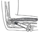
respond to nonsurgical treatment. Usually injected
up to three injections deep in Manila recess which
is sub-tendon positioned (Figure 2).
Figure 4. Fist extension
Figure 1. Forearm supporter
The downside is that steroid injections as the
organism adapts over time, so the answer to the
injection weaker and increases the risk of possible
complications (subcut atrophy, weakening the
surrounding tissue). The next day, after the
injection, pain can be intensified, but by the next Figure 5. Fist extension against resistance
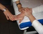
day pain decreases. Exercise 1: Stretching
exercises of extensor muscles: the patient
performs the movement can easily be seen from
the 90=, then completely extends elbow and
palmar inflecting hand while pressing the other
hand increase the palmar flexion fists up to the
occurrence of pain (Fig. 3). At this point, the
maximum painless stretching patient keep
stretching 15:25 s exercise should be repeated 4-
5 times a day, 2 sets of 10 reps, but always only
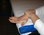
to the occurrence of pain. Exercise 2: Placing the
patient in the position of full forearm pronation on
the table so that his hands hanging over the edge Figure 6. hands extension with small weights
of the table in this position performs the extension
and flexion of the hand (Figure 4).
115Gudelj, J. and Kosinac, Z.: Prevention and treatment of ‘tennis elbow’ Sport Science 6 (2013) 1: 113‐117
The same exercise can be performed with an
elastic band that is placed around the fingers and
thumb, alternately making loops. Exercise 8:
Position arm to the horizontal position (900), the
elbow in extension. In a handful put blades, the
patient alternately increased and decreased hand
grip (Figure 10).
Surgical treatment
Figure 7. Fingers extension Indications for surgical treatment are the
persistence of symptoms for more than six months
with the inability to return to normal work and
leisure activities despite adequately conducted
nonsurgical treatment. Operative treatment is also
carried out for recurrent cases where they
diagnosed degenerative and calcification changes
in muscle-tendon structures with the aim of
removing the damaged tissue section affected
muscle.
Figure 8. Extension of the fingers against resistance
The operation proved to be always a part of
musculus extensor carpi radialis brevis, and in 35
% of cases musculus extensor digitorum
communis (Niethard, et al., 2009). The downside
of surgery is a rehabilitation treatment extended,
and success is not always satisfactory. After the
surgery elbow is immobilized for a week with steal
rail in a position of 900. Exercises for strength and
durability on resistance usually begins three weeks
after the surgery. In 85% of cases it is achieved
the disappearance of pain and restoration of the
Figure 9. Extension of the fingers against resistance previous power. Studies have shown recurrence of
18-66 %. The amount of pain before treatment
allows most reliable prediction of recovery: the
greater the pain, the greater the likelihood of
treatment success. After the surgery should take
8-10 weeks before they started to play tennis
again.
Discussion
Figure 10. Padded hand grip
Tennis elbow can be a big problem for players.
In the beginning, these movements are performed The pain can be so intense that the disabled
slowly, so that at the end of palmar flexion injured while performing work activities, or it may
accommodates up to 6, and at the end of interrupt the sport. Because of the lengthy delay
extensions to 3. Exercise 3: When the previous in sports activities it often lead to hypotrophy
movements cease to be painful, the patient extensor muscles and the islands of the affected
gradually increases the speed of execution and the areas, and can lead to a decrease in joint mobility
external resistance. Resistance is achieved by a and mild degree of calcification. When we talk
small weight or resisting physical therapists in the about prevention of tennis elbow, it is necessary
opposite direction of the movement that the to consider in detail the external and internal
patient performed (Figure 5). Exercise 4: The factors that participate in the realization of
initial position is the same as in the previous sporting activities, as well as their interactions,
exercise. Resistance is achieved by a small weight and the mechanisms of formation and the
(Figure 6). Exercise 5: The patient is in a sitting situation in which it comes to sports injury.
position, and forearms and hands were laid on the Technically correct play is the most important
ground in full pronation, while the fingers off base preventive measure, while the asymmetric training
flexion in MCF joints per 900. The patient performs techniques be avoided or reduced to necessarily
the movement extension of the fingers (Figure 7). play. Prevention of injury involves a series of
Exercise 6: The patient was in the same position measures such as: stretching exercises,
as in the previous exercise, only the resistance strengthening, massage and other forms of
applied to the dorsal side of the fingers in the activities that contribute to easier and faster
direction of flexion (Figure 8). Exercise 7: Elastic recovery of the players. For early detection,
rubber is placed around the fingers and thumb, diagnosis and appropriate treatment of overuse
which are collected in the form of the tower injuries is important knowledge of
(Figure 9). etiopathogenesis.
116Gudelj, J. and Kosinac, Z.: Prevention and treatment of ‘tennis elbow’ Sport Science 6 (2013) 1: 113‐117
The first step in treatment is certainly absolute Conclusion
rest and abstinence from basic sports activities
that led to the disease condition until symptoms Epicondylitis lateralis humeri is the damage that
are present (minimum recovery period is 7-10 occurs in the lateral epicondyle of the humerus, or
days). Experience shows that the disease can be in the grips forearm extensor muscle groups. The
kept, so that treatment can extend sometimes disease is one degenerative process resulting from
months to complete resolution of all symptoms. It fatigue due to hamstring injuries, weaknesses and
is important to emphasize that the pain in the likely changes due to poor blood circulation. Basic
elbow should not be underestimated or ignored, movement that causes overtraining is an
because in most cases they will not disappear by extension of elbow flexion with simultaneous
themselves. The basis of treatment is a stretching dorsal hands flexion and forearms supination from
exercise affected muscle group, and after pronation position. The problem in the treatment
stretching exceeds the static and dynamic of overuse injuries are daily load during training,
strengthening exercises with and without which can lead to chronic stages. Proper selection
resistance. Physical procedures are used to reduce of therapeutic procedures as well as early initiation
pain and improve circulation. A particular problem of treatment will lead to a reduction of
is the return of the sport or competitive activity inflammatory conditions and alleviate.
after treatment. If this return so quickly and
without a certain protection, the recurrence of However, for long-term success of therapy and
pain is more than probable. When they return, it is avoidance of chronic changes necessary to
advisable to continue to carry out activities that educate athletes and coaches. Therefore, great
will help prevent injuries. It takes at least 5 to 10 attention is paid to prevention. More than half of
minutes 'warm up' arms and shoulders gently the injuries can be avoided by proper dosing load
moving and stretching before starting the activity. to prevent fatigue and muscle fatigue. Good effect
In addition, it is recommended to change the in preventing tennis elbow can be achieved: by
racquet, as to reduce the stress on the structure changing the tennis equipment, sports mastered
of the elbow and the forearm muscles to put the techniques and adequate fitness and trained coach
corset that their pressure on the muscles reduce with enviable knowledge of the basics of anatomy,
their maximum power, and thus relieve elbow. physiology and kinesiology methodology.
References
Grisogono, V. (1989). Sports Injuries. Johan Murray (pp. 268-276).
Kosinac, Z. (2008). Kineziterapija sustava za kretanje, 3ed [Kinesitheraphy of locomotor system. In
Croatian.]. Zagreb: Gopal (pp. 298-303).
Kuprian, W. (1987). Sport et physiothérapie. Paris, New York: MASSON(pp. 223-227).
Niethard, F.U., Pfeil, J., & Biberthaler, P. (2009). Duale raihe orthopädie und unfallchirurgie. 6. Ed. Berlin:
Thieme Verlag (pp, 457-458).
Renström, P., & Peterson, L. (2002). Verletzungen im Sport. Prävention und Behandlung. 3. Ed. Berlin:
Verlag (pp, 162-167).
PREVENCIJA I LIJEČENJE 'TENISKOG LAKTA'
Sažetak
'Teniski lakat' je sindrom prenaprezanja koji nastaje zbog čestih ponavljanih kontrakcija ekstenzora šake i
prstiju koje rezultiraju kroničnim naprezanjem mioentezijskog aparata. Glavni simptom je bol koja se
pojavljuje na vanjskoj strani lakta ili vanjskoj strani podlaktice. U dijagnostici teniskog lakta koristimo
kompjutoriziranu tomografiju (CT), ultrazvučnu dijagnostiku, artroskopiju, magnetsku rezonanciju (MRI) te
kliničku dijagnostiku koja je osnovna i najvažnija dijagnostička metoda. Ovisno o stadiju bolesti liječenje je
različito. Velika važnost pridaje se prevenciji i kineziterapiji koja obuhvaća vježbe istezanja, izometrijske i
izotoničke vježbe. Indikacije za operativno liječenje je postojanje simptoma više od 6 mjeseci, unatoč
adekvatno provedenom neoperativnom liječenju.
Ključne riječi: teniski lakat, prevencija, neoperativno liječenje, operativno liječenje
Received: April 15, 2013
Accepted: June 10, 2013
Correspondence to:
Prof.Zdenko Kosinac, PhD
21000 Split, Croatia
Phone: +385 (0)21 547 103
E-mail: zkosinac@gmail.com
117You can also read


