Preparation, characterrization, and in- vitro cytotoxicity of nanoliposomes loaded with anti-tuberculous drugs and TGF-β1 siRNA for improving ...
←
→
Page content transcription
If your browser does not render page correctly, please read the page content below
Preparation, characterrization, and in-
vitro cytotoxicity of nanoliposomes loaded with anti-
tuberculous drugs and TGF-β1 siRNA for improving
spinal tuberculosis therapy
Zongqiang Yang ( Yzq_nyzy@126.com )
Department of Spinal Surgery, General Hospital of Ningxia Medical University
Ningkui Niu
Department of Spinal Surgery, General Hospital of Ningxia Medical University
Caili Lou
Ningxia Medical University
Xuewei Wang
Department of Spinal Surgery, General Hospital of Ningxia Medical University
Chaoran Wang
Ningxia Medical University
Zhiyun Shi
Department of Medical Experimental Center, General Hospital of Ningxia Medical University
Research Article
Keywords: anti-tuberculosis drugs, TGF-β1 siRNA, nanoliposome, drug delivery, spinal tuberculosis
Posted Date: February 18th, 2022
DOI: https://doi.org/10.21203/rs.3.rs-1223038/v1
License: This work is licensed under a Creative Commons Attribution 4.0 International License.
Read Full License
Page 1/23
Abstract
Background:Tuberculosis (TB) represents a bacterial infection affecting many individuals each year and
potentially leading to death. Over expression of transforming growth factor (TGF)-β1 has a major
immunomodulatory function in human tuberculosis. This work aimed to develop nanoliposomes to
facilitate the delivery of antituberculous products to THP-1-derived human macrophages as
Mycobacterium host cells, and to evaluate drug efficiencies as well as the effects of a TGF-β1-specific
short interfering RNA (siRNA) delivery system employing nanoliposomes.

Methods: In the current study, siTGF-β1 nanoliposomes loaded with the anti-TB drugs HRZ (isoniazid,
rifampicin and pyrazinamide) were prepared, and characterized in vitro, determining the size, zeta
potential, morphology, drug encapsulation efficiency (EE), cytotoxicity, and gene silencing efficiency of
TGF-β1 siRNA.

Results: HRZ/siTGF-β1 nanoliposomes appeared as smooth spheres showing size and positive zeta
potential of 168.135±0.5444 nm and +4.03±1.32 mV, respectively. Drug EEs were 90%, 88%, and 37% for
INH, RIF, and PZA, respectively. Meanwhile, the nanoliposomes were weakly cytotoxic towards human
macrophages as assessed by the MTT assay. Nanoliposomal siTGF-β1 could significantly downregulate
TGF-β1 in THP-1-derived human macrophages in vitro.

Conclusion: These findings suggested that HRZ-loaded nanoliposomes with siTGF-β1 have the potential
for improving spinal tuberculosis chemotherapy via nano-encapsulation of anti-TB drugs.

Introduction
Tuberculosis (TB), due to infection by the bacterial pathogen Mycobacterium tuberculosis (Mtb),
represents one of the 10 most powerful killers and the deadliest disease due to a single pathogen, more
than HIV/AIDS, totaling 10.4 million newly diagnosed cases and approximating 1.7 million deaths in
2017[1]. Isoniazid (INH) is employed to treat TB, in combination with other antituberculous drugs such as
rifampicin (RIF) and pyrazinamide (PZA)[2]. TB treatment is intricate because of noncompliant patients
complaining of adverse effects of current drugs, regular doses, and prolonged treatment duration[3].
Meanwhile, anti-TB drugs with low quality and limited bioavailability promote the occurrence of drug-
resistant (DR), multidrug-resistant (MDR), and extensively drug-resistant (XDR) TB[4].

Spinal tuberculosis (STB) comprises 50% of all bone and joint TB cases, as the commonest
extrapulmonary TB, frequently and irreversibly causing neurological damage, which results in severe
socioeconomic problems [5]. STB was treated with first-line anti-TB therapeutics such as INH (H), RIF (R),
and PZA (Z) in 1998. Furthermore, the histopathology of TB, the pharmacokinetics of anti-TB drugs, and
the drug resistance mechanism of Mtb have been studied in depth [6–16]. A significant difference was
observed in the distribution of anti-TB drugs in STB, and these drugs were at extremely low or
undetectable levels in the vertebral sclerosis area and enclosed TB lesions. The conventional dosage

                                                  Page 2/23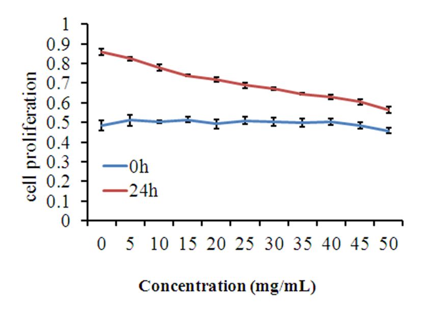
forms of drugs hardly persist in the lesion area for a long period of time, making it difficult to maintain the
effective drug concentration, which is the main cause of prolonged recurrence observed in STB.

In the medical field, nanotechnology has led to significant improvements in cancer therapy[17], diagnostic
imaging of diseases[18], tissue engineering[19], and most importantly drug and gene delivery
systems[20]. Although developing new TB molecules remains critical in curving the TB epidemic, altering
novel therapeutics in nanoparticle-based delivery systems represents an achievable, cost-effective, and
readily available option [21]. Hitherto, multiple nanodelivery systems for administering anti-TB products to
the lung have been widely assessed, and suggested as alternatives to conventional TB therapy.
Nanoparticles can selectively deliver into macrophages, which primarily host TB, greatly increasing the
therapeutic index by enabling high drug levels right where Mtb replicates while reducing systemic toxicity.
An additional advantage is that nanoparticles for TB drugs shield them from liver catabolism and renal
clearance; consequently, these products are safer and more effective in comparison with free drugs,
decreasing treatment time and drug resistance occurrence[2, 4, 22–24]. Therefore, the development of
new dosage forms of high-efficiency anti-TB drugs, improving their biodistribution in diseased vertebrae
and effectively killing Mtb in the target tissue, represents one of the most critical measures for applying
current anti-TB therapeutics in the treatment of STB.

In humans, transforming growth factor (TGF)-β1 plays an essential immunomodulatory role in TB [25].
TGF-β1 with excessively high activity is found in both lung lavages and macrophages from individuals
suffering from pulmonary TB[26, 27]. In addition, TGF-β1 potently deactivates macrophages, reducing
their effectiveness in containing Mtb [28]. Furthermore, TGF-β1 and other cytokines (e.g., TNF-α) may be
involved in tissue damage described in TB patients [29, 30]. Thus, silencing the TGF-β1 gene by the RNA
interference (RNAi) technology [31], reducing the secretion of the TGF-β1 protein in macrophages, and
combining first-line anti-TB drugs to facilitate Mtb clearance are tools that could increase the efficacy of
anti-TB drugs.

Here, an anti-TB nanodelivery system was engineered employing nanoliposomes as the carrier, for
biocompatibility and biodegradability, impressive drug loading rate, organ targeting potential and slow
release, elevated oral bioavailability, and prolonged half-life in circulation [32, 33]. Then, H, R, and Z were
selected as first-line oral drugs for the treatment of TB. The positively-charged nanoliposomes loaded
with HRZ (isoniazid/rifampicin/pyrazinamide) for the treatment of TB were successfully developed by
reverse-phase evaporation and further bound to the negatively-charged siTGF-β1 in order to reduce the TB
granuloma wrapped in Mtb and increase the efficacy of the drugs. Finally, the particle size, zeta potential,
particle shape, and encapsulation efficiency (EE) of nanoliposomes loaded with HRZ/siTGF-β1 were
characterized, evaluating their in vitro cytotoxicity as a potential alternative for the treatment of STB.

Materials And Methods
Preparation of HRZ-loaded nanoliposomes.
                                                   Page 3/23
2,3-dioleoyl-3-trimethylammonium-

Propane(DOTAP) and 1, 2-distearoyl-sn-glycero-3-hosphoethanolamine-n-[methoxy (polyethylene glycol)
2000] (DSPE-PEG 2000) were provided by Sigma-Aldrich (USA). Cholesterol, INH, RIF, and PZA (> 98%
purity) were manufactured by Tokyo Chemical Industry (Japan). RPMI 1640 medium, trypsin, and fetal
bovine serum (FBS) were provided by Hyclone (USA). Anti-TGF-1 (Cat No. ab 92486) was from Abcam
(UK). SYBR® Premix Ex Taq, PrimeScript™ RT reagent Kit with gDNA Eraser, and RNAiso Plus were
manufactured by TaKaRa Biotechnology (Japan). 3-(4,5-dimethylthiazol-2-yl)-3,5-diphenyltetrazolium
bromide salt (MTT) was provided by Biosharp (China). Annexin V-FITC/PI Apoptosis Detection kit and
propidium iodide (PI) staining solution were provided BD Biosciences (USA).

HRZ-loaded nanoliposomes were prepared by the reverse-phase evaporation method. Briefly, DOTAP (36
mg), DSPE-PEG2000 (50 mg), cholesterol (1 mg), INH (7.2 mg), RFP (10.9 mg), and PZA (1.8 mg) at the
molar ratio 20:10:1:21:5.3:5.9 were solubilized in chloroform/methanol (4:1, v/v). After solvent
evaporation (rotary evaporator, 37°C), further drying was performed under vacuum for 1 h. The resulting
inclusion complex was dissolved in 5 ml of deionized water, and a clear orange-red solution was obtained
post-filtration.

The resulting nanoliposome solution was transferred into a 10 kDa ultrafiltration tube, and subjected to
ultrafiltration at 5000 ×g for 10 min and repeated 5 times until a colorless filtrate was obtained. The upper
layer of the preserved orange-red liquid encompassed cationic liposomes containing the anti-TB drugs.
Then, 10% mannitol was added to the liquid and lyophilized to obtain 67 mg of an orange-red oily HRZ-
loaded nanoliposome product.

Conjugation of HRZ-loaded nanoliposomes with siTGF-β1.
The siRNA oligo nucleotides targeting TGF-β1 (siTGF-β1) were manufactured by Biomics Biotechnologies,
and their sequences were as follows: siTGF-β1: sense 5’-GGA GUC AGA UCC UCA GCA AGC-3’ and
antisense 5’-UUG CUG AGG AUC UGA CUC CUG-3’; non-coding control siRNA (siNC), sense 5’-GAA GGC
CCA TAG CCA GTG ACT-3’ and antisense 5’-AGU CAC UGG CUA UGG GCC UUC-3’. Cationic HRZ
nanoliposomes were mixed with siTGF-β1 in weight ratios of 2:1, 5:1, 10:1 and 20:1, respectively, and
further underwent incubation at ambient for 30 min. The binding efficiency of the HRZ nanoliposomes
with siTGF-β1 was determined by the gel retardation assay using 1.5% agarose gel (Ultrapure™ agarose,
Life Technologies).

Characterization of HRZ/siTGF-β1 nanoliposomes.
The size and zeta potential of HRZ/siTGF-β1 nanoliposomes were assessed by dynamic light scattering
(DLS) on a Malvern ZetasizerNano ZS (Malvern Instruments, UK) in triplicate at ambient, after dilution
with double-distilled water.

                                                  Page 4/23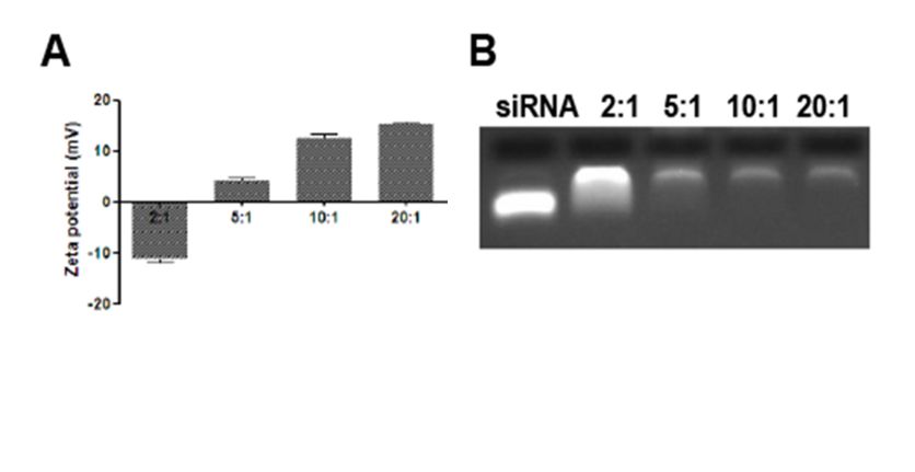
The surface morphology of HRZ/siTGF-β1 nanoliposomes was assessed by transmission electron
microscope (TEM) (TEM Jeol JEM-1400; JEOL, Japan). To prepare TEM samples, cationic
nanoliposomes and siRNA at a mass ratio of 5:1 were spread over a copper grid and air-dried for 30 min
before detection.

INH, RIF and PZA loading in HRZ/siTGF-β1 nanoliposomes was assessed as reported previously[34], with
slight modifications. Briefly, the mobile phase was formulated to an optimal concentration to detect the
EE of HRZ/siTGF-β1 nanoliposomes on a high-performance liquid chromatography (HPLC) system
(Agilent Technologies, USA). After the loading procedure, the suspensions were submitted to
centrifugation at 16873 g for 20 min (Centrifuge 5418; Eppendorf AG, Germany) and unencapsulated
drugs that remained in the supernatant were quantitated by UV detection at 334 nm[35].

Entrapment efficiency (%) was derived as [(weight of drug loaded initially − weight of unencapsulated
drug)/weight of drug loaded initially] × 100%

In-vitro cytotoxicity assays.
Human monocytes THP-1 cells provided by American Type Culture Collection (ATCC) underwent culture at
2×10⁵ cells/ml in RPMI 1640 medium containing 10% FBS, penicillin (100 U/ml) and streptomycin (100
g/ml). The media were replaced twice or thrice weekly, and the cells were sub-cultured until 80–90%
confluency. THP-1 cell differentiation into adherent macrophages was performed with 100 nM phorbol
12-myristate 13-acetate (PMA) for 48 h in RPMI 1640 containing 10% FBS[36]. Then, the PMA media were
removed, followed by three PBS rinses and incubation in fresh medium for 3 h.To assess the developed
nanoliposomes for cytotoxic effects on human macrophages, the 3-(4,5-dimethylthiazol-2-yl)-2,5-
diphenyltetrazolium bromide (MTT) assay was carried out as directed by the manufacturer. In brief, 5×10³
THP-1 cells were added into each well of a 96-well plate, and allowed to differentiate into macrophages
by PMA induction at 100 ng/ml for 48 h. Then, they were incubated with HRZ/siTGF-β1 nanoliposomes at
0, 5, 10, 15, 20, 25, 30, 35, 40, 45, and 50 mg/ml at 37°C in 5% CO2 for 24 h. Subsequently, the medium
was replaced by MTT containing culture medium. Incubation was carried out for an additional 4 h, and
the reaction was stopped with an equivalent volume of DMSO for formazan crystal solubilization. Optical
density was obtained at 570 nm. Cell viability was quantitated as described in a previous report,
determining the percentages of viable cells and inhibitory potency (IC50) values[37]. Triplicate assays
were carried out.

Assessment of cell cycle distribution and apoptosis.
Flow cytometry (FCM) was carried out to assess cell cycle distribution and apoptosis upon treatment with
nanoliposomes. PMA-induced macrophages were added at 5×10³ cells per well of a 96-well plate. Upon
overnight incubation, the siNC group was treated with HRZ/siNC nanoliposomes, while the siTGF-β1
groups were administered various amounts of HRZ/siTGF-β1 nanoliposomes (35 and 40 mg/ml); the
                                                 Page 5/23
HRZ group was treated with HRZ nanoliposomes. On the other hand, control cells were administered an
identical volume of cell culture medium. Upon treatment, the cells underwent trypsinization, centrifugation
(1000 rpm, 5 min), and staining with Annexin V-FITC/PI double-labeling kit (eBioscience, USA) before
analysis for cell apoptosis. Next, cell resuspension was performed in PBS with 40 µg/ml PI followed by a
30-min incubation at 37°C away from light for assessing cell cycle distribution. After filtering through 35
µm nylon meshes, FCM on FACSCalibur (BD Biosciences) was performed for analysis. Then, the rates of
apoptosis in various cell cycle phases were determined.
Gene knockdown efficiency of TGF-β1 siRNA.
THP1-derived macrophages were administered different nanoliposomes containing HRZ, HRZ/siNC, and
HRZ/siTGF-β1 (35 and 40 mg/ml) for 6 h. Total RNA from human macrophages was obtained using
TRIzol, and reverse transcription was performed with PrimeScript Reverse Transcriptase Kit (TaKaRa), as
directed by the manufacturer. RNA quality and amounts were assessed spectrophotometrically on a
NanoDrop 1000 (Thermo Fisher Scientific). Then, qRT-PCR was carried out on an ABI PRISM Real-Time
PCR system (Applied Biosystems) with the QuantiTect SYBR Green Master Mix kit (Qiagen). PCR was
performed at 95°C (10 min), followed by 40 cycles of 95°C (5 s) and 60°C (1 min), with the melting curve
obtained at 95°. Fluorescence was collected at 60°C every 0.3°C until 95°C. The primers employed were:
TGF-β1, Forward 5’-GTC CTG GTG GAA TGG GTT ATA C-3’ and reverse 5’-GTT GAG TGT TCT TTG GCT
TGA C-3’; GAPDH, Forward 5’‑GGT GTG AAC CAT GAG AAG TAT GA-3’ and reverse 5’-GAG TCC TTC CAC
GAT ACC AAA G-3’. The 2−ΔΔCq method[38] was employed for the analysis of triplicate assays,
normalizing the data to GAPDH expression.
TGF-β1 protein amounts were determined by Western blot assays. After the treatment of THP1-derived
macrophages with nanoliposomes containing HRZ, HRZ/siNC, and HRZ/siTGF-β1 (35 and 40 mg/ml),
respectively, total protein was obtained with Total Protein Extraction Kit (Bestbio, China) and quantitated
by the Bradford assay (Bio-Rad, USA) as described by the manufacturer. Equal amounts of total protein
were resolved by 10% SDS-PAGE. Rabbit polyclonal anti-TGF-β1 (Abcam) and anti-GAPDH (Wuhan Boster
Biological Technology, China) primary antibodies were reacted overnight at 4°C, followed by incubation
with secondary antibodies linked to horseradish peroxidase (HRP) (Wuhan Boster Biological Technology)
at ambient for 2 h. Immunoreactive bands were detected with an enhanced chemiluminescence system
(Sino-American Biotechnology, China), and quantitated with Image J version 1.441 (National Institutes of
Health, USA).
Data analysis. Data are mean ± standard deviation (SD). Descriptive statistics and one-way analysis of
variance (ANOVA) were performed for analysis. Independent sample Student’s t-test was carried out for
group pair compassions. P < 0.05 indicated statistical significance.
Results
Page 6/23Particle size.
The particle size was measured by DLS according to the principle that particles move in a random
fashion under Brownian motion. Particle size is highly important in determining the cell’s absorption rate.
Liposomes of about 200 nm in size could induce membrane fusion with target cells, delivering the
encapsulated products into cells with high efficiency[39]. Here, the particle sizes of HRZ/siTGF-β1
nanoliposomes were determined by DLS (Table 1, Fig. 1). The product composed of HRZ nanoliposomes
and siTGF-β1 with a weight ratio around 5:1 displayed an average diameter of 168.135 ± 0.5444 nm,
which was adequate for alveolar epithelium deposition and macrophage internalization[40]. We also
found that with increasing weight ratio, the particle size of HRZ/siTGF-β1 nanoliposomes decreased from
237.885 to 91.46 nm.
Table 1
Mean particle size (nm) of HRZ/siTGF-β1 nanoliposomes with different weight ratios
Particle size (nm) HRZ nanoliposomes:siTGF-β1
2:1 5:1 10:1 20:1
237.885 ± 7.1912 168.135 ± 0.5444 115.265 ± 6.0033 91.46 ± 1.2445
Zeta potential.
Zeta potential provides information regarding the electrostatic potential of the particle in solution [40].
The zeta potential of HRZ/siTGF-β1 nanoliposomes with different weight ratios was measured
immediately after preparation (Table 2, Fig. 2A). The HRZ nanoliposomes carried a positive charge, and
absorbed the negatively-charged oligonucleotides by easily mixing the siTGF-β1 to produce the final
HRZ/siTGF-β1 nanoliposomes. Zeta potential analysis revealed that the surface charge of HRZ
nanoliposomes was 28.13 ± 2.4 mV in aqueous solution. Upon conjugation of siTGF-β1 (weight ratio of
HRZ nanoliposomes/siTGF-β1 = 5:1), zeta potential was reduced to 4.03 ± 1.32 mV, thereby implying
successful conjugation of the components that consumed the surface amino groups. For evaluating the
siRNA loading capacity of nanoliposomes, the gel retardation assay was carried out (Fig. 2B).
HRZ/siTGF-β1 nanoliposomes were generated at various weight ratios of HRZ nanoliposomes to siTGF-
β1 between 0:1 and 20:1. Gel electrophoresis showed a small amount of bands at weight ratios > 5:1,
indicating that most of the siRNA was absorbed by HRZ nanoliposomes. These findings corroborated
surface charge data obtained by DLS. By adjusting HRZ nanoliposome-to-siTGF-β1, surface charges
varied between 15.33 ± 0.55 mV and + 28.13 ± 2.4 mV. In addition, a gradual increasing trend of zeta
potential was observed with increasing amounts of HRZ nanoliposomes, in agreement with findings
obtained by agarose gel electrophoresis.
Page 7/23Table 2
Zeta potential (mV) of HRZ/siTGF-β1 nanoliposomes with different weight ratios
Zeta potential HRZ TGF-β1 HRZ nanoliposomes:TGF-β1 siRNA
(mV) nanoliposomes siRNA
2:1 5:1 10:1 20:1
28.13 ± 2.4 -18 ± 1.77 -11.07 ± 4.03 ± 12.5 ± 15.33 ±
1.32 1.32 1.4 0.55
Cells internalize nanoparticles with positive charges faster than neutral or negatively charged
counterparts[41]. Moreover, excessive surface charge causes cell cytotoxicity[42]. This phenomenon can
be prevented by ensuring an efficient loading of siRNA while maintaining a slight positive charge on the
surface. Thus, a weight ratio of 5:1 for HRZ nanoliposomes to siTGF-β1 was applied in subsequent
assays.
Morphology.
The appearance and morphological properties of the particles were examined by imaging air-dried
HRZ/siTGF-β1 nanoliposomes under a transmission electron microscope (TEM) (Fig. 3; scale bar 0.5
µm). TEM images exhibited a spherical shape for the nanoliposomes with a homogenous surface
morphology (in the range of 200–300 nm), which was consistent with DLS data.
EE.
EE is a valuable index with respect to nanodrug delivery. Adequate drug amounts are required in a given
polymer for sustained release to the target site[43]. EE is calculated as the percentage amount of drug
that is entrapped in the form of nanoliposomes. INH, RIF, and PZA loading of HRZ nanoliposomes
showed EE values of 90%, 88%, and 37%, respectively (Figs. 4A and 4B).
In-vitro cytotoxicity.
In order to achieve macrophage targeting and effective concentrations of anti-TB products at the
infection site, interaction of HRZ/siTGF-β1 nanoliposomes with macrophages was established using a
human macrophage model. Appropriate amounts of nanoliposomes for subsequent research were
selected cytotoxicity data according to the MTT assay. This test was carried out to assess increasing
concentrations (from 0 to 50 mg/ml) of samples, comparatively to untreated nanoliposomes. As shown
in Fig. 5, the human macrophage cell line exhibited a gradual proliferation reduction with increasing levels
of HRZ/siTGF-β1 nanoliposomes compared with untreated cells, and the IC50 value for nanoliposomes in
macrophages was 37.47 mg/ml. Therefore, 35 and 40 mg/ml nanoliposomes were selected for
subsequent experiments. Since nanoliposome components are considered safe or lowly cytotoxic at the
levels assessed, the concentration-dependent cytotoxic effects of loaded HRZ/siTGF-β1 nanoliposomes
Page 8/23were likely due to significant and heterogeneous particle aggregation decreasing cellular activity and
promoting cell death [44]. This phenomenon could be ascribed to the toxicity imparted by the positive
surface charge of nanoliposomes[45].
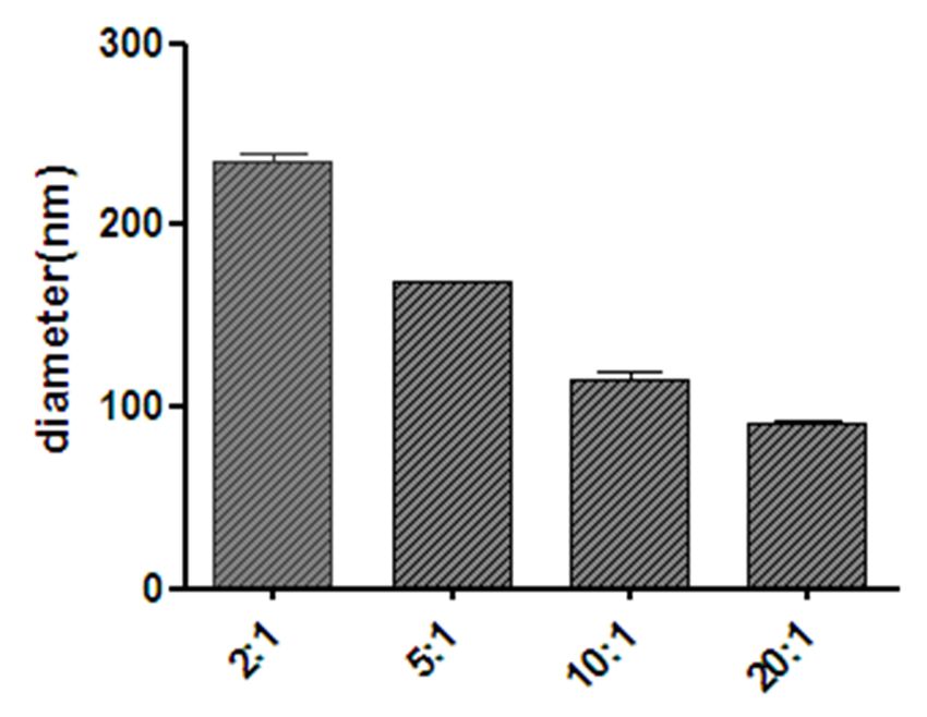
Effects of nanoliposomes on cell cycle distribution and
apoptosis.
Macrophages were exposed to different groups of nanoliposomes containing HRZ, HRZ/siNC, and
HRZ/siTGF-β1 (35 and 40 mg/ml) for 24 h. The percentages of cells in G1, S, and G2 are shown in Table
3; live, apoptotic (early and late) and necrotic cells were also quantitated (Table 4). The results exhibited
that the percentage of G2 cells significantly increased from 21.26–38.54% after HRZ/siTGF-β1 (40
mg/ml) treatment compared to 17.83% and 17.90% for cells treated with HRZ and HRZ/siNC
nanoliposomes, respectively (Table 3, Fig. 6A), suggesting that HRZ/siTGF-β1 nanoliposomes (40 mg/ml)
induced cell accumulation in the G2 phase of the cell cycle in human macrophages. However, no
significant differences were detected upon treatment with HRZ/siTGF-β1 nanoliposomes (35 mg/ml) in
other cell cycle phases.
Table 3
Percentage of cell populations in different stages of the cell cycle following exposure to different groups
of nanoliposomes for 24 h
Treatment group G1 phase S phase G2 phase
Untreated cells 51.73 ± 1.17 27.00 ± 3.44 21.26 ± 3.41
HRZ 55.13 ± 1.59 27.10 ± 5.93 17.83 ± 3.01
HRZ/siNC 54.06 ± 2.93 26.90 ± 4.38 17.90 ± 2.43
HRZ/siTGF-β1 46.95 ± 1.15 25.20 ± 3.22 26.84 ± 1.58
(35 mg/ml)
HRZ/siTGF-β1 41.07 ± 1.22 20.37 ± 2.34 38.54 ± 2.02**
(40 mg/ml)
The results are presented as the average percentage of the cell population (%) ± standard deviation; P
< 0.05*, *P < 0.01 vs. untreated cells.
Page 9/23Table 4
Percentage of populations of viable and non‑viable cells exhibiting structural properties of different cell
death types following exposure to different groups of nanoliposomes for 24 h
Treatment group Viable cells Non-viable cells
Necrosis Late apoptosis Early apoptosis
Untreated cells 91.56 ± 0.87 2.65 ± 0.16 4.52 ± 0.72 1.26 ± 0.12
HRZ 90.83 ± 1.65 1.04 ± 0.20 4.48 ± 1.31 3.64 ± 0.55
HRZ/siNC 91.36 ± 1.27 1.45 ± 0.84 3.83 ± 0.60 3.36 ± 0.32
HRZ/siTGF-β1 84.16 ± 1.53** 1.77 ± 0.29 9.56 ± 0.51 4.48 ± 1.28
(35 mg/ml)
HRZ/siTGF-β1 78.53 ± 1.13** 2.74 ± 0.39 12.50 ± 0.75 6.20 ± 1.16
(40 mg/ml)
The results are presented as the average percentage of the cell population (%) ± standard deviation;
*P < 0.05 *, *P < 0.01 vs. untreated cells.
Moreover, cells treated with HRZ/siTGF-β1 had a slightly higher apoptotic rate in comparison with
untreated cells (Table 4, Fig. 6B), while no obvious cytotoxicity was observed for HRZ and HRZ/siNC
nanoliposomes, which might be due to the high uptake of siTGF-β1. Taken together, these findings
suggested that HRZ/siTGF-β1 nanoliposomes possessed adequate biocompatibility, although TGF-β1
siRNA conjugation slightly increased cytotoxicity.
TGF-β1 silencing analysis.
To confirm the knockdown efficiency of siTGF-β1, total mRNA and protein were obtained 24 h after
transfection from human macrophages treated with or without HRZ/siTGF-β1 nanoliposomes. Negative
and blank control cells were treated with non-coding siRNA (siNC) and PBS, respectively. As shown in Fig.
7, TGF-β1 mRNA and protein amounts were markedly reduced in the HRZ/siTGF-β1 group compared with
the negative and blank control groups. These data indicated that siTGF-β1 successfully repressed TGF-β1
expression at the gene and protein levels.
Discussion
STB, also termed Pott’s disease, encompasses 50% of all musculoskeletal TB cases[46]. Left untreated, it
causes paraspinal abscesses, spinal cord compression, spine deformities, and neurological defects[46,
47]. Severe bone TB can be effectively treated by combining surgery with anti-TB drugs administered for
an optimal duration[48–50]. According to WHO guidelines, long-term administration of multiple anti-TB
drugs is essential for treating bone TB [51]. However, high dosages of antituberculous products are
necessary to achieve effective concentrations at target sites due to limited permeability and
Page 10/23metabolism[52, 53]. Innovative anti-TB drug delivery biomaterials have tremendous potential for the
treatment of STB, and could achieve high drug concentrations at the target site with reduced drug
amounts throughout the body, markedly reducing toxicity[54, 55]. Therefore, we developed
nanoliposomes that encapsulated first-line anti-TB medicines, i.e. INH, RIF, and PZA, and conjugated them
to TGF-β1 siRNA. Successful production of nanoliposomes was verified by multiple properties, and the
end-products were evaluated for drug encapsulation efficiency, cytotoxicity, and TGF-β1 siRNA silencing
effects in THP-1-derived human macrophages.
Particle shape exerts great effects on cellular uptake, distribution within the cell, and cytotoxicity.
Nanoparticles are taken up according to the following order based on shape: sphere > cube > rod > disk;
this is likely because the cell membrane is flexible around low-aspect ratio particles[40, 56]. Microscopy
revealed a spherical shape of the engineered nanoparticles, with the particle size ranging from 100 to 200
nm, which allows a wide distribution in most of organs[57]. Another important parameter is zeta potential,
which depicts the charge and stability of the prepared nanoparticles[58]. Reportedly, high surface charge
reduces the aggregation of particles[59]. The degree and rate of macrophage uptake show direct
associations with particles’ net charge; the physiological compatibility of a negatively charged surface is
greater than that of the positively charged counterpart[40]. Moreover, localization in lysosomes, in which
Mtb does survive, is more pronounced in negatively charged particles than positively charged ones[60]. In
the present study, the zeta-potential values of HRZ nanoliposomes and TGF-β1 siRNA were 28.13 and −
18 mV, respectively, whereas those of HRZ/siTGF-β1 nanoliposomes with different weight ratios ranged
from − 11.07 to 15.33 mV.
EE in liposomes is impacted by various parameters, including the preparative method as well as the
features of liposomes and loaded molecules[61]. Hydrophobic and hydrophilic substances have high
(reaching 100%) and low EE values, respectively[62]. Substances with intermediate hydrophilicity and
lipophilicity generally distribute between the water and lipid phases, and any solubility alteration affects
their partitioning, thereby modifying the EE[61]. In this study, the 100–200 nm HRZ/siTGF-β1
nanoliposomes showed high drug encapsulation efficiencies of 90%, 88%, and 37% for INH, RIF, and PZA,
respectively.
Nanoparticles have potential toxic to the liver, kidney, neurons, and cardiovascular system, which would
limit their application in clinic. Therefore, using reduced nanoparticle quantities is preferable, and low-
toxicity or concentration particles should be utilized[63]. Herein, the MTT assay and FCM indicated that
HRZ/siTGF-β1 nanoliposomes had low cytotoxicity and acted in a concentration-dependent manner in
human macrophages. Interestingly, cell cycle distribution in THP-1-derived macrophages was unaltered
upon drug administration at 35 mg/ml, while the cells treated with HRZ/siTGF-β1 at a concentration of 40
mg/ml showed a higher apoptotic rate than untreated cells.
Macrophages are key cells in the immune response against mycobacteria, and also provide a niche for
Mtb replication[64, 65]. Coordinated events among immune factors, especially macrophages and T cells,
play essential roles in inhibiting TB infection[26]. In addition, macrophage and T cell functions are mostly
Page 11/23regulated by local cytokines, which are essential for developing immune reactions against Mtb. Several
reports revealed that elevated TGF-β1 amounts suppress immune responses targeting Mtb by modulating
proliferation, differentiation, and functions in particular immune cells [25]. In addition, TGF-β1 is
expressed in non-necrotizing granulomas of sarcoidosis as well as TB granulomas[27, 66, 67]. Thanks to
TGF-β1’s essential function in TB pathogenesis, this infection could be controlled by TGF-β1 suppression
while administering anti-TB drugs. Currently, siRNA-mediated gene knockdown is considered a robust
approach for reducing aberrantly elevated amounts of target genes, rendering it putative for clinical
therapy[68]. The wide application of siRNAs for treatment is based on well-designed systems delivering
siRNAs into target cells with high efficiency[69]. Nanoliposomes efficiently carry and deliver siRNAs in
vivo [70]. In this study, TGF-β1 siRNA-mediated gene knockdown downregulated TGF-β1, decreasing the
formation of tuberculous granulomas. The above data also showed that the developed HRZ/siTGF-β1
nanoliposomes significantly reduced TGF-β1 mRNA and protein expression levels in THP-1-derived
macrophages.
Collectively, HRZ (anti-TB drugs INH, RIF, and PZA)-loaded nanoliposomes with siTGF-β1 were
successfully developed with high encapsulation efficacy, and were characterized by a spherical shape
within nanometer size. These formulations had low cytotoxicity and potent TGF-β1 gene silencing,
thereby laying the foundation for in vivo studies.
In conclusion, the urgency of effective treatment of TB, which is among the nine leading causes of death
around the world, was tackled by developing nanoliposomes for the delivery of anti-TB drugs directly to
the infection site. In the present work, we successfully developed nanoliposomes loaded with HRZ,
followed by TGF-β1 siRNA encapsulation. These nanoliposomes were in the nanometer range, with a
diameter averaging 168 nm as determined by DLS. Additionally, they had elevated zeta potential,
suggesting high physical stability. Morphologically, they were spherical and uniform, with a smooth
surface. INH and RIF had elevated encapsulation percentages, with > 80% drug encapsulation efficiencies.
Finally, the developed nanoliposomes had low cytotoxicity and affected the viability of THP-1-derived
human macrophages in a concentration-dependent manner. Overall, the novel nanoliposomes exhibited a
potential as great vehicles for delivering drugs, e.g., antituberculous medicines. This system would
significantly impact the design of therapeutic regimens, improving patient compliance. Nevertheless,
these nanoliposomes should be further investigated in animal models to obtain supportive in vivo data
for potential clinical applications in the future.
Declarations
All procedures were performed in accordance with relevant guidelines’ in the manuscript.
Ethics approval and consent to participate
Not applicable.
Page 12/23Consent for publication
Not applicable.
Availability of data and materials
All data generated or analysed during this study are included in this published article [and its
supplementary information files].
Competing interest
The authors declare that they have no conflict of interest.
Funding
The present study was funded by the National Natural Science Foundation of China (grant no. 81501903
and 81860395), Ningxia Natural Science Foundation (grant no. 2020AAC03391), and the Autonomous
Region health system research project (grant no. 2019-NW-011).
Authors’ contributions
Ningkui Niu and Zongqiang Yang designed the study. Caili Lou and Chaoran Wang were involved in the
manuscript writing. Xuewei Wang collected the data. Zongqiang Yang and Zhiyun Shi analyzed the data.
Ningkui Niu and Zongqiang Yang revised the draft. All authors read and approved the final manuscript.
Acknowledgements
Not applicable.
Author details
a
Department of Spinal Surgery, General Hospital of Ningxia Medical University, Yinchuan, Ningxia Hui
Autonomous Region 750004, China;
b
Department of Medical Experimental Center, General Hospital of Ningxia Medical University, Yinchuan,
Ningxia Hui Autonomous Region 750004, China;
c Ningxia Medical University, Yinchuan, Ningxia Hui Autonomous Region 750004, China
Page 13/23References
1. Zumla A, George A, Sharma V, Herbert RH, Oxley A, Oliver M. The WHO 2014 global tuberculosis
report--further to go. Lancet Glob Health. 2015;3(1):e10-12.
2. Costa A, Pinheiro M, Magalhaes J, Ribeiro R, Seabra V, Reis S, et al. The formulation of
nanomedicines for treating tuberculosis. Adv Drug Deliv Rev. 2016;102:102-115.
3. Saifullah B, El Zowalaty ME, Arulselvan P, Fakurazi S, Webster TJ, Geilich BM, et al. Synthesis,
characterization, and efficacy of antituberculosis isoniazid zinc aluminum-layered double hydroxide
based nanocomposites. Int J Nanomedicine. 2016;11:3225-3237.
4. Ivancic A, Macaev F, Aksakal F, Boldescu V, Pogrebnoi S, Duca G. Preparation of alginate-chitosan-
cyclodextrin micro- and nanoparticles loaded with anti-tuberculosis compounds. Beilstein J
Nanotechnol. 2016;7:1208-1218.
5. Broderick C, Hopkins S, Mack DJF, Aston W, Pollock R, Skinner JA, et al. Delays in the diagnosis and
treatment of bone and joint tuberculosis in the United Kingdom. Bone Joint J. 2018;100-b(1):119-
124.
6. Niu N, Wang Q, Shi J, Zhang X, Geng G, Zhou S, et al. Clinical and genomic responses to ultra-short
course chemotherapy in spinal tuberculosis. Exp Ther Med. 2017;13(5):1681-1688.
7. Jin W, Wang Q, Wang Z, Geng G. Complete debridement for treatment of thoracolumbar spinal
tuberculosis: a clinical curative effect observation. Spine J. 2014;14(6):964-970.
8. Si J, Geng G, Wang Z. Detection of Mycobacterium tuberculosis DNA in the sclerotic spinal wall.
Orthopedics. 2012;35(3):e409-413.
9. Shi J, Wang Z, Li H, Yuan H. Diagnostic performance of the urinary deoxypyridinoline in spinal
tuberculosis. Orthopedics. 2012;35(6):e922-926.
10. Geng G, Wang Q, Shi J, Yan J, Niu N, Wang Z. Establishment of a New Zealand rabbit model of spinal
tuberculosis. J Spinal Disord Tech. 2015;28(3):E140-145.
11. Shi JD, Wang ZL, Geng GQ, Niu NK. Intervertebral focal surgery for the treatment of non-contiguous
multifocal spinal tuberculosis. Int Orthop. 2012;36(7):1423-1427.
12. Ge Z, Wang Z, Wei M. Measurement of the concentration of three antituberculosis drugs in the focus
of spinal tuberculosis. Eur Spine J. 2008;17(11):1482-1487.
13. Shi JD, Wang Q, Wang ZL. Primary issues in the selection of surgical procedures for thoracic and
lumbar spinal tuberculosis. Orthop Surg. 2014;6(4):259-268.
14. Si J, Wang Z, Wang Z, Li H. Sequencing-based detection of drug-resistant Mycobacterium
tuberculosis in patients with spinal tuberculosis. Arch Orthop Trauma Surg. 2012;132(7):941-945.
15. Wang Z, Ge Z, Jin W, Qiao Y, Ding H, Zhao H, et al. Treatment of spinal tuberculosis with ultrashort-
course chemotherapy in conjunction with partial excision of pathologic vertebrae. Spine J.
2007;7(6):671-681.
16. Wang Z, Shi J, Geng G, Qiu H. Ultra-short-course chemotherapy for spinal tuberculosis: five years of
observation. Eur Spine J. 2013;22(2):274-281.
Page 14/2317. Farooq MA, Aquib M, Farooq A, Haleem Khan D, Joelle Maviah MB, Sied Filli M, et al. Recent progress
in nanotechnology-based novel drug delivery systems in designing of cisplatin for cancer therapy: an
overview. Artif Cells Nanomed Biotechnol. 2019;47(1):1674-1692.
18. Karimi M, Zare H, Bakhshian Nik A, Yazdani N, Hamrang M, Mohamed E, et al. Nanotechnology in
diagnosis and treatment of coronary artery disease. Nanomedicine (Lond). 2016;11(5):513-530.
19. Parpura V. Tissue engineering: Nanoelectronics for the heart. Nat Nanotechnol. 2016;11(9):738-739.
20. Karimi M, Ghasemi A, Sahandi Zangabad P, Rahighi R, Moosavi Basri SM, Mirshekari H, et al. Smart
micro/nanoparticles in stimulus-responsive drug/gene delivery systems. Chem Soc Rev.
2016;45(5):1457-1501.
21. Saifullah B, Chrzastek A, Maitra A, Naeemullah B, Fakurazi S, Bhakta S, et al. Novel Anti-Tuberculosis
Nanodelivery Formulation of Ethambutol with Graphene Oxide. Molecules. 2017;22(10).
22. Gao Y, Sarfraz MK, Clas SD, Roa W, Lobenberg R. Hyaluronic Acid-Tocopherol Succinate-Based Self-
Assembling Micelles for Targeted Delivery of Rifampicin to Alveolar Macrophages. J Biomed
Nanotechnol. 2015;11(8):1312-1329.
23. Hwang AA, Lee BY, Clemens DL, Dillon BJ, Zink JI, Horwitz MA. pH-Responsive Isoniazid-Loaded
Nanoparticles Markedly Improve Tuberculosis Treatment in Mice. Small. 2015;11(38):5066-5078.
24. Mohseni M, Gilani K, Mortazavi SA. Preparation and characterization of rifampin loaded mesoporous
silica nanoparticles as a potential system for pulmonary drug delivery. Iran J Pharm Res.
2015;14(1):27-34.
25. Toossi Z, Ellner JJ. The role of TGF beta in the pathogenesis of human tuberculosis. Clin Immunol
Immunopathol. 1998;87(2):107-114.
26. Bonecini-Almeida MG, Ho JL, Boechat N, Huard RC, Chitale S, Doo H, et al. Down-modulation of lung
immune responses by interleukin-10 and transforming growth factor beta (TGF-beta) and analysis of
TGF-beta receptors I and II in active tuberculosis. Infect Immun. 2004;72(5):2628-2634.
27. Aung H, Toossi Z, McKenna SM, Gogate P, Sierra J, Sada E, et al. Expression of transforming growth
factor-beta but not tumor necrosis factor-alpha, interferon-gamma, and interleukin-4 in
granulomatous lung lesions in tuberculosis. Tuber Lung Dis. 2000;80(2):61-67.
28. Hirsch CS, Yoneda T, Averill L, Ellner JJ, Toossi Z. Enhancement of intracellular growth of
Mycobacterium tuberculosis in human monocytes by transforming growth factor-beta 1. J Infect Dis.
1994;170(5):1229-1237.
29. Kumar NP, Moideen K, George PJ, Dolla C, Kumaran P, Babu S. Coincident diabetes mellitus
modulates Th1-, Th2-, and Th17-cell responses in latent tuberculosis in an IL-10- and TGF-beta-
dependent manner. Eur J Immunol. 2016;46(2):390-399.
30. Kalita J, Misra UK, Bhoi SK, Chauhan PS, Sagar B. Possible role of transforming growth factor beta in
tuberculous meningitis. Cytokine. 2017;90:124-129.
31. Fire A, Xu S, Montgomery MK, Kostas SA, Driver SE, Mello CC. Potent and specific genetic interference
by double-stranded RNA in Caenorhabditis elegans. Nature. 1998;391(6669):806-811.
Page 15/2332. Mirahadi M, Ghanbarzadeh S, Ghorbani M, Gholizadeh A, Hamishehkar H. A review on the role of
lipid-based nanoparticles in medical diagnosis and imaging. Ther Deliv. 2018;9(8):557-569.
33. Kong X, Liu Y, Huang X, Huang S, Gao F, Rong P, et al. Cancer Therapy Based on Smart Drug Delivery
with Advanced Nanoparticles. Anticancer Agents Med Chem. 2019;19(6):720-730.
34. Ivashchenko O, Coy E, Peplinska B, Jarek M, Lewandowski M, Zaleski K, et al. Influence of silver
content on rifampicin adsorptivity for magnetite/Ag/rifampicin nanoparticles. Nanotechnology.
2017;28(5):055603.
35. Kumar A, Sharma R, Nair A, Saini G. Development and validation of RP-HPLC method for
simultaneous estimation of nimesulide, phenylephrine hydrochloride, chlorpheniramine maleate and
caffeine anhydrous in pharmaceutical dosage form. Acta Pol Pharm. 2012;69(6):1017-1022.
36. Clemens DL, Lee BY, Xue M, Thomas CR, Meng H, Ferris D, et al. Targeted intracellular delivery of
antituberculosis drugs to Mycobacterium tuberculosis-infected macrophages via functionalized
mesoporous silica nanoparticles. Antimicrob Agents Chemother. 2012;56(5):2535-2545.
37. Shaikh MV, Kala M, Nivsarkar M. Formulation and optimization of doxorubicin loaded polymeric
nanoparticles using Box-Behnken design: ex-vivo stability and in-vitro activity. Eur J Pharm Sci.
2017;100:262-272.
38. Livak KJ, Schmittgen TD. Analysis of relative gene expression data using real-time quantitative PCR
and the 2(-Delta Delta C(T)) Method. Methods. 2001;25(4):402-408.
39. Chay SY, Tan WK, Saari N. Preparation and characterisation of nanoliposomes containing winged
bean seeds bioactive peptides. J Microencapsul. 2015;32(5):488-495.
40. Patel B, Gupta N, Ahsan F. Particle engineering to enhance or lessen particle uptake by alveolar
macrophages and to influence the therapeutic outcome. Eur J Pharm Biopharm. 2015;89:163-174.
41. Hirsch V, Kinnear C, Moniatte M, Rothen-Rutishauser B, Clift MJ, Fink A. Surface charge of polymer
coated SPIONs influences the serum protein adsorption, colloidal stability and subsequent cell
interaction in vitro. Nanoscale. 2013;5(9):3723-3732.
42. Schaeublin NM, Braydich-Stolle LK, Schrand AM, Miller JM, Hutchison J, Schlager JJ, et al. Surface
charge of gold nanoparticles mediates mechanism of toxicity. Nanoscale. 2011;3(2):410-420.
43. Kurmi BD, Kayat J, Gajbhiye V, Tekade RK, Jain NK. Micro- and nanocarrier-mediated lung targeting.
Expert Opin Drug Deliv. 2010;7(7):781-794.
44. Chen R, Han Z, Huang Z, Karki J, Wang C, Zhu B, et al. Antibacterial activity, cytotoxicity and
mechanical behavior of nano-enhanced denture base resin with different kinds of inorganic
antibacterial agents. Dent Mater J. 2017;36(6):693-699.
45. Kedmi R, Ben-Arie N, Peer D. The systemic toxicity of positively charged lipid nanoparticles and the
role of Toll-like receptor 4 in immune activation. Biomaterials. 2010;31(26):6867-6875.
46. Jain AK. Tuberculosis of the spine: a fresh look at an old disease. J Bone Joint Surg Br.
2010;92(7):905-913.
Page 16/2347. Gehlot PS, Chaturvedi S, Kashyap R, Singh V. Pott's Spine: Retrospective Analysis of MRI Scans of 70
Cases. J Clin Diagn Res. 2012;6(9):1534-1538.
48. Li L, Xu J, Ma Y, Tang D, Chen Y, Luo F, et al. Surgical strategy and management outcomes for
adjacent multisegmental spinal tuberculosis: a retrospective study of forty-eight patients. Spine
(Phila Pa 1976). 2014;39(1):E40-48.
49. Sequeira W, Co H, Block JA. Osteoarticular tuberculosis: current diagnosis and treatment. Am J Ther.
2000;7(6):393-398.
50. Dunn RN, Ben Husien M. Spinal tuberculosis: review of current management. Bone Joint J. 2018;100-
b(4):425-431.
51. Falzon D, Jaramillo E, Schunemann HJ, Arentz M, Bauer M, Bayona J, et al. WHO guidelines for the
programmatic management of drug-resistant tuberculosis: 2011 update. Eur Respir J.
2011;38(3):516-528.
52. Yew WW, Leung CC. Antituberculosis drugs and hepatotoxicity. Respirology. 2006;11(6):699-707.
53. Schaberg T. The dark side of antituberculosis therapy: adverse events involving liver function. Eur
Respir J. 1995;8(8):1247-1249.
54. Huang D, Li D, Wang T, Shen H, Zhao P, Liu B, et al. Isoniazid conjugated poly(lactide-co-glycolide):
long-term controlled drug release and tissue regeneration for bone tuberculosis therapy. Biomaterials.
2015;52:417-425.
55. Li D, Li L, Ma Y, Zhuang Y, Li D, Shen H, et al. Dopamine-assisted fixation of drug-loaded polymeric
multilayers to osteoarticular implants for tuberculosis therapy. Biomater Sci. 2017;5(4):730-740.
56. Xie X, Liao J, Shao X, Li Q, Lin Y. The Effect of shape on Cellular Uptake of Gold Nanoparticles in the
forms of Stars, Rods, and Triangles. Sci Rep. 2017;7(1):3827.
57. Hiroi K, Kura H, Ogawa T, Takahashi M, Sato T. Magnetic ordered states induced by interparticle
magnetostatic interaction in alpha-Fe/Au mixed nanoparticle assembly. J Phys Condens Matter.
2014;26(17):176001.
58. Nigam K, Kaur A, Tyagi A, Manda K, Gabrani R, Dang S. Baclofen-Loaded Poly (D,L-Lactide-Co-
Glycolic Acid) Nanoparticles for Neuropathic Pain Management: In Vitro and In Vivo Evaluation.
Rejuvenation Res. 2019;22(3):235-245.
59. Kumar S, Ali J, Baboota S. Design Expert((R)) supported optimization and predictive analysis of
selegiline nanoemulsion via the olfactory region with enhanced behavioural performance in
Parkinson's disease. Nanotechnology. 2016;27(43):435101.
60. Bhattacharjee S, de Haan LH, Evers NM, Jiang X, Marcelis AT, Zuilhof H, et al. Role of surface charge
and oxidative stress in cytotoxicity of organic monolayer-coated silicon nanoparticles towards
macrophage NR8383 cells. Part Fibre Toxicol. 2010;7:25.
61. Kulkarni SB, Betageri GV, Singh M. Factors affecting microencapsulation of drugs in liposomes. J
Microencapsul. 1995;12(3):229-246.
Page 17/2362. Barenholz Y. Relevancy of drug loading to liposomal formulation therapeutic efficacy. J Liposome
Res. 2003;13(1):1-8.
63. Gorgizadeh M, Azarpira N, Dehdari Veis R, Sattarahmady N. Repression of melanoma tumor in vitro
and in vivo by photothermal effect of carbon xerogel nanoparticles. Colloids Surf B Biointerfaces.
2019;176:449-455.
64. Amaral EP, Lasunskaia EB, D'Imperio-Lima MR. Innate immunity in tuberculosis: how the sensing of
mycobacteria and tissue damage modulates macrophage death. Microbes Infect. 2016;18(1):11-20.
65. Sia JK, Georgieva M, Rengarajan J. Innate Immune Defenses in Human Tuberculosis: An Overview of
the Interactions between Mycobacterium tuberculosis and Innate Immune Cells. J Immunol Res.
2015;2015:747543.
66. Niimi T, Sato S, Sugiura Y, Yoshinouchi T, Akita K, Maeda H, et al. Transforming growth factor-beta
gene polymorphism in sarcoidosis and tuberculosis patients. Int J Tuberc Lung Dis. 2002;6(6):510-
515.
67. Limper AH, Colby TV, Sanders MS, Asakura S, Roche PC, DeRemee RA. Immunohistochemical
localization of transforming growth factor-beta 1 in the nonnecrotizing granulomas of pulmonary
sarcoidosis. Am J Respir Crit Care Med. 1994;149(1):197-204.
68. Burnett JC, Rossi JJ. RNA-based therapeutics: current progress and future prospects. Chem Biol.
2012;19(1):60-71.
69. Ozpolat B, Sood AK, Lopez-Berestein G. Liposomal siRNA nanocarriers for cancer therapy. Adv Drug
Deliv Rev. 2014;66:110-116.
70. Zhou J, Shum KT, Burnett JC, Rossi JJ. Nanoparticle-Based Delivery of RNAi Therapeutics: Progress
and Challenges. Pharmaceuticals (Basel). 2013;6(1):85-107.
Figures
Page 18/23Figure 1
Particle size measured by DLS.
Page 19/23Figure 2
(A) Zeta potential values for different weight ratios of HRZ nanoliposomes to loaded siTGF-β1. (B) Gel
retardation data for different weight ratios of HRZ nanoliposomes to loaded siTGF-β1.
Figure 3
TEM images of HRZ/siTGF-β1 nanoliposomes.
Page 20/23Figure 4
HPLC data for (A) pre-preparation mixture of liposomes; (B) filtrate after liposome preparation.
Page 21/23Figure 5
Cell viability of THP-1-derived macrophages after treatment with HRZ/siTGF-β1 nanoliposomes as
assessed by the MTT assay.
Notes: Survival of THP-1 derived macrophages treated with 0–50 mg/ml HRZ/siTGF-β1 nanoliposomes
for 24 h. Data are mean±SD of three independent experiments.
Page 22/23Figure 6 Mechanisms of HRZ/siTGF-β1 nanoliposome-mediated inhibition of macrophage growth. (A) Cell cycle distribution and (B) apoptosis in macrophages after treatment with different nanoliposomes. *P
You can also read