Autophagic response in the Rabbit Hemorrhagic Disease, an animal model of virally-induced fulminant hepatic failure
←
→
Page content transcription
If your browser does not render page correctly, please read the page content below
Vallejo et al. Veterinary Research 2014, 45:15
http://www.veterinaryresearch.org/content/45/1/15
                                                                                                                          VETERINARY RESEARCH

 RESEARCH                                                                                                                                         Open Access

Autophagic response in the Rabbit Hemorrhagic
Disease, an animal model of virally-induced
fulminant hepatic failure
Daniela Vallejo1, Irene Crespo1,2, Beatriz San-Miguel1, Marcelino Álvarez3, Jesús Prieto2,4, María Jesús Tuñón1,2*
and Javier González-Gallego1,2

  Abstract
  The Rabbit Hemorrhagic Disease Virus (RHDV) induces a severe disease that fulfils many requirements of an animal
  model of fulminant hepatic failure. However, a better knowledge of molecular mechanisms contributing to liver
  damage is required, and it is unknown whether the RHDV induces liver autophagy and how it relates to apoptosis.
  In this study, we attempted to explore which signalling pathways were involved in the autophagic response
  induced by the RHDV and to characterize their role in the context of RHDV pathogenesis. Rabbits were infected
  with 2 × 10⁴ hemmaglutination units of a RHDV isolate. The autophagic response was measured as presence of
  autophagic vesicles, LC3 staining, conversion of LC3-I to autophagosome-associated LC3-II and changes in expression
  of beclin-1, UVRAG, Atg5, Atg12, Atg16L1 and p62/SQSTM1. RHDV-triggered autophagy reached a maximum at 24
  hours post-infection (hpi) and declined at 30 and 36 hpi. Phosphorylation of mTOR also augmented in early periods of
  infection and there was an increase in the expression of the endoplasmic reticulum chaperones BiP/GRP78, CHOP and
  GRP94. Apoptosis, measured as caspase-3 activity and expression of PARP-1, increased significantly at 30 and 36 hpi in
  parallel to the maximal expression of the RHDV capsid protein VP60. These data indicate that RHDV infection initiates a
  rapid autophagic response, perhaps in an attempt to protect liver, which associates to ER stress development and is
  independent from downregulation of the major autophagy suppressor mTOR. As the infection continues and the
  autophagic response declines, cells begin to exhibit apoptosis.

Introduction                                                                           regeneration [8,9] are constant features in rabbits expe-
The Rabbit Hemorrhagic Disease Virus (RHDV) is a                                       rimentally infected with the RHDV. This model could
positive-strand RNA virus, member of the Caliciviridae                                 therefore be useful to improve our insight into the patho-
family, that causes in wild and domestic rabbits an acute                              physiology of viral FHF and to facilitate the development
highly fatal disease first reported three decades ago [1].                             and evaluation of new therapeutic modalities.
Hepatic damage plays a central pathogenic role and is his-                               Macroautophagy (thereafter referred to as autophagy)
tologically similar to fatal viral hepatitis causing fulminant                         pathway is a bulk degradation system which controls the
hepatic failure (FHF) in humans [2]. We have shown by                                  clearance and recycling of intracellular constituents for
data on animal survival, clinical features, histopathological                          the maintenance of cellular survival [10] and can partici-
findings, changes in blood chemistry and intracranial pres-                            pate in the host response to infection [11]. Autophagy
sure monitoring that the RHD fulfils many of the require-                              starts with the formation of a doubled-membrane-bound
ments of an animal model of FHF [3]. Moreover, loss of                                 vacuole, known as the autophagosome, that engulfs frac-
the oxidant/antioxidant balance [4], presence of apoptosis                             tions of the cytoplasm in an either unselective or select-
and endoplasmic reticulum (ER) stress [5-7], and lack of                               ive manner via the activity of the autophagy adaptors,
                                                                                       such as sequestrosome 1 (p62/SQSTM1), that forms a
* Correspondence: mjtung@unileon.es                                                    bridge between the target and the growing autophago-
1
 Institute of Biomedicine (IBIOMED), University of León, 24071 León, Spain
2
 Centro de Investigación Biomédica en Red de Enfermedades Hepáticas y                  some membrane [12]. After being formed, most autop-
Digestivas (CIBERehd), Spain                                                           hagosomes receive input from the endocytic vesicles to
Full list of author information is available at the end of the article

                                         © 2014 Vallejo et al.; licensee BioMed Central Ltd. This is an Open Access article distributed under the terms of the Creative
                                         Commons Attribution License (http://creativecommons.org/licenses/by/2.0), which permits unrestricted use, distribution, and
                                         reproduction in any medium, provided the original work is properly cited. The Creative Commons Public Domain Dedication
                                         waiver (http://creativecommons.org/publicdomain/zero/1.0/) applies to the data made available in this article, unless otherwise
                                         stated.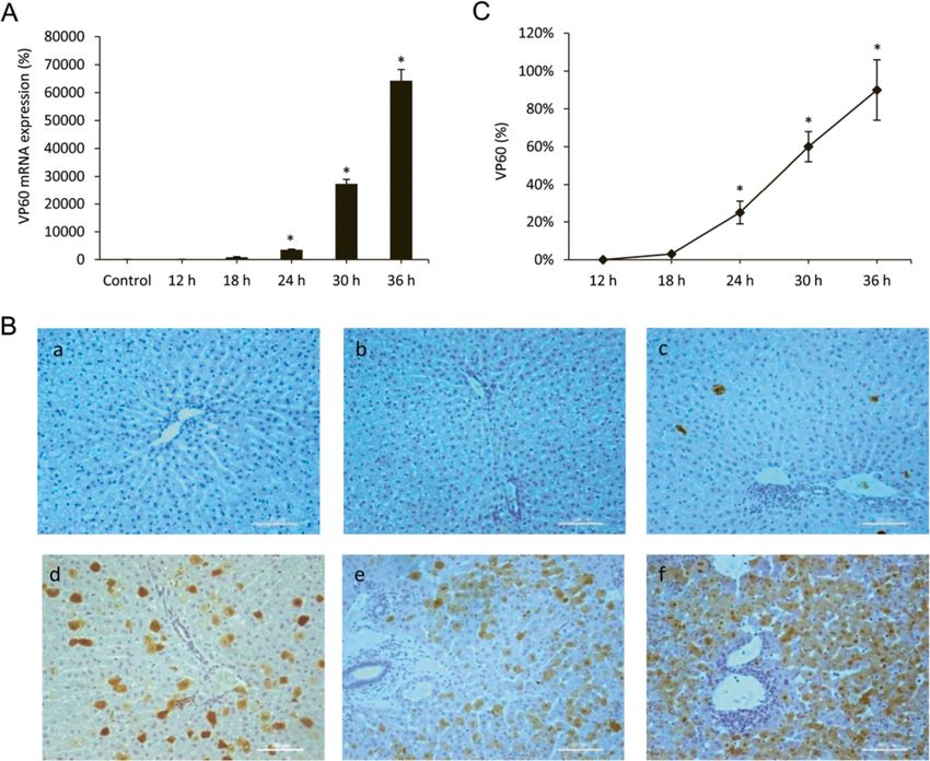
Vallejo et al. Veterinary Research 2014, 45:15                                                                     Page 2 of 13
http://www.veterinaryresearch.org/content/45/1/15

form an amphisome, in which the autophagic cargo is               cells through apoptotic mechanisms [24]. However, the
degraded and delivered into the lysosomal lumen [13].             autophagy-dependent modulation of cell death is a
The first step in the initiation of autophagy is the activation   complex phenomenon and it has also been reported
of a molecular complex containing the serine/threonine            that autophagy can prolong survival of virus-infected
kinase ULK1. The activation of this complex is down-              cells by counteracting the apoptotic response [25,26].
regulated by mammalian target of rapamycin (mTOR),                   We have previously reported that RHDV leads to the
which integrates multiple signalling pathways sensitive           activation of the different branches of the UPR [7] and
to the availability of amino acids, ATP, growth factors,          induces apoptotic death in the last stages of the disease
or level of reactive oxygen species [14]. The nucleation          [5,6]. However it is unknown whether the RHDV in-
of the autophagosomal membrane is controlled by an-               duces autophagy in the liver of infected rabbits and how
other molecular complex containing Bcl-2-interacting              it relates to ER stress and apoptosis. In this study, we
protein (beclin)-1, which allows the production of phos-          attempted to explore which signalling pathways were in-
phatidylinositol 3-phosphate (PI3P) to occur [15]. Several        volved in the autophagic response induced by the RHDV
interacting proteins which participate in this complex            and to characterize the role of autophagy in the context
include positive factors such as UV radiation resistance-         of RHDV pathogenesis.
associated gene (UVRAG) [16]. In the elongation step,
two distinct ubiquitin-like conjugation systems are in-           Materials and methods
volved. The ubiquitin-like autophagy-related (Atg)12 is           Virus and experimental model
conjugated to Atg5 and then forms a complex with                  Nine-week-old male New Zealand white rabbits were
Atg16L1, which is required in the elongation of the               kept in the animal facility of the University of León with
autophagosome membrane and determines its curvature.              12-h light cycle at 21–22 °C and 50% relative humidity.
The main player in the second conjugation system is               They were given a standard dry rabbit food and water
microtubule-associated protein 1 light chain (LC)3, which         ad libitum. Rabbits were injected intramuscularly with
is cleaved to generate the LC3-I form. LC3-I conjugates           2 × 10⁴ hemagglutination units of the RHDV isolate
with phosphatidylethanolamine to LC3-II, which localizes          AST/89 [3,4] at 21 h pm. We have previously reported
to the autophagosomal membrane and is suited to serving           that during experimental RHDV-infection biochemical
as an autophagy-specific marker [10].                             data and expression of genes involved in injury, apop-
  Autophagy primarily fulfills a pro-survival role during         tosis, ER stress and regeneration change remarkably at
adaptation to unfavourable growth conditions or follow-           36–48 hpi, with a 10-15% survival rate by 48 hpi [6-8].
ing cellular stress. In addition, autophagy contributes to        So, we decided to study the effects of infection on the
innate immunity by degrading intracellular pathogens,             mechanisms of autophagy in rabbits that were infected
and its inhibition results in an increased replication of         with the RHDV and sacrificed at 12, 18, 24, 30 and 36
virulence of different viruses such as herpex simplex virus       hpi (n = 6 each). The study was carried out in strict ac-
1 (HSV1) or vesicular stomatitis virus (VSV) [17]. How-           cordance with the recommendations in the Guide for
ever, many viruses, including hepatitis C virus (HCV),            the Care and Use of Laboratory Animals of the National
Dengue virus, or human immunodeficiency virus (HIV)-1,            Institutes of Health, and was specifically approved by the
have evolved mechanisms to evade autophagy and in some            Ethics Committee of the University of León.
cases manage to be even more subversive, using the
autophagic response for enhanced replication and viral re-        Transmission electron microscopy
lease [18,19]. ER stress, which is one of the typical stress      For transmission electron microscopy (TEM) analysis, liver
responses initiated in cells after viral infection, is import-    tissues were dissected into 1-mm3 pieces for good pene-
ant in maintaining the physiology of healthy cells and            tration of the fixative, and then immersed in a modified
functions to down-regulate protein synthesis through              Karnovsky fixative (2% glutaraldehyde + 4% buffered forma-
the unfolded protein response (UPR) [20]. It has been             lin (0.1 mol/L phosphate buffer)) overnight. The samples
reported that autophagy is activated upon ER stress as a          were post-fixed in 2% osmium tetroxide for 2 h at 4 °C and
defensive mechanism for survival [21], and it is known            dehydrated with ascending grades of alcohol. The tissue
that some viruses stimulate signalling pathways from              block was then infiltrated and embedded in epon resin at
UPR to autophagy [22]. Interference of the autophagic             60 °C for 72 h. Ultrathin sections (70 nm) were cut with an
response with cell death mechanisms plays an import-              automatic ultra-microtome (Reichert Ultracut E, Vienna,
ant role in determining the fate of infected cells, and re-       Austria) using a diamond knife. The sections were collected
cent data suggest the existence of a cross talk between           on copper grids (200 meshes) and stained with uranyl acet-
autophagic and apoptotic pathways [23]. For example               ate and lead citrate solutions. TEM images were obser
some studies have demonstrated that the autophagy                 ved under a transmission electron microscope (JEOL Ltd,
process can contribute to the death of virus-infected             Tokyo, Japan) operating at an accelerating voltage of 80 kV.
Vallejo et al. Veterinary Research 2014, 45:15                                                                         Page 3 of 13
http://www.veterinaryresearch.org/content/45/1/15

Real-time RT-PCR                                                      anti-p62/SQSTM1, PARP-1, Bcl-2, Bcl-xL (Santa Cruz
Total RNA was extracted from frozen rabbit liver using a              Biotechnology, Santa Cruz, CA, USA), LC3I/II, and
Trizol reagent (Life Technologies, Madrid, Spain) and quan-           phospho-mTOR (Abcam, Cambridge, UK) antibodies
tified using Nano Drop1000 spectrophotometer (Thermo                  at 1:200–1:1000 dilution with PBST containing 2.5%
Scientific, Wilmington, DE, USA). Residual genomic DNA                non-fat dry milk. Equal loading of protein was demon-
was removed by incubating RNA with RQ1 RNase-free                     strated by probing the membranes with a rabbit anti-
DNase (Promega, Madison, WI, USA). RNA integrity                      GAPDH polyclonal antibody (1:2000 Sigma, St Louis,
was confirmed by formaldehyde gel electrophoresis.                    MO, USA). After washing with TBST, the membranes
Total RNA (1 μg) was reverse transcribed as described                 were incubated for 1 h at room temperature with
[7] and mRNA was determined by real-time PCR ana-                     secondary HRP conjugated antibody (Dako, Glostrup,
lysis using SYBR Green I Master (Roche Diagnostics                    Denmark, 1:5000), and visualized using ECL detection
GmbH, Mannheim, Germany) and the appropriate primers                  kit (Amersham Pharmacia, Uppsala, Sweden) [7]. The
(Table 1). Relative changes in gene expression levels were            density of the specific bands was quantified with an
determined using the 2-DDCt method [27]. The cycle                    imaging densitometer (Scion Image J Software 1.46a,
number at which the transcripts were detectable (Ct) was              Bethesda, MD, USA).
normalized to the cycle number of β-Actin gene detection,
referred to as ΔCt.                                                   Immunohistochemistry
                                                                      Tissue samples were recovered, fixed in 10% buffered for-
Western blot analysis                                                 malin and embedded in paraffin. Sections (4 μm) were
For Western blot analysis liver tissue (25 mg) was homo-              dewaxed and hydrated through graded ethanol, cooked in
genised in 1 mL from RIPA buffer containing protease                  25 mM citrate buffer, pH 6.0, in a pressure cooker for
and phosphatase inhibitor cocktails (Roche Diagnostics                10 min, transferred into boiling deionized water and let
GmbH). Further disrupt and homogenize tissue with a                   to cool for 20 min. Tissue sections were then treated
manual homogenizer, maintaining temperature at 4 °C                   with 3% hydrogen peroxide to inactivate endogenous
throughout all procedures. Then the homogenate was                    peroxidase activity. The slides were incubated with
incubated on ice for 30 min and finally the samples were              rabbit anti-VP60 and anti-LC3 antibodies (Ingenasa,
centrifuged at 13 000 g for 30 min at 4 °C [28]. The                  Madrid, Spain and Abcam, respectively) overnight at
supernatant fraction was recollected and stored at −80 °C             4 °C. Subsequently, the sections were incubated for 30 min
in aliquots until use. Protein concentration was mea-                 using the EnVision + system and developed with a
sured by Bradford assay. Equal amounts of protein ex-                 solution of 3-3-diaminobenzidine (DAB) (Vector Lab,
tracts (30 μg) were separated by 7-12% sodium dodecyl                 Burlingame, CA, USA). The slides were stained with
sulphate (SDS)-polyacrylamide gel electrophoresis and                 hematoxylin for 10 s and mounted. The specificity of
transferred electrically to polyvinyllidene difluoride mem-           the technique was evaluated by negative controls (omit-
branes (Millipore, Bedford, MA, USA). The membranes                   ting the incubation with the primary antibody and incu-
were then blocked with 5% non-fat dry milk in Tris-                   bating it with non-immune sera). Pathological findings
buffered saline containing 0.05% Tween 20 (TBST) for                  were assessed by one of the authors blinded to the
30 min at 3 °C and probed overnight at 4 °C with polyclonal           group allocations.

Table 1 Primers used in this study
Gene             Sense primer (5′-3′)               Antisense primer (5′-3′)          Accession number     Fwd start      Rev start
Beclin-1         CATGCAATGGTGGCTTTCC                TCTCGCCCTTTTCAACCTCTT             XM_002719409.1       936            994
UVRAG            GCGGCGTCTTCGACATCT                 GATGGCCGTTTCTATTAACAATGTT         XM_002708684.1       117            178
Atg5             CGTCCTGTGGCTGCAGATG                AAGGACACACTTCTTTGAGGAGATC         XM_002714882         417            479
Atg12            TGCTGAAGGCTGTGGGAGAT               TGTTCGCTCTACAGCCCATTT             XM_002712042.1       176            237
Atg16L1          CCACCAAACCGGCATGAG                 CTTGCAGCTGGCTGTCATTC              XM_002721435.1       190            250
p62/SQSTM1       AACAGAGGTGACCACCCTTCA              AGCACAGACTGGCTGGAAGTC             XM_002710315.1       738            798
RHDV             TAGCCCAACAGAAGCACAAG               AAACAAGTCGTCAACCTCCC
BiP              ATTGACAATGGTGTCTTCGAAGTC           CCCCGCCCAGGTGAGT                  XM_002720437.1       709            766
CHOP             ATACATCACCACACCTGAAAGCA            GCACTCGGCTGCCATCTC                XM_002720915.1       103            160
GRP94            TGCTTAATTGGATGAAAGACAAA            GCTGAGACACCACAGCCTTTT             XM_002711230.1       1772           1834
β−Actin          TGGCATCCTGACGCTCAA                 TCGTCCCAGTTGGTCACGAT              NM_001101683         262            317
Vallejo et al. Veterinary Research 2014, 45:15 Page 4 of 13
http://www.veterinaryresearch.org/content/45/1/15
Caspase activity of LC3 enables the detection of autophagosomes. The
Lysates were prepared by homogenizing liver tissue TEM examination in this study reveals that autophagic
in 0.25 mM sucrose, 1 mM EDTA, 10 mM Tris and a structures were present in liver cells from 12 hpi (Figure 2).
protease inhibitor cocktail (Roche Diagnostics GmbH). Phagophore structure, double-membrane autophagosomes
The lysates were then centrifuged at 14 000 g for with engulfed damaged organelles, and autolysosomes
10 min at 4 °C, and supernatants (50 μg protein) were with a large vacuole containing large amount of cellular
incubated for 1 h at 37 °C in HEPES buffer containing debris were present at 18 and 24 hpi. Severe cytoplasmic
100 μM concentrations of the specific fluorogenic sub- biliary necrosis was observed in the periportal rather than
strate (7-amino-4-methylcoumarin N-acetyl-L-aspartyl- centrilobular hepatocytes, characterized by the accumu-
L-glutamyl-L-valyl-l-aspartic acid amide, Ac-DEVD-AMC). lation of electron-dense biliary materials and markedly
Cleavage of the caspase substrate was monitored using a increased number of lysosomes. The bile canalicular mi-
spectrofluorimeter (Hitachi F-2000 fluorimeter, Hitachi crovilli were very swollen and stunted. Condensation of
LTD, Tokyo, Japan) at excitation/emission wavelengths of nuclear chromatin and cytoplasmic vacuolization, which is
380/460 nm. Activity was expressed as fluorescence units a typical sign of apoptosis, were observed at late infection
per milligram of protein per min of incubation. periods, 30 and 36 hpi (Figure 2).
Statistical analysis RHDV infection induced autophagy molecular signalling
Results are expressed as mean values ± standard error Monitoring static levels of autophagosomes is not suffi-
of the mean (SEM). Data were compared by analysis of cient to elucidate effects of RHDV on autophagy, because
variance (ANOVA); when the analysis indicated the accumulation of autophagosomes may result either from
presence of a significant difference, the means were an increased in their formation or a decrease in their
compared with the Newman-Keul’s test. Significance fusion with lysosomes [31]. Thus, to examine autophagy
was accepted when p was less than 0.05. Values were in RHDV-infected rabbits and to avoid misinterpretation,
analyzed using the statistical package SPSS 19.0 (IBM in this research we combined the TEM study with immu-
Corporation, Armonk, NY, USA). nohistochemical analysis, Western blot and RT-PCR of
different autophagy markers.
Results LC3 is a major marker of the autophagosome formation
Expression of the capsid protein VP60 and a protein widely used as a hallmarker of autophagy
The RHDV is a single positive stranded RNA virus with [32]. As shown in Figure 3A-B, immunoreactivity for LC3
a 40 nm icosaedric capsid composed by 90 dimers of the was negative in liver sections from control rabbits. LC3
capsid protein VP60, and a minor structural protein VP2 antigen was detected in hepatocytes as soon as 12 hpi.
which regulates capsid protein levels [29]. The expres- At 18 hpi LC3 immunolabelling increased significantly,
sion level of the VP2 protein is very low and has been reaching a peak at 24 hpi. At 30 and 36 hpi, hepatic sec-
estimated to be one-fifth of that of VP60; therefore, to tions revealed a decrease in the number of labelled hepa-
determine the presence of the virus in infected hepato- tocytes. Stressors, such as some viruses, upregulate LC3
cytes we employed VP60 mRNA as a viral marker, and expression and promote the binding of cytosolic LC3-I to
its expression was analysed in liver extracts by quantita- PE to form autophagosome-specific lipidated form LC3-II,
tive real-time PCR (Figure 1A). As might be expected which remains attached to the inner membrane, making it
[29], the relative VP60 mRNA expression increased ex- a good marker of autophagosomes. Thus, the conversion
ponentially after 18 hpi in RHDV-infected rabbits. Viral of LC3-I to LC3-II is a certain and specific marker of
VP60 antigen was also examined by immunohistochemi- autophagy and necessary for the autophagosome forma-
cal techniques too (Figure 1B-C). Labelling was not tion [10]. When liver homogenates were analyzed by
found in infected rabbits from the group of animals Western blot to detect the different forms of LC3, a sig-
killed at 12 hpi. Viral VP60 antigen was first detected in nificant increase in protein expression of LC3-II was ob-
hepatocytes from animals killed at 18 hpi. The extent of served at 18 and 24 hpi, with a later decrease at 30 and 36
labelling increased markedly at 24 hpi, with the labelled hpi (Figure 3C-D). The results from TEM studies, LC3
hepatocytes mainly found in the periportal area. At 30 hepatocyte labelling and LC3-II protein expression un-
and 36 hpi, the liver revealed extensive viral VP60 anti- equivocally demonstrate that the autophagy was induced
gen immunolabelling. at an early stage in rabbits infected with the RHDV.
In addition to the LC3 system there is a second
Autophagy vesicles were detected in RHDV-infected ubiquitine-like system essential for autophagosome for-
hepatocytes by transmision electron microscopy mation which is formed by the Atg12-Atg5-Atg16L1
One standard method to monitor autophagy is TEM, complex, which is situated in the outer layer of the iso-
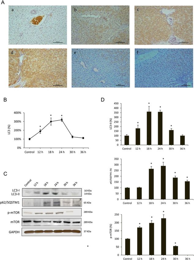
which together with immunohistochemical localization lation membrane [33]. To confirm that RHDV infectionVallejo et al. Veterinary Research 2014, 45:15 Page 5 of 13 http://www.veterinaryresearch.org/content/45/1/15 Figure 1 Liver expression of the RHDV capsid protein VP60. A: Levels of mRNA analyzed by real-time PCR assay and normalized against β-Actin. Data are presented as percentage change from the control group. Values are expressed as means SEM (n = 6). B-C: Immunohistochemical labeling in hepatocytes (a) Control, (b) RHDV 12 hpi; (c) RHDV 18 hpi; (d) RHDV 24 hpi; (e) RHDV 30 hpi; (F) RHDV 36 hpi. Paraffin-embedded sections were immunostained with a VP60 antibody. Original magnification: 200×. The graph shows evolution of the percentage of positively labeled hepatocytes over time. Values are expressed as means S.E.M (n = 6). *P < 0.05, compared with Control. Image analysis was performed using the ImageJ software v3.91 [30]. triggers autophagy activation we quantified mRNA expres- mRNA expression revealed a peak at 24 hpi coinciding with sion of the complex components at different infection pe- changes in beclin-1 mRNA expression, and then began to riods. Results obtained indicate that mRNA levels increase decrease. We further studied specific autophagy substrate from 12 hpi for Atg12 and Atg5 and from 18 hpi for p62/SQSTM1, an adaptor protein which plays an essential Atg16L1, reach a maximum at 18 hpi, and still remain sig- role in mediating selective autophagy, and serves as an nificantly elevated at 24 hpi; values return to basal levels autophagy receptor targeting ubiquitin proteins to autopha- or even lower at 30 and 36 hpi (Figure 4). The beclin-1- gosomes for degradation [12]. p62/SQSTM1 mRNA and PI3K complex is a critical element in the autophagy signal- protein expression increased from 12 to 24 hpi, with de- ling pathway [15]. We observed that beclin-1 mRNA levels creases at 30 and 36 hpi (Figures 3C-D, 4). increased at 18 and 24 hpi with a decrease in later periods, in parallel to the changes detected in both ubiquitine-like Effects of RHDV infection on pathways regulating systems (Figure 4). Beclin-1-PI3K -mediated autophagy is autophagy induction positively regulated by UVRAG, which interacts with One of the major pathways regulating autophagy involves beclin-1 in the early steps, leading to activation of autoph- mTOR. It is known that activation of mTOR in nutrient- agy through the autophagosome maturation [16]. UVRAG proficient cells acts as a negative regulator of autophagy,
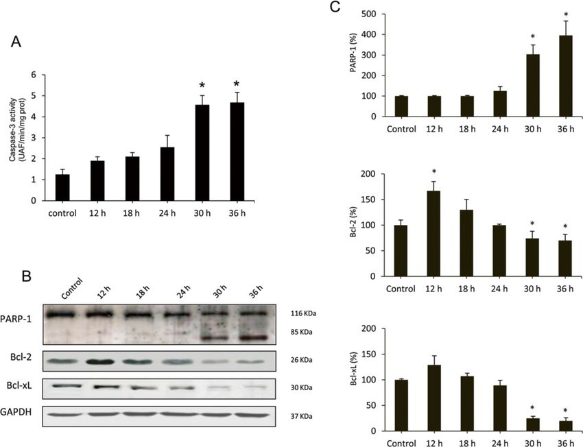
Vallejo et al. Veterinary Research 2014, 45:15                                                                                     Page 6 of 13
http://www.veterinaryresearch.org/content/45/1/15

 Figure 2 Liver transmission electron microscopy of RHDV-infected rabbits. (A) Control, (B) RHDV 12 hpi; (C) RHDV 18 hpi; (D) RHDV 24 hpi;
 (E) RHDV 30 hpi; (F) RHDV 36 hpi. Electron micrographs of the liver from control rabbits (A) and infected rabbits (B-F). Normal appearance of
 hepatocytes was observed in rabbits from the control group (A). Early disease periods (B-D) showed increased levels of lysosomes and
 mitochondria as well as an augmentation of their density. Images revealed a great number of autophagic vacuoles (black arrows) in different
 stages. In more advanced disease periods (E-F) the chromatin was condensed and aggregated at the periphery of the nuclear membrane and
 hepatocytes showed vacuolization of the cytoplasm (black arrows). Original magnification: 5000 – 15 000×.

while repression of mTOR by nutrient deprivation or                       94 (GRP94). BiP is an ER chaperone protein which is re-
rapamycin treatment induces autophagy [14]. However,                      quired for protein folding and has been recently shown to
the cross talk between mTOR pathway and autophagy in-                     play a central role modulating the sensitivity and duration
duction during viral infection is complex, and it has been                of the UPR [35]. Hepatic expression of BiP was measured
reported that some viruses activate mTOR signalling                       by RT-PCR (Table 2). Results showed a progressive in-
[23,34]. We analyzed the hepatic expression of phospho-                   crease in the values at different time infection periods
mTOR by Western blot at different RHDV-infection pe-                      until 24 hpi. Activation of UPR in infected rabbits was
riods (Figure 3C-D). A progressive increased hepatic                      confirmed by quantification of the mRNA level of
expression of phospho-mTOR was observed at 12, 18, and                    CHOP, a major marker of the ER stress response, and
24 hpi in RHDV-infected animals. However, at 30 hpi                       GRP94, a molecular chaperone and resident protein of
phospho-mTOR hepatic level decreased to values below                      the ER that aids in the folding of secretory and mem-
the control group, and it was undetectable at 36 hpi.                     brane proteins [7]. Results showed a peak of mRNA
  Although the role of autophagy in normal ER function                    expression for both chaperones at 24 hpi (Table 2).
is not established, there are some studies that have
shown that autophagy is associated with the ER and                        Apoptotic death in RHDV-infected liver cells
maybe an important part of normal ER function [21]. ER                    Autophagy has a complex interaction with apoptosis. It
stress-induced autophagy plays an important role in main-                 can inhibit or cause cell death, and, on the other hand,
taining cellular homeostasis through alleviating stress                   apoptosis is known to inhibit the genesis of autophagy
and can also be used as an alternative degradation                        [36]. Moreover, it is know that autophagy plays a major
mechanism to process misfolded proteins that have ac-                     role in determining the fate of virally infected cells by
cumulated in the ER lumen [7]. During ER stress different                 blocking or promoting apoptotic mechanisms [23]. We
transcription factors regulate the expression of ER cha-                  have previously reported that apoptotic liver damage de-
perones that enhance the folding capacity of the ER,                      velops in rabbits infected by the RHDV and the effect is
including CCAAT/enhancer-binding protein homo-                            attenuated by treatment with antioxidants [5,6]. In this
logous protein (CHOP), immunoglobulin-heavy-chain-                        study we analyzed changes with time in the activation
binding protein (BiP/GRP78) and glucose-regulated protein                 of caspase-3, the common event initiated by multiple
Vallejo et al. Veterinary Research 2014, 45:15      Page 7 of 13
http://www.veterinaryresearch.org/content/45/1/15

 Figure 3 (See legend on next page.)
Vallejo et al. Veterinary Research 2014, 45:15                                                                                           Page 8 of 13
http://www.veterinaryresearch.org/content/45/1/15

 (See figure on previous page.)
 Figure 3 Effects of RHDV infection on markers of autophagy. A-B: Immunohistochemical labeling of the autophagy marker LC3 in the liver
 of RHDV-infected rabbits. (a) Control; (b) RHDV 12 hpi; (c) RHDV 18 hpi; (d) RHDV 24 hpi; (e) RHDV 30 hpi; (F) RHDV 36 hpi. Paraffin-embedded
 sections were immunostained with a LC3 antibody. Original magnification: 200×. The graph shows evolution of the percentage of positively labeled
 hepatocytes over time. Values are expressed as means S.E.M (n = 6). *P < 0.05, compared with Control. Image analysis was performed using the ImageJ
 software v3.91 [30]. C-D: Western blot of markers of autophagy. Proteins from liver extracts were separated by sodium dodecyl sulfate
 polyacrylamide gel electrophoresis, followed by immunoblotting. Equal loading of proteins is illustrated by GAPDH bands. The graph shows
 densitometric quantification. Values are expressed as means S.E.M (n = 6). *P < 0.05, compared with Control.

different stimuli that induces apoptosis [37]. Samples were                  of Bcl-2 and Bcl-xL (Figure 5B-C), two antiapoptotic pro-
incubated with a specific fluorigenic substrate whose cleav-                 teins involved in the intrinsic pathway of apoptosis [6].
age indicated that infection resulted in a marked increase of
caspase-3 activity only at 30 and 36 hpi (Figure 5A). Fur-                   Discussion
thermore, Western blot analysis demonstrated that at later                   This research examined the occurrence of autophagy
periods of infection there was a marked proteolysis of                       during experimental infection by the RHDV. Similarly to
PARP-1 (Figure 5B-C), a nuclear enzyme whose cleavage                        other studies conducted with viruses that promote au-
into a 85-kDA fragment by caspase-3 confirms that cells                      tophagy, TEM analysis showed that number and content
are undergoing apoptosis [5]. Our data also show that at 30                  of autophagy vesicles increased in RHDV-infected livers.
and 36 hpi there is a significant inhibition of the expression               We further analyzed the impact of RHDV infection of

 Figure 4 Effect of Rabbit Hemorrhagic Disease virus (RHDV) infection on mRNA levels of genes related to autophagy (A) and ER stress
 (B). Levels of mRNA were analyzed by real-time PCR assays. Data, normalized against β-Actin, are presented as percentage change from the control
 group. Values are expressed as means S.E.M (n = 6). *P < 0.05, compared with Control.
Vallejo et al. Veterinary Research 2014, 45:15                                                                                                  Page 9 of 13
http://www.veterinaryresearch.org/content/45/1/15

Table 2 Effect of Rabbit Hemorrhagic Disease virus (RHDV) infection on mRNA levels of genes related to autophagy
                           Control                 12 h                    18 h                    24 h                    30 h                     36 h
Beclin-1                   100 ± 11                118 ± 8                 203 ± 16*               253 ± 36*               105 ± 6                  87 ± 7
                                                                                         *                                        *
UVRAG                      100 ± 6                 104 ± 8                 227 ± 19                131 ± 15                81 ± 9                   99 ± 5
Atg5                       100 ± 5                 125 ± 11*               163 ± 19*               139 ± 7*                87 ± 7                   63 ± 5
Atg12                      100 ± 9                 173 ± 12*               296 ± 41*               177 ± 9*                104 ± 8                  82 ± 7
                                                                                     *
Atg16L1                    100 ± 10                106 ± 9                 223 ± 6                 140 ± 12*               130 ± 7*                 106 ± 10
                                                              *                      *                       *
p62/SQSTM1                 100 ± 4                 145 ± 6                 192 ± 8                 202 ± 8                 86 ± 6                   69 ± 4*
                                                                                         *                       *                    *
BiP                        100 ± 7                 118 ± 17                177 ± 41                304 ± 29                241 ± 39                 184 ± 18*
CHOP                       100 ± 8                 169 ± 10*               211 ± 19*               540 ± 79*               158 ± 9*                 161 ± 13*
                                                          *                          *                           *                    *
GRP94                      100 ± 4                 80 ± 4                  163 ± 5                 412 ± 54                314 ± 13                 226 ± 21*
Levels of mRNA were analyzed by real-time PCR assays. Data, normalized against β-Actin, are presented as percentage change from the control group. Values are
expressed as means±S.E.M (n = 6). *P
Vallejo et al. Veterinary Research 2014, 45:15 Page 10 of 13 http://www.veterinaryresearch.org/content/45/1/15 several proteins that regulate distinct molecular events cells after infection with the Newcastle virus [44], and in leading to autophagy vesicle formation, including its ini- bovine kidney cells infected with the bovine herpesvirus tiation (beclin-1), and maturation by the Atg12 and LC3 type-4 [40]. Our data demonstrate that mTOR is not a conjugation systems. Data obtained demonstrate an early negative regulator during RHDV-induced autophagy, increased expression of the Atg16L1 complex compo- and could indicate that induction of autophagy occurs nents, together with enhanced LC3 immunostaining and upstream of mTOR signalling or that both processes act conversion of soluble cytosolic LC3-I to its lipidated, concurrently. In HCV-infected hepatocytes it has been autophagosome-associated form LC3-II, which unequivo- suggested that mTOR activation is necessary for cell cally demonstrates that the autophagy was induced at an growth through regulation of phospho-eukaryotic trans- early stage in rabbits infected with the RHDV. Real-time lation initiation factor 4E-binding protein (EBP)1 [34]. PCR confirmed that the key autophagy gene beclin-1 was Further work would be necessary to identify if there is a also activated, a fact which suggests a crucial role for similar requirement following infection with the RHDV. this protein in the induction of the autophagic response Autophagy is also triggered in response to ER stress by the RHDV. Although beclin-1 up-regulation is a fre- through the induction of the UPR [45]. In mammalian quent finding following viral infection [13], there are cells, knockdown of the upstream UPR regulator BiP in- data of beclin-1-independent autophagy induction by hibits autophagosome formation, but does not affect the enterovirus 71 [38] and it has been reported a late and conversion of LC3-I to LC3-II, suggesting that ER stress rather limited increase in the expression of this proau- induction is an obligatory factor for autophagy and may tophagic protein by HSV-1 [26]. function at the phagophore expansion rather than induc- In our experiments, p62/SQSTM1 expression increased tion step [46]. Previous studies have shown that induction from 12 hpi and remained elevated until 24 hpi. p62/ of autophagosomes by the HCV virus depends on the UPR SQSTM1 is a multifunctional protein, involved in the de- [43], and the three branches of the UPR contribute to regu- livery of ubiquitin-bound cargo to the autophagosome, late HCV replication via modulation of autophagy [22]. It that interacts with LC3 and is specifically degraded by the has also been reported that the tobacco mosaic virus RNA autophagic-lysosome pathway, being commonly measured induces ER stress-related autophagy in HeLa cells [47], to detect autophagic flux [12]. Viral infection with dif- and it is known that autophagosome formation during ferent herpes viruses has been reported to result in a de- varicella-zoster virus infection follows ER stress and the crease of p62/SQSTM1 in parallel to increase in the UPR [48]. In a previous work, our research group, using protein LC3-II [39,40]. However, upregulated expres- the RHDV model of FHF, reported that ER stress was in- sion of both p62/SQSTM1 and LC3 has been shown duced in RHDV infected rabbits through a modulation of to exist in different types of tumours, whose growth is sig- the three branches of the UPR [7]. Here, it is shown that nificantly inhibited by p62/SQSTM1 down-regulation [41]. mRNA levels of the molecular chaperones CHOP, BiP and Moreover, the expression of p62/SQSTM1 and LC3-II also GRP94 reached a peak at 24 hpi, in parallel to the increase increases in livers from patients with primary biliary cir- of the expression of beclin-1 and the components of rhosis and cultured biliary epithelial cells treated with the two ubiquitin-like conjugation systems Atg12-Atg5- hydrogen peroxide, with an accumulation of p62-positive Atg16L1 and LC3. Our data suggest that autophagy could aggregates [42]. In Huh 7.5 cells it has been reported that be provoked at least in part upon ER stress. This hypoth- after the transfection of the HCV RNA there is a continu- esis is further supported by the RHDV-induced increase in ous increase of p62/SQSTM1 which indicates that HCV the upregulation of beclin-1, whose expression is required does not enhance autophagic protein degradation [43]. Re- for ER-stress induced autophagy [46]. sults from the present research suggest a similar response The interplay between autophagy and programmed cell to RHDV infection, with an upregulation of p62/SQSTM1 death is complex. Autophagy is a cytoprotective mechan- which may reflect a dysfunctional process in which the ism which enables cells to survive unfavourable growth capacity of autophagy is not much enough to process the conditions and can prevent cell death by apoptosis. How- damaged proteins bound to p62/SQSTM1. ever, some studies have demonstrated that autophagy may mTOR is an important signalling molecule which in have an active contribution to cell death in virus infected nutrient-proficient cells acts as a negative regulator of au- cells. Thus, it has been reported that pharmacological in- tophagy [26]. When the expression of phospho-mTOR was hibition of autophagy efficiently suppresses apoptosis in- monitored by Western blot assay we observed an increased duced by human adenovirus type 5 Delta-24-RGD mutant expression between 12 and 24 hpi, showing that infection in mouse fibroblast or human U251 glioma cells [49]. with the RHDV stimulates the mTOR signalling pathway Blocking of autophagy also attenuates cell death caused by in parallel to the development of the autophagic process. A the avian influenza A H5N1 virus both in vitro and in vivo similar unexpected result has been previously reported in [50], and it is known that knockdown of beclin-1 or Atg5 HCV-infected human hepatocytes [34], in U251 glioma protects human rhabdomyosarcoma cells from enterovirus
Vallejo et al. Veterinary Research 2014, 45:15                                                                                     Page 11 of 13
http://www.veterinaryresearch.org/content/45/1/15

71-induced apoptotic death [24]. We and others have pre-         dominates. Although it is necessary to be cautious, consid-
viously reported that RHDV infection induces in rabbits a        ering that autophagy is also involved in modulation of viral
marked apoptotic response at 36–48 hpi with increased            replication and recognition/presentation of viral antigens
caspase-3 activity and immunoexpression and a marked             [26], therapeutic potential of autophagy modulation in con-
proteolysis of PARP-1 [5,6,8,51]. Results from the present       trolling RHDV-induced cell death is worthy to be explored,
study indicate that apoptosis is present in the late stages of   considering the importance of RHDV infection as a model
the disease, with no significant increase in caspase-3 activ-    of human FHF of viral origin. Findings from the present
ity and PARP-1 degradation or decreased expression of            study could contribute to the search for new pharmaco-
the antiapoptotic proteins Bcl-2 or Bcl-xL occurring in          logical strategies to protect livers from FHF injury.
early periods. The fact that autophagy develops in hepato-
cytes at early stage and cells begin to exhibit apoptosis in     Competing interests
parallel to the decline of the autophagy response, suggests      The authors declare that they have no competing interests.

that autophagy play a beneficial role in an attempt to pro-
                                                                 Authors’ contributions
tect cells from the impending noxious effects of the virus.
                                                                 DV, IC, MA and BS carried out the experiments. JP, JGG and MJT interpreted
It has been recently shown that cardiomyocites exposed to        the results and contributed to the discussion. MJT and JGG were responsible
angiotensin II exhibit a similar behavior, with autophagy        for overall supervision. All authors read and approved the final manuscript.
occurring at early stages whereas apoptosis occurs late
[36]. A number of studies have also demonstrated the abil-       Acknowledgements
                                                                 CIBERehd is funded by Instituto de Salud Carlos III, Spain.
ity of virally-induced autophagy to prevent or delay death
of infected cells. For example, apoptotic death of hepa-         Author details
                                                                 1
toma cells expressing the oncogenic HBV X protein in-             Institute of Biomedicine (IBIOMED), University of León, 24071 León, Spain.
                                                                 2
                                                                  Centro de Investigación Biomédica en Red de Enfermedades Hepáticas y
creases when autophagy is blocked [52], and the infection        Digestivas (CIBERehd), Spain. 3Department of Animal Health, University of
with Japanese encephalitis virus increases caspase activa-       León, 24071 León, Spain. 4Division of Hepatology and Gene Therapy, Center
tion and cell death in beclin-1 or Atg5-deficient cells [53].    for Applied Medical Research (CIMA), University of Navarra, Pamplona, Spain.

It has also been shown that in HSV-1-infected U251 gli-          Received: 4 November 2013 Accepted: 22 January 2014
oma cells the autophagic response markedly delayed               Published: 4 February 2014
caspase activation and other hallmarks of apoptotic cell
death [26]. Data here obtained also point to a role of           References
                                                                 1. Liu SJ, Xue HP, Pu BQ, Quia NH: A new viral disease in rabbits. Anim Husb
virally-induced autophagy to support survival of the in-             Vet Med 1984, 16:253–255.
fected cells and suggest that autophagy might contrib-           2. Mikami O: Hepatic lesions in young rabbits experimentally infected with
ute to limit the pathological consequences associated                rabbit haemorrhagic disease virus. Res Vet Sci 1999, 66:237–242.
                                                                 3. Tuñón MJ, Sánchez-Campos S, García-Ferreras J, Álvarez M, Jorquera F,
with cell death triggered by the RHDV infection. Con-                González-Gallego J: Rabbit hemorrhagic viral disease: characterization of
firmation of the connection between autophagy and                    a new animal model of fulminant liver failure. J Lab Clin Med 2003,
RHDV pathogenecity should require the use of cell cul-               141:272–278.
                                                                 4. Sánchez-Campos S, Álvarez M, Culebras JM, González-Gallego J, Tuñón MJ:
ture systems, that are unavailable at present [54]. An               Pathogenic molecular mechanisms in an animal model of fulminant
additional interesting finding concerns the increased                hepatic failure: rabbit hemorrhagic viral disease. J Lab Clin Med 2004,
expression of p62/SQSTM1 observed in liver cells. This               144:215–222.
                                                                 5. San-Miguel B, Álvarez M, Culebras JM, González-Gallego J, Tuñón MJ:
reflects inhibition of autophagosome-lysosomal func-                 N-acetyl-cysteine protects liver from apoptotic death in an animal model
tion and dysfunctional autophagy, which may contrib-                 of fulminant hepatic failure. Apoptosis 2006, 11:1945–1957.
ute to altered signal transduction pathway and liver             6. Tuñón MJ, San Miguel B, Crespo I, Jorquera F, Santamaría E, Álvarez M,
                                                                     Prieto J, González-Gallego J: Melatonin attenuates apoptotic liver damage
damage [55]. In fact, it is known that upregulation of               in fulminant hepatic failure induced by the rabbit hemorrhagic disease
p62/SQSTM1 positively controls apoptosis by polyubi-                 virus. J Pineal Res 2011, 50:38–45.
quitination and aggregation of the key initiator caspase         7. Tuñón MJ, San-Miguel B, Crespo I, Laliena A, Vallejo D, Álvarez M, Prieto J,
                                                                     González-Gallego J: Melatonin treatment reduces endoplasmic reticulum
8 [56,57], thus playing a potential role in the cross-               stress and modulates the unfolded protein response in rabbits with
regulation between autophagy and apoptosis.                          lethal fulminant hepatitis of viral origin. J Pineal Res 2013, 55:221–228.
   In summary, experiments here reported were aimed to           8. García-Lastra R, San-Miguel B, Crespo I, Jorquera F, Álvarez M, González-Gallego
                                                                     J, Tuñón MJ: Signalling pathways involved in liver injury and regeneration in
enhance our understanding of the interplay between the               rabbit hemorrhagic disease, an animal model of virally-induced fulminant
RHDV and the host liver cells. The most important find-              hepatic failure. Vet Res 2010, 41:2.
ing is that RHDV infection in vivo initiates a rapid autoph-     9. Laliena A, San Miguel B, Crespo I, Álvarez M, González-Gallego J, Tuñón MJ:
                                                                     Melatonin attenuates inflammation and promotes regeneration in
agic response, perhaps in an attempt to protect liver,               rabbits with fulminant hepatitis of viral origin. J Pineal Res 2012,
which associates to ER stress development and is inde-               53:270–278.
pendent from down-regulation of the major autophagy              10. Deretic V, Levine B: Autophagy, immunity, and microbial adaptations.
                                                                     Cell Host Microbe 2009, 5:527–549.
suppressor mTOR. As the infection continues and the              11. Dreux M, Chisari FV: Viruses and the autophagy machinery. Cell Cycle 2010,
autophagic response declines, the process of apoptosis               9:1295–1307.
Vallejo et al. Veterinary Research 2014, 45:15 Page 12 of 13
http://www.veterinaryresearch.org/content/45/1/15
12. Pankiv S, Clausen TH, Lamark T, Brech A, Bruun JA, Outzen H, Overvatn A, 33. Fukuda M, Itoh T: Direct link between Atg protein and small GTPase Rab:
Biorkoy G, Johansen T: p62/SQSTM1 binds directly to Atg8/LC3 to facilite Atg16L functions as a potential Rab33 effector in mammals. Autophagy
degradation of ubiquitinated protein aggregates by autophagy. J Biol 2008, 4:824–826.
Chem 2007, 28:24131–24145. 34. Shrivastava S, Bhanja Chowdhury J, Steele R, Ray R, Ray RB: Hepatitis C virus
13. Kudchodkar SB, Levine B: Viruses and autophagy. Rev Med Virol 2009, upregulates Beclin1 for induction of autophagy and activates mTOR
19:359–378. signaling. J Virol 2012, 86:8705–8712.
14. Jung CH, Ro SH, Cao J, Otto NM, Kim DH: mTOR regulation of autophagy. 35. San-Miguel B, Crespo I, Cuevas MJ, González-Gallego J, Tuñón MJ:
FEBS Lett 2010, 584:287–1295. Glutamine treatment attenuates endoplasmic reticulum stress and
15. Wirawan E, Lippens S, Vanden Berghe T, Romagnoli A, Fimia GM, Piacentini apoptosis in TNBS-induced colitis. PLoS One 2012, 7:e50407.
M, Vandenabeele P: Beclin: a role in membrane dynamics and beyond. 36. Wang X, Dai Y, Ding Z, Khaidakov M, Mercanti F, Mehta JL: Regulation
Autophagy 2012, 8:6–17. of autophagy and apoptosis in response to angiotensin II in HL-1
16. Zhao Z, Ni D, Ghozalli I, Pirooz SD, Ma B, Liang C: UVRAG: at the crossroad cardiomyocytes. Biochem Biophys Res Commun 2013, 440:696–700.
of autophagy and genomic stability. Autophagy 2012, 8:1392–1393. 37. Mauriz JL, González P, Jorquera F, Olcoz JL, González-Gallego J: Caspase
17. Maier HJ, Britton P: Involvement of autophagy in coronavirus replication. inhibition does not protect against liver damage in hemorrhagic shock.
Viruses 2012, 30:3440–3451. Shock 2003, 19:33–37.
18. Blanchet FP, Moris A, Nikolic DS, Lehmann M, Cardinaud S, Stalder R, Garcia 38. Huang SC, Chang CL, Wang PC, Tsai Y, Liu HS: Enterovirus 71- induced
E, Dinkins C, Leuba F, Wu L, Schwartz O, Deretic V, Piguet V: Human autophagy detected in vitro and in vivo promotes viral replication.
immunodeficiency virus-1 inhibition of immunoamphisomes in dendritic J Med Virol 2009, 81:1241–1252.
cells impairs early innate and adaptive immune responses. Immunity 39. Takahashi MN, Jackson W, Laird DT, Culp TD, Grose C, Haynes JI 2nd, Benetti L:
2010, 32:654–669. Varicella-zoster virus infection induces autophagy in both cultured cells
19. Crawford SE, Hyser JM, Utama B, Estes MK: Autophagy hijacked through and human skin vesicles. J Virol 2009, 83:5466–5476.
viroporin-activated calcium/calmodulin-dependent kinase kinase-β 40. Montagnaro S, Ciarcia R, Pagnini F, De Martino L, Puzio MV, Granato GE,
signalling is required for rotavirus replication. Proc Natl Acad Sci USA Avino F, Pagnini U, Iovane G, Giordano A: Bovine herpesvirus type 4
2012, 109:E3405–E3413. infection modulates autophagy in a permissive cell line. Cell Cycle 2013,
20. Crespo I, San Miguel B, Prause C, Marroni N, González-Gallego J, Tuñón MJ: 12:6839–6848.
Glutamine treatment attenuates endoplasmic reticulum stress and 41. Ren F, Shu G, Liu G, Liu D, Zhou J, Yuan L, Zhou J: Knockdown of p62/
apoptosis in TNBS-induced colitis. PLoS One 2012, 7:e50407. sequestosome 1 attenuates autophagy and inhibits colorectal cancer
21. Ogata M, Hino S, Saito A, Morikawa K, Kondo S, Kanemoto S, Murakami T, cell growth. Mol Cell Biochem 2014, 385:95–102.
Taniguchi M, Tanii I, Yoshinaga K, Shiosaka S, Hammarback JA, Urano F, 42. Sasaki M, Miyakoshi M, Sato Y, Nakanuma Y: A possible involvement of
Imaizumi K: Autophagy is activated for cell survival after endoplasmic p62/sequestosome-1 in the process of biliary epithelial autophagy and
reticulum stress. Mol Cell Biol 2006, 26:9220–9231. senescence in primary biliary cirrhosis. Liver Int 2012, 32:4877–4899.
22. Shinohara Y, Imajo K, Yoneda M, Tomeno W, Owaga Y, Kirikoshi H, 43. Sir D, Chen WL, Choi J, Wakita T, Yen TS, Ou JH: Induction of incomplete
Funakoshi K, Ikeda M, Kato N, Nakajima A, Saito S: Unfolded protein autophagic response by hepatitis C virus via the unfolded protein
response pathways regulate hepatitis C virus replication via modulation response. Hepatology 2008, 148:1054–1061.
of autophagy. Biochem Biophys Res Commun 2013, 432:326–333. 44. Meng C, Zhou Z, Jiang K, Yu S, Jia L, Wu Y, Liu Y, Meng S, Ding C:
23. Tovilovic G, Ristic B, Milenkovic M, Stanojevic M, Trakjovic V: The role Newcastle disease virus triggers autophagy in U251 glioma cells to
and therapeutic potential of autophagy modulation in controlling enhance virus replication. Arch Virol 2012, 157:1011–1018.
virus-induced cell death. Med Res Rev. in press. 45. Ding WX, Ni HM, Gao W, Yoshimori T, Stolz DB, Ron D, Yin XM: Linking of
24. Xi X, Zhang X, Wang B, Wang T, Wang H, Huang H, Wang J, Jin Q, Zhao Z: autophagy to ubiquitin-proteasome system is important for the
The interplays between autophagy and apoptosis induced by regulation of endoplasmic reticulum stress and cell viability. Am J Pathol
enterovirus 71. PLoS One 2013, 8:e56966. 2007, 171:513–524.
25. Joubert PE, Werneke SW, de la Calle C, Guivel-Benhassine F, Giodini A, 46. Li J, Ni M, Lee B, Barron E, Hinton DR, Lee AS: The unfolded protein
Peduto L, Levine B, Schwartz O, Lenschow DJ, Albert ML: Chikungunya response regulator GRP78/BiP is required for endoplasmic reticulum
virus-induced autophagy delays caspase-dependent cell death. J Exp Med integrity and stress-induced autophagy in mammalian cells. Cell Death
2012, 209:1029–1047. Differ 2008, 15:12460–12471.
26. Tovilovic G, Ristic B, Siljic M, Nikolic V, Kravic-Stevovic T, Dulovic M, Milenkovic M, 47. Li L, Wang L, Xiao R, Zhu G, Li Y, Liu C, Yang R, Tang Z, Li J, Huang W, Chen
Knezevic A, Bosnjak M, Bumbasirevic V, Stanojevic M, Trajkovic V: L, Zheng X, He Y, Tan J: The invasion of tobacco mosaic virus RNA
mTOR-independent autophagy counteracts apoptosis in herpes simplex induces endoplasmic reticulum stress-related autophagy in HeLa cells.
virus type 1-infected U251 glioma cells. Microbes Infect 2013, 15:615–624. Biosci Rep 2012, 32:171–184.
27. Lima-Cabello E, García-Mediavilla V, Miquilena-Colina E, Vargas-Castrillón J, 48. Carpenter JE, Jackson W, Benetti L, Grose C: Autophagosome formation
Lozano-Rodríguez T, Fernández-Bermejo M, Olcoz JL, González-Gallego J, during varicella-zoster virus infection following endoplasmic reticulum
García-Monzón C, Sánchez-Campos S: Enhanced expression of pro stress and the unfolded protein response. J Virol 2011, 85:1914–1924.
inflammatory mediators and liver X-receptor-regulated lipogenic genes 49. Jiang H, White EJ, Rios-Vicil CI, Xu J, Gomez-Manzano C, Fueyo J: Human
in non-alcoholic fatty liver disease and hepatitis C. Clin Sci (Lond) 2011, adenovirus type 5 induces cell lysis through autophagy and autophagy-
120:239–250. triggered caspase activity. J Virol 2011, 85:4720–4729.
28. Kretzmann NA, Fillmann H, Mauriz L, Marroni CA, Marroni N, González-Gallego 50. Sun Y, Li C, Shu Y, Ju X, Zou Z, Wang H, Rao S, Guo F, Liu H, Nan W, Zhao Y,
J, Tuñón MJ: Effects of glutamine on proinflammatory gene expression and Yan Y, Tang J, Zhao C, Yang P, Liu K, Wang S, Lu H, Li X, Tan L, Gao R, Song
activation of nuclear factor kappa B and signal transducers and J, Gao X, Tian X, Qin Y, Xu KF, Li D, Jin N, Jiang C: Inhibition of autophagy
activators of transcription in TNBS-induced colitis. Inflam Bowel Dis 2008, ameliorates acute lung injury caused by avian influenza A H5N1 infection.
14:1504–1513. Sci Signal 2012, 5:ra16.
29. Chen L, Liu G, Ni Z, Yu B, Yun T, Song Y, Hua J, Li S, Chen J: Minor 51. Niedźwiedzka-Rystwej P, Deptuła W: Apoptosis of peripheral blood
structural protein VP2 in rabbit hemorrhagic disease virus downregulates leukocytes from rabbits infected with non-haemagglutinating strains of
the expression of the viral capsid protein VP60. J Gen Virol 2009, rabbit haemorrhagic disease virus (RHDV). Vet Immunol Immunopathol
90:2952–2955. 2012, 149:54–57.
30. ImageJ [http://rsbweb.nih.gov/ij/] 52. Mao Y, Da L, Tang H, Yang J, Lei Y, Tiollais P, Li T, Zhao M: Hepatitis B virus
31. Lussignol M, Queval C, Bernet-Camard MF, Cotte-Laffitte J, Beau I, Codogno X protein reduces starvation-induced cell death through activation of
P: The herpes simplex virus 1 Us11 protein inhibits autophagy through autophagy and inhibition of mitochondrial apoptotic pathway. Biochem
its interaction with the protein kinase PKR. J Virol 2013, 87:859–871. Biophys Res Commun 2011, 415:68–74.
32. Wang J, Pan XL, Ding LJ, Liu DY, Da-Peng L, Jin T: Aberrant expression of 53. Jin R, Zhu W, Cao S, Chen R, Jin H, Liu Y, Wang S, Wang W, Xiao G:
beclin-1 and LC3 correlates with poor prognosis of human hypopharyngeal Japanese encephalitis virus activates autophagy as a viral immune
squamous cell carcinoma. PLoS One 2013, 8:e69038. evasion strategy. PLoS One 2013, 8:e52909.Vallejo et al. Veterinary Research 2014, 45:15 Page 13 of 13
http://www.veterinaryresearch.org/content/45/1/15
54. Abrantes J, De Loo W, Le Pendu J, Esteves PJ: Rabbit haemorrhagic
disease (RHD) and rabbit haemorrhagic disease vitrus (RHDV): a review.
Vet Res 2012, 43:12.
55. Mathew R, Karp CM, Beaudoin B, Vuong N, Chen G, Chen HY, Bray K, Reddy A,
Bhanot G, Gelinas C, Dipaola RS, Karantza-Wadsworth V, White E: Autophagy
suppresses tumorigenesis through elimination of p62. Cell 2009,
137:1062–1075.
56. Huang S, Okamoto K, Yu C, Sinicrope FA: p62/sequestosome-1 upregulation
promotes ABT-263-induced caspase-8 aggregation/activation on the
autophagosome. J Biol Chem 2013, 288:33654–33666.
57. Jin Z, Li Y, Pitti R, Lawrence D, Pham VC, Lill JR, Ashkenazi A: Cullin 3-based
polyubiquitination and p62-dependent aggregation of caspase-8
mediate extrinsic apoptosis signaling. Cell 2009, 137:729–735.
doi:10.1186/1297-9716-45-15
Cite this article as: Vallejo et al.: Autophagic response in the Rabbit
Hemorrhagic Disease, an animal model of virally-induced fulminant
hepatic failure. Veterinary Research 2014 45:15.
Submit your next manuscript to BioMed Central
and take full advantage of:
• Convenient online submission
• Thorough peer review
• No space constraints or color figure charges
• Immediate publication on acceptance
• Inclusion in PubMed, CAS, Scopus and Google Scholar
• Research which is freely available for redistribution
Submit your manuscript at
www.biomedcentral.com/submitYou can also read