Effects of neonatal exposure to a glyphosate-based herbicide on female rat reproduction
←
→
Page content transcription
If your browser does not render page correctly, please read the page content below
REPRODUCTION
  RESEARCH

Effects of neonatal exposure to a glyphosate-based herbicide
on female rat reproduction
Paola I Ingaramo, Jorgelina Varayoud, María M Milesi, Marlise Guerrero Schimpf,
Mónica Muñoz-de-Toro and Enrique H Luque
Instituto de Salud y Ambiente del Litoral (ISAL), Facultad de Bioquímica y Ciencias Biológicas, Universidad
Nacional del Litoral – Consejo Nacional de Investigaciones Científicas y Técnicas (CONICET), Santa Fe, Argentina
Correspondence should be addressed to E H Luque; Email: eluque@fbcb.unl.edu.ar

Abstract
In this study, we investigated whether neonatal exposure to a glyphosate-based herbicide (GBH) alters the reproductive performance
and the molecular mechanisms involved in the decidualization process in adult rats. Newborn female rats received vehicle or
2 mg/kg/day of a GBH on postnatal days (PND) 1, 3, 5 and 7. On PND90, the rats were mated to evaluate (i) the reproductive
performance on gestational day (GD) 19 and (ii) the ovarian steroid levels, uterine morphology, endometrial cell proliferation,
apoptosis and cell cycle regulators, and endocrine pathways that regulate uterine decidualization (steroid receptors/COUP-TFII/Bmp2/
Hoxa10) at the implantation sites (IS) on GD9. The GBH-exposed group showed a significant increase in the number of resorption sites
on GD19, associated with an altered decidualization response. In fact, on GD9, the GBH-treated rats showed morphological changes
at the IS, associated with a decreased expression of estrogen and progesterone receptors, a downregulation of COUP-TFII (Nr2f2)
and Bmp2 mRNA and an increased expression of HOXA10 and the proliferation marker Ki67(Mki67) at the IS. We concluded that
alterations in endometrial decidualization might be the mechanism of GBH-induced post-implantation embryo loss.
Reproduction (2016) 152 403–415

Introduction                                                        (Dallegrave et al. 2003, Romano et al. 2012). Regarding
                                                                    the effects of postnatal exposure to GBH on reproductive
In Argentina, transgenic crops (soybean, maize and
                                                                    female parameters, female offspring rats born to mothers
cotton) account for three quarters of the total cultivated
                                                                    exposed to different doses of GBH (50, 150 and
land (Aparicio et al. 2013). In this country, glyphosate
is the most commonly used herbicide, with around                    450 mg/kg) during pregnancy and lactation show a delay
180–200 million liters applied every year (Aparicio et al.          in vaginal opening (Dallegrave et al. 2007).
2013). Although regulatory agencies have asserted that                 It is well documented that early postnatal exposure
glyphosate-based herbicides (GBH) are relatively safe,              to endocrine-disrupting chemicals might lead to long-
some reports suggest that they may have harmful effects             lasting female reproductive disorders, such as altered
on health (Dallegrave et al. 2003, Caglar & Kolankaya               cyclicity, decreased conception rates, endometriosis
2008, Astiz et al. 2012). In a study performed in a farm            and pregnancy loss (Varayoud et al. 2011, Milesi
population of Ontario, Canada, preconception exposure               et al. 2015, Ingaramo et al. 2016). In the last decade,
to glyphosate in women increased the risk of abortions              growing data have demonstrated that GBH may have
(Arbuckle et al. 2001).                                             endocrine-disrupting effects (Gasnier et al. 2009, Clair
   The most adverse effect of GBH on the reproductive               et al. 2012, Romano et al. 2012, Thongprakaisang
tract has been found in male rats, but there are few reports        et al. 2013). In rodents, once embryos attach to the
in females. GBH are aromatase disruptors in different               uterine luminal epithelium, endometrial stromal cells
tissues and species (Richard et al. 2005, Benachour et al.          initiate a specialized morphological and functional
2007, Gasnier et al. 2009, Clair et al. 2012). Male rats            transformation named decidualization, accompanying
treated with a GBH during the prepubertal period show               the angiogenic and inflammatory responses (Fonseca
a decrease in serum testosterone levels together with               et al. 2012). Decidualization starts in the vicinity
alterations in the morphology of seminiferous tubules               of the implanting embryo, on the antimesometrial
(Romano et al. 2010). Similar reproductive effects have             (AM) pole of the uterine endometrium, giving
been observed in male offspring rats born to mothers                rise to the AM decidua and, on the opposite side,
exposed to a GBH during pregnancy and lactation                     cells differentiate to the mesometrial (M) decidua
© 2016 Society for Reproduction and Fertility                                                                  DOI: 10.1530/REP-16-0171
ISSN 1470–1626 (paper) 1741–7899 (online)                                                  Online version via www.reproduction-online.org
                                                                                              Downloaded from Bioscientifica.com at 06/17/2020 05:22:56AM
                                                                                                                                            via free access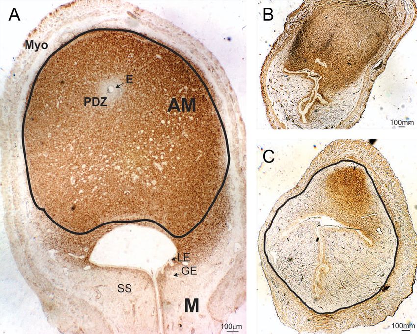
404     P I Ingaramo and others

(Fonseca et al. 2012). In mice, between gestational         Materials and methods
day (GD)5 and GD6, stromal cells situated at
                                                            Animals
the AM decidua cease proliferation and undergo
differentiation into decidual cells, forming the primary    All the procedures used in this study were approved by the
and secondary decidual zone (Tan et al. 2002). In rats,     Institutional Ethics Committee of the School of Biochemistry
the AM decidua achieves its maximum development             and Biological Sciences (Universidad Nacional del Litoral,
on GD10 (Correia-da-Silva et al. 2004).                     Santa Fe, Argentina) and performed in accordance with the
   The onset of stromal cell proliferation and              principles and procedures outlined in the Guide for the Care
differentiation is largely controlled by various cell       and Use of Laboratory Animals issued by the U.S. National
cycle regulatory molecules at different stages of the       Academy of Sciences. Rats of an inbred Wistar-derived strain
cell cycle, and an imbalance of the differentiation or      from the Department of Human Physiology (Universidad
                                                            Nacional del Litoral, Santa Fe, Argentina) were used. The
proliferation process might promote failures in uterine
                                                            animals were maintained under a controlled environment
decidualization (Das 2009, Tan et al. 2002). One of
                                                            (22 ± 1°C; lights on from 06:00 to 20:00 h) and had free
these regulatory molecules is Hoxa10. Hoxa10 is
                                                            access to pellet laboratory chow (Nutrición Animal, Santa Fe,
involved in mediating stromal cell proliferation and        Argentina) and tap water. The concentration of phytoestrogens
differentiation through the regulation of hundreds          in the diet was not evaluated. However, because food intake of
of genes, and a mutation of this gene can lead to           control and GBH-treated rats was equivalent, we assumed that
implantation and decidualization defects (Lim et al.        all animals were exposed to the same levels of phytoestrogens
1999, Lu et al. 2008). Among the key molecules              (see Kass et al. (2012) for more information regarding food
involved in the signaling pathway that regulates            composition). To minimize additional exposures to endocrine-
decidualization are progesterone (P) and progesterone       disrupting chemicals, rats were housed in stainless steel cages
receptors (PR), which act by activating a cascade of        with wood bedding, and tap water was supplied in glass bottles
several factors (Szekeres-Bartho et al. 2009, Zuo et al.    with rubber stoppers surrounded by a steel ring.
2014). PR regulate COUP-TFII (chicken ovalbumin
upstream promoter transcription factor II) through
Ptch/Smo (patched gene and smoothened gene)                 Experimental design
signaling, and then COUP-TFII (NR2F2) regulates bone        Adult female rats (90 days old) were housed with males of
morphogenetic protein 2 (BMP2), which promotes              proven fertility. The day on which sperm was found in the
uterine decidualization (Kurihara et al. 2007). COUP-       vaginal smear was designated as GD1. Pregnant rats were
TFII, also known as NR2F2, is a member of the nuclear       housed singly and, at delivery, pups were sexed according
receptor superfamily and is highly expressed in the         to the anogenital distance. To minimize the use of siblings
uterine stroma during early pregnancy (Kurihara et al.      and avoid potential litter effects, offspring of the same litter
2007). Bmp2, which belongs to the transforming growth       were distributed between different mothers. Cross-fostered
factor β (TGF-β) superfamily, regulates proliferation and   litters were adjusted to eight pups, prioritizing a maximum of
differentiation and is induced downstream of P action       eight female pups per litter when possible. When fewer than
in the mouse uterine stroma during decidualization          eight females were available, an appropriate number of males
(Li et al. 2007). Ablation of the COUP-TFII and Bmp2        were retained. Female pups from each foster mother were
genes in the uterine stroma results in decidualization      assigned to one of the following neonatal treatment groups:
failure, whereas a decrease in the uterine PR and BMP2      (1) control group (C), receiving saline solution (n = 34); and
expression would be related to increased incidence of       (2) GBH group, receiving a commercial formulation of
fetal resorptions (Kurihara et al. 2007, Wetendorf &        glyphosate dissolved in saline solution at 2 mg/kg (n = 38).
DeMayo 2012, Mestre-Citrinovitz et al. 2015).               The glyphosate formulation used was a liquid water-soluble
                                                            formulation containing 66.2% of glyphosate potassium salt,
   In the present work, we hypothesized that
                                                            as its active ingredient, coadjuvants and inert ingredients.
early postnatal exposure to a commercial GBH
                                                            Treatments were given on postnatal days (PND) 1, 3, 5 and
alters reproductive parameters in adult female rats
                                                            7 by sc injections in the nape of the neck. The rodent uterus
and promotes failure of the endocrine-regulated             is not fully developed or differentiated at birth. Uterine
decidualization process. To test this hypothesis, we        development continues during the first postnatal days and
investigated the effects of neonatal GBH exposure on        due to the high sensitivity to chemical compounds, a brief
(1) the reproductive performance by determining the         exposure to endocrine-disrupting chemicals may produce
pregnancy rate, the number of corpora lutea (CLs), and      permanent morphological or functional changes (Zama &
the number of implantation sites (IS) and resorption        Uzumcu 2010, Spencer et al. 2012). The dose of GBH was
sites (RS) on GD19; and (2) the ovarian steroid levels,     selected based on the reference dose (RfD) of glyphosate of
uterine morphology, endometrial cell proliferation,         2 mg/kg/day established by the US Environmental Protection
apoptosis and cell cycle regulators (p27 and cyclin         Agency (USA EPA 1993), which is an indicative and
G1), and the endocrine pathways that regulate uterine       representative value of levels that are usually not harmful in
decidualization (steroid receptors/COUP-TFII/Bmp2/          humans and is in the order of magnitude of the environmental
Hoxa10) at the IS on GD9.                                   levels detected in our country (Arregui et al. 2004,

Reproduction (2016) 152 403–415                                                                        www.reproduction-online.org

                                                                                    Downloaded from Bioscientifica.com at 06/17/2020 05:22:56AM
                                                                                                                                  via free access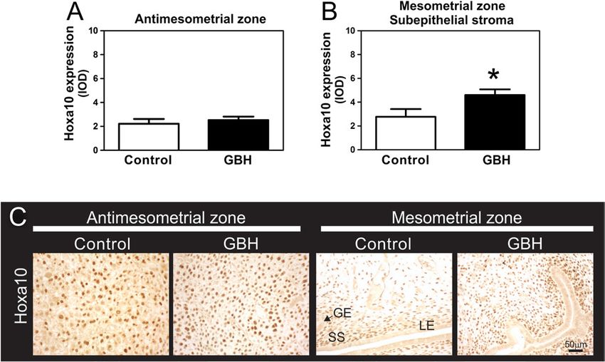
Effects of glyphosate on female reproduction            405

Peruzzo et al. 2008, Aparicio et al. 2013). Although the GBH      and visually inspected to identify RS and IS, following criteria
administered by the subcutaneous route might be metabolized       previously described in Varayoud et al. (2011). The RS were
faster if taken orally, this route is the only administration     defined as endometrial sites with an appended amorphous
route that warrants the whole incorporation of a chemical         mass without a fetus and the number of IS as the result of the
compound when an early postnatal exposure model is used.          total number of placentae with fetuses plus the total number of
The early postnatal model of exposure to endocrine disruptors     RS. The rate of pre-implantation loss was calculated as follows:
has been extensively used in our laboratory in both rodents       (number of CLs − number of IS/number of CLs) × 100, whereas
(Ramos et al. 2007, Monje et al. 2007, 2009, 2010, Varayoud       the rate of post-implantation loss was calculated as (number
et al. 2008, 2011, Bosquiazzo et al. 2010, Rodriguez et al.       of IS – number of live fetuses)/number of IS × 100 (Perobelli
2010, Milesi et al. 2015) and lambs (Rivera et al. 2011) and      et al. 2012).
has been demonstrated as a persuasive paradigm to study
short- and long-term consequences of neonatal exposure to
                                                                  Evaluation of uterine histoarchitecture, gene expression
hormonally active substances (Rivera et al. 2015). No signs of
                                                                  and steroid hormone levels on GD9
acute or chronic toxicity were observed, and no significant
differences in weight between GBH-exposed and control             Samples
pups were recorded during the experiment. No alterations
in maternal care were detected between the experimental           Control pregnant female rats (n = 8) and another group of GBH-
groups. Female pups were weaned on PND21 and held                 exposed pregnant female rats (GBH, n = 8) were killed in the
without further treatment until PND90. On PND90, female           morning of GD9 (post-implantation period), and trunk blood
rats neonatally exposed to GBH or vehicle were housed             was collected for the hormone assays. For each rat, all IS were
for two consecutive weeks with sexually mature untreated          collected, weighed and randomly distributed to be processed
males of the same strain and of proven fertility to allow         for different experimental purposes. For immunohistochemistry,
several possible matings. Every morning, vaginal smears were      the IS were fixed in 10% buffered formalin for 6 h at room
obtained to check for the presence of spermatozoa (Montes         temperature and embedded in paraffin. For RNA extraction,
& Luque 1988). The first day on which a sperm-positive            the dissected IS were immediately frozen in liquid nitrogen
smear was detected was considered GD1. Pregnant female            and stored at −80°C.
rats were assigned to reproductive performance (GD19) or
post-implantation (GD9) studies. In summary, only four            Hormone assays
female pups from any given mother were used in this study,        Serum estradiol (E2) levels and P were determined by
two in each experimental group (control and GBH) and one          radioimmunoassay after ethyl ether and hexane (Merck)
from each group at each time point (GD19 and GD9). The            extraction respectively (Varayoud et al. 2011). The antibodies
experimental design is shown in Fig. 1.                           were provided by G. D. Niswender, and labeled hormones
                                                                  were purchased from PerkinElmer Life and Analytical Sciences
Evaluation of reproductive performance                            (Boston, MA, USA). Assay sensitivities were 4 pg/mL and
                                                                  1.2 ng/mL for E2 and P respectively. The intra- and inter-assay
Control (n = 26 rats) and GBH-treated female rats (n = 30 rats)   coefficients of variation were 3.2 and 11% for E2 and 9 and
with a sperm-positive smear were housed separately, and           14.3% for P respectively.
their reproductive performance was evaluated on GD19. The
pregnancy rates were calculated as the number of pregnant
                                                                  Study of the histoarchitecture of the IS
females/number of females housed with a male × 100.
Sperm-positive females were killed on GD19. The ovaries           The IS were morphologically studied using routine histological
were dissected, and the numbers of profusely irrigated            techniques. At least three histological cross sections paraffin-
CLs were counted by direct visualization with the aid of a        embedded (5 µm thickness) at different depths of the IS
stereomicroscope (Leica). The two-horned uteri were removed       from both the control and GBH groups were stained with

                                                                                      Figure 1 Schematic representation of the
                                                                                      experimental protocol used to investigate the
                                                                                      effects of neonatal exposure to a glyphosate-
                                                                                      based herbicide (GBH) on female fertility and
                                                                                      implantation. sc, subcutaneous.

www.reproduction-online.org                                                                             Reproduction (2016) 152 403–415

                                                                                            Downloaded from Bioscientifica.com at 06/17/2020 05:22:56AM
                                                                                                                                          via free access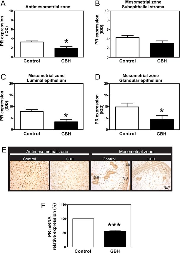
406 P I Ingaramo and others
hematoxylin–eosin (H&E) to provide an overall view of were incubated for 30 min with anti-fluorescein antibody
the tissue. Apoptotic features such as shrinkage of the cell, conjugated with POD and the reaction was developed
presence of apoptotic bodies and punch images due to rapid using diaminobenzidine (Sigma-Aldrich) as a chromogenic
phagocytosis by neighboring cells were morphologically substrate. Samples were counterstained with Mayer’s
evaluated. hematoxylin and then dehydrated and mounted. Cells
containing fragmented nuclear chromatin exhibited dark
Immunohistochemistry and TUNEL assay brown staining. For positive control, the involuting rat
prostate after the second day of castration was processed in
Immunohistochemistry was performed to evaluate protein the same way as the experimental samples; a consecutive
expression of estrogen receptor-α (ERα (ESR1)), PR (PGR), Ki67 tissue section processed without TdT was used as a negative
(Mki67), HOXA10, cyclin G1, p27 and desmin. At least three control of the TUNEL assay (Ramos et al. 2002).
cross sections paraffin-embedded (5 µm thickness) at different
depths at the middle point from each IS were immunostained.
Sections were mounted on 3-aminopropyl triethoxysilane Quantification of protein expression
(Sigma-Aldrich)-coated slides and microwave pretreatment Desmin as a decidualization marker Desmin immuno
for antigen retrieval was performed (Varayoud et al. 2011). staining was used as a marker of decidualized endometrium
The endogenous peroxidase activity and non-specific binding in cross sections of the IS on GD9 (Halperin et al. 1991). The
sites were blocked. The samples were incubated in a humid decidualized area (DA) and endometrial area (EA) at the IS
chamber first with the specific primary antibody (for 14–16 h on GD9 were measured using the software Image Pro-Plus
at 4°C) and then with the corresponding biotin-conjugated 5.0.2.9 system (Media Cybernetics, Silver Spring, MD, USA).
secondary antibody (for 30 min at room temperature) (Table 1). The images were captured with a Spot Insight version 3.5 color
The reactions were developed using a streptavidin–biotin video camera and attached to an Olympus BH2 microscope
peroxidase method and diaminobenzidine (Sigma-Aldrich) using a Dplan ×10 objective (complete structures of the IS
as a chromogenic substrate. Each immunohistochemical run were recorded in each section, and three sections per IS were
included positive controls (sections from tissues known to evaluated). The measurement system was spatially calibrated
express the proteins of interest) and negative controls (in which using a Neubauer Chamber. Results are expressed as square
the primary antibody was replaced by non-immune serum of millimeters (mm2). For each image, a manual selection was
the species used to generate the primary antibody) (Ingaramo performed as described by Brey et al. (2003). In each image,
et al. 2016). For Ki67 immunodetection, the samples were desmin-positive areas corresponding to DA and EA, delimited
counterstained with Mayer’s hematoxylin (Biopur, Rosario, by myometrium (Myo), were selected. In addition, the DA:EA
Argentina). Samples were mounted with permanent mounting ratio at the IS on GD9 was calculated.
medium (Eukitt, Sigma-Aldrich).
Apoptotic cells in sections of IS were evaluated by the
TUNEL assay using the In Situ Cell Death Detection Kit, ERα, PR, HOXA10, cyclin G1 and p27 proteins The protein
POD (Roche), following the manufacturer’s instructions. expression of ERα, PR, Hoxa10, cyclin G1 and p27 was
Tissue sections were pretreated with microwave at 350W evaluated by image analysis, using the Image Pro-Plus 5.0.2.9
(citrate 0.01 M, pH 6). Thereafter, sections were rinsed system, as described previously (Ramos et al. 2002). Images of
in PBS, immersed in a buffer containing 3% BSA (Sigma- immunostained tissues were recorded with a Spot Insight V3.5
Aldrich) and 20% normal horse serum for 30 min to block color video camera, attached to a microscope (Olympus) and
non-specific binding sites. Then, samples were incubated converted to a gray scale. An automated standard sequence
with TUNEL reaction mixture: terminal deoxynucleotidyl operation was created to measure the integrated optical
transferase (TdT) and fluorescein (FITC)-labeled nucleotide density (IOD) as a linear combination between the average
mixture (fluorescein-dUTP) for 60 min at 37°C in a gray intensity and the relative area occupied by positive cells.
humidified chamber in the darkness. Then, the samples Because IOD is a dimensionless parameter, the results are
expressed as arbitrary units (Milesi et al. 2015).
The AM and M zones of the IS were assessed. The AM
Table 1 Antibodies used for immunohistochemistry.
zone was delimited by the myometrium and the adjacent M
Antibodies Dilution Supplier area. In the M zone, each compartment was differentially
Primary delimited and quantified: luminal and glandular epithelium
Anti-PR (clone A0098) 1/500 Dako and subepithelial stroma (a 200-µm-wide area adjacent to the
Anti-ER (clone 6F-11) 1/200 Novocastra (Newcastle epithelium, from the basement membrane toward the outer
upon Tyne, UK) layers). A representation of the different areas quantified is
Anti-Hoxa10 (sc-17159) 1/50 Santa Cruz Biotechnology
Anti-Ki67 (clone MIB-5) 1/25 Dako shown in Fig. 3A. The protein expressions at the AM or M
Anti-desmin (clone DE-R-11) 1/6400 Novocastra (Newcastle zones were quantified on at least ten fields per section and
upon Tyne, UK) two sections per rat (separated 50 μm from each other).
Anti-cyclin G1 (sc-7865) 1/600 Santa Cruz Biotechnology
Anti-p27 (sc-528) 1/1200 Santa Cruz Biotechnology
Secondary Ki67 as a proliferation marker Ki67 expression was
Anti-mouse 1/100 Sigma evaluated in counterstained tissue sections. In the glandular
Anti-rabbit 1/200 Sigma and luminal epithelium of the M zone, a minimum of 2000
Anti-goat 1/200 Sigma
nuclei per compartment in each uterine section were counted
Reproduction (2016) 152 403–415 www.reproduction-online.org
Downloaded from Bioscientifica.com at 06/17/2020 05:22:56AM
via free accessEffects of glyphosate on female reproduction 407
and the results are expressed as the percentage of Ki67- transcriptase (300 units; Promega) using 200 pmol of random
positive nuclei. Expression of Ki67 in the subepithelial stroma primers (Promega). Twenty units of ribonuclease inhibitor
and AM zone was evaluated by a point counting procedure, as (RNAout; Invitrogen) and 100 nmol of a deoxynucleotide
described previously (Ramos et al. 2002). To record the data, triphosphate (dNTP) mixture were added to each reaction tube
a glass disk with a square grid was inserted into a focusing at a final volume of 30 µL of 1 × reverse transcriptase buffer.
eyepiece of the microscope and the volume fractions (Vv) of Reverse transcription was performed at 37°C for 90 min and at
the Ki67 (+) cells were calculated by applying the method 42°C for 15 min. Reactions were stopped by heating at 80°C
described by Weibel (1969). for 5 min and cooling on ice.
Reverse transcription and real-time quantitative Real-time quantitative PCR Each reverse-transcribed
PCR analysis product was diluted with ribonuclease-free water to a final
volume of 60 µL and further amplified using the Real-Time
RNA extraction and reverse transcription Total RNA was Rotor-Gene Q (Qiagen; Tecnolab; Buenos Aires, Argentina).
individually extracted from the IS using TRIZOL reagent (Life L19 was used as a housekeeping gene. The primer sequences
Technologies) according to the manufacturer’s instructions. used are described in Table 2. For cDNA amplification, 5 µL
The concentration and purity of total RNA was determined by cDNA were combined with HOT FIREPol EvaGreen qPCR Mix
measuring the optical density at 260 and 280 nm. All samples Plus (Solis BioDyne; Biocientífica, Rosario, Argentina) and
were precipitated with ethanol and then dissolved in distilled 10 pmol of each primer (Invitrogen) in a final volume of 20 µL.
water, and their quality was verified by gel electrophoresis. After initial denaturation at 95°C for 15 min, the reaction
Equal quantities (4 µg) of total RNA were reverse transcribed mixture was subjected to successive cycles of denaturation at
into cDNA with Moloney Murine Leukemia Virus reverse 95°C for 15 s, annealing at 52°C (Bmp2), 57°C (COUP-TFII)
or 60°C (ERα, PR and L19) for 7, 4 and 15 s and extension at
72°C for 20 s. Product purity was confirmed by dissociation
curves, and random samples were subjected to agarose gel
electrophoresis. Controls containing no template DNA were
included in all assays, yielding no consistent amplification.
Relative gene expression data were calculated using the
comparative cycle threshold (CT) method (Higuchi et al. 1993).
Figure 3 Low-magnification photomicrographs of representative IS of
pregnant rats on GD9 immunostained for desmin. (A) Control IS
showing different decidualized zones. The antimesometrial (AM) zone
Figure 2 Reproductive performance in female rats neonatally exposed is marked with a continuous line. In the AM zone, it is possible to
to GBH. (A) Percentage of pregnant females vs the total number of observe the primary decidual zone (PDZ) adjacent to the embryo (E).
females that were housed with a fertile male. (B) Number of corpora Surrounding the AM zone, there is a thin layer of smooth muscle
lutea (CLs) and (C) implantation sites (IS) evaluated on GD19 (each (Myo). The mesometrial zone (M) is adjacent to the AM zone. In the
column represents the mean ± s.e.m.). (D) Number of fetuses per dam mesometrial (M) zone, it is possible to observe the luminal epithelium
evaluated on GD19. (E) Percentage of pre-implantation and (LE), the subepithelial stroma (SS) and the glandular epithelium (GE).
(F) post-implantation loss (results were calculated as described in In the right panel, it is possible to observe two representative IS: (B)
‘Materials and methods’ section). (G) The numbers of resorption sites from a control and (C) from a GBH-treated rat. The endometrial area
on GD19 in each pregnant rat were plotted; the horizontal lines are is indicated with a continuous line. Note the significant reduction of
the mean of each experimental group. *P < 0.05 vs the control group. the desmin-positive area in the GBH-treated group.
www.reproduction-online.org Reproduction (2016) 152 403–415
Downloaded from Bioscientifica.com at 06/17/2020 05:22:56AM
via free access408 P I Ingaramo and others
Table 2 Primers and PCR products for gene expression analysis by real-time QPCR.
Gene Primer sequence Product size (bp) GenBank accession number
Bmp2 Forward: TCCATCACGAAGAAGCCATC 95 NM_017178.1
Reverse: CTCATCAGTAGGGACAGAACTTAAA
COUP-TFII Forward: CCAAGAGCAAGTGGAGAAGC 116 NM_080778.2
Reverse: CGTGGGCTACATCAGACAGA
Progesterone receptor (PR) Forward: GACCAGTCTCAACCAACTAGGC 137 NM_022847.1
Reverse: ACACCATCAGGCTCATCCAG
Estrogen receptor (ERα) Forward: ACTACCTGGAGAACGAGCCC 153 NM_012689
Reverse: CCTTGGCAGACTCCATGATC
Ribosomal protein L19 (L19) Forward: AGCCTGTGACTGTCCATTCC 99 NM_031103.1
Reverse: TGGCAGTACCCTTCCTCTTC
For each sample, CT was calculated as the difference in CT G respectively). As the neonatal exposure to GBH did
between target mRNA and L19 mRNA. The CT for each not affect the number of IS but increased the number of
sample was calculated using Rotor-Gene Q-Pure Detection RS, we decided to study uterine morphological changes,
software (Version 1.7, Qiagen; Tecnolab). Accordingly, the ovarian steroid levels and uterine markers of endometrial
fold expression over control values was calculated for each proliferation and differentiation on GD9. Because during
target by relative standard curve methods, which are designed the post-implantation period the uterine proliferation
to analyze data from real-time PCR (Cikos et al. 2007). and differentiation processes are regulated through
For all experimental samples, the relative target quantity was PR action, we focused our assessment in elucidating
determined from the standard curve, normalized to the relative
whether defective uterine decidualization is associated
quantity of the reference gene and finally divided by the
with a failure in key molecules regulated by P.
normalized target value of the control sample. No significant
differences in CT values were observed for the ribosomal
protein L19 between the different experimental groups. Ovarian and uterine markers on GD9
Ovarian steroid levels
Statistical analysis
Pregnant rats were evaluated on GD9 to test ovarian
Data are expressed as the mean ± s.e.m. All statistical
steroidogenesis. No differences were observed in the
determinations were performed using GraphPad Prism version
serum levels of E2 and P between the experimental
5.03 for Windows (GraphPad Software). The pregnancy rates
were analyzed by the Fisher’s exact test. The number of RS
groups (E2 levels: control 96.4 ± 17.1 pg/mL vs GBH
was analyzed using a generalized linear model with a negative 69.7 ± 17.9 pg/mL; P levels: control 40.8 ± 3.0 ng/mL vs
binomial response, using the glm.nb function of the statistical GBH 49.3 ± 4.5 ng/mL).
software R (The R Foundation for Statistical Computing). For
the other variables, the Mann–Whitney U test was applied. Morphological and morphometric analysis of the IS
Differences were considered significant at a P < 0.05. and decidual cells
At first sight, no macroscopic differences were observed
Results between the IS from the control and GBH-exposed
groups. On GD9, all IS appeared to be healthy and
Reproductive performance on GD19 no signs of abnormality were observed. However, the
Neonatal exposure to GBH did not affect the pregnancy weights of the IS from the GBH-exposed group were
rates and, on GD19, all rats were pregnant (Fig. 2A). significantly lower than those from controls (Table 3).
Besides, exposure to GBH did not modify the number To further investigate the IS at this early developmental
of CLs in pregnant females (CLs/rat, 12–14) (Fig. 2B), the stage, the immunohistochemical expression of desmin
number of IS (Fig. 2C) or the number of fetuses per dam was used as a marker of decidualized endometrium.
(Fig. 2D). However, the GBH group evidenced a higher A representative IS (two IS were evaluated/rat) from a
number of RS among the IS than the control group. The control rat, showing the DA divided in AM and M zones,
RS were seen as yellow small scars in areas of the IS. It is shown in Fig. 3A. In the middle of the AM decidua,
was not possible to establish when the embryo stopped
growing, but according to the size of the RS, the event Table 3 Measurements at the implantation sites (IS).
could have taken place after GD9. The percentage Experimental groups
of pre-implantation loss (i.e., number of oocytes not Determinations at the IS Control GBH-treated
fertilized or embryo loss before implantation) in the GBH Weight (mg) 63.33 ± 2.32 53.37 ± 3.52†
group was increased but without reaching statistical Decidualized area, DA (mm2) 1.55 ± 0.32 0.71 ± 0.18†
significance (Fig. 2E), whereas the percentage of post- Endometrial area, EA (mm2) 3.01 ± 0.28 3.12 ± 0.19
DA/EA 0.52 ± 0.10 0.22 ± 0.06†
implantation loss as well as the number of rats with more
than one RS were significantly increased (Fig. 2F and †
P < 0.05 vs control.
Reproduction (2016) 152 403–415 www.reproduction-online.org
Downloaded from Bioscientifica.com at 06/17/2020 05:22:56AM
via free accessEffects of glyphosate on female reproduction 409
the embryo zone (E) and a primary decidual zone (PDZ) compartment of the IS. No differences were observed
adjacent to the embryo are observed. Surrounding the in the expression level of ERα in the AM zone (Fig. 4A).
AM zone, there is a thin layer of smooth muscle (Myo). In the M zone, we found a significant decrease in ERα
Adjacent to the AM zone, the M zone exhibits luminal (LE) expression in the glandular epithelium of the GBH group,
and glandular epithelium (GE) and subepithelial stroma but no changes in the remaining compartments (Fig. 4B,
(SS). The AM zone of both control and GBH-treated rats C and D). In addition, no differences were observed in
showed high expression of desmin in the DA (Fig. 3). We the expression of ERα mRNA between experimental
found no significant differences in the endometrial area groups (Fig. 4E).
(EA) between GBH-treated and control rats (Table 3).
Nevertheless, a significant decrease in the DA and in PR/COUP-TFII/BMP2 To study whether the P
the DA:EA ratio was found at the IS of the GBH-treated signaling pathway in the uteri during decidualization
group on GD9 (Table 3). Moreover, a marked decrease was affected by neonatal exposure to GBH, we
in the desmin-positive area was observed at the IS from measured PR protein expression and mRNA at the IS
the GBH group (Fig. 3B). This feature may be a hallmark on GD9. PR protein expression was decreased in the
of GBH action, which is associated with the reduction GBH-treated group vs the control group, both in the
of the DA.
Markers associated with endometrial cell
decidualization
ERα Immunohistochemistry was used to assess the
protein expression of several markers in each uterine
Figure 5 Effects of neonatal GBH exposure on PR protein and mRNA
expression at the IS of pregnant rats on GD9. (A, B, C and D)
Figure 4 Effects of neonatal GBH exposure on ERα protein and Quantification of PR immunostaining in the AM and M zones is
mRNA expression at the IS of pregnant rats on GD9. (A, B, C and D) expressed as integrated optical density (IOD). Note that the
Quantification of ERα immunostaining in the M and AM zones is expression of PR in both zones was affected in GBH-treated rats.
expressed as integrated optical density (IOD). Note that ERα (E) Representative photomicrographs of PR expression on sections of
expression was lower in the glandular epithelium of the GBH-treated the IS. (F) PR relative mRNA levels of pregnant rats at the IS on GD9
rats. (E) ERα relative mRNA levels of pregnant rats at the IS on GD9 were measured via real-time quantitative RT-PCR. Control values
were measured via real-time quantitative RT-PCR. Control values were assigned to a reference level of 100 and values of the GBH
were assigned to a reference level of 100 and values of the GBH group are given as mean ± s.e.m. The ribosomal protein L19 was used
group are given as mean ± s.e.m. The ribosomal protein L19 was used as an internal control. Each column represents the mean ± s.e.m. (n = 8
as an internal control. Each column represents the mean ± s.e.m. (n = 8 per group). *P < 0.05; ***P < 0.005. GE, glandular epithelium; LE,
rats per group). *P < 0.05 vs the control group. luminal epithelium; SS, subepithelial stroma. Scale bar, 50 µm.
www.reproduction-online.org Reproduction (2016) 152 403–415
Downloaded from Bioscientifica.com at 06/17/2020 05:22:56AM
via free access410 P I Ingaramo and others
AM and M zones (in the LE and GE) (Fig. 5A, C and D).
No changes in PR protein expression were observed
in the subepithelial stroma of the M zone (Fig. 5B).
Representative photomicrographs of PR expression
in all uterine compartments are shown (Fig. 5E). PR
mRNA levels at the IS on GD9 were measured to
further investigate whether the reduction in PR protein
expression was extended to mRNA transcription. The
mRNA levels of PR at the IS of rats neonatally exposed
to GBH was significantly lower (Fig. 5F).
Subsequently, the PR/COUP-TFII/Bmp2 signaling
pathway was evaluated in depth to better understand the
increased number of RS found in the GBH-treated group.
Therefore, we performed additional measurements of Figure 7 Effects of neonatal GBH exposure on Hoxa10 protein
mRNA expression of key molecules participating in the expression at the IS of pregnant rats on GD9. (A and B)
PR pathway. Interestingly, we found that the GBH group Quantification of Hoxa10 immunostaining in the AM and M zones is
expressed as the IOD. Hoxa10 immunostaining is increased in the
showed lower mRNA levels of COUP-TFII and Bmp2 at
subepithelial stroma of GBH-exposed rats. (C) Representative
the IS on GD9 (Fig. 6A and B). photomicrographs of Hoxa10 expression on sections of the IS. Each
column represents the mean ± s.e.m. (n = 8 per group). *P < 0.05 vs the
Hoxa10 No changes were observed in the expression control group. GE, glandular epithelium; LE, luminal epithelium; SS,
of Hoxa10 in the AM zone between both experimental subepithelial stroma. Scale bar, 50 µm.
groups (Fig. 7A); however, the expression of Hoxa10
was significantly increased in the subepithelial stroma cells was detected by the TUNEL in situ assay of IS and
of the M zone (Fig. 7B). Hoxa10 immunohistochemical no differences between experimental groups were found
expression was negative in the epithelium. Representative (Fig. 8K). Representative photomicrographs of Ki67,
photomicrographs of Hoxa10 protein expression in all cyclin G1 and p27 protein expression and the TUNEL
uterine compartments are shown in Fig. 7C. in situ assay are shown in Fig. 8J and K.
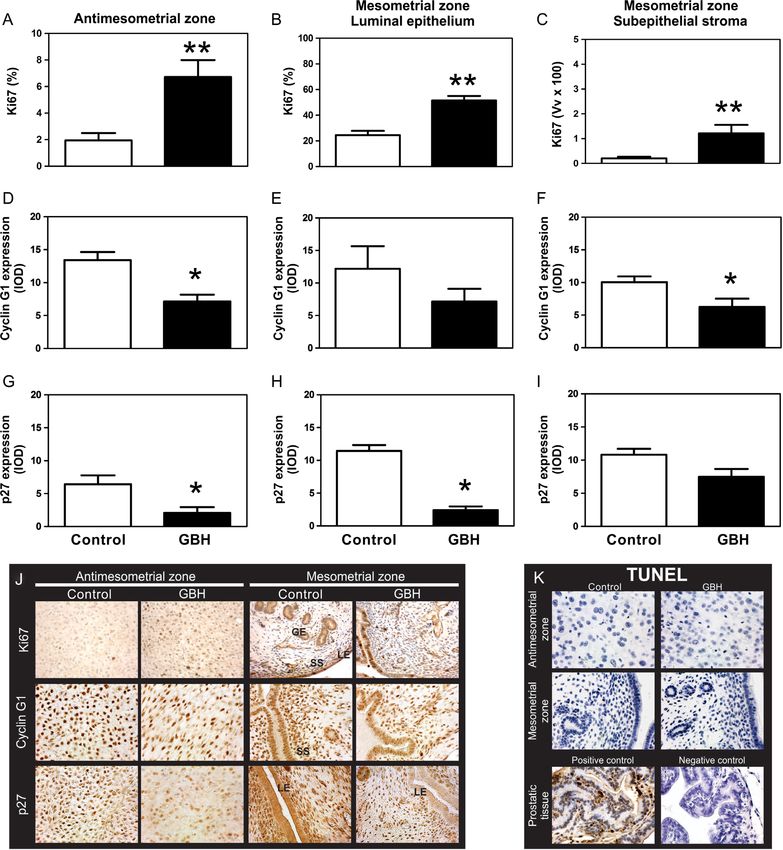
Endometrial cell proliferation, apoptosis and cell Discussion
cycle regulators
In this work, we assessed the effects of the neonatal
To better understand the increase in the number of RS treatment of GBH on the aspects of the female rat
and the decrease in the DA following GBH treatment, reproduction. Reproductive parameters were analyzed
we assessed the expression of the proliferation marker on GD19 by counting the number of resorptions vs
Ki67 at the IS on GD9. A remarkable increase in Ki67 that of live fetuses in the uterine horn. The number
expression levels in all compartments was observed of RS in the group neonatally treated with GBH was
(Fig. 8A, B and C). Ki67 was negative in the glandular significantly increased, thus suggesting an adverse effect
epithelium of both experimental groups. of the herbicide on embryo development. On the other
In the AM zone, the expression of cyclin G1 and hand, exposure to GBH did not affect the ovulation
p27 was significantly decreased (Fig. 8D, E, F and G), rate (evaluated by the number of CLs) or the ovarian
whereas in the M zone, we found a significant decrease steroidogenesis in accordance with the serum levels of
in cyclin G1 in the SS and in p27 in the LE of the GBH E2 and P. As the herbicide did not affect the number of
group (Fig. 8F, G and H). A low percentage of apoptotic IS, we suggest that miscarriage occurs after implantation
and that the adverse effects of GBH may be directed to
the uterus.
Few studies have tested the effects of GBH on the
female reproductive tract. Thus, the present results
using an animal model are interesting because they
allow suggesting a link between the reproductive
failures observed in women living in rural zones and
the massive use of herbicides (Arbuckle et al. 2001,
Figure 6 Effects of neonatal GBH exposure on the mRNA levels of Greenlee et al. 2003, Kumar 2011). Despite the
COUP-TFII and Bmp2 at the IS of pregnant rats on GD9, quantified well-established safety of glyphosate for humans by
by real-time RT-PCR. Control values were assigned to a reference
level of 100 and values of the GBH group are given as mean ± s.e.m.
regulatory agencies (US EPA 1993, WHO 1994), the
The ribosomal protein L19 was used as an internal control. Each International Agency for Research on Cancer (IARC
column represents the mean ± s.e.m. (n = 8 per group). **P < 0.01 and 2015), based on studies that are available in the
***P < 0.005 vs the control group. scientific literature, has recently classified this herbicide
Reproduction (2016) 152 403–415 www.reproduction-online.org
Downloaded from Bioscientifica.com at 06/17/2020 05:22:56AM
via free accessEffects of glyphosate on female reproduction 411
Figure 8 Effects of neonatal GBH exposure on Ki67, cyclin G1 and p27 protein expression at the IS of pregnant rats on GD9. (A, B and C)
Quantification of Ki67, (D, E and F) cyclin G1 and (G, H and I) p27 immunostaining in the AM and M zones is expressed as the IOD. Ki67
showed that there is an increased proliferation in both zones of the IS in GBH-treated rats. Cyclin G1 and p27 showed decreased expression in
the AM zone of GBH-treated rats as well as decreased expression in the SS and LE of the M zone respectively. (J) Representative
photomicrographs of Ki67, cyclin G1 and p27 expression on sections of the IS. Each column represents the mean ± s.e.m. (n = 8 per group).
*P < 0.05 and **P < 0.01 vs the control group. (K) Representative photomicrographs of the TUNEL assay. No apoptotic cell was observed in the IS
of pregnant rats on GD9 of either the GBH or control groups. Apoptosis of prostatic cells with cytoplasmic and nuclear condensation and
nuclear fragmentation are observed in the positive control, while no staining was detected in a consecutive tissue section when TdT was avoided
(negative control of TUNEL assay). GE, glandular epithelium; LE, luminal epithelium; SS, subepithelial stroma. Scale bar, 50 µm.
www.reproduction-online.org Reproduction (2016) 152 403–415
Downloaded from Bioscientifica.com at 06/17/2020 05:22:56AM
via free access412 P I Ingaramo and others
as probably carcinogenic to humans (Group 2A). It has development (Lee et al. 2007). The intraluminal
also been suggested that chronic, low-level exposure injection of recombinant Bmp2 in one horn of the uterus
may lead to developmental and reproductive health in the ablated mouse can partially rescue the decidual
problems, particularly for men and women residing response, suggesting that the observed phenotype is
in agricultural areas associated with heavy herbicide due to the developmental ablation of Bmp2 (Lee et al.
use (Williams et al. 2012). In a study performed in an 2007). In this study, Bmp2 mRNA was clearly decreased
Ontario Farm Population, women provided information at the IS of the GBH group on GD9. Thus, in GBH-
on spontaneous abortions and it was demonstrated that treated rats, P signaling failed due to the altered PR/
preconception exposure to glyphosate increased the Bmp2 pathway.
risk of abortions (Arbuckle et al. 2001). COUP-TFII makes a link between PR and Bmp2.
In several species, it has been recognized that the Bmp2 expression in COUP-TFII mutants is greatly
synchrony between the needs of developing embryos reduced (Kurihara et al. 2007). This molecule is
and the secretions of the uterus are critical for regulated through PR in the decidual tissue and
implantation and to maintain a successful pregnancy deficiency of COUP-TFII produces failures in the
(Pope 1988, Bourdiec & Akoum 2014). Following decidualization process and early embryonic lethality
embryo implantation, one of the critical steps to establish due to cardiovascular defects (Takamoto et al. 2005,
pregnancy in rodents and humans is the optimal Kurihara et al. 2007). During the peri-implantation
decidualization of endometrial stromal cells (ESCs) period, COUP-TFII regulates embryo attachment and
(Kusama et al. 2014). This step is intricately regulated decidualization through controlling ERα activity. In
by E2 and P, which activate the transcription of target the post-implantation period, COUP-TFII expression is
genes through the binding of their cognate receptors still required to facilitate placentation (Lee et al. 2010).
(Wetendorf & DeMayo 2012). Here, we detected no The decidualization process is under the control of P
alterations in ovarian steroid hormone levels on GD9 and E2 in the presence of blastocysts or deciduogenic
or in the number of CLs on GD19 in GBH-exposed stimuli. Pawar et al. (2015) showed that if ERα is
animals. Therefore, our results suggest that the uterus conditionally deleted from both the epithelial and
rather than the ovary is the principal organ involved in stromal compartments of the uterus, using PR-Cre, the
the reproductive failure in GBH-exposed rats. resulting ERαd/d mice display complete loss of decidual
In this work, we described morphological differences response. In the present work, in the post-implantation
between the experimental groups at the IS on GD9. period (GD9), we detected a decrease in ERα protein
Effects similar to those described here have been levels in the uterine glands from the GBH group and
observed by other authors when uterine PR and Bmp2 no changes in the rest of the uterine compartments.
signaling was altered (Lee et al. 2007, Mestre-Citrinovitz Although some authors have suggested that estrogenic
et al. 2015). A decrease in the decidual zone at the IS of influence via ERα is minimal for the induction of
GBH-treated rats on GD6 and GD7 has been reported decidualization in pregnant mice on GD8 (Paria et al.
in rats treated with onapristone (PR antagonist) (Mestre- 1999, Tan et al. 1999), others have shown a strong
Citrinovitz et al. 2015). Moreover, Lee et al. (2007) positive ERα expression in the uterine glands and a
demonstrated that, after a decidualization stimulus, mice more intense expression for ERα mRNA at the M pole
with conditional ablation of Bmp2 failed to increase (Tan et al. 1999). Additionally, in mice in which ERα
horn size. is ablated in uterine luminal and glandular epithelia
An adequate paracrine signaling is required for but retained in the stroma, the decidualization defect
proper differentiation of the embryo surrounding the could be due to the lack of secretion of an ERα-
stroma within the uterus. Paracrine signaling is essential regulated paracrine factor from the glands (Pawar et al.
in the support and development of the implanted 2015). Although we did not demonstrate a specific role
embryo in decidualization (Wetendorf & DeMayo of ERα in decidualization on GD9, we might suggest
2012). As PR signaling is critical for a successful that the abnormal glandular ERα expression on GD9
pregnancy (Wetendorf & DeMayo 2012), the decreased in the GBH group could contribute to the failure in
PR expression in most of the uterine compartments decidualization. Moreover, studies have indicated that
of the GBH group on GD9 suggests that the failure uterine ERα-PR signaling is vulnerable to exposure to
in maintaining the pregnancy might be explained by environmental endocrine-disrupting chemicals with
defects in the uterine PR signaling pathway. either estrogenic or antiestrogenic activity (Li et al.
Knockout mice have been pivotal in demonstrating 2016). Accordingly, the defective decidualization
that members of the Bmp family are critical within observed in the GBH group may be explained by a
early pregnancy (Wetendorf & DeMayo 2012). In mice dysregulation of ERα-PR signaling.
with conditional ablation of Bmp2 in uterine cells but Besides the changes described in the PR pathway,
which normally express uterine PR, embryo attachment we evaluated the proliferation of decidual tissue. In
is normal, but the uterine stroma is unable to undergo the uterus, Hoxa10 is regulated in response to E2 and
the decidual reaction to support further embryonic P and promotes proliferation of human ESCs through
Reproduction (2016) 152 403–415 www.reproduction-online.org
Downloaded from Bioscientifica.com at 06/17/2020 05:22:56AM
via free accessEffects of glyphosate on female reproduction 413
the regulation of hundreds of genes (Lu et al. 2008). Declaration of interest
For stromal cell differentiation, the different regulatory
The authors declare that there is no conflict of interest that
mechanisms of the cell cycle should be appropriately could be perceived as prejudicing the impartiality of the
balanced; PR and Hoxa10 are key molecules that research reported.
participate in proliferation and differentiation during
decidualization (Das 2009). Increased Hoxa10
expression in the stroma and M region and increased
Funding
proliferation (by Ki67 expression) in the uteri of the
GBH group allow us to assume that the cell cycle is This work was supported by Universidad Nacional del Litoral
dysregulated. During stromal cell decidualization, (Santa Fe, Argentina) (CAI+D program), Argentine Council for
HOXA10 is downregulated and ESCs exit the cell Scientific and Technological Research (CONICET), and the
cycle and enter differentiation (Qian et al. 2005). Argentine Agency for the Promotion of Science and Technology
Accordingly, cyclin G1 and p27 downregulation (ANPCyT).
may impair the exit of ESCs from the cell cycle,
allowing these cells to remain proliferating. Hoxa10
is a key player in the regulation of cyclin Gs in the Acknowledgements
uterus. Expression of cyclin G1, a negative regulator P I I, M M M, J V and E H L are career investigators and M G M
of the cellular cycle, is also controlled by P via its is a fellow of the CONICET.
nuclear receptor. Cyclin G1 is primarily associated
with stromal cell proliferation and differentiation
during decidualization (Yue et al. 2005). On the other References
hand, the expression of p27 in the peri-implantation
Aparicio VC, De Geronimo E, Marino D, Primost J, Carriquiriborde P
uterus has been closely associated with the onset of & Costa JL 2013 Environmental fate of glyphosate and amino
decidualization (Tan et al. 2002). Thus, the increased methylphosphonic acid in surface waters and soil of agricultural basins.
expression of Hoxa10, together with a dysregulated Chemosphere 93 1866–1873. (doi:10.1016/j.chemosphere.2013.06.041)
expression of PR and a decrease in cyclin G1 and p27 Arbuckle TE, Lin Z & Mery LS 2001 An exploratory analysis of the effect
of pesticide exposure on the risk of spontaneous abortion in an Ontario
activity in the GBH groups, could be the molecular farm population. Environmental Health Perspectives 109 851–857.
mechanism to explain the proliferation/differentiation (doi:10.1289/ehp.01109851)
balance. It has been described that P appears to Arregui MC, Lenardon A, Sanchez D, Maitre MI, Scotta R & Enrique S
2004 Monitoring glyphosate residues in transgenic glyphosate-resistant
regulate apoptosis of stromal cells by modulating Bax soybean. Pest Management Science 60 163–166. (doi:10.1002/
and Bcl2 expression (Dai et al. 2000, Joswig et al. (ISSN)1526-4998)
2003). Although the P pathway was altered in GBH- Astiz M, de Alaniz MJ & Marra CA 2012 The oxidative damage and
exposed rats, we found no differences in apoptosis inflammation caused by pesticides are reverted by lipoic acid in rat
brain. Neurochemistry International 61 1231–1241. (doi:10.1016/j.
between control and treated rats on GD9. Therefore, neuint.2012.09.003)
the increase in the number of RS found in the GBH Benachour N, Sipahutar H, Moslemi S, Gasnier C, Travert C & Seralini GE
group could be explained by defective uterine signaling 2007 Time- and dose-dependent effects of roundup on human embryonic
of PR/COUP-TFII/Bmp2, ER and Hoxa10, which lead and placental cells. Archives of Environmental Contamination and
Toxicology 53 126–133. (doi:10.1007/s00244-006-0154-8)
to an imbalance between differentiation/proliferation Bosquiazzo VL, Varayoud J, Muñoz-de-Toro M, Luque EH & Ramos JG
and consequently, defective decidualization. 2010 Effects of neonatal exposure to bisphenol A on steroid regulation
In summary, we found a significant increase in the of vascular endothelial growth factor expression and endothelial cell
proliferation in the adult rat uterus. Biology of Reproduction 82 86–95.
number of RS on GD19 associated with an altered (doi:10.1095/biolreprod.109.078543)
decidualization response, probably due to a defective Bourdiec A & Akoum A 2014 [Embryo implantation: role of interleukin
differentiation/proliferation uterine process. In fact, 1 family members]. Médecine Sciences 30 644–650. (doi:10.1051/
on GD9, the GBH-treated rats showed an increased medsci/20143006014)
Brey EM, Lalani Z, Johnston C, Wong M, McIntire LV, Duke PJ &
expression of the proliferation marker Ki67 and Patrick CW Jr 2003 Automated selection of DAB-labeled tissue for
Hoxa10, an increased expression of cyclin G1 and p27, immunohistochemical quantification. Journal of Histochemistry and
a decreased expression of estrogen and progesterone Cytochemistry 51 575–584. (doi:10.1177/002215540305100503)
Caglar S & Kolankaya D 2008 The effect of sub-acute and sub-chronic
receptors – key regulators of decidualization – and exposure of rats to the glyphosate-based herbicide Roundup.
a downregulation of COUP-TFII and Bmp2 mRNA Environmental Toxicology and Pharmacology 25 57–62. (doi:10.1016/j.
at the IS. etap.2007.08.011)
Pregnancy loss may occur for many reasons, not Cikos S, Bukovska A & Koppel J 2007 Relative quantification of mRNA:
comparison of methods currently used for real-time PCR data analysis.
all of which can be identified. Some of these causes BMC Molecular Biology 8 113. (doi:10.1186/1471-2199-8-113)
include genetic, uterine or hormonal abnormalities; Clair E, Mesnage R, Travert C & Seralini GE 2012 A glyphosate-based
reproductive tract infections; and tissue rejection. Based herbicide induces necrosis and apoptosis in mature rat testicular cells
on the present results, we suggest that exposure to low in vitro, and testosterone decrease at lower levels. Toxicology in Vitro 26
269–279. (doi:10.1016/j.tiv.2011.12.009)
levels of GBH may also be associated with pregnancy Correia-da-Silva G, Bell SC, Pringle JH & Teixeira NA 2004 Patterns of
loss in humans and animals. uterine cellular proliferation and apoptosis in the implantation site
www.reproduction-online.org Reproduction (2016) 152 403–415
Downloaded from Bioscientifica.com at 06/17/2020 05:22:56AM
via free access414 P I Ingaramo and others
of the rat during pregnancy. Placenta 25 538–547. (doi:10.1016/j. pathway involving Wnt4 to regulate uterine decidualization in the mouse
placenta.2003.11.007) and the human. Journal of Biological Chemistry 282 31725–31732.
Dai D, Moulton BC & Ogle TF 2000 Regression of the decidualized (doi:10.1074/jbc.M704723200)
mesometrium and decidual cell apoptosis are associated with a shift Lim H, Ma L, Ma WG, Maas RL & Dey SK 1999 Hoxa-10 regulates uterine
in expression of Bcl2 family members. Biology of Reproduction 63 stromal cell responsiveness to progesterone during implantation and
188–195. (doi:10.1095/biolreprod63.1.188) decidualization in the mouse. Molecular Endocrinology 13 1005–1017.
Dallegrave E, Mantese FD, Coelho RS, Pereira JD, Dalsenter PR & (doi:10.1210/mend.13.6.0284)
Langeloh A 2003 The teratogenic potential of the herbicide glyphosate- Lu Z, Hardt J & Kim JJ 2008 Global analysis of genes regulated by HOXA10
Roundup in Wistar rats. Toxicology Letters 142 45–52. (doi:10.1016/ in decidualization reveals a role in cell proliferation. Molecular Human
S0378-4274(02)00483-6) Reproduction 14 357–366. (doi:10.1093/molehr/gan023)
Dallegrave E, Mantese FD, Oliveira RT, Andrade AJ, Dalsenter PR Mestre-Citrinovitz AC, Kleff V, Vallejo G, Winterhager E & Saragueta P
& Langeloh A 2007 Pre- and postnatal toxicity of the commercial 2015 A suppressive antagonism evidences progesterone and estrogen
glyphosate formulation in Wistar rats. Archives of Toxicology 81 receptor pathway interaction with concomitant regulation of Hand2,
665–673. (doi:10.1007/s00204-006-0170-5) Bmp2 and ERK during early decidualization. PLoS ONE 10 e0124756.
Das SK 2009 Cell cycle regulatory control for uterine stromal cell (doi:10.1371/journal.pone.0124756)
decidualization in implantation. Reproduction 137 889–899. Milesi MM, Alarcon R, Ramos JG, Muñoz-de-Toro M, Luque EH &
(doi:10.1530/REP-08-0539) Varayoud J 2015 Neonatal exposure to low doses of endosulfan induces
Fonseca BM, Correia-da-Silva G & Teixeira NA 2012 The rat as an implantation failure and disrupts uterine functional differentiation at the
animal model for fetoplacental development: a reappraisal of the post- pre-implantation period in rats. Molecular and Cellular Endocrinology
implantation period. Reproductive Biology 12 97–118. (doi:10.1016/ 401 248–259. (doi:10.1016/j.mce.2014.11.028)
S1642-431X(12)60080-1) Monje L, Varayoud J, Luque EH & Ramos JG 2007 Neonatal exposure to
Gasnier C, Dumont C, Benachour N, Clair E, Chagnon MC & Seralini GE bisphenol A modifies the abundance of estrogen receptor alpha transcripts
2009 Glyphosate-based herbicides are toxic and endocrine disruptors with alternative 5′-untranslated regions in the female rat preoptic area.
in human cell lines. Toxicology 262 184–191. (doi:10.1016/j. Journal of Endocrinology 194 201–212. (doi:10.1677/JOE-07-0014)
tox.2009.06.006) Monje L, Varayoud J, Muñoz-de-Toro M, Luque EH & Ramos JG 2009
Greenlee AR, Arbuckle TE & Chyou PH 2003 Risk factors for female Neonatal exposure to bisphenol A alters estrogen-dependent mechanisms
infertility in an agricultural region. Epidemiology 14 429–436. governing sexual behavior in the adult female rat. Reproductive
(doi:10.1097/01.ede.0000071407.15670.aa) Toxicology 28 435–442. (doi:10.1016/j.reprotox.2009.06.012)
Halperin R, Fleminger G, Kraicer PF & Hadas E 1991 Desmin as an Monje L, Varayoud J, Muñoz-de-Toro M, Luque EH & Ramos JG 2010
immunochemical marker of human decidual cells and its expression in Exposure of neonatal female rats to bisphenol A disrupts hypothalamic
menstrual fluid. Human Reproduction 6 186–189. LHRH pre-mRNA processing and estrogen receptor alpha expression
Higuchi R, Fockler C, Dollinger G & Watson R 1993 Kinetic PCR analysis: in nuclei controlling estrous cyclicity. Reproductive Toxicology 30
real-time monitoring of DNA amplification reactions. Biotechnology 11 625–634. (doi:10.1016/j.reprotox.2010.08.004)
1026–1030. (doi:10.1038/nbt0993-1026) Montes GS & Luque EH 1988 Effects of ovarian steroids on vaginal smears
IARC Working Group 2015 Glyphosate. In Some Organophosphate in the rat. Acta Anatomica 133 192–199. (doi:10.1159/000146639)
Insecticides and Herbicides: Diazinon, Glyphosate, Malathion, Parathion, Paria BC, Tan J, Lubahn DB, Dey SK & Das SK 1999 Uterine decidual
and Tetrachlorvinphos, vol 112. IARC Monographs. Prog, 1–92. response occurs in estrogen receptor-alpha-deficient mice. Endocrinology
Ingaramo PI, Milesi MM, Schimpf MG, Ramos JG, Vigezzi L, 140 2704–2710. (doi:10.1210/en.140.6.2704)
Muñoz-de-Toro M, Luque EH & Varayoud J 2016 Endosulfan affects Pawar S, Laws MJ, Bagchi IC & Bagchi MK 2015 Uterine epithelial estrogen
uterine development and functional differentiation by disrupting receptor-alpha controls decidualization via a paracrine mechanism.
Wnt7a and beta-catenin expression in rats. Molecular and Cellular Molecular Endocrinology 29 1362–1374. (doi:10.1210/me.2015-1142)
Endocrinology 425 37–47. (doi:10.1016/j.mce.2016.02.011) Perobelli JE, Alves TR, de Toledo FC, Fernandez CD, Anselmo-Franci JA,
Joswig A, Gabriel HD, Kibschull M & Winterhager E 2003 Apoptosis in Klinefelter GR & Kempinas Wde G 2012 Impairment on sperm
uterine epithelium and decidua in response to implantation: evidence quality and fertility of adult rats after antiandrogen exposure during
for two different pathways. Reproductive Biology and Endocrinology 1 prepuberty. Reproductive Toxicology 33 308–315. (doi:10.1016/j.
44. (doi:10.1186/1477-7827-1-44) reprotox.2011.12.011)
Kass L, Altamirano GA, Bosquiazzo VL, Luque EH & Muñoz-de-Toro M Peruzzo PJ, Porta AA & Ronco AE 2008 Levels of glyphosate in surface
2012 Perinatal exposure to xenoestrogens impairs mammary gland waters, sediments and soils associated with direct sowing soybean
differentiation and modifies milk composition in Wistar rats. Reproductive cultivation in north pampasic region of Argentina. Environmental
Toxicology 33 390–400. (doi:10.1016/j.reprotox.2012.02.002) Pollution 156 61–66. (doi:10.1016/j.envpol.2008.01.015)
Kumar S 2011 Occupational, environmental and lifestyle factors associated Pope WF 1988 Uterine asynchrony: a cause of embryonic loss. Biology of
with spontaneous abortion. Reproductive Sciences 18 915–930. Reproduction 39 999–1003. (doi:10.1095/biolreprod39.5.999)
(doi:10.1177/1933719111413298) Qian K, Chen H, Wei Y, Hu J & Zhu G 2005 Differentiation of endometrial
Kurihara I, Lee DK, Petit FG, Jeong J, Lee K, Lydon JP, DeMayo FJ, stromal cells in vitro: down-regulation of suppression of the cell
Tsai MJ & Tsai SY 2007 COUP-TFII mediate progesterone regulation of cycle inhibitor p57 by HOXA10? Molecular Human Reproduction 11
uterine implantation by controlling ER activity. PLoS Genetics 3 e102. 245–251. (doi:10.1093/molehr/gah147)
(doi:10.1371/journal.pgen.0030102) Ramos JG, Varayoud J, Bosquiazzo VL, Luque EH & Muñoz-de-Toro M
Kusama K, Yoshie M, Tamura K, Daikoku T, Takarada T & Tachikawa E 2014 2002 Cellular turnover in the rat uterine cervix and its relationship to
Possible roles of the cAMP-mediators EPAC and RAP1 in decidualization estrogen and progesterone receptor dynamics. Biology of Reproduction
of rat uterus. Reproduction 147 897–906. (doi:10.1530/REP-13-0654) 67 735–742. (doi:10.1095/biolreprod.101.002402)
Lee DK, Kurihara I, Jeong JW, Lydon JP, DeMayo FJ, Tsai MJ & Tsai SY 2010 Ramos JG, Varayoud J, Monje L, Moreno-Piovano G, Muñoz-de-Toro M &
Suppression of ERalpha activity by COUP-TFII is essential for successful Luque EH 2007 Diethylstilbestrol alters the population dynamic of neural
implantation and decidualization. Molecular Endocrinology 24 precursor cells in the neonatal male rat dentate gyrus. Brain Research
930–940. (doi:10.1210/me.2009-0531) Bulletin 71 619–627. (doi:10.1016/j.brainresbull.2006.12.004)
Lee KY, Jeong JW, Wang J, Ma L, Martin JF, Tsai SY, Lydon JP & DeMayo FJ Richard S, Moslemi S, Sipahutar H, Benachour N & Seralini GE 2005
2007 Bmp2 is critical for the murine uterine decidual response. Molecular Differential effects of glyphosate and roundup on human placental
and Cellular Biology 27 5468–5478. (doi:10.1128/MCB.00342-07) cells and aromatase. Environmental Health Perspectives 113 716–720.
Li Q, Davila J, Kannan A, Flaws JA, Bagchi MK & Bagchi IC 2016 Chronic (doi:10.1289/ehp.7728)
exposure to bisphenol A affects uterine function during early pregnancy Rivera OE, Varayoud J, Rodriguez HA, Muñoz-de-Toro M & Luque EH
in mice. Endocrinology 157 1764–1774. (doi:10.1210/en.2015-2031) 2011 Neonatal exposure to bisphenol A or diethylstilbestrol alters the
Li Q, Kannan A, Wang W, Demayo FJ, Taylor RN, Bagchi MK & Bagchi IC ovarian follicular dynamics in the lamb. Reproductive Toxicology 32
2007 Bone morphogenetic protein 2 functions via a conserved signaling 304–312. (doi:10.1016/j.reprotox.2011.06.118)
Reproduction (2016) 152 403–415 www.reproduction-online.org
Downloaded from Bioscientifica.com at 06/17/2020 05:22:56AM
via free accessYou can also read