Phosphatidylcholine Enhances Homeostasis in Peach Seedling Cell Membrane and Increases Its Salt Stress Tolerance by Phosphatidic Acid - MDPI
←
→
Page content transcription
If your browser does not render page correctly, please read the page content below
International Journal of
               Molecular Sciences

Article
Phosphatidylcholine Enhances Homeostasis in Peach Seedling
Cell Membrane and Increases Its Salt Stress Tolerance by
Phosphatidic Acid
Maoxiang Sun, Xiaolong Liu, Huaifeng Gao, Binbin Zhang, Futian Peng * and Yuansong Xiao *

                                          State Key Laboratory of Crop Biology, College of Horticulture Science and Engineering,
                                          Shandong Agricultural University, Tai’an 271018, China; maoxiangs0514@163.com (M.S.);
                                          lxl17860721016@163.com (X.L.); gaohuaifeng1992@163.com (H.G.); zhangbinbin199212@163.com (B.Z.)
                                          * Correspondence: pft@sdau.edu.cn (F.P.); ysxiao@sdau.edu.cn (Y.X.); Tel.: +86-13563821651 (F.P.);
                                            +86-15163873786 (Y.X.)

                                          Abstract: Salt stress is a major adverse abiotic factor seriously affecting fruit tree growth and develop-
                                          ment. It ultimately lowers fruit quality and reduces yield. Phosphatidylcholine (PC) is an important
                                          cell membrane component that is critical for cell structure and membrane stability maintenance. In
                                          this study, we found that the addition of external PC sources significantly increased the tolerance of
                                          one-year-old peach trees, Prunus persica (L.) Batsch., to salt stress and attenuated their damage. The
                                          effect of exogenous application of 200 mg/L PC exerted the most significant positive effect. Its use
                                          caused seedling leaf stomatal opening, contributing to normal gas exchange. Moreover, beneficial
                                          effects were exerted also to the root system, which grew normally under salt stress. Meanwhile,
                                phospholipase D activity in the cell was promoted. The production of phosphatidic acid (PA) was

                                          enhanced by increased decomposition of phospholipids; PA serves as a secondary messenger in-
Citation: Sun, M.; Liu, X.; Gao, H.;
                                          volved in plant biological process regulation and the reduction in the reactive oxygen species- and
Zhang, B.; Peng, F.; Xiao, Y.
                                          peroxide-induced damage caused by salt stress. The possible mechanism of action is via promoted
Phosphatidylcholine Enhances
                                          plant osmotic regulation and tolerance to salt stress, reducing salt stress-induced injury to plants.
Homeostasis in Peach Seedling Cell
Membrane and Increases Its Salt
Stress Tolerance by Phosphatidic
                                          Keywords: salt stress; phosphatidylcholine; cell membranes; peach; phosphatidic acid
Acid. Int. J. Mol. Sci. 2022, 23, 2585.
https://doi.org/10.3390/
ijms23052585
                                          1. Introduction
Academic Editors: Jianhua Zhang
and Biao Gong                                   More than 6% of the world’s total land area (approximately 800 million hectares) is
                                          affected by salinity [1]. Poor irrigation techniques, inappropriate fertilizer application,
Received: 27 January 2022                 and the excessive accumulation of industrial pollutants have contributed to an increase
Accepted: 23 February 2022                in soil salinity [2]. Under salt stress, plant chloroplasts are destroyed, which decreases
Published: 26 February 2022
                                          the activity of related photosynthetic enzymes, reducing plant photosynthetic rate [3].
Publisher’s Note: MDPI stays neutral      Meanwhile, the accumulation of salt ions diminishes the content of thylakoid membrane
with regard to jurisdictional claims in   glycolipids and unsaturated fatty acids, which affects the photosynthetic characteristics
published maps and institutional affil-   of the cell membrane [3]. Due to the increase in the sodium ion level, plants suffer from
iations.                                  oxidative stress and ion damage, which increases the permeability of the cell membrane and
                                          disrupts the ion balance in the plant. Protein and cell membrane structures and functions
                                          are disrupted, which hinders plant growth and development. Ultimately, a continuous
                                          high-salt environment causes plant death, resulting in considerably reduced crop yields [4].
Copyright: © 2022 by the authors.
                                                The cell membrane serves as a barrier and interface for physical–chemical and informa-
Licensee MDPI, Basel, Switzerland.
                                          tion exchange with their external environment. The stability of its structural integrity and
This article is an open access article
                                          its functions are the basis for normal cell metabolism and overall physiology [5]. Adverse
distributed under the terms and
                                          conditions, such as ion poisoning and low-temperature and drought stress, initially and
conditions of the Creative Commons
                                          directly attack the plant cell membrane, destroying its structure and reducing its fluidity.
Attribution (CC BY) license (https://
creativecommons.org/licenses/by/
                                                Meanwhile, the lipid composition and content in the cell membrane directly affects
4.0/).
                                          the cell membrane structure stability and fluidity [6,7]. Continuous adverse influence

Int. J. Mol. Sci. 2022, 23, 2585. https://doi.org/10.3390/ijms23052585                                         https://www.mdpi.com/journal/ijms
Int. J. Mol. Sci. 2022, 23, 2585                                                                                           2 of 17

                                   disrupts cell membrane integrity, fluidity, and selective permeability, causing loss of basic
                                   functions of the entire plant cell [8]. Under adverse stress conditions, plants strive to find
                                   new ways to reduce the damage caused through long-term adaptive evolution. One of the
                                   most important plant survival mechanisms is changing the content and composition of
                                   membrane lipids for adaptation to the action of adverse factors [9].
                                         Phosphatidylcholine (PC) is the most abundant phospholipid of all eukaryotic cell mem-
                                   brane components. It is present in high levels in the photosynthetic and non-photosynthetic
                                   organs of Arabidopsis thaliana [10]. In addition being a major phospholipid in the cellular
                                   membranes of most eukaryotes [11], PC is an important precursor in lipid signaling or serves
                                   as a regulatory protein ligand [12].
                                         Phospholipase D (PLD) can hydrolyze phospholipids, and its products are directly
                                   used as signal molecules involved in stress response signal transduction [13]. PLD regulates
                                   cell physiological processes by governing the spatial distribution, temporal and spatial
                                   expression, and the content of its product, phosphatidic acid (PA) [14]. Hong et al. [15]
                                   presented data on PLD and PA signaling in response to drought and salinity. PA can be
                                   directly produced by PLD hydrolysis of phospholipids. Earlier genetic and pharmacological
                                   research found that PA plays an important role in the regulation of stomatal movement,
                                   root growth, and plant tolerance to salinity and water stress [16].
                                         Reduced PC content and defective plant growth was recently reported in a dou-
                                   ble mutant of A. thaliana, phospho-baseN-methyltransferase1 (PMT1) and PMT3 [17].
                                   Additionally, Shimojima et al. established that, under exposure to environmental stress
                                   factors, plants reduce the phospholipid content of their cell membranes to maintain their
                                   integrity [18]. Plants then replace the missing phospholipids by increasing the content of
                                   glycolipids in the cell membrane. With the advances in the technologies for research on
                                   membrane lipids, increasingly more scholars have devoted to studying the relationship
                                   between the changes in cell membrane lipid content and cell membrane structure homeosta-
                                   sis. However, few reports are available on the associations between the changes in plant PC
                                   content and plant tolerance to environmental stress. Therefore, we used peach seedlings
                                   as experimental material to examine the effects of exogenous PC on the growth of the
                                   root and photosynthetic organs of peach seedlings under salt stress. We also investigated
                                   their influence on the membrane lipid content and the composition of the leaves and roots.
                                   We aimed to elucidate whether exogenous PC treatment can enhance the activity of PLD,
                                   improve the salt tolerance of plants, and reduce the damage caused by salt stress exposure
                                   to plants.

                                   2. Results
                                   2.1. Exogenous Application of PC Improved the Net Photosynthetic Rate and Chlorophyll Content
                                   of Peach Leaves
                                         Leaves are the main photosynthetic organs in higher plants. The chloroplast in the
                                   leaf consists of three parts: envelope, stroma, and thylakoid, which is the main place where
                                   photosynthesis occurs. Phospholipid is an important component of the chloroplast membrane.
                                         We found that 200 mg/L PC had a better alleviation effect on NaCl concentration
                                   of 70 mmol/L, so NaCl + 200 mg/L PC was selected as one of the treatments. The pos-
                                   sible reason is that 200 mg/L PC is the optimum concentration for peach seedlings to
                                   resist 70 mmol/L NaCl. As can be observed in Figure 1A, the salt stress treatment not
                                   only hindered the development of the peach plants, but also caused leaf yellowing and
                                   negatively affected the photosynthetic system of the plant. Our research found that the
                                   net photosynthetic rate value in the control treatment increased slightly over time. Com-
                                   pared with the control treatment, the net photosynthetic rate value of the NaCl treatment
                                   gradually decreased. The treatments of NaCl + 100 mg/L PC, NaCl + 200 mg/L PC, and
                                   NaCl + 400 mg/L PC showed that the net photosynthetic rate was always lower than the
                                   control treatment and higher than the NaCl treatment. The net photosynthetic rate in the
                                   salt treatments supplemented with 200 mg/L PC and 400 mg/L PC slowly increased from
                                   the first to the fifth day and then stabilized from the fifth to the ninth day. The treatment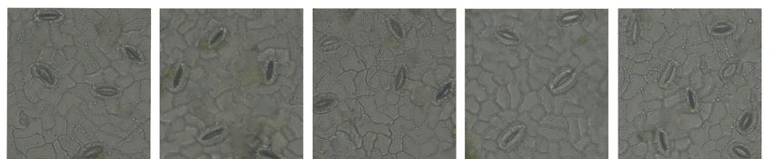
Int. J. Mol. Sci. 2022, 23, x FOR PEER REVIEW 3 of 18
mg/L PC showed that the net photosynthetic rate was always lower than the control treat‐
Int. J. Mol. Sci. 2022, 23, 2585 ment and higher than the NaCl treatment. The net photosynthetic rate in the salt treat‐ 3 of 17
ments supplemented with 200 mg/L PC and 400 mg/L PC slowly increased from the first
to the fifth day and then stabilized from the fifth to the ninth day. The treatment with
NaCl + 200+ mg/L
with NaCl PC alleviated
200 mg/L more
PC alleviated significantly
more significantlythethe
adverse
adverseinfluence
influenceofofsalt
saltstress.
stress.
Among them, the treatment with NaCl NaCl ++ 200 mg/Lmg/L PC PC alleviated
alleviated more
more significantly
significantly the
adverse
adverse influence
influence ofof salt stress. From
From thethe third
third to
to the
the ninth
ninth day,
day, the net photosynthetic
rate
rate of the NaCl + 200 mg/L mg/L PC PC treatment
treatment increased
increased by by 9.0%,
9.0%, 19.5%,
19.5%, 21.7%,
21.7%, and
and 22.9%
22.9%
compared withwiththetheNaCl
NaCltreatment
treatment (Figure
(Figure 1B).
1B). As As
cancan be seen
be seen in Figure
in Figure 1C,stress
1C, salt salt stress
had a
had a greater
greater impactimpact
on the on the chlorophyll
chlorophyll contentcontent
of peachofleaves.
peach Salt
leaves. Salt
stress stress exposure
exposure pro‐
prolongation
longation
gradually gradually
decreased decreased the chlorophyll
the chlorophyll content in content in the
the leaves. leaves. Compared
Compared with NaClwith NaCl
treatment,
adding PCadding
treatment, can significantly reduce the reduce
PC can significantly decrease theindecrease
chlorophyll content. We
in chlorophyll measured
content. We
chlorophyll
measured a, chlorophyll
chlorophyll b and carotenoids
a, chlorophyll on the ninth
b and carotenoids onday
theof salt day
ninth stress,
of and found and
salt stress, that
the treatment
found that theoftreatment
NaCl + 200 of mg/L
NaCl +PC had
200 higher
mg/L PCvalues than the
had higher otherthan
values treatments.
the otherChloro-
treat‐
phyll a,Chlorophyll
ments. chlorophyll a, b and carotenoids
chlorophyll b and ofcarotenoids
the NaCl + of 200the
mg/L
NaClPC treatment
+ 200 mg/L PC increased
treatment by
78.9%, 133.3%, and 68.1% compared with the NaCl treatment (Figure
increased by 78.9%, 133.3%, and 68.1% compared with the NaCl treatment (Figure 1D–F). 1D–F).
Figure 1. Plant photosynthetic system parameters under salt stress. (A) Peach seedlings under salt
Figure 1. Plant photosynthetic system parameters under salt stress. (A) Peach seedlings under salt
stress; (B) Net photosynthetic rate; (C) Chlorophyll content in 1–9 days; (D–F) Chlorophyll a, b, and
stress; (B) Net photosynthetic rate; (C) Chlorophyll content in 1–9 days; (D–F) Chlorophyll a, b, and
carotenoid content on the ninth day. The photos in Figure 1A were taken on the 11th day under salt
carotenoid content on the ninth day. The photos in Figure 1A were taken on the 11th day under salt
stress. The
stress. The error
error bar
bar represents
represents the
the standard
standard deviation
deviation of
of the mean (n
the mean (n == 3).
3). Different
Different lowercase
lowercase letters
letters
indicate significant
indicate significant differences
differences among
among treatments
treatments (Duncan
(Duncan test,
test, 21.7%, 22.9%; pp
Int. J. Mol. Sci. 2022, 23, x FOR PEER REVIEW 4 of 18
Int. J. Mol. Sci. 2022, 23, 2585 2.2. Effect of PC on Stomatal Density and Size 4 of 17
The stomata are pores through which plants exchange gas with the outside environment;
transpiration intensity is also controlled by adaptive changes in their structure. The opening
and
and closing
closingof ofthe
thestomata
stomataareareclosely
closelyrelated
relatedtoto
plant
plantphotosynthesis
photosynthesis since thethe
since COCO2 needed for
2 needed
photosynthesis can enter the plant organism only when the stomata
for photosynthesis can enter the plant organism only when the stomata are open. are open.
Our observationunder
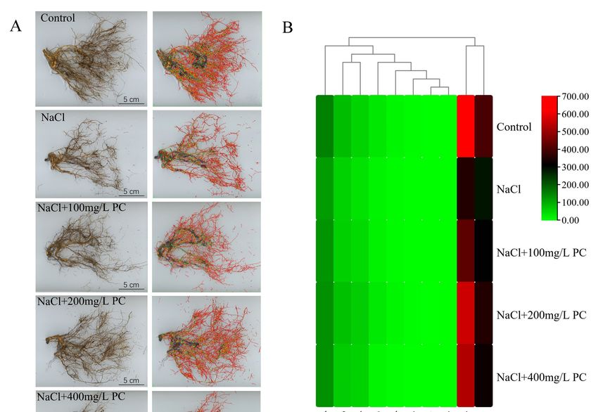
Our observation under400x
400x magnification
magnification showed
showed thatthat the stomata
the stomata of theof the peach
peach leaves
leaves in the control treatment were open and the shape of the
in the control treatment were open and the shape of the stomata guard cells was stomata guard cells was
round
round and full. The NaCl treatment resulted in guard cell closure.
and full. The NaCl treatment resulted in guard cell closure. On the contrary, the shapeOn the contrary, the
shape
of the of the stomata
stomata treatedtreated with three
with three salt stress
salt stress supplemented
supplemented withwith
PC isPC is relatively
relatively full,full,
but
but there are also stomata in the closed state (Figure 2). Of them, the stomata
there are also stomata in the closed state (Figure 2). Of them, the stomata treated with treated with
200
200 mg/L
mg/LPC PCwaswasin inthe
the open
open state,
state, indicating
indicating that
that the
the gas
gas normally
normally interacted
interacted withwith the
the
outside environment. The data of the length, width, and area of the stomata
outside environment. The data of the length, width, and area of the stomata are presented are presented
in
in Table
Table 1.1. As
As can
can be
be seen,
seen, the
the average
average area
area of
of the
the stomata
stomata in in the
the salt
salt treatments
treatments was was
smaller than thethe untreated
untreated control, whereas the area of the stomata in the the treatments
treatments with
PC
PC supplementation
supplementation was was larger.
larger. The
The largest
largest guard
guard cell
cell area
area was
was established
established in in the
the PC‐
PC-
supplemented
supplemented treatment with 200 mg/L. mg/L.Compared
Comparedwith withthethe NaCl
NaCl treatment,
treatment, the the NaCl
NaCl ++
200 mg/L
mg/LPC PCtreatment
treatmenthadhad28.3%
28.3%larger
largerguard
guardcell
cellwidth
widthand and 37.4%
37.4% greater
greater area.
area.
Figure 2.
Figure The cell
2. The cell stomatal
stomatal size
size of
of peach
peach tree
tree leaves
leaves among
among different
different treatments.
treatments. Measurements were
Measurements were
performed using a light microscope to visualize peeled impressions of peach tree leaf epidermis. Peach
tree
treeleaf
leafcell
cellstomata
stomata were sampled
were sampledand and
observed at 10:30
observed at am on a.m.
10:30 the 10th
onday
theof10th
the treatment experiment.
day of the treatment
All samples are
experiment. All measured
samples areat measured
approximately the same time
at approximately theofsame
day. time
Bars of
correspond to 30 μm. to 30 µm.
day. Bars correspond
Table 1. Stomatal
Table 1. Stomatal length,
length, width,
width, and
and area.
area.
Prunus persica (L.) Batsch. Stomatal Measurements
Prunus persica (L.) Batsch. Stomatal Measurements
Length (μm) Width (μm) Area (μm2)
Length (µm) Width (µm) 2
Control 22.8 ± 3.7 a 15.4 ± 1.5 a 329.1 (µm
Area ± 0.51) a
NaCl
Control 22.8 ±21.8
3.7 a± 2.1 b 11.3
15.4 ± 1.3
± 1.5 ad 213.5±± 0.51
329.1 0.21 ad
NaClNaCl
+ 100 mg/L PC 21.8 ±21.3
2.1 b± 2.5 b ± 1.3
11.312.8 dc
± 1.2 245.3±±0.21
213.5 0.20dc
NaClNaCl
+ 100+ mg/L PC PC
200 mg/L 21.3 ±21.7
2.5 b± 3.5 b 12.8 ± 1.2
14.5 cb
± 1.8 293.3±± 0.20
245.3 0.61 cb
NaClNaCl
+ 200+ mg/L PC PC
400 mg/L 21.7 ±21.5
3.5 b± 2.3 b ± 1.8
14.513.1 bc
± 1.6 267.9±±0.61
293.3 1.01bc
NaCl + 400 mg/L PC 21.5 ± 2.3 b 13.1 ± 1.6 c 267.9 ± 1.01 c
Note: Mean ± standard deviation (n = 3). The different letters indicate significant differences at a
Note: of
level p
Int. J. Mol. Sci. 2022, 23, x FOR PEER REVIEW                                                                                                      5 of 18

                                    the attenuation of the reduction in the number of absorbing roots. The total length, total
 Int. J. Mol. Sci. 2022, 23, 2585   surface area, total volume, and the tips and forks of the root system were 65.23%, 41.01%,
                                                                                                                          5 of 17
                                    20.36%, 66.99%, and 84.1% higher than those in the NaCl treatment, respectively.

                                     Figure 3. Structural changes in the peach root system under salt stress. (A) Photo of the peach tree
                                    Figure 3. Structural changes in the peach root system under salt stress. (A) Photo of the peach tree
                                     rootstructure,
                                    root     structure,thethefigures
                                                               figuresofofroots
                                                                           rootsininthe
                                                                                     thesecond
                                                                                          secondcolumn
                                                                                                   columnare  arewhere
                                                                                                                  wherethe
                                                                                                                         thedata
                                                                                                                              datawere
                                                                                                                                   werecollected
                                                                                                                                         collectedby bythe
                                                                                                                                                         the
                                     rootanalysis
                                    root     analysissoftware.
                                                        software.TheThecollected
                                                                         collecteddata
                                                                                     dataare
                                                                                           areroot
                                                                                               rootlength,
                                                                                                     length,surface
                                                                                                              surfacearea,
                                                                                                                      area,root
                                                                                                                            rootvolume,
                                                                                                                                  volume,tips,
                                                                                                                                           tips,and
                                                                                                                                                 andforks.
                                                                                                                                                       forks.
                                     (B)Fine
                                    (B)    Fineroot
                                                  rootdistribution
                                                        distributionheat
                                                                       heatmap.
                                                                             map.TheThevast
                                                                                         vastmajority
                                                                                               majorityofofroot
                                                                                                             rootlengths
                                                                                                                  lengthswere
                                                                                                                           weredistributed
                                                                                                                                 distributedininthe
                                                                                                                                                  therange
                                                                                                                                                      range
                                    ofof00<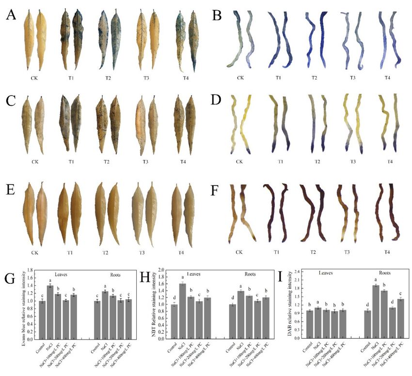
Int. J. Mol. Sci. 2022, 23, x FOR PEER REVIEW 6 of 18
Int. J. Mol. Sci. 2022, 23, 2585 control group was the best, and the structural integrity of root cells under NaCl treatment 6 of 17
was the worst. The root cells of the three treatments with PC added had higher cell integ‐
rity than those treated with NaCl. Among them, the cells treated with NaCl + 200 mg/L
PC hadintegrity.
highest the highest integrity.
It can be seen Itfrom
canFigure
be seen4Bfrom Figure
that the root4B
cellthat the rootstructure
membrane cell membrane
under
structure under NaCl treatment has been severely damaged, and
NaCl treatment has been severely damaged, and the cell fluid leaks, resulting the cell fluid in
leaks,
severere‐
sulting in severe damage to cell function. The addition of PC salt treatment
damage to cell function. The addition of PC salt treatment can protect the cell membrane can protect
the cell membrane
structure. structure.
Among them, Among them,
the addition of 200the addition
mg/L of 200
and 400 mg/Lmg/L of and 400 mg/L ofcell
PC maintained PC
maintainedintegrity
membrane cell membrane
under theintegrity
applied under the applied
salt stress salt stress conditions.
conditions.
Figure4.4. Transmission
Figure Transmissionelectron
electronmicrograph
micrograph of ofpeach
peachtreetreeroot
rootcells
cellsand
andcell
cellmembranes.
membranes. (A) (A)Root
Root
cell
cellintegrity.
integrity.Bars
Barscorrespond
correspondtoto 2 µm;
2 μm;(B)(B)
Root cellcell
Root membrane
membrane integrity. BarsBars
integrity. correspond
correspondto 500to nm.
500
The
nm.redThearrows in theinNaCl-treated
red arrows figures
the NaCl‐treated represent
figures irregular
represent cells and
irregular cellscell
andmembrane
cell membranestructures.
struc‐
tures.
The redThe red arrows
arrows in the control
in the control and +NaCl
and NaCl + 200 mg/L
200 mg/L PC‐treated
PC-treated figures
figures indicate
indicate intactintact
cells cells and
and cell
cell membrane
membrane structures.
structures. Measurements
Measurements werewere performed
performed usingusing the tips
the root rootoftips of peach
peach plants.plants. The
The data
data sampled
were were sampled at a.m.
at 11:00 11:00onamthe
on11th
the 11th day after
day after thestress
the salt salt stress treatment.
treatment.
2.5.
2.5. PC
PC and
and PA
PAContent,
Content,and
andPLD
PLDActivity
Activityininthe
theLeaves
LeavesandandRoots
Rootsunder
underSalt
SaltStress
Stress
The plant cell membrane is the first that is attacked under salt stress
The plant cell membrane is the first that is attacked under salt stress exposure. exposure. PCPC is is
a
phospholipid with the highest content in the phospholipid bilayer of the
a phospholipid with the highest content in the phospholipid bilayer of the cell membrane. cell membrane.
Therefore,
Therefore, the
the content
content of
of PC
PC in
in the
the cell
cellmembrane
membrane isis essential
essential for
for plant
plant tolerance
tolerance to to salt
salt
stress
stress(Figure
(Figure5A).
5A).
We
Wefound
foundthat
thatunder
undersalt
saltstress,
stress,the
theleaves
leavesand
androots
rootsofof peach
peach seedlings
seedlingscould
could absorb
absorb
exogenous
exogenous PC (Figure 5B), and the activity of PLD in the cell was significantly enhanced
PC (Figure 5B), and the activity of PLD in the cell was significantly enhanced
(Figure
(Figure5D).
5D).OfOfthe
theexamined
examinedtreatment,
treatment,NaCl NaCl++200 200mg/L
mg/L PCPC had
had the
the most
most pronounced
pronounced
impact on the PLD activity in the leaves and roots. PLD decomposes
impact on the PLD activity in the leaves and roots. PLD decomposes PC, producing PC, producing PA.PA.
As
can be seen in Figure 5C, the content of PA, which is a secondary messenger,
As can be seen in Figure 5C, the content of PA, which is a secondary messenger, was sig‐ was significantly
increased.
nificantly PA plays also
increased. PAan important
plays also anrole in the tolerance
important role in the oftolerance
plants to of
salt stress.to salt stress.
plants
2.6. Exogenous Application of PC Enhances Root Cell Activity
Evans blue-stained leaves and roots under salt stress are displayed in Figure 6A,B. The
application of PC increased the activity of the leaf and root cells. The relative Svensland
staining intensity visible in Figure 6G shows that the highest leaf cell activity was observed
at the PC concentration of 200 mg/L; the root cell activity was the highest at the PC
concentrations of 200 mg/L and 400 mg/L.
SOD is present in animals and plants. Its function is to scavenge superoxide anion
free radicals. NBT staining is widely used for the determination of SOD activity due to
its simple operation and high-sensitivity [19]. As illustrated in Figure 6C,D, the relative
staining intensity of the leaves and roots of the plants treated with exogenous PC was
lower than that of the only salt stress-treated plants. The relative staining intensity of
NaCl + 200 mg/L PC was the lowest, and the relative staining intensity of leaves is 31.88%
less than NaCl treatment. The relative staining intensity of roots was lower by 20.63%
compared with NaCl treatment (Figure 6H).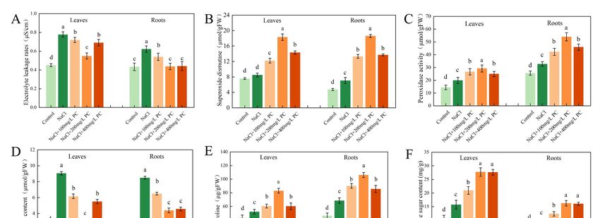
Int.
Int. J.J. Mol.
          Mol. Sci. 2022, 23, 2585
                              x FOR PEER REVIEW                                                                                         77 of
                                                                                                                                           of 17
                                                                                                                                              18

                                     Figure 5. Cell membrane structure, PC, PA content, and PLD activity. (A) Effect of salt stress on the
                                     Figure 5. Cell membrane structure, PC, PA content, and PLD activity. (A) Effect of salt stress on the
                                     phospholipid bilayer of the plant cell membrane; (B) PC content; (C) PLD activity; (D) PA content.
                                     phospholipid bilayer of the plant cell membrane; (B) PC content; (C) PLD activity; (D) PA content.
                                     The
                                     The error  bar represents
                                          error bar  representsthe
                                                                 thestandard
                                                                     standarddeviation
                                                                              deviationofof
                                                                                          thethe mean
                                                                                               mean (n =(n3).= Different
                                                                                                               3). Different lowercase
                                                                                                                         lowercase       letters
                                                                                                                                   letters indi‐
                                     indicate significant differences  among treatments (Duncan   test, p <
                                     cate significant differences among treatments (Duncan test, p < 0.05).   0.05).

                                      2.6. Exogenous Application of PC Enhances Root Cell Activity
                                          Evans blue‐stained leaves and roots under salt stress are displayed in Figure 6A,B.
                                      The application of PC increased the activity of the leaf and root cells. The relative Svens‐
                                      land staining intensity visible in Figure 6G shows that the highest leaf cell activity was
                                      observed at the PC concentration of 200 mg/L; the root cell activity was the highest at the
                                      PC concentrations of 200 mg/L and 400 mg/L.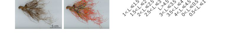
POD decomposes hydrogen peroxide (H2O2) to produce water and release oxygen.
DAB staining is used to detect the active peroxidase sites in cells. As shown in Figure 6E,
the difference in DAB staining of peach tree leaves was small. As illustrated in Figure 6F,
the DAB staining of roots of the studied peach trees produced significantly different re‐
Int. J. Mol. Sci. 2022, 23, 2585 sults. The relative staining intensity of the 200 mg/L PC treatment was the lowest, which
8 of 17
was 42.10% lower than that of the NaCl treatment (Figure 6I).
Figure 6.
Figure Leaves and
6. Leaves and roots
roots staining.
staining. (A)
(A) Evans
Evans blue‐staining
blue-staining of of leaves;
leaves; (B)
(B) Evans
Evans blue‐staining
blue-staining of of
roots; (C)
roots; (C) NBT
NBT staining
staining of
of leaves;
leaves; (D)
(D) NBT
NBT staining
staining ofof roots;
roots; (E)
(E) DAB
DAB staining
staining of
of leaves;
leaves; (F)
(F) DAB
DAB
staining of
staining ofroots;
roots;(G)
(G)Relative
RelativeEvans
Evansblue‐staining
blue-staining intensity;
intensity; (H)(H) Relative
Relative NBTNBT staining
staining intensity;
intensity; (I)
DAB
(I) DABrelative staining
relative intensity.
staining Control
intensity. Controltreatment
treatment (CK,
(CK,a anegative
negativecontrol
controlgroup);
group);tree
tree treatment
treatment
with
with only 70 mmol/L
mmol/L NaCl
NaCl (T1);
(T1); tree
tree treatment with 70 mmol/L
mmol/L NaClNaCl and
and 100 mg/L
mg/L PC PC (T2);
(T2); tree
tree
treatment with 70 mmol/L NaCl and 200 mg/L PC (T3); tree treatment with
treatment with 70 mmol/L NaCl and 200 mg/L PC (T3); tree treatment with 70 mmol/L NaCl and 70 mmol/L NaCl and
400
400 mg/L
mg/LPC PC(T4).
(T4).The
Thedata
datawere
weresampled
sampled atat 8:00
8:00 am
a.m.ononthe
the12th
12thday
dayafter
afterthe
thesalt
saltstress
stresstreatment.
treatment.
Measurements were performed using the root tips of peach plants. Error bar represents standard
Measurements were performed using the root tips of peach plants. Error bar represents standard
deviation of the mean (n = 3). Different lowercase letters indicate significant differences among dif‐
deviation of the mean (n = 3). Different lowercase letters indicate significant differences among
ferent treatments (Duncan test, p < 0.05).
different treatments (Duncan test, p < 0.05).
POD decomposes hydrogen peroxide (H2 O2 ) to produce water and release oxygen.
DAB staining is used to detect the active peroxidase sites in cells. As shown in Figure 6E,
the difference in DAB staining of peach tree leaves was small. As illustrated in Figure 6F,
the DAB staining of roots of the studied peach trees produced significantly different results.
The relative staining intensity of the 200 mg/L PC treatment was the lowest, which was
42.10% lower than that of the NaCl treatment (Figure 6I).
2.7. Electrolyte Leakage Rate and SOD, POD, MDA, Proline, and Soluble Sugar Contents
Notably, salt stress exposure increased the cell electrolyte leakage rate, whereas the
PC treatment reduced the leaf and root electrolyte leakage rate (Figure 7A). Therefore, the
application of PC can protect plant cells from the adverse effects of salt stress and attenuate
their damage. As visible in Figure 7B, the SOD activity of 200 mg/L PC was the highest.
The SOD activity of the leaves in that treatment was 115.82% higher than that of the control;
the SOD activity of the roots was 164.06% higher than that of the control. The results of
the NBT staining and SOD showed that exogenous application of PC under salt stress
Int. J. Mol. Sci. 2022, 23, 2585 9 of 17
Int. J. Mol. Sci. 2022, 23, x FOR PEER REVIEW 10 of 18
can remove the superoxide anions accumulated in the plant, protecting plant cells and
reducing damage.
Antioxidant
Figure7.7.Antioxidant
Figure enzyme
enzyme activity
activity and and osmotic
osmotic balance
balance in leaves
in leaves andand roots.
roots. (A) Electrolyte
(A) Electrolyte ex‐
extravasation
travasation rate;
rate; (B)(B) Superoxide
Superoxide dismutase;
dismutase; (C)(C) Peroxidase
Peroxidase activity;
activity; (D)Malondialdehyde
(D) Malondialdehydecontent;
content;
(E)
(E)Proline
Prolinecontent;
content;(F)(F)
Soluble
Solublesugar
sugarcontent. TheThe
content. error bar bar
error represents the standard
represents deviation
the standard of theof
deviation
the mean (n = 3). Measurements were performed using the root tips of peach plants. The datawere
mean (n = 3). Measurements were performed using the root tips of peach plants. The data were
sampled
sampledatat9:00
9:00am onon
a.m. thethe
12th day
12th after
day the
after salt
the stress
salt treatment.
stress treatment.Different
Differentlowercase
lowercaseletters
lettersindicate
indicate
significant differences among different treatments (Duncan test, p < 0.05).
significant differences among different treatments (Duncan test, p < 0.05).
3. Discussion
As can be seen in Figure 7C, compared with 100 mg/L PC and 400 mg/L PC, exoge-
nous
3.1. PC,application
PA Contents, of 200
andmg/L PC had the
PLD Activity in themost significant
Cells Exposed to increase in POD activity in leaves
Salt Stress
and High
roots.salinity
The POD activity of the leaves in that treatment
is commonly due to high‐concentrations of Na and Cl was 47.47%
+ higher
− in the than
soilthat
solu‐of
tion, resulting in hyperosmotic and hyperionic conditions that impede soil water andNaCl
the NaCl treatment; the POD activity of the roots was 66.05% higher than that of the nu‐
treatment.
trient Its effect
absorption by is beneficial
plants as H2 O
[20]. Plant 2 ismembrane
cell released incontrols
the cell,the
which reduces
entry and exit theofdamage
most
to plants
ions by POD. The high
and macromolecular amount ofItMAD
substances. is thein plants indicates
interface between aplants
severeand degree of plant
the outside
cell membrane damage as it is a manifestation of the degree of
environment that serves for information and material exchange; it is also the first barrier peroxidation. As can be
observed in Figure 7D, the content of MDA in the salt stress treatment was higher than that
used by plants to resist external damage [21]. The cell membrane is a viable organelle,
in the control group, indicating that salt stress had increased the degree of peroxidation in
which can adjust the membrane structure in different states by changing its chemical
the leaf and root cell membranes. However, the addition of PC significantly reduced the
structure or molecular morphology [5]. The membrane system is a plant part that is sen‐
peroxidation degree of the plant cell membrane. In the NaCl + 200 mg/L PC treatment,
sitive to injury. PC is an important component of membrane lipids. The steady state of its
the MDA contents in the leaves was 64.53% lower, respectively, than those in the NaCl
content determines the normal biological function of membranes [22]. Plant salt tolerance
treatment. The 200 mg/L PC and 400 mg/L PC treatments could significantly reduce MDA
is closely related to the structure and function of cell membranes, and the lipid content
content in roots, which were 48.41% and 46.29% lower than NaCl treatments, respectively.
and composition of plant cell membranes under salt stress had undergone considerable
To adapt to adverse conditions, such as salt stress, plants actively accumulate proline
changes [23,24]. Our research revealed that PC not only is the main component of the
and soluble sugars, reduce their osmotic potential, and promote water absorption by
structural basis of biological membranes, but its metabolites are also used as signal sub‐
the root system to adapt to the external environmental changes. The accumulation of
stances
anotherinvolved
important in the growth
osmotic and development
adjustment substance ofinplants and their
the vacuole, response
proline, also toplays
salt stress.
a role
in regulating the cytoplasmic osmotic balance. As illustrated in Figure 7E, under the[14].
PA is an important phospholipid messenger that has been recently discovered salt
Itstress
is involved
treatmentsin a variety of adversity the
of our experiment, responses
contents and of hormone
proline ininformation transmission
the plant increased. The
processes.
plants in the In contrast
NaCl + 200 to the
mg/Lhigh PC levels of structural
treatment phospholipids
accumulated more proline in the
andeukaryotic cell,
soluble sugars
PA content is low, accounting for only 1–2% of the total phospholipids,
than those in the treatment with NaCl. NaCl + 200 mg/L PC treatment had the most in the cell [13,25].
Previous
significant studies found thatofPA
accumulation is involved
proline in leavesnot and
onlyroots,
in various
which cellular processes,
were 59.05% andsuch
54.93%as
cytoskeleton rearrangement, vesicle transport, and membrane
higher than NaCl treatment, respectively. As illustrated in Figure 7F, NaCl + 200 mg/L lipid biosynthesis [26,27],
but
PCittreatment
is also a regulator
and NaClthat participates
+ 400 mg/L PCintreatment the physiological
were theresponse of plantsfor
most significant to soluble
a vari‐
ety of biotic
sugar and abiotic
accumulation stresses.
in leaves and These
roots.adverse abioticsugar
The soluble conditions
contents include low‐tempera‐
in leaves and roots
ture, freezing,
of NaCl + 200dehydration, drought,and
mg/L PC treatment saltNaCl
stress,+ nutrient
400 mg/L deficiency,
PC treatment and mechanical
were 76.46% dam‐
and
age [28–30]. Some scholars believe that the functions of PA messenger
75.76% higher, 71.59% and 69.27% higher than those of NaCl treatment, respectively. This and classic second
messenger
accumulation (Ca2+had
and cAMP) areeffect
a beneficial equally
on theimportant
plants as [31]. Here, weplant
it adjusted established
osmoticthat the ex‐in
potential
ogenous application of PC under
response to the salt stress conditions. salt stress promotes PLD activity. In turn, this enhanced
activity leads to the decomposition of intracellular phospholipids and the production of
PA, thereby regulating and improving the tolerance of the peach seedlings to the exposure
to salt stress.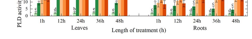
Int. J. Mol. Sci. 2022, 23, 2585                                                                                            10 of 17

                                   3. Discussion
                                   3.1. PC, PA Contents, and PLD Activity in the Cells Exposed to Salt Stress
                                          High salinity is commonly due to high-concentrations of Na+ and Cl− in the soil
                                   solution, resulting in hyperosmotic and hyperionic conditions that impede soil water and
                                   nutrient absorption by plants [20]. Plant cell membrane controls the entry and exit of most
                                   ions and macromolecular substances. It is the interface between plants and the outside en-
                                   vironment that serves for information and material exchange; it is also the first barrier used
                                   by plants to resist external damage [21]. The cell membrane is a viable organelle, which
                                   can adjust the membrane structure in different states by changing its chemical structure
                                   or molecular morphology [5]. The membrane system is a plant part that is sensitive to
                                   injury. PC is an important component of membrane lipids. The steady state of its content
                                   determines the normal biological function of membranes [22]. Plant salt tolerance is closely
                                   related to the structure and function of cell membranes, and the lipid content and composi-
                                   tion of plant cell membranes under salt stress had undergone considerable changes [23,24].
                                   Our research revealed that PC not only is the main component of the structural basis of
                                   biological membranes, but its metabolites are also used as signal substances involved in
                                   the growth and development of plants and their response to salt stress.
                                          PA is an important phospholipid messenger that has been recently discovered [14].
                                   It is involved in a variety of adversity responses and hormone information transmission
                                   processes. In contrast to the high levels of structural phospholipids in the eukaryotic cell,
                                   PA content is low, accounting for only 1–2% of the total phospholipids, in the cell [13,25].
                                   Previous studies found that PA is involved not only in various cellular processes, such as
                                   cytoskeleton rearrangement, vesicle transport, and membrane lipid biosynthesis [26,27], but
                                   it is also a regulator that participates in the physiological response of plants to a variety of
                                   biotic and abiotic stresses. These adverse abiotic conditions include low-temperature, freezing,
                                   dehydration, drought, salt stress, nutrient deficiency, and mechanical damage [28–30]. Some
                                   scholars believe that the functions of PA messenger and classic second messenger (Ca2+ and
                                   cAMP) are equally important [31]. Here, we established that the exogenous application
                                   of PC under salt stress promotes PLD activity. In turn, this enhanced activity leads to the
                                   decomposition of intracellular phospholipids and the production of PA, thereby regulating
                                   and improving the tolerance of the peach seedlings to the exposure to salt stress.

                                   3.2. Effect of the Exogenous Application of PC on the Photosystem under Salt Stress Exposure
                                        Under salt stress, plant chloroplasts are destroyed, and the related photosynthetic
                                   enzyme activities decrease, resulting in a decline in the photosynthetic rate of plants [3].
                                   Previous studies have found that phospholipids are essential for the photosynthesis in
                                   higher plants and cyanobacteria. The content of phospholipids in plant cell membranes
                                   changes in response to stress to retard or reduce the damage [32,33]. The chloroplast in
                                   the leaves is composed of three parts: envelope, matrix, and thylakoid, which is the main
                                   location in plants where photosynthesis occurs [34]. Kim found that phosphatidylcholine
                                   is required for the efficient formation of photosynthetic membrane and B800-850 light-
                                   harvesting complex in rhodobacter sphaeroides [35]. It can be seen from Figure 1D–F that
                                   the exogenous application of PC under salt stress can protect the contents of chlorophyll a,
                                   chlorophyll b, and carotenoids in leaves. Our research found that the application of PC
                                   under salt stress can stabilize the phospholipid content in leaf cells, ensure the normal
                                   function of cells, and facilitating the maintenance of normal photosynthesis under salt
                                   stress. Compared with breeding and biotechnological methods to alleviate salt stress
                                   damage, the application of PC is simple and convenient, and can save a lot of human and
                                   financial resources.
                                        Stomata are the channels through which higher plants exchange water and gas with
                                   the outside environment. More than 90% of the water loss from plants is via leaf stomatal
                                   transpiration. Investigations of the mechanism of stomatal movement are of substantial
                                   significance for the in-depth understanding of plant stress adaptation, water use, and signal
                                   transduction mechanisms. Jiang et al. evidenced that the abscisic acid (ABA)-induced
Int. J. Mol. Sci. 2022, 23, 2585 11 of 17
stomatal closure process in Arabidopsis required the participation of PA in the regulation of
microtubule depolymerization [36]. Furthermore, Zhang et al. found that PLD-derived PA
was essential in microtubule tissue process regulation under salt stress; PA and MAP65-1
interacted to regulate the microtubule structural organization and salt tolerance [37]. Our
present study revealed that under salt stress, PA, which is a signal molecule produced by
PC decomposition, is critically involved in the ABA-induced stomatal opening and closing.
The exogenous application of PC under salt stress can thus regulate the opening of stomata
and ensure that cells can normally communicate with the outside world. This can also
explain the normal entry of CO2 into the mesophyll by opening the stomata, enabling the
maintenance of normal photosynthesis in the leaves.
3.3. Effects of PC on the Root Structure, Growth, Cell Components, and Cell Membrane Integrity
under Salt Stress
In plants, PLD and PA are considered to be involved in the promotion of cell elongation
of pollen tubes and root hairs [38,39]. Additionally, they participate in the adaptation
processes associated with plant growth under phosphorus deficiency and high osmotic
stress [28,40]. PLD activation was found to occur under high permeability conditions [31,41].
Moreover, Hong et al. established that PLD can also promote plant growth under high-
salinity and high-osmotic stress caused by the lack of water [42]. Under these adverse
conditions, PLD can enhance the plant nitrogen absorption and increase the root surface
area, promoting greater water absorption and utilization by plants. The lipid profile
analysis of root tissues in a previous study indicated that PA molecules may participate in
sensing nutrition and osmotic signals to regulate root growth.
In agricultural practice, grafting has been described to increase salt tolerance by ex-
cluding or restricting toxicion accumulation in shoots [43]. Previous research found that
the influence of rootstock on a scion’s salt and water stress tolerance is due to: using a
larger and vigorous root systems capable of absorbing water and nutrients much more effi-
ciently [44]. However, the selection of rootstocks is limited, and it is difficult to concentrate
multiple excellent traits on one rootstock. Therefore, the selection of rootstocks to resist
salt stress not only consumes a lot of time and money, but also it is difficult to retain the
original excellent characters of the species [45]. Our present research evidenced that under
salt stress, the exogenous application of PC enhanced the activity of PLD and increased
the content of PA in the cells. Notably, the exogenous application of PC increased the root
growth of the treated peach seedlings as compared with that in the control, especially the
number of fine roots (Figure 3B). Using transmission electron microscope observations
of the root cells, we found that the exogenous administration of PC had a positive effect
on maintaining cell integrity and cell-membrane structure. Bewley (1979) showed that
plant drought tolerance depends on the ability to limit membrane damage during water
stress [46]. It can be seen from Figure 4 that the absorption of PC by peach can stabilize the
cell membrane structure and enhance the integrity of the cell membrane, thereby enhancing
the tolerance to salt stress. The potential underlying mechanism of action could be through
the maintenance of the cell membrane phospholipid homeostasis by PC and the enhanced
salt tolerance of the cell membrane structure via PA regulation. Therefore, the application
of PC to alleviate salt stress damage can save the financial resources and time of rootstock
selection, and the operation is simple and the excellent characters of the species are retained
to a greater extent.
3.4. Application of PC on Maintaining Cell Activity under Salt Stress
Leaves are the main photosynthetic organs in higher plants. The chloroplast in the
leaf consists of three parts: envelope, stroma, and thylakoid, which is the main place for
photosynthesis of plants [47]. In trees and other perennial woody species, the role of the
root system is even more important than that in small annual plants [48]. Therefore, the
activity of plant leaf and root cells under salt stress determines whether plants can maintain
normal growth and development. By staining the leaves and roots with Evans blue, weInt. J. Mol. Sci. 2022, 23, 2585 12 of 17
found that the application of 200 mg/L PC under salt stress can protect the physiological
activity of leaf and root cells to the greatest extent, significantly reducing the electrolyte
extravasation rate, which is important for protecting the integrity of cell membranes and
the maintenance of normal cell function.
Under salt stress, plants adjust accordingly their physiological and biochemical pro-
cesses to adapt, including ion and osmotic homeostasis regulation, as well as the control
and repair (detoxification) of stress-induced damage [49]. SOD is an antioxidant metal
enzyme that catalyzes the dismutation of superoxide anion radicals to generate oxygen
and H2 O2 . It plays an important role in plant tolerance against biotic stress. Under adverse
conditions or senescence, H2 O2 accumulates in the plant due to increased active oxygen
metabolism. H2 O2 can directly or indirectly oxidize nucleic acids, proteins, and other
biological macromolecules in the cell and damage the cell membrane, thereby accelerating
plant senescence and the degradation of cells. POD is another antioxidant enzyme located
in the peroxisome of the cell, which plays an important role in preventing the damage by
oxygen metabolites [50]. It can be seen from Figures 6 and 7 that the exogenous application
of 200 mg/L PC under salt stress increased the SOD activity of the leaves and roots, and
the activity of root POD. We speculate that PA produced by PC can activate the activities
of SOD and POD. It exerted a positive effect in plants by removal of superoxide anions
and peroxide.
3.5. Effect of PC Application on MDA, Proline, and Soluble Sugar Contents
Plant organs are damaged under salt stress, and membrane lipid peroxidation often
occurs. MDA is the final decomposition product of membrane lipid peroxidation, and its
content can reflect the degree of damage to plants [51]. Another important aspect of salt
tolerance mechanisms is the capacity of plant cells to adapt osmotically by the plant tissue
accumulation of organic solutes, such as free proline, endogenous glycine betaine, soluble
sugar, and protein. Hence, their increased contents might indicate the degree of tolerance
to osmoregulation-induced stress [52]. These osmotic solutes maintain osmotic balance
and normal turgor and control water influx (reduce the efflux). It can be seen from Figure 7
that exogenous application of PC under salt stress reduced the MDA content and the cell
membrane damage in the experimental plants. The content of proline and soluble sugar
increased significantly. PLD-produced PA increases rapidly in cell-suspension cultures of
tomato and alfalfa subject to salt stress and in the dehydrated leaves of the resurrection
plant Craterostigma plantagineum [53]. We speculate that PLD decomposes PC to generate
organic solutes and maintain the osmotic balance of intracellular organic solutes and part
of the PA is produced by PLD to regulate the salt tolerance of plants.
4. Materials and Methods
4.1. Plant Material and Exogenous PC Treatment
Experiments were performed in the experiment center of Shandong Agricultural
University (Tai’an, China) in June 2020. The 101-year-old ‘lu xing’ peach, Prunus persica (L.)
Batsch., seedlings were planted in 2.5 L soil pots. These pots were then placed in a glass
greenhouse with a day/night temperature regime of 26 ◦ C/18 ◦ C, a natural photoperiod of
around 12.5 h, and constant relative humidity of 30%.
The experiment set up included five treatments, and each treatment was repeated in
20 pots (replicates). In the preliminary salt stress screening test, we found that salt-induced
damage to the peach seedlings began to appear after their treatment with 70 mmol/L NaCl
for 10 days, which met the test requirements. Therefore, in this experiment, we chose to
use 70 mmol/L NaCl as the salt stress treatment concentration. We used the following
treatments when the plants grew to a stage of having from 10 to 12 fully expanded leaves:
control treatment (CK, a negative control group); tree treatment with only 70 mmol/L
NaCl (T1); tree treatment with 70 mmol/L NaCl and 100 mg/L PC (T2); tree treatment
with 70 mmol/L NaCl and 200 mg/L PC (T3); tree treatment with 70 mmol/L NaCl and
400 mg/L PC (T4). In this experiment, PC and NaCl were directly applied to the soil at aInt. J. Mol. Sci. 2022, 23, 2585 13 of 17
reagent dosage of 100 mL per tree. All PC treatments were applied in soil pots at 5 p.m. to
avoid excessively high-temperatures.
4.2. PC and PA Content and PLD Enzyme Activity in Plants
Nine leaves and six roots were randomly selected from each treatment, ground under
liquid nitrogen and used for later use. To determine the amounts of plant PC in the
samples, cover microtiter plate wells with purified plant PC antibody, produce solid-phase
antibody, and then add PC to wells. After thorough washing, a combined Horseradish
Peroxidase (HRP)-labeled PC antibody was used as an antibody-antigen-enzyme-antibody
complex. Upon addition of 3,30 ,5,50 -Tetramethylbenzidine (TMB) substrate solution, the
TMB substrate turned blue. The reaction catalyzed by the HRP enzyme was terminated
by the addition of sulphuric-acid solution, leading to a color change that was detected
spectrophotometrically at 450 nm. We measured the concentration of plant PC/PA in the
samples by comparing the OD of the samples to the standard curve. To compute the sample
concentration, we substituted the sample OD value into the equation.
A PA level dynamic assay was performed as described previously [54].
The PLD activity was then determined following the method previously described
by Wang [55]. PLD catalyzes the hydrolysis of the phosphatidyl diester bond at the end of
PC to produce PA and choline. Choline is next catalyzed by choline oxidase to generate
betaine and hydrogen peroxide. Further, 4-aminoantipyrine and double-distilled phenol
were oxidized to a pink substance with a characteristic absorption peak at 500 nm. W is the
sample mass of 0.1 g, PLD enzyme activity (nmol/min/g) = 16.7 × A measuring tube/A
standard tube/W.
4.3. Measurements of the Net Photosynthetic Rate and SPAD Values
On the first day of our experiment and every two days following the salt stress
treatment (until the ninth day), we assessed the net photosynthetic rate and chlorophyll
content of peach leaves. We measured the leaf net photosynthetic rate in the leaves of peach
trees from the treatments using a CIRAS-3 Portable Photosynthesis System (PP Systems,
Amesbury, MA, USA). All leaves were equilibrated at a constant flow rate of 500 mL min−1
and a CO2 concentration of approximately 400 µmol mol−1 at PAR of 1000 µmol M−2 S−1 .
Between 9:30 and 11:00 a.m., a SPAD-502 PLUS chlorophyll meter was used to measure
the amount of chlorophyll in fully expanded leaves (Spectrum Technologies, Aurora, IL,
USA). On the ninth day, we used a previously reported procedure to determine the content
of chlorophyll a, chlorophyll b, and carotenoids [56].
4.4. Determination of Stomatal Area, Root Growth, and Root Cell Integrity
Three pots of peach seedlings were randomly selected for each treatment, and three
leaves were selected from each peach seedling, with a total of nine leaves for each treatment.
Nine leaves of the same developmental stage were chosen on the tenth-day post-salt stress
treatment to measure the size and density of the stomata. To perform the measurements, we
stained the leaf epidermis with clear acrylic nail polish. The nail polish was taken off with
forceps once it had dried. The solid polish was then put on a microscope slide and examined
with a fluorescence microscope at 400× magnification (AXI0, Carl Zeiss, Jena, Germany). We
make nine slides per treatment. We randomly selected three 3.2-mm2 areas on each slide and
took a picture. We used ImageJ version 1.48 to count the number of stomata and quantify
their length and width (National Institutes of Health, Bethesda, MD, USA).
Next, we randomly collected six roots of fresh peach seedlings, rinsed them with
water, and determined the root configuration parameters using the professional version of
WinRHIZO Root Analysis System (Epson V 700).
Transmission electron microscopy was further employed to examine the plant root
cells’ integrity, as well as the membrane structure of the cells. Samples were then embedded
in Epon812 epoxy resin before incubation and three sequential curing phases at 37 ◦ C,Int. J. Mol. Sci. 2022, 23, 2585 14 of 17
45 ◦ C, and 65 ◦ C, each for 24 h. We sectioned samples and used lead uranyl acetate to stain
them. Finally, we put samples in the machine for testing and capture images.
4.5. Measurements of Root Cell Activity, NBT and DAB Staining, and Electrolyte Leakage Rate
Nine root tips from each treatment were selected for staining. Evans blue staining
examination has been confirmed as a reliable method for microscopic cell death determina-
tion. We used that method to determine cell death, as previously described by Baker and
Mock [57], with slight changes. Root samples were vacuum infiltrated in 0.1 % Evans blue
(w/v) solution for 5 min before staining at room-temperature for 3 to 5 h. The samples were
then rinsed in phosphate-buffered saline (PBS) containing 0.05% (v/v) Tween-20. ImageJ
was utilized to evaluate the relative staining intensity after an image was taken using an
electron microscope. A darker root image indicated a lower cell activity.
Further, we prepared 0.5 mg/mL of nitroblue tetrazolium chloride (NBT) staining
solution, poured the staining solution into an Erlenmeyer flask, added leaves and roots,
and inserted a vacuum pump. The samples were incubated in the dark at 28 ◦ C for 8 h.
Next, we discarded the staining solution, and boil in ethanol for 5 min: lactic acid: glycerol
(6 mL:2 mL:2 mL) fixing solution until all chlorophyll is removed, cooled, and 10 mL of
absolute alcohol is added. Finally, we perform observation staining.
We prepared 1 mg/mL of diaminobenzidine (DAB) staining solution and adjusted
its pH to 5.8 with NaOH. Then, we transferred the staining solution into an Erlenmeyer
flask, added pretreated plant tissues, and inserted a vacuum pump. We next incubated the
mixture in the dark for 8 h at 28 ◦ C. Further, we discarded the staining solution and boiled
the sample in a fixative solution containing ethanol: lactic acid: glycerol (6 mL:2 mL:2 mL)
fixative solution for 5 min until complete removal of the chlorophyll was performed. Then,
we cooled the samples, added absolute alcohol 10 mL, and observed the staining.
A simple approach was implemented to determine the electrolyte leakage rate (MII),
as reported earlier [58]. Three pots of peach seedlings were randomly selected from each
treatment, and three leaves and three roots were randomly selected from each treatment.
Peach tree leaves and roots were cleansed thoroughly, and little discs of root tissue were
punched out. The initial electrical conductivity (initial EC) of the solution was measured
using a DDS-307 conductivity meter after this immersion (Shanghai Precision Scientific
Instrument Co., Ltd., Shanghai, China). After heating the samples at 100 ◦ C for 20 min,
we utilized the same apparatus to determine the final EC of the solution. The following
formula was used to calculate MII: MII (%) = (initial EC/final EC) × 100.
4.6. Determination of the Antioxidant system
Measurements were performed using the leaves and root tips of peach plants. Three
pots are required for each treatment. The data were sampled at 9:00 a.m. on the 12th day
after the salt stress treatment. Superoxide dismutase (SOD) and peroxidase (POD) activities
were measured following the protocol reported previously by Liu [59]. Malondialdehyde
(MDA) and proline contents were measured as described by Tang [60]. We conducted the
determination of soluble sugar content as described by Zhao [56].
4.7. Statistical Analysis
We collected three biological replicates for each treatment. Origin version 9.4 was used
to conduct all statistical analyses. Duncan multiple range tests, which are included in SPSS
version 20.0, were performed to detect any statistically significant differences in the mean
values (IBM SPSS, Chicago, IL, USA). The threshold of statistical significance used for all
tests was p < 0.05.
5. Conclusions
In this study, the growth and development of peach seedlings were severely negatively
affected by salt stress. The photosynthetic system of the seedlings failed to function
normally, the root development was blocked, root cell morphology was abnormal, andInt. J. Mol. Sci. 2022, 23, 2585 15 of 17
the cell membranes were damaged. The exogenous application of PC alleviated the toxic
effects of salt stress; the most significant positive effect was observed at a PC concentration
of 200 mg/L. We can make the following speculations. First, the exogenous PC absorbed by
plants under salt stress protects the homeostasis of cell membrane phospholipids, ensuring
normal plant growth and development. Second, PLD can decompose the remaining PC to
produce PA, which acts as a secondary messenger to regulate plant tolerance against salt
stress. However, the regulation of PA levels in the plant under salt stress conditions needs
further research.
Author Contributions: M.S.: Conceptualization, Methodology, Software, Data curation, Writing orig-
inal draft; X.L.: Visualization, Software; H.G.: Validation; B.Z.: Investigation; F.P.: Conceptualization,
Methodology, Software, Supervision, Writing—review and editing; Y.X.: Methodology, Supervision,
Funding acquisition. All authors have read and agreed to the published version of the manuscript.
Funding: This work was supported by National Key R&D Program of China (Nos. 2019YFD1000103
and 2020YFD1000203) and the National Modern Agroindustry Technology Research System Fund
(No. CARS-30-2-02).
Institutional Review Board Statement: Not applicable.
Informed Consent Statement: Not applicable.
Data Availability Statement: Not applicable.
Conflicts of Interest: The authors declare that they have no known competing financial interests or
personal relationships that could have appeared to influence the work reported in this paper.
References
1. Munns, R.; Tester, M. Mechanisms of salinity tolerance. Annu. Rev. Plant Biol. 2008, 59, 651–681. [CrossRef] [PubMed]
2. Ouhibi, C.; Attia, H.; Rebah, F.; Msilini, N.; Chebbi, M.; Aarrouf, J.; Urban, L.; Lachaal, M. Salt stress mitigation by seed priming
with UV-C in lettuce plants: Growth, antioxidant activity and phenolic compounds. Plant Physiol. Biochem. 2014, 83, 126–133.
[CrossRef]
3. Parida, A.K.; Das, A.B. Salt tolerance and salinity effects on plants: A review. Ecotoxicol. Environ. Saf. 2005, 60, 324–349. [CrossRef]
[PubMed]
4. Chen, S.; Lin, H. Signal transduction pathways in response to salt stress in plants. Plant Physiol. J. 2011, 47, 119–128.
5. McMahon, H.T.; Gallop, J.L. Membrane curvature and mechanisms of dynamic cell membrane remodelling. Nature 2005, 438,
590–596. [CrossRef]
6. Gigon, A.; Matos, A.-R.; Laffray, D.; Zuily-Fodil, Y.; Pham-Thi, A.-T. Effect of drought stress on lipid metabolism in the leaves of
Arabidopsis thaliana (ecotype Columbia). Ann. Bot. 2004, 94, 345–351. [CrossRef] [PubMed]
7. Li, F.; Xing, S.; Guo, Q.; Zhao, M.; Zhang, J.; Gao, Q.; Wang, G.; Wang, W. Drought tolerance through over-expression of the
expansin gene TaEXPB23 in transgenic tobacco. J. Plant Physiol. 2011, 168, 960–966. [CrossRef]
8. Zhao, J.; Wang, C.; Bedair, M.; Welti, R.; Sumner, L.W.; Baxter, I.; Wang, X. Suppression of phospholipase Dγs confers increased
aluminum resistance in Arabidopsis thaliana. PLoS ONE 2011, 6, e28086. [CrossRef]
9. Harwood, J.L. Recent advances in the biosynthesis of plant fatty acids. Biochim. Biophys. Acta Lipids Lipid Metab. 1996, 1301, 7–56.
[CrossRef]
10. Devaiah, S.P.; Roth, M.R.; Baughman, E.; Li, M.; Tamura, P.; Jeannotte, R.; Welti, R.; Wang, X. Quantitative profiling of polar
glycerolipid species from organs of wild-type Arabidopsis and a Phospholipase Dα1 knockout mutant. Phytochemistry 2006, 67,
1907–1924. [CrossRef]
11. Botella, C.; Jouhet, J.; Block, M.A. Importance of phosphatidylcholine on the chloroplast surface. Prog. Lipid Res. 2017, 65, 12–23.
[CrossRef]
12. Nakamura, Y.; Andrés, F.; Kanehara, K.; Liu, Y.-C.; Dörmann, P.; Coupland, G. Arabidopsis florigen FT binds to diurnally
oscillating phospholipids that accelerate flowering. Nat. Commun. 2014, 5, 1–7. [CrossRef] [PubMed]
13. Zhang, W.; Qin, C.; Zhao, J.; Wang, X. Phospholipase Dα1-derived phosphatidic acid interacts with ABI1 phosphatase 2C and
regulates abscisic acid signaling. Proc. Natl. Acad. Sci. USA 2004, 101, 9508–9513. [CrossRef] [PubMed]
14. Li, M.; Hong, Y.; Wang, X. Phospholipase D-and phosphatidic acid-mediated signaling in plants. Biochim. Biophys. Acta Mol. Cell
Biol. Lipids 2009, 1791, 927–935. [CrossRef] [PubMed]
15. Hong, Y.; Zhang, W.; Wang, X. Phospholipase D and phosphatidic acid signalling in plant response to drought and salinity. Plant
Cell Environ. 2010, 33, 627–635. [CrossRef]
16. Wang, X.; Devaiah, S.P.; Zhang, W.; Welti, R. Signaling functions of phosphatidic acid. Prog. Lipid Res. 2006, 45, 250–278.
[CrossRef]You can also read