Mast Cells and Acupuncture Analgesia - Review - MDPI
←
→
Page content transcription
If your browser does not render page correctly, please read the page content below
cells
Review
Mast Cells and Acupuncture Analgesia
Yingchen Li, Yi Yu, Yuhang Liu and Wei Yao *

                                             Shanghai Key Laboratory of Acupuncture Mechanism and Acupoint Function,
                                             Department of Aeronautics and Astronautics, Fudan University, 220 Handan Road, Shanghai 200433, China;
                                             20110290014@fudan.edu.cn (Y.L.); 17110290008@fudan.edu.cn (Y.Y.); 21210290018@m.fudan.edu.cn (Y.L.)
                                             * Correspondence: weiyao@fudan.edu.cn

                                             Abstract: Mast cells are widely distributed in various parts of the human body and play a vital
                                             role in the progression of many diseases. Recently, the close relationship between mast cells and
                                             acupoints was elucidated, and the role of mast cells in acupuncture analgesia has attracted the
                                             attention of researchers worldwide. Using mast cells, acupuncture analgesia and acupoint as key
                                             words to search CNKI, PubMed, Web of Science and other databases, combining the representative
                                             articles in these databases with the published research papers of our group, we summarized: The
                                             enrichment of mast cells and the dense arrangement of collagen fibers, microvessels, and nerves form
                                             the basis for acupoints as the reaction sites of acupuncture; acupuncture can cause the deformation of
                                             collagen fibers and activate TRPV channels on mast cells membrane, so as to stimulate mast cells to
                                             release bioactive substances and activate nerve receptors to generate analgesic effect; system biology
                                             models are set up to explain the quantitative process of information initiation and transmission at
                                             acupuncture points, and indicate that the acupuncture effect depends on the local mast cells density.
                                             In a conclusion, this review will give a scientific explanation of acupuncture analgesia from the
                                             material basis of acupoints, the local initiation, and afferent biological mechanism.

                                   Keywords: mast cell; acupuncture analgesia; acupoint sensitization; mechanical stimuli; TRPV
                                      channels; system biology model
Citation: Li, Y.; Yu, Y.; Liu, Y.; Yao, W.
Mast Cells and Acupuncture
Analgesia. Cells 2022, 11, 860.
https://doi.org/10.3390/                     1. Introduction
cells11050860
                                                  Mast cells, important immune cells widespread in various areas of the human body,
Academic Editors: Yoshimichi Okayama         play a vital role in the progression of many diseases. Mast cells were previously thought
and Alessandro Poggi                         to cause allergic reaction by releasing cytokines, chemokines, proteases, and biogenic
                                             amines after activation. At present, mast cells are also considered to be related to protective
Received: 31 December 2021
Accepted: 17 February 2022
                                             host immunity, acting as the sentry of innate immunity and the regulator of adaptive
Published: 2 March 2022
                                             immunity [1,2]. Recently, the close relationship between mast cells and acupoints has
                                             been elucidated [3]. The migration, the aggregation, and the activation of mast cells under
Publisher’s Note: MDPI stays neutral         acupuncture stimuli have been reported [3–5].
with regard to jurisdictional claims in
                                                  The efficacy of acupuncture analgesia is recognized worldwide. Acupuncture (punch-
published maps and institutional affil-
                                             ing a tiny needle into the skin and giving mechanical stimulations manually) can relieve the
iations.
                                             pain caused by many diseases. The effect can be evaluated through observation of animal
                                             behaviors, typically the tail flick and the paw withdraw. Zhang et al. observed an increase
                                             of pain threshold (PT) after acupuncture at zusanli acupoint (ST36) in adjuvant arthritis
Copyright: © 2022 by the authors.
                                             (AA) rat models [3]. The acupuncture effect is a complex process, which involves multiple
Licensee MDPI, Basel, Switzerland.           physiological systems from the periphery to the central. Acupoint is the reaction point of
This article is an open access article       disease, and also the stimulation point of acupuncture treatment. Acupoints are enriched
distributed under the terms and              in mast cells [4]. Zhu proposed that the local formation of “acupoint sensitization pools”
conditions of the Creative Commons           induces a pathological reaction process of “neuropeptide-mast cell-sensitizer release” [6].
Attribution (CC BY) license (https://        Acupoint sensitization is the acupoint transition from its physiological “resting state” to
creativecommons.org/licenses/by/             the pathological “active state”. Various sensitization phenomena occur after this transition,
4.0/).                                       including the feelings of acid, swelling, itching, numbness, and pain. These feelings make

Cells 2022, 11, 860. https://doi.org/10.3390/cells11050860                                                       https://www.mdpi.com/journal/cells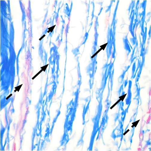
Cells 2022, 11, 860                                                                                            2 of 13

                      people involuntarily seek local stimuli such as friction, scratching, pinching, and heat. This
                      process initializes homeostasis regulation and activates the cascade reaction. By promoting
                      homeostasis regulation, acupuncture and moxibustion cure the disease.
                            The contribution of mast cells in acupuncture analgesia was gradually revealed and
                      became a hotspot, with the proposal of the mechanical signal transduction theory, the
                      humoral theory, and the nerve-humoral theory [7]. The mechanical signal transduction
                      assumes that mast cells can react to mechanical force signals, acting on them through
                      the extracellular matrix. Later, the finding of mechanical sensitive channels on mast cells
                      laid the material basis for theoretical foresight [8–10]. Humoral theory means that the
                      active substances released by mast cell degranulation diffuse along the meridian channel
                      through tissue fluid, causing mast cell degranulation. Nerve-humoral theory holds that
                      the active substances released by mast cells can also stimulate nerve endings, enter the
                      center through afferent nerve, and then act on target organs, effectors, or endocrine glands
                      through pituitary or autonomic nerves. Nowadays, it is found that acupuncture can activate
                      mast cells and cause their degranulation (releasing biological substances), inducing an
                      analgesic effect [11–13]. With a comprehensive understanding of the previous theories,
                      it is possible to build a bottom-up model and simulate the calcium and mediator signals
                      in the whole system/network, including mast cell degranulation and mast cell-nerve
                      interactions [14–16]. To summarize, the effect of mast cells on acupuncture analgesia may
                      bring new insights to the mechanism of acupuncture therapy.
                            Focusing on the function of mast cells in acupoints, this paper reviews the mechanism
                      of acupuncture analgesia by discussing the following topics: (1) mast cell degranulation and
                      its function, (2) the relationship between mast cells and acupoint sensitization, (3) the phys-
                      iological responses of mast cells under mechanical stimulations, (4) the synergistic effect of
                      mast cells with collagen fibers and nerves during acupuncture, and (5) the system biology
                      model to quantitatively explain acupuncture information initiation and transmission.

                      2. Characteristics of Mast Cells and Its Function
                      2.1. Origin and Distribution of Mast Cells
                            Mast cells were discovered by von Recklinghausen in 1863 [17]. Afterwards, Paul
                      Ehrlich detailly described the histological observations of this granules contained cell, and
                      gave it the name [18]. Since then, tremendous progress has been made in the research on
                      the origin, the distribution, and the function of mast cells.
                            Mast cells are originated from hematopoietic cells in the bone marrow. They migrate
                      into peripheral tissues and differentiate into mature mast cells under the influence of vari-
                      ous factors such as monocyte chemoattractant protein-1 (MCP-1), stem cell factor (SCF),
                      transforming growth factor type-β (TGF-β), activin (ACT), etc [19]. Furthermore, the adhe-
                      sion and the migration of mast cells are influenced by integrin and cytoskeleton, etc. [20,21].
                      According to the surrounding tissue, mast cells are classified into the mucosal mast cell
                      (granules containing abundant tryptase) and the connective tissue-type mast cell (granules
                      containing tryptase and chymotrypsin) [22].
                            Mast cells are widely distributed in connective tissues and mucosal layers, especially
                      at the interface of the inner and the outer environments such as the skin [23], the digestive
                      tract [24], the airways [25], and other borders where the interaction with the external
                      environment occurs. Besides, mast cells can also be found in organs such as the heart, the
                      liver, and the lung [26]. This paper focuses on mast cells at acupoints, which are distributed
                      in the connective tissue of the skin. Mast cells in the skin are often scattered on the intima,
                      perimysium, and adventitia of the nerve tract; they also distribute around small blood
                      vessels, hair follicles and sweat glands [5,23]. Moreover, mast cells are closely associated
                      with fibroblasts, vascular endothelial cells, cardiomyocytes, etc. A “transgranulation”
                      cellular behavior was also reported [27]. Figure 1 shows the origin and the distribution of
                      mast cells in the skin [28]. Existing in a complex stromal environment, mast cells play a
                      vital role in the circulatory, the neurological, the endocrine, and the immune systems. Like
                      a linking node in a network, mast cells communicate with these physiological systems and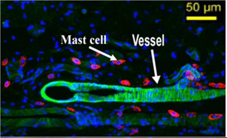
Cells 2022, 11, 860                                                                                                                           3 of 13

                      couple them together. This review aims to elucidate in detail the mechanism of mast cells’
                      involvement in acupuncture analgesia.

                                                                                                                        immature mast cell
                                                                                                                               (in vessel)
                                                                                                                          mature mast cell
                                                   Hair                                                                         (in tissue)

                                                                                                                             red blood cell

                                                                                                                           endothelial cell
                       Epidermis
                                                                                                                                other
                                                                                                                             immunocytes

                                                                                                                                  other
                                                                                                                               tissue cells

                                       Hair follicle
                                                                                                         Blood vessel
                                                          MT        Mast cell                                                       Bone marrow
                                                            H                   Maturing
                        Dermis                                                                 Settle
                                                                                               in skin

                                                                TN
                                                                M
                                   Nerve
                                                                                MTV

                                                                            F
                                                                          MT
                                                                                      MT
                                                                                           M
                                           Fat tissue

                        Muscle

                      Figure 1. Mast cells in the skin. Mast cell progenitors originate from bone marrow. Under certain
                      conditions, immature mast cells (colored in pink) migrate into peripheral tissues and settle down,
                      mainly in the dermis. Mature mast cells (colored in purple) then migrate to vessels (MTV), nerves
                      (MTN), hair follicles (MTH), muscle tissues (MTM), and adipose tissues (MTA). Mast cells modulate
                      the neighboring cell behaviors by releasing multiple mediators, typically by degranulation after
                      stimulation (marked in red circles), for example, mast cells distributed along vessels can increase
                      vascular permeability, and mast cells distributed along nerves can active nerves. Adapted from [28].

                      2.2. Mast Cell Degranulation and Its Function
                           The function of mast cells is mainly fulfilled by the degranulation process. Under
                      degranulation, the cell is activated, and the cell membrane breaks, releasing rich mediators,
                      including histamine, platelet activating factor (PAF), interleukins (IL-1, IL-13, IL-4, and
                      IL-5 etc.), prostaglandin D2 (PGD2), substance P, tryptase, serotonin, bradykinin, heparin,
                      chemokines, and so on [29]. Thanks to the distribution of mast cells, these mediators
                      will quickly act on the neighboring nerves, blood vessels, and muscles, forming a neural-
                      endocrine-immune network. For example, the histamine and platelet activating factor will
                      cause the relaxation of blood vessels and increase the capillary permeability, leukotriene can
                      cause smooth muscle contraction and vasodilation, interleukins work as pro-inflammatory
                      and inflammatory cytokines that activate and recruit inflammatory cells, including granulo-
                      cytic leukocytes (neutrophils, basophils, eosinophils) and agranular leukocytes (monocytes,
                      lymphocytes) to the site of inflammation, contributing to the development of allergic dis-
                      eases such as allergic rhinitis. Histamine, substance P, and serotonin etc. can regulate
                      nerve activities.
                           Mast cells are rich in receptors and in responses to a wide range of stimuli. This allows
                      mast cells to function differently in the presence of different stimuli, having both positive
                      and negative effects in the body depending on specific conditions [30,31]. The activation
                      in the physiological state and body repair state will adjust the homeostasis of the internal
                      environment, which is beneficial to body health by defending against the entering viruses
                      and bacteria. Abnormal activation in a pathological state will cause body discomfort and
                      threaten health. That is, in the case of weak responses, mast cells can aggravate local
                      pathological manifestations [32–34]. For example, interleukin-33 (IL-33), produced by
                      epithelial cells, could stimulate mast cells to secrete histamine, which is the main mediator
                      that stimulates nasal rubbing and sneezing in ovalbumin (OVA) induced allergic rhinitis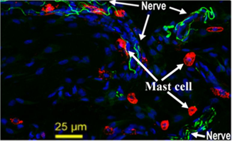
Cells 2022, 11, 860                                                                                                   4 of 13

                      (AR) [35]. Another well-known example of this is the reports that mast cells are activated by
                      COVID-19 and lead to inflammation and fibrosis in the lung. The virus causes mast cells to
                      release pro-inflammatory molecules, thereby contributing to SARS-CoV-2 infection [36,37];
                      in other settings, especially in severe cutaneous hypersensitivity, mast cells may suppress
                      the process, in part by producing interleukin-10 [38,39]. Mast cells in acupoint sensitization
                      and acupuncture analgesia may reflect their negative and positive effects, respectively.

                      2.3. Mast Cells and Acupoint Sensitization
                           The conception of acupoint is introduced in Traditional Chinese Medicine (TCM), as
                      effective targets for acupuncture therapy. Acupoints are a series of special points (about
                      360 in human) in the skin. These points may become sensitive to mechanical or thermal
                      stimulus under various pathologies [40]. Yuan et al. found that acupoints were mainly
                      collagen fiber-rich areas such as intermuscular connective tissue, peri-neurovascular con-
                      nective tissue, and organ portal and peri-neural connective tissue [41]. Through magnetic
                      resonance imaging (MRI) and X-ray computed tomography (XCT), Fei et al. also noticed
                      the enrichment of connective tissues in acupoints [42], they found that mast cells, blood
                      vessels, nerves tracts, and lymph vessels, together with the connective tissue as the base,
                      form a very complex structural system. Figure 2 shows the distribution of collagen fibers
                      and mast cells at the zusanli acupoint.

                                        a                                                          b
                      Figure 2. The structure of zusanli acupoint in rats. (a) The mallory staining of zusanli acupoint;
                      collagen fibers are in blue, indicated by black solid line arrows. Note that collagen fibers are rich
                      and have a parallel arrangement at the acupoint. Myofibers and blood cells are in red, indicated
                      by black dotted arrows. (b) The toluidine blue staining of zusanli acupoint. The scale bar is 10 µm;
                      mast cells are in blue, indicated by solid line arrows. Note that mast cells gather in large numbers at
                      the acupoint [3,43].

                           In a pioneering histomorphological observation in amputated limbs, Song found
                      that the number of mast cells was significantly larger at acupoints than non-acupoints,
                      she also found that mast cells were located near nerve endings and blood vessels [44,45].
                      Crivellato’s study gave similar results, finding the abundantly presence of mast cells in
                      the dermal tissue of acupoints area, distributed diffusely or in clusters [18]. Zhang et al.
                      found “synaptic-like” connections between mast cells and nerve endings in the Yang Ming
                      meridian [46,47]. In a histomorphological observation of tissues in acupoints, Luo et al.
                      found a composite strip structure of mast cells, blood vessels, and nerves [4]. The close
                      anatomic relation between mast cells, blood vessels, and nerves implies their reciprocity
                      correlation. Figure 3 shows the distribution of mast cells along blood vessels and nerves of
                      zusanli acupoint in rats.
Cells 2022, 11, x FOR PEER REVIEW 5 of 13
Cells 2022, 11, 860 5 of 13
Figure 3 shows the distribution of mast cells along blood vessels and nerves of zusanli
acupoint in rats.
(a) (b)
Figure
Figure3.3.The
Thedistribution
distributionofofmast
mastcells,
cells,nerves,
nerves,and
andblood
bloodvessels
vesselsat at
zusanli acupoint
zusanli acupointin in
rats after
rats after
immunofluorescence staining. (a) The distribution of mast cells around blood vessels.
immunofluorescence staining. (a) The distribution of mast cells around blood vessels. (b) The (b) The dis-
tribution of mast
distribution cellscells
of mast around nerve
around fibers
nerve [28].[28].
fibers
Acupoints
Acupointsare arereaction
reactionsites
sitesofofdisease
diseaseand andtargets
targetsforforacupuncture.
acupuncture. Many
Many chemicals
chemicals
are involved in acupoint sensitization, they form the so-called “acupoint
are involved in acupoint sensitization, they form the so-called “acupoint sensitization pool”. sensitization
pool”.
He et Heal. et al. revealed
revealed that that the high
the high expression
expression of local
of local allergic
allergic substancesand
substances andnociceptive
nocicep-
tive neuropeptides, such as substance P, calcitonin gene related peptide
neuropeptides, such as substance P, calcitonin gene related peptide (CGRP), histamine, (CGRP), hista-
sero-
mine, serotonin, and tryptase, are responsible for the acupoint sensitization
tonin, and tryptase, are responsible for the acupoint sensitization [48]. By releasing these [48]. By re-
leasing
importantthese importantmast
chemicals, chemicals,
cells aremast cellsassociated
closely are closelywithassociated
both the with both thesensitization
acupoint acupoint
sensitization and the acupuncture
and the acupuncture effect
effect [49]. Ding [49].
et al. Ding etthat
reported al. reported
the releasethat
ofthe release histamine,
serotonin, of sero-
tonin, histamine,
and tryptase and mast
during tryptase
cell during mast cellregulated
degranulation degranulation regulated
the acupoint the acupoint
sensitization sen-
[12]. The
sitization [12]. The released substance
released substance P may also be involved [50].P may also be involved [50].
Moreover,
Moreover,He Heetetal.
al.found
foundthatthatthetheconcentration
concentration ofofsubstance
substance P, P,
aa calcitonin
calcitoningene
gene
related peptide, at the same acupoint, is different under normal, pathological,
related peptide, at the same acupoint, is different under normal, pathological, and acupunc- and acu-
puncture conditions
ture conditions [51]. Using
[51]. Using high-performance
high-performance liquidliquid chromatography
chromatography (HPLC) (HPLC) to
to measure
measure the adenosine
the adenosine concentration
concentration at acupoints,
at acupoints, Wang et al. Wangfoundet al. found significant
significant differencesdiffer-
before
ences before
and after and afterand
modeling modeling and acupuncture
acupuncture [52]. These [52]. These
findings findings
imply imply avariation
a dynamic dynamic of
variation
chemicalsofatchemicals at the acupoints,
the acupoints, manipulated manipulated
by the inner by the inner environment
environment or the ex-
or the external stim-
ternal stimulation. Therefore, a further investigation of the relationship
ulation. Therefore, a further investigation of the relationship between mast cells and thebetween mast cells
and the acupoint
acupoint sensitization
sensitization is the
is the key keymechanism
to the to the mechanism of acupuncture
of acupuncture analgesia.
analgesia.
3.
3.Activation
Activationand
andMechanical
MechanicalSensitivity
SensitivityofofMast
MastCells
Cells
3.1.Degranulation
3.1. DegranulationofofMast
MastCells
Cellsunder
underMechanical
MechanicalStimulations
Stimulations
Mast cells
Mast cells are activated
activatedby byaavariety
varietyofofpathways,
pathways,such such asasIgEIgEantibody-antigen
antibody-antigen complexes,
com-
pathogens
plexes, in the environment,
pathogens physical stimuli
in the environment, physical (pressure,
stimuli heat, electricity,
(pressure, heat,and light), etc.
electricity, andThe
mechanical
light), etc. The sensitivity
mechanical is one of the main
sensitivity is one factors
of thefor
mainmast cells for
factors activation.
mast cellsForactivation.
example, a
mechanical
For example,removal
a mechanicalof theremoval
airway epithelium
of the airway disrupts
epitheliumthe mast cell the
disrupts structure
mast cellandstruc-
causes
the degranulation,
ture and causes the which influences
degranulation, the airway
which function
influences [25]. Shimbori
the airway functionet[25].
al. found
Shimborithatetthe
cyclic
al. mechanical
found stressmechanical
that the cyclic induced mast cellinduced
stress degranulation
mast cell in degranulation
the rat lung, whichin thecontributed
rat lung,
which contributed to the pulmonary fibrosis [53]. The mechanical sensitivity ofacupuncture
to the pulmonary fibrosis [53]. The mechanical sensitivity of mast cells under mast cells
has been
under widely recognized.
acupuncture has been widelyZhang et al. found
recognized. that the
Zhang et al.degranulation
found that therate of mast cells
degranulation
increased
rate of mastsignificantly
cells increased after the mechanical
significantly after stimulation
the mechanical of acupuncture
stimulation of [3]. They argued
acupuncture
thatThey
[3]. the argued
releasedthatbiological mediators
the released would
biological effectively
mediators would acteffectively
on the neighboring nerves,
act on the neigh-
blood vessels,
boring and muscles,
nerves, blood vessels, andpotentially
muscles,impacting
potentially onimpacting
the endocrine, on thethe immune,the
endocrine, and
the neurological
immune, systems. In systems.
and the neurological this way,Inthe thismechanical stimulation
way, the mechanical was interpreted
stimulation was inter-into
the biological
preted into the information [54]. Yang[54].
biological information et al. found
Yang et al.that
foundshear thatstress
shearinduced the calcium
stress induced the
calcium changes in rat basophilic leukemia cells (RBL-2H3, a model cell line for mast and
changes in rat basophilic leukemia cells (RBL-2H3, a model cell line for mast cells) cells)led
to histamine
and release release
led to histamine [55]. Wang[55].et al. further
Wang confirmed
et al. further the existence
confirmed of membrane
the existence currents
of membrane
during mast cell degranulation under the mechanical stimulation [56]. To conclude, a
reasonable assumption is that the acupuncture analgesia effect may begin with the mast
cell activation under the mechanical stimulation.Cells 2022, 11, 860 6 of 13
3.2. Mechanosensitive Channels of Mast Cells
Mast cell membranes are enriched with receptors and ion channels, including im-
munoglobulin E receptor (IgE–FcεRI), Toll-like receptors, immunoglobulin receptor (Ig–
FcγRIII), stem cell factor receptors, G protein-coupled receptors, the ATP-sensitive
receptors, etc [57]. Nowadays, transient receptor potential vanilloid channels have been
reported to be responsible for the mechanical sensitivity of mast cells [8,54,55,58]. Besides,
the stretch-activated (SA) chlorine channels also play a role [56]. Transient receptor poten-
tial vanilloid channels are reported to exist in HMC-1 (human leukemia cells), RBL-2H3 (rat
basophils), and other model cells for the in-vitro study of mast cells. Though not identical,
these model cells demonstrate the main characteristics of mast cells.
Members of the transient receptor potential vanilloid family consist of TRPV1 to
TRPV6, of which TRPV1 to TRPV4 are sensitive to mechanical or thermal stimulation.
Transient receptor potential vanilloid channels are activated to induces a calcium flow into
the cell [58]. Zhang et al. convinced the expression of TRPV1, TRPV2, and TRPV4 in HMC-1
cells. They also found that the TRPV2 channel can be activated under mechanical, heat, and
laser stimulations. Meanwhile, an increase of histamine release was detected. Moreover, the
channel currents (measured by a patch clamp) could be inhibited by the transient receptor
potential vanilloid specific inhibitor ruthenium red (RuR) [54]. Stokes et al. convinced the
existence of TRPV1, 2 and 6 channels in RBL-2H3 cells. They also detected the current flow
through the TRPV2 channel under mechanical and thermal stimulations [8]. Yang et al. also
observed the increase of the intracellular calcium concentration and the release of histamine
when shear stress was applied to RBL-2H3 cells [55]. They reported a participation of
the TRPV4 channel. To conclude, the inflow of calcium seems the key to the mechanical
activation of mast cells. The transient receptor potential vanilloid channels, expressed in
mast cell membranes, are the main receptors and sensors for mechanical stimulation, such
as acupuncture.
The intracellular signaling pathways from transient receptor potential vanilloid chan-
nels opening to mast cell degranulation remain unclear. The TRPV2- Protein kinase A
(PKA)-Calcium-Inositol triphosphate (IP3) pathway may be involved [8,16]. Moreover, the
cytoskeleton also plays a role in the mechanical sensitivity of mast cells. Fowlkes et al.
found that a mechanical stretching of 3-dimensional cultured RBL-2H3 cells could induce
degranulation, but after blocking the RGD-Integrin by Echistatin, the degranulation was
significantly inhibited [59]. Stretch-activated chlorine channels are also associated with
mast cell degranulation. Wang et al. found that osmotic stress activated stretch-activated
chlorine channels in HMC-1 cells, generated membrane currents, and caused cell degranula-
tion. The degranulation could be inhibited by DIDS (a chloride channel blocker) [56]. They
presumed that the activation of stretch-activated chlorine channels induced the chlorine
influx and caused cell hyperpolarization, then the increased cross-membrane potential
drove the calcium inflow and consequently induced the cell degranulation.
4. Mast Cells and Acupuncture Analgesia
4.1. Mast Cells in Acupuncture Analgesia
Zhang et al. found the increase of pain threshold after acupuncture at zusanli acupoint
in adjuvant arthritis rat models depended on mast cell degranulation in the neighboring
tissue. After reducing the mast cell degranulation with disodium cromolyn (DSCG, mast
cell membrane stabilizer), the analgesic effect of acupuncture was inhabited [3]. Cui et al.
found that the analgesic effect was closely related with the intensity of mechanical stimu-
lation, and mast cell deficiency in rat attenuated the analgesic effect of acupuncture [60].
Therefore, they concluded that mast cells were essential in acupuncture analgesia. As
discussed previously, the TRPV2 channel on mast cells also affects the analgesic effect of
acupuncture. Huang et al. observed a great reduction in mast cell degranulation rate at
zusanli acupoint in the TRPV2 gene knockout mice (comparing with wild-type animals), as
well as a suppression of acupuncture analgesic effect [61].Cells 2022, 11, 860 7 of 13
Mast cells release multiple kinds of biological substances, some of which may be
involved in acupuncture analgesia [5]. Huang et al. found that both histamine injection and
acupuncture at zusanli acupoint in adjuvant arthritis rats increased the pain threshold, and
also promoted the mast cell degranulation. The pretreatment with clemastine (histamine
H1 receptor antagonist) could suppress the analgesic effect of acupuncture and decrease the
mast cell degranulation rate induced by histamine, while the degranulation rate induced
by acupuncture was not affected. Moreover, the pretreatment with disodium cromolyn
reduced the mast cell degranulation in both conditions, but the analgesic effect remained
in the histamine injection group. These experiments indicated a key role of histamine in
the activation of mast cells and the fulfillment of acupuncture analgesia, with a positive
feedback effect [62]. Through microdialysis sampling and high-performance liquid chro-
matography detection of acupoint tissues, Goldman et al. reported an increase in ATP, ADP,
AMP, and adenosine induced by acupuncture. Adenosine could induce the anti-nociceptive
effect by activating the adenosine A1 receptor. The anti-nociceptive effect could also be
reproduced with a direct injection of an agonist to the receptor. Acupuncture treatment fails
to suppress pain in the mice lacking adenosine A1 receptors. These observations indicate
that adenosine mediates acupuncture analgesia effects [63].
4.2. Function of Mast Cells and Collagen at Acupoint
The clinical criterion for achieving acupuncture effect is the acquisition of sensations
(a concept called De Qi in Chinese), which is a feeling, including tingling, numbness, and
heaviness, elicited by acupuncture. Acupuncturists feel the needle sink tightly, like a fish
swallowing a hook [64]. Researchers are attempting to reveal the biophysical basis of
this subjective, vague, and incomprehensible concept. Liu et al. performed a biopsy on
a patient with the meridian pathology, and they assumed that De Qi is related with the
tubular structure formed by the interconnected collagen fibers [65]. The bundles of collagen
fibers arrange in parallel at the zusanli acupoint, having a high transmittance of 9–20 um
infrared rays [42]. Based on magnetic resonance imaging and X-ray computed tomography
observations of the acupoint, Langevin et al. concluded that De Qi is a manifestation of the
mechanical coupling between the subcutaneous collagen fibers and the needle body [66].
Collagen fibers are intertwined and interlaced, forming a three-dimensional network in
the connective tissue. In the De Qi state, the mechanical stimulation of the needle (lifting,
thrusting, and rotation) effectively causes tissue deformation, and the signal is easily
transferred to the mast cell, inducing its degranulation. This hypothesis is supported
by animal experiments. Yu et al. destroyed the collagen at zusanli acupoint in rats by
collagenase, and they found that acupuncture could not cause mast cell degranulation
effectively, thus the analgesic effect was significantly weakened. Moreover, the lifting and
twisting force of the needle body on the acupoint was dramatically reduced [43]. Therefore,
effective coupling of the needle body to the collagen at the acupoint is the key to De Qi
during acupuncture.
4.3. Mast Cell-Nerve Cell Interaction at Acupoint
Nerves play an essential role in the acupuncture process. The acupuncture analgesic
effect is significantly attenuated by either blocking the peripheral nerves at acupoints, or
blocking the nerve pathways, or damaging part of the central nervous system. Zhu et al.
suggested that the nerve excitation at acupoints was necessary for acupuncture effect [67].
Sa et al. observed discharges of the peripheral nerve tracts when stimulating the zusanli
acupoint in rats. The injection of disodium cromolyn (blocking mast cell degranulation)
at the acupoint weakened the discharges [68]. This experiment verified the mast cell
participation in the changing of neural electrical signals during acupuncture. The changing
of neural electrical signals could also be detected at the dorsal root of the spinal cord [69],
indicating the existence of an afferent signal pathway. Yin et al. further proved that the
histamine released by mast cell degranulation participated in the activation process of
acupuncture neuroelectric signals [70].Cells 2022, 11, 860 8 of 13
Mast cell-nerve cell spatial contacts has been verified both in vitro and in vivo [71].
The functional associations between mast cells and nerves have been proven at both the
anatomic and the molecular levels [72–74]. The interaction between nerve cells and mast
cells is mutual. Immune activation of mast cells by the injection of antigen into sensitized
animals causes the release of histamine to excite neurons, which can be inhibited by
histamine H2 receptor blockers [75]. The stimulated nerve cells will also affect the activity
of mast cells. Studies have found that stimulating the enteric nerve in rats caused histamine
release, and reduced mast cell degranulation [76]. Prolonged electrical stimulation of
sensory nerves can lead to degranulation of mast cells and an increase of the vascular
permeability in the rat [77].
To conclude, mast cells and nerves interact with each other. The mediators released by
mast cells induce neuroelectric activities (both locally and centrally), and the transmitters
released from sympathetic neurons manipulate mast cell activation in turn, forming a feed-
back network. One possible advantage of the network, although remaining mysterious, is
the capability to link different parts of the body together and cause collaborative responses.
5. Mathematical Model of Mast Cell Involvement in Acupuncture Analgesia
The acupoint response to mechanical stimulation includes local mast cell degranulation
and the cascade reaction of biological transmitters. The acupuncture is a complex and
multi-scale process, involving biochemical and biophysical factors. Mathematical modeling
provides an effective method to help systematically understand and quantitatively analyze
the process. In this review, we will give a very brief introduction to those models. One of the
important advantages of these models is the convenience to giving deductive but reasonable
quantitative results that are not possible to measure with current techniques. Yannick et al.
analyzed the effect of mast cells density on acupuncture by numerical simulation [78].
Shi et al. established a mathematical model to simulate the intracellular calcium signal
and degranulation of a mast cell [79]. Yao et al. proposed a series of mathematical models
that demonstrated the biophysical and biochemical processes during acupuncture. The
calcium rise in a mast cell was described using differential equations based on behaviors
of the ion channels [16], and the calcium signal propagation in mast cells network was
investigated [14]. Numerical simulation results showed that the acupuncture effect is not
only dependent on the mast cells at the acupoints, but is also influenced by the local mast
cell density. The chain reactions of mast cell degranulation and neuroreceptor activation
are not elicited where mast cell density is low. The vast majority of acupoints in the human
body are enriched in mast cells, so acupuncture at these acupoints is easier in order to
produce acupuncture effects. Furthermore, the mast cell and nerve interaction was also
modeled mathematically [14–16].
The second advantage of mathematical models is the ability to synthesize the complex,
multiscale process with a framework of combined abstract blocks (or stages). The dynamic
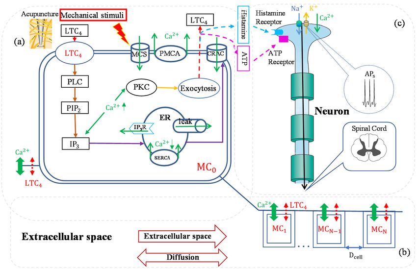
process of the mast cell activation is illustrated in Figure 4 [14,15]. In the first stage, mechan-
ical stimulations activate the mechanical sensitive ion channels on mast cells membrane
and allow calcium entry; the intracellular calcium increase activates protein kinase C (PKC)
and increases the sensitivity of secretory granules to calcium, thus driving exocytosis and
mediators release. In the second stage, the released mediators trigger cellular responses
through the G-protein linked receptors. These receptors bind to phospholipase C (PLC),
and phospholipase C catalyzes the hydrolysis of phosphatidylinosital biphosphate (PIP2)
and the release of inositol triphosphate. Inositol triphosphate acts on receptors (IP3R) of the
endoplasmic reticulum (ER) and leads the stored calcium release; the depletion of calcium
in endoplasmic reticulum triggers calcium entry through calcium release-activated calcium
(CRAC) channels. In the third stage, mediators diffuse or flow in extracellular space (ECS)
and activate other mast cells. Mediators can also bind to receptors of adjacent nerve termi-
nals (sensory neuron) and trigger action potentials, which induce passive electrical flow
from primary sensory neurons to spinal cord neurons.receptors (IP3R) of the endoplasmic reticulum (ER) and leads the stored calcium release;
the depletion of calcium in endoplasmic reticulum triggers calcium entry through calcium
release-activated calcium (CRAC) channels. In the third stage, mediators diffuse or flow
in extracellular space (ECS) and activate other mast cells. Mediators can also bind to re-
Cells 2022, 11, 860 9 of 13
ceptors of adjacent nerve terminals (sensory neuron) and trigger action potentials, which
induce passive electrical flow from primary sensory neurons to spinal cord neurons.
Figure4.4. The
Figure The bio-mathematical
bio-mathematical model
model ofof acupuncture
acupunctureeffecteffectbybythethesynergistic
synergisticaction
actionmechanism
mechanism of
interstitial
of interstitialsubstances.
substances.The
Thekinetic
kineticmodels
modelsdescribe
describethethe biochemical
biochemical response
response of of individual
individual cell
cellto
to
stimulation. The conductance models describe the transmission of electrical signals
stimulation. The conductance models describe the transmission of electrical signals in continuous in continuous
nerve fibers. The mast cells network models describe the information response between mast cells.
nerve fibers. The mast cells network models describe the information response between mast cells.
(a) The representation of single mast cell. (b) The representation of mast cells network. (c) The rep-
(a) The representation of single mast cell. (b) The representation of mast cells network. (c) The
resentation of nerve cells. Unidirectional arrows represent substance transport directions, the green
representation of nerve arrows
solid line bidirectional cells. Unidirectional arrows
represent calcium, andrepresent
the red dashsubstance transport directions,
line bidirectional the
arrows repre-
green solid line bidirectional arrows represent calcium, and the red dash line bidirectional
sent mediators such as leukotriene c-4 (LTC4) and histamine, etc. MC0 is the mast cell activated by arrows
represent
mechanicalmediators
stimuli;such as leukotriene
the steps c-4 (LTC
leading from 4 ) and histamine,
mechanical sensitivityetc. MC0 is(MSC)
channel the mast cell activated
activation to cal-
cium release from the calcium store (Endoplasmic Reticulum) into the cytosol
by mechanical stimuli; the steps leading from mechanical sensitivity channel (MSC) activation and mediators release
to
into extracellular
calcium release fromspace
thebycalcium
exocytosis
storedescribed in MC0Reticulum)
(Endoplasmic . A lane of model mast
into the cells (MC
cytosol means the
and 1mediators
first mast
release intocell from MC0 in
extracellular the flow
space direction; described
by exocytosis MC−1 means in the
MCfirst mast cell from MC0 in the contra-
0 . A lane of model mast cells (MC1
flow direction; MC N means the Nth mast cell from MC0 in the flow direction; and MC−N means the
means the first mast cell from MC0 in the flow direction; MC− 1 means the first mast cell from MC0 in
Nth mast cell from MC0 in the contra-flow direction) are separated by Dcell. Each cell exchanges bio-
the contra-flow direction; MCN means the Nth mast cell from MC0 in the flow direction; and MC− N
logical messengers through cell membrane with extracellular space. Extracellular space is regarded
means the Nth mast cell from MC0 in the contra-flow direction) are separated by Dcell . Each cell
as continuous, and diffusion and convection are included. Adapted from [14].
exchanges biological messengers through cell membrane with extracellular space. Extracellular space
is regarded
Tableas1 continuous,
shows the and diffusion
response and convection
results of mast cells are included.
and nerves. Adapted
Nerve from [14].
cells at Dist (dis-
tance from mast cell) of 200 μm, 400 μm, 600 μm, and 800 μm can be activated by media-
Table 1 shows the response results of mast cells and nerves. Nerve cells at Dist (distance
tors released from the stimulated mast cell. The response time of membrane potential (Em)
from mast cell) of 200 µm, 400 µm, 600 µm, and 800 µm can be activated by mediators
activation of the nerve cell increases with distance, causing this signal intensity to de-
released from the stimulated mast cell. The response time of membrane potential (Em )
crease. However, when the Dist is 1 × 10⁻⁴ m, the nerve cells are no longer activated, in
activation of the nerve cell increases with distance, causing this signal intensity to decrease.
other words, when the distance between the mast cell and the nerve cell is too large, there
However, when the Dist is 1 × 10⁻⁴ m, the nerve cells are no longer activated, in other
is no acupuncture
words, analgesia
when the distance effect.the mast cell and the nerve cell is too large, there is no
between
acupuncture analgesia effect.
Table 1. Response time of Em and [Ca2+ ]i (calcium concentration) peak of different Dist s [15].
Dist Response Time of Em [Ca2+ ]i Peak (µM)
2 × 10⁻⁵ m 22 s 0.33
4 × 10⁻⁵ m 24 s 0.31
6 × 10⁻⁵ m 27 s 0.29
8 × 10⁻⁵ m 33.1 s 0.27
1 × 10⁻⁴ m No 0.24Cells 2022, 11, 860 10 of 13
6. Conclusions and Discussion
Acupuncture analgesia is an internationally accepted effective treatment in Traditional
Chinese Medicine and has a wide range of applications. However, the lack of scientific
elucidation of the background mechanism has hindered its modern development, as well
as its application in mainstream medicine. In this paper, we reviewed the literature on mast
cells and acupuncture analgesia, which is a major concern in revealing the acupuncture
mechanism. These research efforts in the past decades have contributed to a scientific
explanation of acupuncture effect in all aspects: from the material basis of acupoints and
acupuncture (in anatomical, cellular, and molecular levels), to the initiation, transformation,
and propagation of acupuncture signals. Mast cells play a key role—without doubt.
Acupuncture in a broader context includes mechanical (acupuncture), electrical (elec-
troacupuncture), and heat (moxibustion) treatments. This review only focuses on acupunc-
ture. Because the main mechanical sensitive channel TRPV2 can also be activated by heat
stimulation [57], we suppose that the therapeutic mechanism of moxibustion is similar to
that of acupuncture: activate mast cells by heat or mechanism stimulation, which leads to an
analgesia effect. Some works on moxibustion support this hypothesis [80]. The mechanism
of electroacupuncture is complex; electrical stimulation may activate both mast cells and
nerve cells. Acupuncture mainly activates the local mast cell, while electroacupuncture not
only caused degranulation of mast cells at the zusanli acupoint, but also in the abdominal
cavity on the same meridian [81]. On one hand, the effect of electroacupuncture on mast
cells and mast cell-mediated analgesia is not as good as that of acupuncture. On the other
hand, unlike what was observed in acupuncture, the analgesia effect of electroacupuncture
cannot be totally blocked by the mast cell membrane stabilizer (disodium cromolyn) [82].
It reminds us that there is another mechanism involved in electroacupuncture analgesia
besides mast cell activation.
Author Contributions: Conceptualization, W.Y.; methodology, Y.L. (Yingchen Li), Y.Y., and W.Y.;
formal analysis, Y.L. (Yingchen Li), Y.Y.; resources, Y.L. (Yingchen Li), Y.Y., and Y.L. (Yuhang Liu);
validation, Y.L. (Yingchen Li), Y.Y., and W.Y.; writing—original draft preparation, Y.L. (Yingchen Li),
Y.Y., and W.Y.; writing—review and editing, Y.Y., W.Y., Y.L. (Yingchen Li), and Y.L. (Yuhang Liu),
supervision, W.Y.; project administration, W.Y.; funding acquisition, W.Y. All authors have read and
agreed to the published version of the manuscript.
Funding: This research was funded by National Natural Science Foundation of China (grant num-
ber: 12172092, 82174488) and Shanghai Key Laboratory of Acupuncture Mechanism and Acupoint
Function (grant number: 21DZ2271800).
Institutional Review Board Statement: Not applicable.
Informed Consent Statement: Not applicable.
Data Availability Statement: Not applicable.
Acknowledgments: We appreciate Mingzhu Sun for his meaningful advice on our manuscript.
Conflicts of Interest: The authors declare no conflict of interest.
References
1. Heib, V.; Becker, M.; Taube, C.; Stassen, M. Advances in the understanding of mast cell function. Br. J. Haematol. 2008, 142, 683–694.
[CrossRef]
2. Pejler, G.; Ronnberg, E.; Waern, I.; Wernersson, S. Mast cell proteases: Multifaceted regulators of inflammatory disease. Blood
2010, 115, 4981–4990. [CrossRef] [PubMed]
3. Zhang, D.; Ding, G.H.; Shen, X.Y.; Yao, W.; Zhang, Z.Y.; Zhang, Y.Q.; Lin, J.; Gu, Q.B. Role of mast cells in acupuncture effect: A
pilot study. Explore 2008, 4, 170–177. [CrossRef] [PubMed]
4. Luo, M.F.; Dong, X.T.; Song, X.J.; Jiang, J.L.; Zhan, J.; Han, Y. Study on the dynamic compound structure composed of mast cells,
blood vessels, and nerves in rat acupoint. Evid.-Based Complement. Altern. Med. 2013, 2013, 160651.
5. Wu, M.L.; Xu, D.S.; Bai, W.Z.; Cui, J.J.; Shu, H.M.; He, W.; Wang, X.Y.; Shi, H.; Su, Y.S.; Hu, L.; et al. Local cutaneous nerve terminal
and mast cell responses to manual acupuncture in acupoint li4 area of the rats. J. Chem. Neuroanat. 2015, 68, 14–21. [CrossRef]
6. Zhu, B. The sensitization phenomenon of acupoint and biological significances. Chin. Acupunct. Moxibustion 2019, 39, 115–121.Cells 2022, 11, 860 11 of 13
7. He, T.F.; Chen, Y.F. Advances in studies on the correlation between acupuncture-moxibustion treatment and mast cells.
Chin. Acupunct. Moxibustion 2010, 30, 84–87.
8. Stokes, A.J.; Shimoda, L.M.N.; Koblan-Huberson, M.; Adra, C.N.; Turner, H. A trpv2–pka signaling module for transduction of
physical stimuli in mast cells. J. Exp. Med. 2004, 200, 137–147. [CrossRef]
9. Turner, H.; Del Carmen, K.A.; Stokes, A. Link between TRPV Channels and Mast Cell Function. In Transient Receptor Potential
(TRP) Channels; Springer: Berlin/Heidelberg, Germany, 2007; pp. 457–471.
10. Solís-López, A.; Kriebs, U.; Marx, A.; Mannebach, S.; Liedtke, W.B.; Caterina, M.J.; Freichel, M.; Tsvilovskyy, V.V. Analysis of
trpv channel activation by stimulation of fcεri and mrgpr receptors in mouse peritoneal mast cells. PLoS ONE 2017, 12, e171366.
[CrossRef]
11. Huang, M.; Xie, Y.Y.; Ding, G.H. Acupoint-injection of histamine induced analgesic effect in acute adjuvant-induced-arthritis rats.
Acupunct. Res. 2010, 35, 99–103.
12. Ding, N.; Jiang, J.; Qin, P.P.; Wang, Q.X.; Hu, J.T.; Li, Z.G. Mast cells are important regulator of acupoint sensitization via the
secretion of tryptase, 5-hydroxytryptamine, and histamine. PLoS ONE 2018, 13, e194022. [CrossRef] [PubMed]
13. Zhang, X.; Li, F.; Qi, Y.; Ming, C.; Li, Y.; Pan, S.; Liu, S.; Ma, T. Electroacupuncture improves cutaneous allergic reaction
by inhibiting degranulation of intrape-ritoneal mast cells, mapk signaling and inflammatory factor levels in urticaria rats.
Acupunct. Res. 2020, 45, 299–304.
14. Yao, W.; Yang, H.W.; Li, Y.; Ding, G.H. Dynamics of calcium signal and leukotriene c-4 release in mast cells network induced by
mechanical stimuli and modulated by interstitial fluid flow. Adv. Appl. Math. Mech. 2016, 8, 67–81. [CrossRef]
15. Yao, W.; Yang, H.W.; Yin, N.; Ding, G.H. Mast cell-nerve cell interaction at acupoint: Modeling mechanotransduction pathway
induced by acupuncture. Int. J. Biol. Sci. 2014, 10, 511–519. [CrossRef]
16. Yao, W.; Huang, H.X.; Ding, G.H. A dynamic model of calcium signaling in mast cells and ltc_4 release induced by mechanical
stimuli. Chin. Sci. Bull. 2014, 59, 956–963. [CrossRef]
17. Blank, U.; Falcone, F.H.; Nilsson, G. The history of mast cell and basophil research—Some lessons learnt from the last century.
Allergy 2013, 68, 1093–1101. [CrossRef]
18. Crivellato, E.; Beltrami, C.; Mallardi, F.; Ribatti, D. Paul Ehrlich’s doctoral thesis: A milestone in the study of mast cells.
Br. J. Haematol. 2003, 123, 19–21. [CrossRef]
19. Jamur, M.C.; Oliver, C. Origin, maturation and recruitment of mast cell precursors. Front. Biosci. 2011, 3, 1390–1406.
20. Xiang, M.; Lv, J.; Zhu, X. Advances in the research about recruitment of mast cell. Med. Recapitul. 2006, 588–590.
21. Draber, P.; Sulimenko, V.; Draberova, E. Cytoskeleton in mast cell signaling. Front. Immunol. 2012, 3, 130. [CrossRef]
22. Metcalfe, D.D.; Baram, D.; Mekori, Y.A. Mast cells. Physiol. Rev. 1997, 77, 1033–1079. [CrossRef] [PubMed]
23. Metz, M.; Siebenhaar, F.; Maurer, M. Mast cell functions in the innate skin immune system. Immunobiology 2008, 213, 251–260.
[CrossRef]
24. Kraft, S.C.; Kirsner, J.B. Mast cells and the gastrointestinal tract: A review. Gastroenterology 1960, 39, 764–770. [CrossRef]
25. Franconi, G.M.; Rubinstein, I.; Levine, E.H.; Ikeda, S.; Nadel, J.A. Mechanical removal of airway epithelium disrupts mast-cells
and releases granules. Am. J. Physiol. 1990, 259, 372–377. [CrossRef] [PubMed]
26. Yong, L.C.J. The mast cell: Origin, morphology, distribution, and function. Exp. Toxicol. Pathol. 1997, 49, 409–424. [CrossRef]
27. Greenberg, G.; Burnstock, G. A novel cell-to-cell interaction between mast cells and other cell types. Exp. Cell Res. 1983, 147, 1–13.
[CrossRef]
28. Yang, H.W.; Liu, X.Y.; Shen, Z.F.; Yao, W.; Gong, X.B.; Huang, H.X.; Ding, G.H. An investigation of the distribution and location of
mast cells affected by the stiffness of substrates as a mechanical niche. Int. J. Biol. Sci. 2018, 14, 1142–1152. [CrossRef]
29. Mukai, K.; Tsai, M.; Saito, H.; Galli, S.J. Mast cells as sources of cytokines, chemokines, and growth factors. Immunol. Rev. 2018,
282, 121–150. [CrossRef]
30. Bulfone-Paus, S.; Nilsson, G.; Draber, P.; Blank, U.; Levi-Schaffer, F. Positive and negative signals in mast cell activation.
Trends Immunol. 2017, 38, 657–667. [CrossRef]
31. Christy, A.L.; Brown, M.A. The multitasking mast cell: Positive and negative roles in the progression of autoimmunity. J. Immunol.
2007, 179, 2673–2679. [CrossRef]
32. Gonzalez-Espinosa, C.; Odom, S.; Olivera, A.; Hobson, J.P.; Martinez, M.E.C.; Oliveira-dos-Santos, A.; Barra, L.; Spiegel, S.;
Penninger, J.M.; Rivera, J. Preferential signaling and induction of allergy-promoting lymphokines upon weak stimulation of the
high affinity ige receptor on mast cells. J. Exp. Med. 2003, 197, 1453–1465. [CrossRef] [PubMed]
33. Galli, S.J.; Gaudenzio, N.; Tsai, M. Mast cells in inflammation and disease: Recent progress and ongoing concerns.
Annu. Rev. Immunol. 2020, 38, 49–77. [CrossRef] [PubMed]
34. Tetè, G.; D’orto, B.; Ferrante, L.; Polizzi, E.; Cattoni, F. Role of mast cells in oral inflammation. J. Biol. Regul. Homeost. Agents 2021,
35, 65–70. [PubMed]
35. Yang, C.; Chen, N.; Tang, X.L.; Qian, X.H.; Cai, C.P. Immunomodulatory effects of IL-33 and IL-25 in an ovalbumin-induced
allergic rhinitis mouse model. J. Biol. Regul. Homeost. Agents 2021, 35, 571–581.
36. Theoharides, T.C.; Conti, P. Dexamethasone for COVID-19? Not so fast. J. Biol. Regul. Homeost. Agents 2020, 34, 1241–1243.
37. Kempuraj, D.; Selvakumar, G.P.; Ahmed, M.E.; Raikwar, S.P.; Thangavel, R.; Khan, A.; Zaheer, S.A.; Iyer, S.S.; Burton, C.;
James, D.; et al. COVID-19, mast cells, cytokine storm, psychological stress, and neuroinflammation. Neuroscientist 2020,
26, 402–414. [CrossRef]Cells 2022, 11, 860 12 of 13
38. Reber, L.L.; Sibilano, R.; Starkl, P.; Roers, A.; Grimbaldeston, M.A.; Tsai, M.; Gaudenzio, N.; Galli, S.J. Imaging protective mast
cells in living mice during severe contact hypersensitivity. JCI Insight 2017, 2, e92900. [CrossRef]
39. Grimbaldeston, M.A.; Nakae, S.; Kalesnikoff, J.; Tsai, M.; Galli, S.J. Mast cell-derived interleukin 10 limits skin pathology in
contact dermatitis and chronic irradiation with ultraviolet B. Nat. Immunol. 2007, 8, 1095–1104. [CrossRef]
40. Li, F.; He, T.; Xu, Q.; Ling, L.T.; Li, H.; Liu, Y.; Shi, G.X.; Liu, C.Z. What is the acupoint? A preliminary review of acupoints.
Pain Med. 2015, 16, 1905–1915. [CrossRef]
41. Yuan, L.; Yao, D.W.; Tang, L.; Huang, W.H.; Jiao, P.F.; Lu, Y.T.; Dai, Y.X.; Zhang, H.; He, Z.Q.; Zhong, S.Z. A study on morphological
basis of chinese acupuncture and moxibustion from digital human body. Acta Anat. Sin. 2004, 35, 337–343.
42. Fei, L.; Cheng, H.S.; Cai, D.H.; Yang, S.X.; Xu, J.R.; Chen, E.Y.; Dang, R.S.; Ding, G.H.; Shen, X.Y.; Tang, Y.; et al. Experimental
exploration and research prospect of physical bases and functional characteristics of meridians. Chin. Sci. Bull. 1998, 43, 1233–1252.
[CrossRef]
43. Yu, X.J.; Ding, G.H.; Huang, H.; Lin, J.; Yao, W.; Zhan, R. Role of collagen fibers in acupuncture analgesia therapy on rats.
Connect. Tissue Res. 2009, 50, 110–120. [CrossRef] [PubMed]
44. Song, J.M. Mast cells and meridian phenomena. Liaoning J. Tradit. Chin. Med. 1977, 4, 59–61.
45. Song, J.M. Preliminary observations on mast cells in acupuncture point tissue. Liaoning J. Tradit. Chin. Med. 1980, 3, 26–28.
46. Zhang, B.; Wang, J. Discovery of nerve-mast cell junctions in the meridian line in human skin II “Afferent” nerve-mast cell
junctions and Xue Wang cells with efferent axons. J. Neuroanat. 1985, 107–111.
47. Zhang, B.; Wang, J. Discovery of nerve-mast cell junctions in the meridian line in human skin I. Efferent nerve-mast cell
connections. J. Neuroanat. 1985, 1, 47–52.
48. He, W.; Wang, X.Y.; Shi, H.; Bai, W.Z.; Chen, B.; Su, Y.S.; Yu, X.C.; Jing, X.H.; Zhu, B. Cutaneous neurogenic inflammation in the
sensitized acupoints induced by gastric mucosal injury in rats. BMC Complementary Altern. Med. 2017, 17, 141. [CrossRef]
49. Mou, Q.J.; JI, B.; Li, Y.J.; Zhao, G.Z.; Ren, J.Y.; Li, Z.G. Research on correlation between acupoint sensitization and mast cells.
J. Clin. Acupunct. Moxibustion 2020, 36, 1–4.
50. Shi, H.; Cheng, B.; Li, J.H.; Chen, S.L.; Tan, Q.W.; Jin, Z.G.; Jing, X.H. Mast cell and substance p are involved in the process of
acupoint sensitization induced by acute gastric mucosal injury. Acupunct. Res. 2010, 35, 323–329.
51. He, W.; Wu, M.L.; Jing, X.H.; Bai, W.Z.; Zhu, B.; Yu, X.C. Entity of acupoint: Kinetic changes of acupoints in histocytochemistry.
Chin. Acupunct. Moxibustion 2015, 35, 1181–1186.
52. Wang, X.Z.; Huang, M.; Yang, H.W.; Zhang, D.; Yao, W.; Xia, Y.; Ding, G.H. Mast cell degranulation and adeno-
sine release:acupoint specificity for effect of electroacupuncture on pituitrin-induced acute heart bradycardia in rabbits.
Evid.-Based Complement. Altern. Med. 2020, 2020, 1348914. [CrossRef] [PubMed]
53. Shimbori, C.; Upagupta, C.; Bellaye, P.S.; Ayaub, E.A.; Sato, S.; Yanagihara, T.; Zhou, Q.; Ognjanovic, A.; Ask, K.; Gauldie, J.; et al.
Mechanical stress-induced mast cell degranulation activates tgf-beta 1 signalling pathway in pulmonary fibrosis. Thorax 2019,
74, 455–465. [CrossRef] [PubMed]
54. Zhang, D.; Spielmann, A.; Wang, L.; Ding, G.; Huang, F.; Gu, Q.; Schwarz, W. Mast-cell degranulation induced by physical stimuli
involves the activation of transient-receptor-potential channel trpv2. Physiol. Res. 2012, 61, 113–124. [CrossRef]
55. Yang, W.Z.; Chen, J.Y.; Zhou, L.W. Effects of shear stress on intracellular calcium change and histamine release in rat basophilic
leukemia (rbl-2h3) cells. J. Environ. Pathol. Toxicol. Oncol. 2009, 28, 223–230. [CrossRef] [PubMed]
56. Wang, L.N.; Ding, G.H.; Gu, Q.B.; Schwarz, W. Single-channel properties of a stretch-sensitive chloride channel in the human
mast cell line hmc-1. Eur. Biophys. J. EBJ 2010, 39, 757–767. [CrossRef] [PubMed]
57. Yang, L.; Li, L.; Chen, G. Progress in the study of mast cell function and mediator release mechanisms. Int. J. Lab. Med. 2010,
31, 834–836.
58. Raboune, S.; Stuart, J.M.; Leishman, E.; Takacs, S.M.; Rhodes, B.; Basnet, A.; Jameyfield, E.; Mchugh, D.; Widlanski, T.;
Bradshaw, H.B. Novel endogenous n-acyl amides activate trpv1-4 receptors, bv-2 microglia, and are regulated in brain in an
acute model of inflammation. Front. Cell. Neurosci. 2014, 8, 195. [CrossRef]
59. Fowlkes, V.; Wilson, C.G.; Carver, W.; Goldsmith, E.C. Mechanical loading promotes mast cell degranulation via rgd-integrin
dependent pathways. J. Biomech. 2013, 46, 788–795. [CrossRef]
60. Cui, X.; Liu, K.; Xu, D.; Zhang, Y.Y.; He, X.; Liu, H.; Gao, X.Y.; Zhu, B. Mast cell deficiency attenuates acupuncture analgesia for
mechanical pain using c-kit gene mutant rats. J. Pain Res. 2018, 11, 483–495. [CrossRef]
61. Huang, M.; Wang, X.Z.; Xing, B.B.; Yang, H.W.; Sa, Z.Y.; Zhang, D.; Yao, W.; Yin, N.; Xia, Y.; Ding, G.H. Critical roles of trpv2
channels, histamine h1 and adenosine a1 receptors in the initiation of acupoint signals for acupuncture analgesia. Sci. Rep.-UK
2018, 8, 6523. [CrossRef]
62. Huang, M.; Zhang, D.; Sa, Z.Y.; Xie, Y.Y.; Gu, C.L.; Ding, G.H. In adjuvant-induced arthritic rats, acupuncture analgesic effects are
histamine dependent: Potential reasons for acupoint preference in clinical practice. Evid.-Based Complementa. Altern. Med. 2012,
2012, 810512. [CrossRef] [PubMed]
63. Goldman, N.; Chen, M.; Fujita, T.; Xu, Q.; Peng, W.; Liu, W.; Jensen, T.K.; Pei, Y.; Wang, F.; Han, X.; et al. Adenosine a1 receptors
mediate local anti-nociceptive effects of acupuncture. Nat. Neurosci. 2010, 13, 883–888. [CrossRef] [PubMed]
64. Sheng, X.; Xing, J.; Han, Y.; Zhang, Y.; Yan, X.; Zhang, X. Brief introduction of Zheng’s “golden hook fishing” technique in
acupuncture. Chin. Acupunct. Moxibustion 2016, 36, 963–966.
65. Liu, Z.G. The Essence of Meridians Observed in A Special Case. Shanghai J. Acupunct. Moxibustion 1998, 17, 21–23.You can also read