Optical imaging of alpha emitters: simulations, phantom, and in vivo results
←
→
Page content transcription
If your browser does not render page correctly, please read the page content below
Optical imaging of alpha emitters:
                 simulations, phantom, and in vivo results

                 Federico Boschi
                 Sergio Lo Meo
                 Pier Luca Rossi
                 Riccardo Calandrino
                 Andrea Sbarbati
                 Antonello E. Spinelli

Downloaded From: https://www.spiedigitallibrary.org/journals/Journal-of-Biomedical-Optics on 18 Feb 2022
Terms of Use: https://www.spiedigitallibrary.org/terms-of-use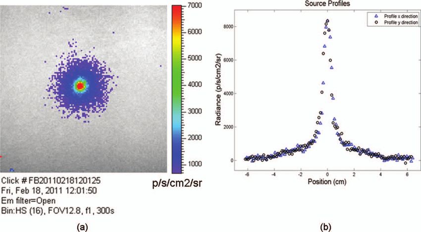
Journal of Biomedical Optics 16(12), 126011 (December 2011)

                Optical imaging of alpha emitters: simulations,
                phantom, and in vivo results
                Federico Boschi,a Sergio Lo Meo,b Pier Luca Rossi,c Riccardo Calandrino,d Andrea Sbarbati,a and Antonello E. Spinellid
                a University of Verona Department of Neurological, Neuropsychological, Morphological and Motor Sciences, Strada Le
                Grazie 8, 37134 Verona, Italy
                b Ionizing Radiation Laboratory, National Institution for Insurance against Accidents at Work (INAIL), Via Fontana
                Candida 1, I-00040, Monte Porzio Catone, Rome, Italy
                c University of Bologna, Department of Physics, Viale Berti Pichat 6/2, Bologna, Italy
                d San Raffaele Scientific Institute, Medical Physics Department, Via Olgettina N. 60, Milan, Italy

                               Abstract. There has been growing interest in investigating both the in vitro and in vivo detection of optical photons
                               from a plethora of beta emitters using optical techniques. In this paper we have investigated an alpha particle
                               induced fluorescence signal by using a commercial CCD-based small animal optical imaging system. The light
                               emission of a 241 Am source was simulated using GEANT4 and tested in different experimental conditions including
                               the imaging of in vivo tissue. We believe that the results presented in this work can be useful to describe a possible
                               mechanism for the in vivo detection of alpha emitters used for therapeutic purposes. C 2011 Society of Photo-Optical
                               Instrumentation Engineers (SPIE). [DOI: 10.1117/1.3663441]

                               Keywords: fluorescence; biology; medicine; biomedical optics.
                               Paper 11519LR received Oct. 18, 2011; revised manuscript received Oct. 28, 2011; accepted for publication Nov. 2, 2011; published
                               online Dec. 1, 2011.

                1 Introduction
                                                                                                       The detection of ultraviolet fluorescence light emission from
                Recently, there has been a growing interest in investigating
                                                                                                   nitrogen due to interactions with alpha particles in the atmo-
                both the in vitro and in vivo detection of optical photons from
                                                                                                   sphere was investigated, for example, in Ref. 7 and applied to
                a plethora of radioisotopes using CCD detectors. In earlier
                                                                                                   nuclear safeguard issues.
                works, particular attention has been focused on the detection
                                                                                                       In this paper we have investigated alpha particle induced flu-
                of Cerenkov photons from beta plus emitters;1–5 these studies
                                                                                                   orescence signal by using GEANT4 Monte Carlo (MC) simula-
                have shown that the spectrum of the emitted optical photons
                                                                                                   tions and a commercial CCD-based small animal optical imag-
                follows the predicted 1/λ2 shape, typical of the Cerenkov radia-
                                                                                                   ing system. More precisely, we measured the light emission
                tion (CR). It is well-known that the emission of CR takes place
                                                                                                   induced by a 241 Am source in different experimental conditions
                only when charged particles travel in the medium with a veloc-
                                                                                                   including the imaging of in vivo tissue. This isotope is interest-
                ity greater than the speed of light in the medium. The energy
                                                                                                   ing since the principal decay modes of 241 Am are mainly alpha
                threshold for CR emission is thus dependent on the medium and,
                                                                                                   emission and low energy gamma radiations. The decay prod-
                for example, the threshold for a beta particle traveling in water
                                                                                                   uct of 241 Am is 237 Np which has a long half-life equal to 2.1
                is 263 keV. In the last year, the investigation of weak optical
                                                                                                   × 106 years. These physical properties of 241 Am are quite useful
                emission coming from a pure gamma emitter such as Tc-99 m
                                                                                                   since they allow us to exclude any possible contribution from
                was also reported.6
                                                                                                   Cerenkov light photons generated by high energy electrons and,
                    One recent study has shown the detection of an intense light
                                                                                                   thus, gave us the possibility to estimate the contribution of alpha
                emission when using an alpha emitter such as 225 Ac.5 In this
                                                                                                   particles induced fluorescence only. We believe that the results
                case given the mass of the alpha particles, the generation of op-
                                                                                                   presented in this work can be useful to describe a possible al-
                tical photons cannot be explained in terms of Cerenkov effect
                                                                                                   ternative mechanism for the in vivo detection of alpha emitters
                and, thus, alternative explanations are needed. For example, in
                                                                                                   used for therapeutic purposes.
                Ref. 5 it has been suggested that even if the origin of optical
                                                                                                       For the sake of clarity, in order to distinguish between
                emission from 225 Ac is uncertain it can be linked to CR emit-
                                                                                                   Cerenkov luminescence imaging and luminescence induced by
                ted from the beta minus short-lived daughter nuclides such as
                213                                                                                non-Cerenkov mechanisms we use here the more general term
                    Bi. This appears to be a plausible and interesting hypothesis;
                                                                                                   of radioluminescence imaging (RLI).
                however, a more general explanation of the intense light emis-
                sion of 225 Ac can be the combination of both Cerenkov photons
                generated by the beta particles emitted by daughter nuclides and                   2 Materials and Methods
                fluorescence induced by alpha particles. The main goal of this                     2.1 RLI Simulation and Acquisition in Air and Under
                proof of principle work was to investigate the latter effect in                        a Plexiglas Slab
                different experimental conditions.
                                                                                                   RLI images of 5-mm diameter 241 Am source (The Radiochemi-
                Address all correspondence to: Federico Boschi, University of Verona, De-
                                                                                                   cal Centre, Amersham) with an activity equal to 70 kBq placed in
                partment of Neurological, Neuropsychological, Morphological and Motor Sci-
                ences, Strada Le Grazie 8, 37134 Verona, Italy. Tel: 0458027272; E-mail:           1083-3668/2011/16(12)/126011/5/$25.00
                                                                                                                                         C 2011 SPIE
                federico.boschi@univr.it.

                Journal of Biomedical Optics                                                126011-1                                      December 2011 r Vol. 16(12)

Downloaded From: https://www.spiedigitallibrary.org/journals/Journal-of-Biomedical-Optics on 18 Feb 2022
Terms of Use: https://www.spiedigitallibrary.org/terms-of-use
Boschi et al.: Optical imaging of alpha emitters: simulations, phantom, and in vivo results
air were acquired by using the IVIS 200 optical imager (Caliper model is empirical and data-driven, and uses the Evaluated Nu-
Life Sciences, Alameda, USA). The IVIS 200 is based on a clear Structure Data File9 to obtain all the information about:
cooled ( − 90◦ C) back-thinned, back-illuminated CCD camera. nuclear half-lives, nuclear level structure for nuclides, decay
The CCD has an active array of 1920 × 1920 pixels with a branching ratios, and energy of the decay process. In the event
dimension of 13 μm. that a nuclide obtained by a nuclear decay is an excited iso-
RLI spectral data of the 241 Am source in air were mea- mer, its prompt nuclear de-excitation is treated by using the
sured by acquiring six images with the narrow band (FWHM G4PhotoEvaporation class. The air fluorescence was simulated
∼20 nm) emission filters (centered on 560, 580, 600, 620, 640, by a light yield factor equal to seven optical photons for each
and 660 nm) with the following parameters: exposure time MeV (Ref. 8) of energy deposited by alpha particles crossing
= 300 s, f = 1, binning B = 8 or 32, and with a field of view the air.
= 12.8 cm. Images were acquired and analyzed with Living
Image 4.0 (Caliper Life Sciences) and were corrected for dark
measurements. 2.2 RLI Acquisition In Vivo
A lateral image of the fluorescence of air induced by alpha
In order to investigate the luminescence in vivo, the 241 Am
particles were also investigated by placing the source close to
source was placed under a mouse shoulder; the animal was a
a 45 deg mirror and the corresponding image was then focused
control nude mouse of 25 g. The amount of tissue above the
on the CCD detector. In order to further understand the origin of
source was 2 to 3 mm. Even if this cannot be considered as a
fluorescence, a small piece of carton (1-mm thick, black carton,
true physiological condition, it allowed us to investigate under
Strathmore) was placed 1 cm above the 241 Am source, to stop
known setting, e.g., source position, radioisotope activity, and
a fraction of the emitted alpha particles. A lateral image of this
tissue thickness, the signal due to tracer accumulation in a su-
experimental setup was acquired to show the missing optical
perficial region of the animal. RLI spectral data in vivo were
signal in the region above the carton.
measured by acquiring six images with the narrow band emis-
Similar to the acquisition of RLI images in air, the 241 Am
sion filters as described in Sec. 2.1. During image acquisition the
source was placed under a 2.5 slab of Plexiglas. The aver-
mouse was kept under gaseous anesthesia (2% of isoflurane and
age radiance (p/s/cm2 /sr) at different wavelengths was then
1 l/min of oxygen). All the animal handling was approved by the
obtained by placing a small region of interest (ROI) over the
Institutional Ethical Committee according to the regulations of
source.
the Italian Ministry of Health and to the European Communities
The Monte Carlo GEANT48 (4.9.0 version) was used for
Council (86/609/EEC) directives. Analogously to Sec. 2.1, the
simulation air fluorescence. The simulated setup consisted of
average radiance at the different wavelengths was obtained by
a cylinder (10 μm height; 2.5 mm radius) homogeneously
tracing on the images a small ROI around the source.
filled with 241 Am placed on a aluminium cylinder (1 mm
height; 7.5 mm radius). A thin detector, with a efficiency of
85% for optical photons in the range from 1.4 to 3.1 eV, is
placed perpendicular to the base of the 241 Am source and in
3 Results
contact with it. The simulation of the electromagnetic inter- 3.1 RLI Simulation and Acquisition in Air and Under
actions was made by using the “EM Standard” library, while a Plexiglas Slab
for nuclear decays GEANT4 provided a G4RadioactiveDecay Figure 1(a) shows a frontal RLI of the 241 Am source placed in
class to simulate the full disintegration of radioactive nuclei by air. Figure 1(b) shows the radiance profiles in the horizontal (x)
α, β + , β − emissions and electron capture. The simulation and vertical (y) directions at the center of the source.
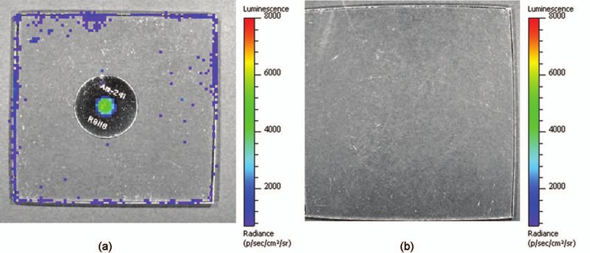
Fig. 1 (a) RLI image of the 241 Am source in air. (b) The corresponding radiance (p/s/cm2 /sr) profiles in the x-y directions at the center of the source.
Journal of Biomedical Optics 126011-2 December 2011 r Vol. 16(12)
Downloaded From: https://www.spiedigitallibrary.org/journals/Journal-of-Biomedical-Optics on 18 Feb 2022
Terms of Use: https://www.spiedigitallibrary.org/terms-of-use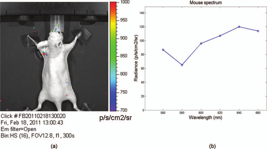
Boschi et al.: Optical imaging of alpha emitters: simulations, phantom, and in vivo results

                Fig. 2 Lateral view (obtained using a mirror) of the 241 Am source in air. (a) and (b) show, respectively, experimental and MC simulated data. (c) The
                resulting fluoresce image obtained by using a 1 mm sheet of carton positioned 1 cm above the source. The arrows point at the area above the sheet
                of carton where there is a clear loss of light signal.

                     By looking at the profiles in Fig. 1(b) one can deduce that the                   The images obtained by covering the alpha source with a
                total luminescence signal is a sum of localized intense emission                   piece of carton in Fig. 3(b) show a negligible signal in the
                in the active part of the source and a much broader signal. The                    Plexiglas; this is clear evidence that the main cause of the light
                broader component of the luminescence signal is about 4-cm                         was the fluorescence of the source.
                long, matching quite well the 241 Am alpha particles range in                          The spectra of the light obtained when placing the source in
                air.                                                                               air and under a 2.5 mm Plexiglas slab are shown in Fig. 4.
                     A lateral view of the air fluorescence induced by the 241 Am                      The spectra in Fig. 4 present different shapes and intensities
                source is presented in Fig. 2. Figure 2(a) shows the light signal                  depending on the materials. It is important to underline here
                generated by alpha particles. The corresponding Monte Carlo                        that because the spectra contains only 6 points this cannot be
                simulations shown in Fig. 2(b) fits qualitatively well with the                    considered to be a detailed precise spectral measurements, and,
                experimental findings. Figure 2(c) shows the fluorescence image                    thus, Fig. 4 provides a rough estimate of the emission spectra of
                obtained by stopping a fraction of alpha particles with a piece of                 both air and Plexiglas.
                carton as described in Sec. 2. As one can notice by comparing
                Fig. 2(a) with Fig. 2(c), there is a clear reduction of the fluores-
                cence light signal above the region covered by the carton. This is                 3.2 In Vivo RLI
                a rather simple but useful way to show that the causes of the air                  An in vivo image of luminescence induced by an alpha particle
                fluorescence were mainly alpha particles emitted by the 241 Am                     source in a mouse is shown in Fig. 5(a). It is interesting to notice
                source.                                                                            that the fluorescence signal can be clearly seen inside the red
                     Figure 3(a) shows the luminescence after covering the alpha                   ROI even if the source activity is quite modest.
                source with a Plexiglas slab. In this case the spread of the alpha                    Another interesting aspect is that the source signal is well
                particles emitted by the 241 Am source is negligible and the light                 localized under the mouse shoulder, suggesting a reasonably
                is localized only above the active part of the source.                             precise spatial delineation of the luminescence emission region.

                      Fig. 3 (a) An RLI image of the 241 Am source under 2.5 mm of Plexiglas. (b) The source was covered with a 1-mm thick piece of carton.

                Journal of Biomedical Optics                                              126011-3                                      December 2011 r Vol. 16(12)

Downloaded From: https://www.spiedigitallibrary.org/journals/Journal-of-Biomedical-Optics on 18 Feb 2022
Terms of Use: https://www.spiedigitallibrary.org/terms-of-use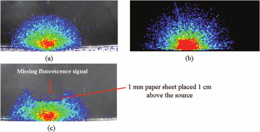
Boschi et al.: Optical imaging of alpha emitters: simulations, phantom, and in vivo results

                                                                                                        The output of Monte Carlo simulations qualitatively agreed
                                                                                                    with the experimental data obtained by imaging the alpha source
                                                                                                    in the air. This is quite an interesting aspect of the work that
                                                                                                    needs to be further developed to simulate, for example, more
                                                                                                    complex conditions. In this paper the main goal of the MC
                                                                                                    simulations was to further investigate the cause of the detected
                                                                                                    fluorescence signal in air. A quantitative comparison between
                                                                                                    the measured and simulated data was not our priority and was
                                                                                                    not possible since we did not have a detailed scheme of the
                                                                                                    imaging apparatus to be implemented in the MC simulations.
                                                                                                        Plexiglas and in vivo tissue showed a different light output
                                                                                                    response both in terms of magnitude and spectral output. As al-
                                                                                                    ready mentioned in Sec. 3 the measured spectra using the IVIS
                                                                                                    200 can be considered as an evaluation of the true emission spec-
                                                                                                    tra. A detailed spectral analysis requires the use of a dedicated
                                                                                                    spectrometer and is well beyond the scope of this paper since
                                                                                                    our main goal here was to investigate the use of a CCD-based
                                                                                                    commercial small animal optical imaging system.
                                                                                                        In vivo investigation of alpha particles tissue interactions is
                 Fig. 4 RLI spectrum of the 241 Am source in air (circles) and Plexiglas
                 (square). The spectra were acquired as described in Sec. 2.1.                      shown in Sec. 3.2, in particular Fig. 5 provides a clear evidence
                                                                                                    that alpha source induces a well localized luminescence signal
                                                                                                    in a mouse tissues. Obviously the generated light photons will
                                                                                                    suffer from scattering analogously to conventional luminescence
                                                                                                    imaging, and, thus, the in vivo spatial resolution will ultimately
                 4 Discussions and Conclusions                                                      be dependent on this effect.
                 The results presented in Sec. 3 showed that the light emission                         In our opinion the most interesting aspect of the work pre-
                 of an alpha particles source can be detected using a commer-                       sented here is the proof of concept that alpha emitters with
                 cial small animal optical imaging system. Such systems are                         no beta emission can be imaged in vivo using standard optical
                 becoming widely available in biology and preclinical imaging                       imaging methods commonly used for preclinical experiments.
                 laboratories and, thus, we believe that the results presented in                       Most of the alpha emitters used for therapeutic applications
                 this work will help to expand their range of applications.                         also emit gamma rays and, thus, they can be imaged using
                     The broad component of the radiance profiles shown in                          SPECT; however the percentage of gamma emission is rather
                 Fig. 1(b) clearly underlines a direct link between the known al-                   low, typically being a few percent of the total decays. This might
                 pha particles range in air and the extension of the region where                   increase significantly the acquisition time and requires expen-
                 the fluoresce light is emitted. The experiment performed by                        sive dedicated hardware with respect to optical imaging.
                 stopping a fraction of the alpha particles (see Fig. 2) confirms                       As shown by Miederer et al.10 the preclinical study of biodis-
                 as well that the origin of the fluorescence signal are the alpha                   tribution of 225Ac-DOTATOC for neuroendocrine tumor model
                 particles emitted by the 241 Am source.                                            was performed by scarifying the animals and then counting

                 Fig. 5 (a) RLI image of the 241 Am source (white arrow) placed under a control nude mouse (25 g). The spectrum of the emitted light by the animal
                 at the center of the red ROI can be found in (b).

                 Journal of Biomedical Optics                                              126011-4                                      December 2011 r Vol. 16(12)

Downloaded From: https://www.spiedigitallibrary.org/journals/Journal-of-Biomedical-Optics on 18 Feb 2022
Terms of Use: https://www.spiedigitallibrary.org/terms-of-use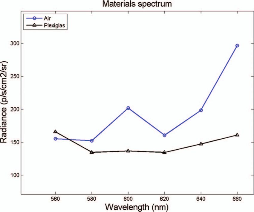
Boschi et al.: Optical imaging of alpha emitters: simulations, phantom, and in vivo results

                the organs with a gamma counter. Our optical imaging method                         4. A. E. Spinelli, F. Boschi, D. D’Ambrosio, L. Calderan, M. Marengo,
                could be used to image at different time points the same animal,                       A. Fenzi, A. Sbarbati, A. Del Vecchio, and R. Calandrino, “Cerenkov
                                                                                                       radiation imaging of beta emitters: In vitro and in vivo results,” Nucl.
                and, thus without scarifying them. This will allow the reduc-
                                                                                                       Instr. Meth. A 648, S310–S312 (2011).
                tion of the number of animals used for preclinical protocols and                    5. A. Ruggiero, J. P. Holland, J. S. Lewis, and J. Grimm, “Cerenkov
                more importantly to provide a better understanding, in a truly                         luminescence imaging of medical isotopes,” J. Nucl. Med. 51, 1123–
                molecular imaging sense, of the in vivo alpha emitters biodistri-                      1130 (2010).
                bution.                                                                             6. A. E. Spinelli, S. Lo Meo, R. Calandrino, A. Sbarbati, and F. Boschi,
                                                                                                       “Optical Imaging of Tc-99m based tracers, in vitro and in vivo results,”
                   This is quite an important and useful aspect because by using
                                                                                                       J. Biomed. Opt. (in press).
                the RLI approach we hypothesize it will be possible to perform                      7. S. M. Baschenko, “Remote optical detection of alpha particle sources,”
                cost effective preclinical studies of new radiopharmaceuticals,                        J. Rad. Prot. 24, 75–82 (2004).
                and to infer more precisely the therapeutic outcomes by imag-                       8. S. Agostinelli, J. Allison, K. Amako, J. Apostolakis, H. Araujo, P. Arce,
                ing directly the biodistribution of molecules labeled with alpha                       M. Asai, D. Axen, S. Banerjee, G. Barrand, F. Behner, L. Bellagamba,
                                                                                                       J. Boudreau, L. Broglia, A. Brunengo, H. Burkhardt, S. Chauvie, J.
                emitters.                                                                              Chuma, R. Chytracek, G. Cooperman, G. Cosmo, P. Degtyarenko, A.
                   Future work will be focused on this direction more precisely                        Dell’Acqua, G. Depaola, D. Dietrich, R. Enami, A. Feliciello, C. Fergu-
                to develop xenograft mouse tumor models and to use the RLI                             son, H. Fesefeldt, G. Folger, F. Foppiano, A. Forti, S. Garelli, S. Giani,
                approach to image the tumor progression.                                               R. Giannitrapani, D. Gibin, J. J. Gómez Cadenas, I. González, G. Gracia
                                                                                                       Abril, G. Greeniaus, W. Greiner, V. Grichine, A. Grossheim, S. Guatelli,
                                                                                                       P. Gumplinger, R. Hamatsu, K. Hashimoto, H. Hasui, A. Heikkinen, A.
                                                                                                       Howard, V. Ivanchenko, A. Johnson, F. W. Jones, J. Kallenbach, N.
                Acknowledgments                                                                        Kanaya, M. Kawabata, Y. Kawabata, M. Kawaguti, S. Kelner, P. Kent,
                The authors would like to acknowledge the Cariverona Founda-                           A. Kimura, T. Kodama, R. Kokoulin, M. Kossov, H. Kurashige, E.
                                                                                                       Lamanna, T. Lampén, V. Lara, V. Lefebure, F. Lei, M. Liendl, W. Lock-
                tion and the Ospedale Sacro Cuore Don Calabria for the financial                       man, F. Longo, S. Magni, M. Maire, E. Medernach, K. Minamimoto, P.
                support.                                                                               Mora de Freitas, Y. Morita, K. Murakami, M. Nagamatu, R. Nartallo,
                                                                                                       P. Nieminen, T. Nishimura, K. Ohtsubo, M. Okamura, S. O’Neale, Y.
                                                                                                       Oohata, K. Paech, J. Perl, A. Pfeiffer, M. G. Pia, F. Ranjard, A. Rybin,
                                                                                                       S. Sadilov, E. Di Salvo, G. Santin, T. Sasaki, N. Savvas, Y. Sawada,
                References
                                                                                                       S. Scherer, S. Sei, V. Sirotenko, D. Smith, N. Starkov, H. Stoecker,
                 1. R. Robertson, M. S. Germanos, C. Li, G. S. Mitchell, S. R. Cherry,                 J. Sulkimo, M. Takahata, S. Tanaka, E. Tcherniaev, E. Safai Tehrani,
                    and M. D. Silva, “Optical imaging of Cerenkov light generation                     M. Tropeano, P. Truscott, H. Uno, L. Urban, P. Urban, M. Verderi, A.
                    from positron-emitting radiotracers,” Phys. Med. Biol. 54, N355–N365               Walkden, W. Wander, H. Weber, and J. P. Wellisch, “Geant4: a simula-
                    (2009).                                                                            tion toolkit,” Nucl. Instrum. Meth. A 506, 250–303 (2003).
                 2. A. E. Spinelli, D. D’Ambrosio, L. Calderan, M. Marengo, A. Sbarbati,            9. J. Tuli, “Evaluated Nuclear Structure Data File,” BNL-NCS-51655-
                    and F. Boschi, “Cerenkov radiation allows in vivo optical imaging of               Rev87 (1987).
                    positron emitting radiotracers,” Phys. Med. Biol. 55, 483–495 (2010).          10. M. Miederer, G. Henriksen, A. Alke, I. Mossbrugger, L. Quintanilla-
                 3. F. Boschi, L. Calderan, D. D’Ambrosio, M. Marengo, A. Fenzi,                       Martinez, R. Senekowitsch-Schmidtke, and M. Essler, “Preclinical eval-
                    R. Calandrino, A. Sbarbati, and A. E. Spinelli, “In vivo (18)F-FDG                 uation of the α-particle generator nuclide 225Ac for somatostatin re-
                    tumour uptake measurements in small animals using Cerenkov radia-                  ceptor radiotherapy of neuroendocrine tumors” Clin. Cancer Res. 14,
                    tion,” Eur. J. Nucl. Med. 38, 120–127 (2011).                                      3555–3561 (2008).

                Journal of Biomedical Optics                                              126011-5                                            December 2011 r Vol. 16(12)

Downloaded From: https://www.spiedigitallibrary.org/journals/Journal-of-Biomedical-Optics on 18 Feb 2022
Terms of Use: https://www.spiedigitallibrary.org/terms-of-use
You can also read