New skulls of the basal sauropodomorph Plateosaurus trossingensis from Frick, Switzerland: Is there more than one species?
←
→
Page content transcription
If your browser does not render page correctly, please read the page content below
Editors' choice
New skulls of the basal sauropodomorph Plateosaurus
trossingensis from Frick, Switzerland: Is there more
than one species?
JENS N. LALLENSACK, ELŻBIETA M. TESCHNER, BEN PABST, and P. MARTIN SANDER
Lallensack, J.N., Teschner, E.M., Pabst, B., and Sander, P.M. 2021. New skulls of the basal sauropodomorph Plateosaurus
trossingensis from Frick, Switzerland: Is there more than one species? Acta Palaeontologica Polonica 66 (1): 1–28.
The Triassic basal sauropodomorph Plateosaurus trossingensis is well-known from mass accumulations at the German
localities of Trossingen and Halberstadt and the Swiss locality of Frick, and is significant especially regarding its ta-
phonomy and proposed developmental plasticity. These implications, however, rely on the assumption that this material
derives from a single species, which has been questioned. Here we describe new skull material from Frick including eight
complete and six partial skulls, more than doubling the number of known skulls of P. trossingensis. This exceptional
sample size allows for gaining a deeper understanding of variability that may occur in a single species. The new mate-
rial includes the first known juvenile skulls of Plateosaurus, allowing for detecting ontogenetic changes. An attempt is
made to distinguish between variability caused by taphonomic plastic deformation and intraspecific variability. Plastic
deformation may shorten, but not widen bones. A number of characters commonly included in phylogenetic analyses of
basal sauropodomorphs are shown to be variable within P. trossingensis, and possibly require re-evaluation. Although
P. trossingensis skulls are highly variable, many of the variable characters include intermediate character states and
therefore are continuous. No groupings based on skull features, locality, or stratigraphy are apparent. Consequently, the
analyzed skull material from the bonebeds of Frick, Trossingen, and Halberstadt bears no evidence for the presence of
more than one species.
K ey w o r d s : Dinosauria, Sauropodomorpha, Plateosaurus, ontogeny, intraspecific variability, preservation, skull ana
tomy, Triassic, Switzerland.
Jens. N. Lallensack [info@dinospuren.de], Section Paleontology, Institute of Geoscience, University of Bonn, Nussallee
8, 53115 Bonn, Germany; School of Natural Sciences and Psychology, Liverpool John Moores University, James Par-
sons Building, Bryon Street, Liverpool L3 3AF, UK.
Elżbieta M. Teschner [eteschner@uni.opole.pl], Section Paleontology, Institute of Geoscience, University of Bonn,
Nussallee 8, 53115 Bonn, Germany; Institute of Biology, University of Opole, Oleska 22, 45-052, Opole, Poland.
Ben Pabst [benpabst@bluewin.ch], Sauriermuseum Aathal, Zürichstrasse 69, 8607 Aathal-Seegräben, Switzerland.
P. Martin Sander [martin.sander@uni-bonn.de], Section Paleontology, Institute of Geoscience, University of Bonn,
Nussallee 8, 53115 Bonn, Germany; Dinosaur Institute, Natural History Museum of Los Angeles County, 900 Exposition
Boulevard, Los Angeles, CA 90007, USA.
Received 29 August 2020, accepted 15 December 2020, available online 26 February 2021.
Copyright © 2021 J.N. Lallensack et al. This is an open-access article distributed under the terms of the Creative
Commons Attribution License (for details please see http://creativecommons.org/licenses/by/4.0/), which permits unre-
stricted use, distribution, and reproduction in any medium, provided the original author and source are credited.
bonebeds sensu Sander, 1992. Large-scale excavations in
Introduction Trossingen and Halberstadt have been conducted during the
first half of the 20th century but have halted since (but see
The basal sauropodomorph Plateosaurus trossingensis Schoch and Seegis 2016). The fossil locality of Frick, the last
Fraas, 1913, from the Late Triassic of Central Europe is one bonebed to be discovered, is continuously being excavated
of the best known Triassic dinosaur species (Galton and since 1976, with the vast majority of Plateosaurus material
Upchurch 2004). The majority of the material, including doz- still awaiting description.
ens of partial to complete skeletons, stems from three local- The known Plateosaurus bonebeds are thought to have
ities: Trossingen and Halberstadt in Germany, and Frick in formed under similar taphonomic conditions. Their mono-
Switzerland (Sander 1992). These localities represent distinc- specific nature, the upright posture of the skeletons with
tive near-monospecific assemblages termed Plateosaurus the feet buried the deepest, the predominance of posterior
Acta Palaeontol. Pol. 66 (1): 1–28, 2021 https://doi.org/10.4202/app.00804.2020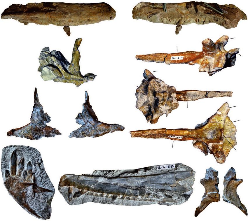
2 ACTA PALAEONTOLOGICA POLONICA 66 (1), 2021
skeletal parts, the completeness of the remains, and the lack fossil Reptilia), Berlin, Germany; MSF, Sauriermuseum
of juveniles has been interpreted as evidence for in situ mir- Frick, Frick, Switzerland; NAAG, Naturama Aargau,
ing in mud (Sander 1992). P. trossingensis may also allow Aarau, Switzerland; SMA, Sauriermuseum Aathal, Aathal-
for general insights into the evolution of endothermy—the Seegräben, Switzerland; SMNS, Staatliches Museum für
lack of a strong correlation between ontogenetic age and Naturkunde, Stuttgart, Germany.
body size indicates that this dinosaur, which was already
endothermic, retained developmental plasticity, which is
otherwise only known from ectothermic animals (Sander Material and methods
and Klein 2005; Klein and Sander 2007).
Galton (1984, 1985) assigned the sauropodomorph ma- Skull material of Plateosaurus is known from the German
terial from the bonebeds of Trossingen, Halberstadt, and localities of Trossingen (four complete and three partial
Frick to a single species, Plateosaurus engelhardti (now skulls), Halberstadt (two complete and at least one partial
P. trossingensis, see ICZN 2019), which was followed by skull), and Erlenberg (one partial skull) (Galton 1985), as
many subsequent studies (e.g., Sander 1992; Moser 2003; well as from the Swiss locality of Frick (eight complete and
Yates 2003). This classification, however, remained vague six partial skulls, described herein). The three German lo-
given the high degree of morphological variability and the calities can probably be referred to the Trossingen Formation
stratigraphic uncertainties. Galton and Kermack (2010) re- (Schoch 2011; Galton 2012), while the Swiss locality is part
ferred the Halberstadt specimens to Plateosaurus longiceps of the Klettgau Formation (Jordan et al. 2016). In the local-
and the Trossingen specimens to P. trossingensis based on ity of Trossingen, two distinct Plateosaurus bonebeds, an
differences in the pterygoid. The question regarding the upper and a lower bonebed, are present at Obere Mühle,
number of species in these bonebeds is therefore far from while Halberstadt and Frick exposed a single bonebed re-
being resolved, despite it being fundamental for the inter- spectively (Sander 1992). All of the skull material from
pretation of these mass assemblages, and therefore requiring Trossingen mentioned herein stems from the lower bonebed
further testing. Most importantly, the presence of more than of Trossingen (Galton 1985). Another skull from Trossingen
one species would question the developmental plasticity hy- (GPIT 18318a) stems from an older unit (middle Löwenstein
pothesis of Sander and Klein (2005), because taxonomic di- Formation at Untere Mühle; Hungerbühler 1998) and has
versity is the most parsimonious explanation to the observed been referred to a separate species, P. gracilis (Yates 2003);
lack of correlation between ontogenetic age and body size. this skull is not included in the present study.
The purpose of the present paper is threefold: We aim to The locality of Frick, which is primarily exposed in the
(i) describe the skull material from Frick for the first time active clay pit “Gruhalde” operated by the Tonwerke Keller
in detail, (ii) assess the contribution of taphonomic defor- AG, is the only Plateosaurus bonebed that is still being ex-
mation, ontogeny, and intraspecific variation to the mor- cavated (Fig. 1). Excavation started in 1976 after first bone
phological variability in Plateosaurus fossils, and (iii) test fragments were discovered in 1962. The yield of the first
whether or not the bonebeds of Trossingen, Halberstadt, and excavations, led by Urs Oberli, has been described by Galton
Frick contain a single Plateosaurus species based on skulls. (1986) and included two partial skulls (SMA 5 and MSF 6),
The material from Frick includes eight complete skulls, a braincase (MSF 2), and an isolated premaxilla and dentary
seven partial skulls, and a number of isolated or disartic- (MSF 1). Since then, a great quantity of material was disco
ulated skull bones, all of which are undescribed except for vered, including a first complete skeleton with skull, disco
two partial skulls and several isolated elements described vered in 1985 by Beat and Thomas Imhof (MSF 23); a com-
by Galton (1986). This material includes the first skulls of plete skull discovered in 1995 by the local museum Naturama
an early-stage as well as a late-stage juvenile, offering the Aargau; and six complete and five partial skulls discovered
first insights into ontogenetic changes of skull morphol- by an excavation team lead by one of us (BP) in annual ex-
ogy in Plateosaurus. This new material from Frick more cavation campaigns between 2004 and 2018. The majority of
than doubles the number of known skulls of Plateosaurus. the material from Frick does belong to the repository of the
Together with two complete skulls from Halberstadt and Sauriermuseum Frick (MSF); the known and prepared skull
four complete skulls from Trossingen, P. trossingensis is material from Frick is listed in Table 1.
now known from a total of 14 complete skulls. The excep- The material described herein includes the first known
tional quantity of available skulls improves our understand- juvenile skulls of Plateosaurus. MSF 12.3 comprises a
ing of preservational and intraspecific variability in basal nearly complete and articulated skull and partial postcra-
sauropodomorphs, which has implications for taxonomy. nium (femur length 44.7 cm); open neurocentral sutures in-
dicate a late juvenile stage. MSF 15.8B is the disarticulated,
Institutional abbreviations.—AMNH FARB, American partial skeleton of an early juvenile stage, as indicated by the
Museum of Natural History, Fossil Amphibian, Reptile, and small size (femur length: 24.3 cm) and the absence of suture
Bird collection, New York, USA; GPIT, Institut und Museum closure (Nau et al. 2020). This specimen was found within a
für Geologie und Paläontologie der Universität Tübingen, circular bone cluster (MSF 15.8) comprising ca. 2000 bones
Germany; MB.R., Museum für Naturkunde (collection of belonging to at least eight individuals. Referral of the post-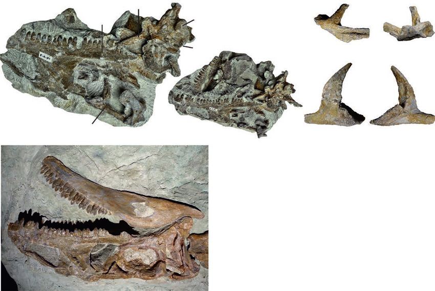
LALLENSACK ET AL.—DEFORMATIONS AND VARIABILITY IN TRIASSIC SAUROPODOMORPH                                                                         3

Fig. 1. A. Stratigraphic section of the Gruhalde Member of the Klettgau Formation as exposed in the Gruhalde Quarry in Frick, redrawn from Jordan et
al. 2016 and Nau et al. 2020 (CC BY). The studied Plateosaurus material comes from the lower and middle bonebeds, which are separated by a dolomite
marker horizon. B. Location of Frick (star) in Switzerland. C. Excavation of the find MSF 15.8 in the Gruhalde Quarry in July 2016.

cranial elements to a single individual was based on the                    fused to its basioccipital, indicating a juvenile state—it could
consistently small size and the lack of elements that are rep-              therefore possibly pertain to a second, larger juvenile indi-
resented twice (Nau et al. 2020). Several skull elements of                 vidual of MSF 15.8 that is known from postcranial remains.
MSF 15.8, including a premaxilla, a dentary, and a parietal,                    To assess the distribution of variable skull features in
can be referred to the specimen based on their small size; the              P. trossingensis as a whole, we also examined the skulls
dentary measures approximately 50% of those of the adult                    GPIT-PV-30784 (previously designated as GPIT/RE/9392,
Plateosaurus specimens studied herein. A prefrontal is com-                 GPIT 1, or UT 1), SMNS 12949, SMNS 13200, SMNS 52967,
paratively larger, which is possibly due to a proportionally                SMNS 52968, and SMNS 12950 from Trossingen, as well
larger orbit (Reisz et al. 2005) if allometry is present in the             as MB.R.1937 (previously HMN XXIV) and MB.R.4430.1
skull; this element is preliminarily referred to MSF 15.8B as               from Halberstadt. Our comparisons to AMNH FARB 6810
well. Two additional elements, a basisphenoid-parasphenoid                  from Trossingen are based on the detailed descriptions
(MSF 15.8.1043) and a squamosal (MSF 15.8.2030), have ini-                  of Galton (1984) and Prieto-Márquez and Norell (2011).
tially been listed as parts of MSF 15.8B (Nau et al. 2020), but             Additional photographs of the examined skulls are provided
are distinctly larger in size and therefore probably pertain to             in the SOM (Supplementary Online Material available at
different individuals. The basisphenoid-parasphenoid is not                 http://app.pan.pl/SOM/app66-Lallensack_etal_SOM.pdf).
4                                                                                  ACTA PALAEONTOLOGICA POLONICA 66 (1), 2021

Table 1. Plateosaurus skull material from the lower and middle “saurian levels” of Frick, Switzerland.
    Specimen number                 Material                        Excavation                           Repository            Reference
                           premaxilla and dentary, iso-
MSF 1                                                            Urs Oberli 1976           Sauriermuseum Frick, Switzerland   Galton 1986
                                      lated
MSF 2                              braincase                     Urs Oberli 1976Sauriermuseum Frick, Switzerland Galton 1986
                                                                                     Sauriermuseum Aathal,
MSF 5                      disarticulated skull            Urs Oberli 1976                                         Galton 1986
                                                                                  Aathal-Seegräben, Switzerland
                       anterior part of skull and                                    Umwelt Arena Schweiz,
MSF 6                                                      Urs Oberli 1976                                         Galton 1986
                                  dentary                                           Spreitenbach, Switzerland
MSF 33                       isolated dentary              Urs Oberli 1976      Sauriermuseum Frick, Switzerland    this study
MSF 23                        complete skull         Beat and Thomas Imhof 1985 Sauriermuseum Frick, Switzerland    this study
NAAG_00011238                 complete skull            Rainer F. Foelix 1995  Naturama Aargau, Aarau, Switzerland this study
NAAG_00011239       partial skull, isolated elements    Rainer F. Foelix 1995  Naturama Aargau, Aarau, Switzerland this study
                                                                                   Museum of Natural Sciences,
MSF 07.M               braincase and lower jaws             Ben Pabst 2007                                          this study
                                                                                        Brussels, Belgium
MSF 08.M              partial skull with braincase          Ben Pabst 2008        University of Bonn, Germany       this study
MSF 08.H                    isolated elements               Ben Pabst 2008      Sauriermuseum Frick, Switzerland    this study
                                                                                     Sauriermuseum Aathal,
SMA 09.1 (SMA 0255)       skull with braincase              Ben Pabst 2009                                          this study
                                                                                  Aathal-Seegräben, Switzerland
MSF 09.2                       partial skull                Ben Pabst 2009      Sauriermuseum Frick, Switzerland    this study
MSF 11.4                      complete skull                Ben Pabst 2011      Sauriermuseum Frick, Switzerland    this study
MSF 12.3                      complete skull                Ben Pabst 2012      Sauriermuseum Frick, Switzerland    this study
MSF 15.4                      complete skull                Ben Pabst 2015      Sauriermuseum Frick, Switzerland    this study
                     juvenile skull (MSF 15.8B);
MSF 15.8                                                Ben Pabst 2015–2016     Sauriermuseum Frick, Switzerland    this study
                            isolated elements
MSF 16.1                      complete skull                Ben Pabst 2016      Sauriermuseum Frick, Switzerland    this study
MSF 17.4                      complete skull                Ben Pabst 2017      Sauriermuseum Frick, Switzerland    this study

Photogrammetry.—Photogrammetric models of the cranial                    concluded that the lower bonebed of Trossingen is late
material from Frick, including all complete skulls, MSF                  Norian, while the upper bonebed is early Rhaetian in age.
08.M, the braincase of MSF 07.M, and NAAG_00011239,                          The dinosaur-bearing section in Frick may be subdivided
were built using Agisoft Metashape Professional (www.agi-                into three horizons. The Plateosaurus material analyzed
soft.com), and are available in the SOM. Orthophotos as                  herein stems from the lower two of these horizons, the lower
well as ambient occlusions, which enhance 3D morphology                  and middle saurian levels, which are separated by a dolomite
by shading points of the surface that are occluded by sur-               marker horizon (Fig. 1A). The time interval represented by
rounding objects, were extracted from the 3D models us-                  these levels is unknown. The Gruhalde Member has been es-
ing MeshLab (meshlab.net) and are shown in orthographic                  timated to represent approximately 20 Myr, while the lower
projection. Shading by low-angled virtual light was used                 and middle saurian levels together account for 20% of the
in some cases to further enhance surface detail. In addi-                total thickness of the Gruhalde Member in the Gruhalde
tion to the direct examination of the skulls, the digital 3D             Quarry (Jordan et al. 2016); however, it cannot be necessarily
models displayed in orthographic projection were used for                assumed that the sedimentation rate was constant, and wide-
comparisons between specimens. Details on these visual-                  spread pedogenesis may indicate sedimentation hiatuses.
ization methods can be found in Lallensack et al. (2020).                    The lower and middle saurian levels are mostly com-
Photogrammetric models of the Plateosaurus skull mate-                   posed of grayish to variegated marl of high carbonate con-
rial from Frick are available at https://doi.org/10.6084/m9.             tent that contain intraclasts and dolomite lenses. Layers are
figshare.12890672.                                                       non-continuous, and the marl is cut by channel structures
                                                                         composed of silty marl to coarse-grained sandstone. While
Geology of the Gruhalde Quarry.—Plateosaurus remains                     the majority of Plateosaurus fossils come from the marl,
in Frick stem from the middle part of the Gruhalde Member                they can also be found in the sandstone. The marl has been
of the Klettgau Formation (Jordan et al. 2016), in a succes-             interpreted as overbank deposits affected by pedogenesis
sion known as the “Obere Bunte Mergel” (Upper Variegated                 within a terrestrial playa environment (Jordan et al. 2016).
Marls). Although stratigraphical correlations are not possi-                 The state of preservation of the Plateosaurus specimens
ble at present, this succession is thought to be at least partly         found in Frick ranges from accumulations of a few bones to
equivalent to Plateosaurus-bearing strata of Trossingen and              complete skeletons; isolated bones are relatively rare (Sander
Halberstadt, which are commonly considered late Norian                   1992). Specimen density is high, and the lateral distance be-
in age (Sander 1992; Jordan et al. 2016). Etzold et al. (2010)           tween specimens is sometimes a few meters only. Exposures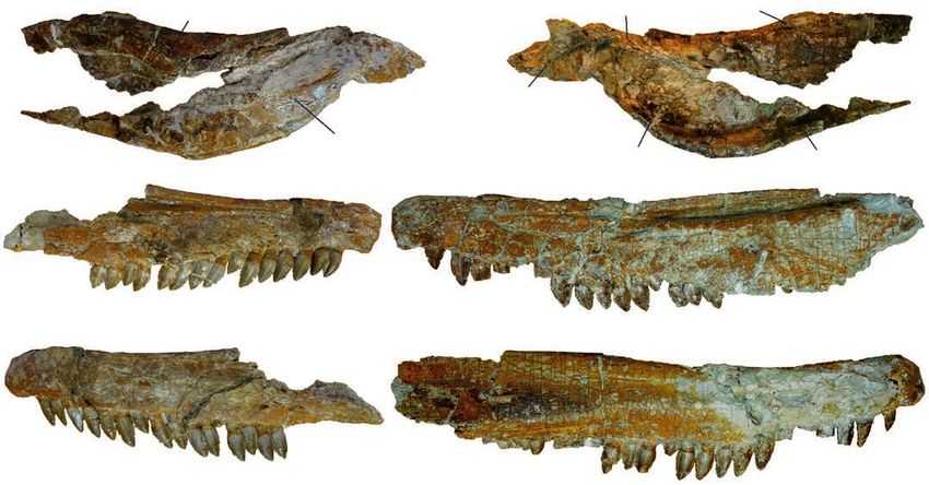
LALLENSACK ET AL.—DEFORMATIONS AND VARIABILITY IN TRIASSIC SAUROPODOMORPH 5
of the horizons at the Frickberg at the other side of the Frick
valley, almost 2 km away from the Gruhalde Quarry, contain
equally abundant Plateosaurus remains and suggest an ex-
tent of the bonebed on the order of square kilometers.
Sauropodomorph remains, as well as the theropod Nota
tesseraeraptor frickensis Zahner and Brinkmann, 2019,
were also found in the upper saurian level of the Gruhalde
Quarry, immediately below the discordantly overlying ma-
rine sediments of the Early Jurassic (Zahner and Brinkmann
2019). The upper saurian level differs in lithology, and the
bone material is black in color, not orange-gray as in the
lower and middle levels, indicating different sedimentolog-
ical conditions. As these sauropodomorph remains possibly
represent a novel taxon (Zahner and Brinkmann 2019), they
will be described elsewhere.
Results
Preservation.—Plateosaurus fossils from Frick are af-
fected by plastic deformation (i.e., deformation without
breakage) to a stronger degree than those from Trossingen
and Halberstadt, complicating interpretation and causing
loss of information (Fig. 2). The impact of such deformation
on features of potential taxonomic relevance can be best
studied by comparing mediolaterally compressed skulls
with those that are compressed obliquely or dorsoventrally,
and by analyzing left-right asymmetry in single skulls. Of
the eight complete skulls, three are compressed mediolater-
ally, three obliquely, and two dorsoventrally. The deforma-
tion can be extreme, as seen in the case of the skull MSF
16.1, which is less than 10% as wide as long due to oblique
lateral compression. In obliquely compressed skulls, the Fig. 2. Bone shortening due to plastic deformation. A. Compression may
dorsal surface of the skull is often projected onto a single lead to substantial shortening along the axis of deformation. Note that no
plane with the lateral surface, exposing the skull roof on widening occurs. B. Right humerus of MSF 12.3 in posterior (B1) and me-
one side of the skull but not on the other. This can be most dial (B2) views, showing extreme anteroposterior compression especially
clearly seen in MSF 16.1 (Fig. 3). of the distal end. Despite this compression, the bone is not significantly
It is important to note, however, that plastic deformation widened in posterior view when compared with the relatively undeformed
humerus of GPIT-PV-30785.
only leads to shortening of fossils, but not to widening,
because the sediment package containing the fossils is de- include changes in aspect ratios, angles, curvature, and relief.
formed as a whole (Fig. 2A; Walton 1936; Harris 1974; Rex For example, mediolaterally compressed skulls will have a
and Chaloner 1983). This may be demonstrated based on higher length-to-width ratio in dorsal and ventral views, and
the distal end of the right humerus of MSF 12.3, which is a lower height-to-width ratio in anterior and posterior views.
flattened anteroposteriorly to an extreme degree (Fig. 2B). Angles may become larger or smaller; for example, the angle
Despite this extreme deformation, no widening is apparent between the paroccipital processes will be more acute in a
in posterior view (parallel to the axis of deformation), and mediolaterally compressed skull. The curvature and relief of
the ratio between the length and mediolateral width of this a ridge is reduced when directed towards the axis of defor-
bone is almost equal to that of the relatively undeformed mation, but exaggerated when directed parallel to it. These
humerus of GPIT-PV-30785 (previously GPIT 2; 3.35 versus effects are determined by two factors: the magnitude of de-
3.37, respectively). Consequently, linear measurements tend formation and the orientation of the fossil relative to the axis
to underestimate, but not overestimate, the original absolute of deformation. The latter factor determines how the original
dimensions, allowing to constrain the influence of deforma- shape is projected onto the flattened fossil, and may be dif-
tion to some extent. ficult to reconstruct, especially when the original orienta-
Below, we describe the effects of a simple compression tion of the fossil within the sediment was not documented
assuming a single axis of deformation. These effects, which three-dimensionally. Slight variations in the orientation of
can be easily explored by transforming 3D models of skulls, the fossils will result in different projections, potentially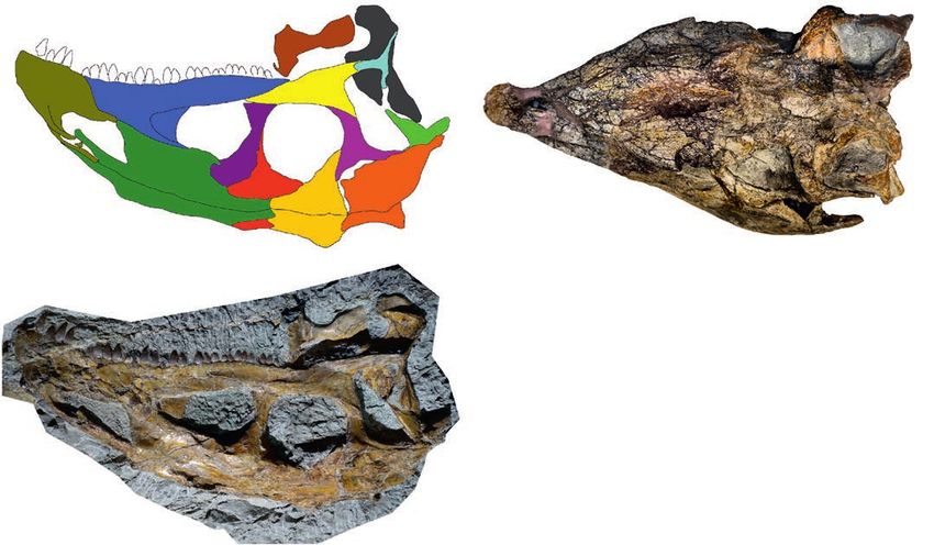
6 ACTA PALAEONTOLOGICA POLONICA 66 (1), 2021
Fig. 3. Skull of Plateosaurus trossingensis (MSF 16.1) from the Triassic of Frick, Switzerland. Photograph of the left side (A1); ambient occlusion of
the 3D-model of the left side (A2); photograph of the right side (A3) and interpretative diagram (A4). Abbreviations: an, angular; d, dentary; f, frontal;
j, jugal; l, lacrimal; mx, maxilla; na, nasal; pf, prefrontal; pmx, premaxilla; po, postorbital; pt, pterygoid; q, quadrate; qj, quadratojugal; sa, surangular;
sq, squamosal; ud, undefined.
leading to significant variation in apparent shape. If both the and therefore is an oversimplification. Although the fossil
magnitude of deformation and the orientation of the fossil bearing layers are primarily composed of mudstone, they
relative to the axis of deformation are known, the original also contain sandstone and dolomite as well as intraclasts,
shape may potentially be received through retrodeformation. and sediment composition may change at a small scale.
This requires finding a combination of the two factors that Bones may also be appressed upon other bones, and the
meets assumptions such as symmetry between the left and degree of ossification of the bone itself might further in-
right sides and known length-to-width ratios of elements. In fluence the magnitude of deformation (Fig. 2B; Hugi and
practice, the magnitude of deformation is not relevant when Scheyer 2012). These factors may result in varying degrees
the direction of view is parallel to the axis of deformation: a of deformation, also within single elements. In addition to
skull that was compressed dorsoventrally and does not show shortening along a single axis, the fossils may be affected
obvious skewing thus may preserve its approximate original by substantial shearing. This is best seen in MSF 11.4, where
shape in dorsal or ventral views, irrespective of the magni- the left side of the skull is displaced anteriorly by almost
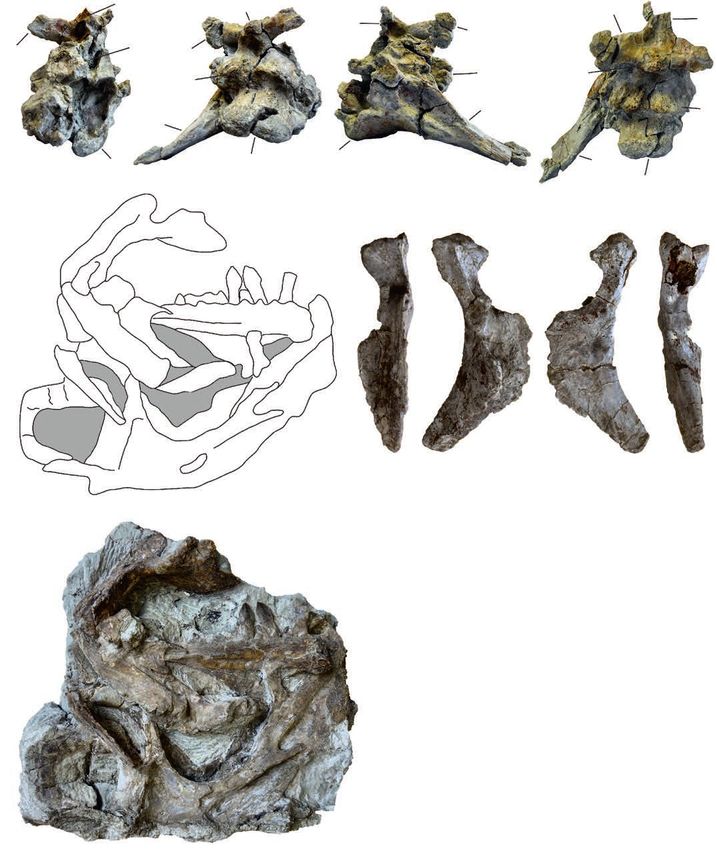
tude of deformation. 2 cm relative to the right side, resulting in an anteroposterior
The model described above, however, can only partly ex- shortening of the left side of the skull by approximately 25%
plain the preservational variability in the examined skulls, in relation to the right side (Fig. 4A5, A6).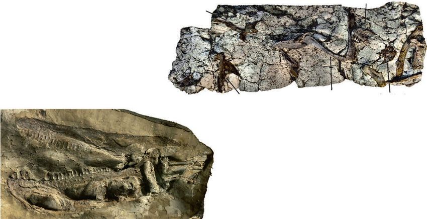
LALLENSACK ET AL.—DEFORMATIONS AND VARIABILITY IN TRIASSIC SAUROPODOMORPH                                                                                          7

Fig. 4. A. Skull of Plateosaurus trossingensis (MSF 11.4) from the Triassic of Frick, Switzerland. Photograph of the right side (A1) and interpretative
diagram (A2); ambient occlusion of the 3D-model of the right side (A3) and interpretative diagram (A4); photograph in dorsal view (A5) and interpreta-
tive diagram (A6). B. The jugal-quadratojugal articulation as seen in GPIT-PV-30784 from Trossingen. Abbreviations: an, angular; cb, ceratobranchial;
d, dentary; f, frontal; j, jugal; l, lacrimal; mx, maxilla; na, nasal; p, parietal; pf, prefrontal; pmx, premaxilla; po, postorbital; pra, prearticular; q, quadrate;
qj, quadratojugal; sa, surangular; sq, squamosal.

    In addition to plastic deformation, mechanical crushing                             Faults frequently occur in the fossil-bearing layers due to
may also occur. This is most evident in MSF 16.1, where                             differential subsidence and may separate fossils into halves
the right mandible is broken at the mandibular fenestra,                            offset from each other, as seen, e.g., in the posterior part of the
with the dentary angled 110° relative to the posterior part                         left side of the skull of MSF 16.1 (Fig. 3A1, A2). At a smaller
of the mandible (Fig. 3). In MSF 15.4, the premaxilla is                            scale, differential compression of separate parts of a bone can
appressed onto an intraclast and fragmented (Fig. 5A1,                              lead to the formation of shear zones whereby parts of the bone
A 2). In MSF 23 (Fig. 6A), the articulated block composed                           surface are depressed relative to other parts, as seen in the
of the postorbital, frontal, parietal, and squamosal was                            premaxilla of NAAG_00011238, for example (Fig. 7A1, A2).
shifted anteriorly and ventrally, exposing the supratem-                                Both articulated skulls and isolated skull elements are
poral fenestra in lateral view of the skull and reducing the                        found at Frick, though the high proportion of articulated and
orbit to a narrow slit while the antorbital fossa retains its                       complete skulls is remarkable even when accounting for the
original shape. A similar foreshortening of the orbit is seen                       fact that some of the more fragmentary material still awaits
on the right side of GPIT-PV-30784. Sander (1992) instead                           preparation (Table 1). The Frick material encompasses eight
suggested that this foreshortening was the result of a char-                        nearly complete and articulated skulls as well as seven par-
acteristic disarticulation pattern.                                                 tial skulls of which three (MSF 09.2, MSF 08.M, MSF 6) are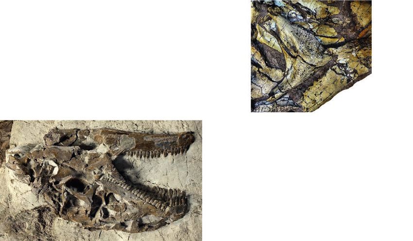
8                                                                                      ACTA PALAEONTOLOGICA POLONICA 66 (1), 2021

Fig. 5. A. Skull of Plateosaurus trossingensis (MSF 15.4) from the Triassic of Frick, Switzerland. Photograph in lateral view (A1); ambient occlusion of
3D-model (A2); interpretative diagram (A3). B. Photograph of SMNS 12949 in dorsal view. Abbreviations: f, frontal; j, jugal; l, lacrimal; mx, maxilla;
na, nasal; par, paroccipital process; pf, prefrontal; pmx, premaxilla; po, postorbital; pt, pterygoid; q, quadrate; qj, quadratojugal; sq, squamosal.

articulated and two are disarticulated (MSF 5 and the juve-                   individual from Frick known from an articulated skull. Even
nile MSF 15.8B). Of the articulated skulls, the degree of ar-                 smaller is MSF 12.3 at 12.5 cm. The early-stage juvenile
ticulation varies. MSF 15.4, MSF 16.1, and NAAG_00011238                      MSF 15.8B (Nau et al. 2020) lacks the maxilla and is not
are best articulated, while in MSF 11.4 most elements are                     included in this comparison; the length of the dentary of
slightly shifted out of their anatomical position.                            this specimen is 8.7 cm, measured from the distal tip to the
    Sander (1992) proposed that disarticulation began at the                  anteriormost point of the gap between the posterodorsal and
nasofrontal suture, and that the posterior skull bones disar-                 posteroventral process of the dentary. Excluding the juvenile
ticulated more rapidly than those of the rostrum, with the na-                skull MSF 12.3, the laterally exposed surface averages at
sal, premaxilla, and maxilla remaining as one unit. However,                  16.7 cm in length in the Frick skulls. Skulls from Trossingen
based on our much larger sample size, we now propose that                     and Halberstadt (SMNS 13200, SMNS 12950, SMNS 12949,
the most frequent disarticulation in the complete skulls is                   GPIT-PV-30784, MB.R.1937, and AMNH FARB 6810) av-
that of the premaxilla (seen in MSF 17.4; MSF 23; MSF 12.3;                   erage at 16.6 cm. There is, therefore, no evidence for differ-
MSF 11.4; SMNS 12949; GPIT-PV-30784); maxilla (seen in                        ences in body size between the three localities.
MSF 17.4; MSF 11.4; MSF 23); and jugal (seen in MSF 23;                           The skull of Plateosaurus is commonly reconstructed
MSF 11.4; SMNS 12949; GPIT-PV-30784; missing in SMA                           as mediolaterally narrow compared to other basal sau-
09.1, MSF 17.4 and on the left side of MSF 16.1).                             ropodomorphs. However, the relative width of the skull is
                                                                              highly variable in known specimens, and mostly depends on
Osteology.—Size, proportions, and skull openings: Skull                       the degree and direction of plastic deformation (Galton and
size was quantified by measuring the anteroposterior length                   Upchurch 2004). Published idealized skull reconstructions
of the laterally exposed surface of the maxilla, because this                 are likewise variable: the length-to-width ratio, measured
measurement was available from the majority of skulls. In                     from the posterior end of the parietal suture to the tip of
the eight articulated skulls from Frick, the lateral surface                  the premaxillae and across the greatest width at the level of
of the maxilla measures 16.1 cm on average. Only three of                     the postorbitals, varies between 2.0 in the reconstruction of
the skulls are notably larger or smaller: NAAG_00011238                       Wilson and Sereno (1998: fig. 5A) and 2.27 in that of Galton
is, at 20.3 cm (total skull length: 36.27 cm), the largest skull              and Upchurch (2004: fig. 12.2A). Because plastic deforma-
known from Frick and the largest of Plateosaurus from any                     tion results in bone shortening but not widening, as was dis-
locality. MSF 23 is, at 14.5 cm, probably the smallest adult                  cussed above, skull width will be underestimated in the vast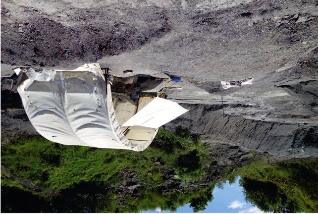
LALLENSACK ET AL.—DEFORMATIONS AND VARIABILITY IN TRIASSIC SAUROPODOMORPH                                                                                     9

Fig. 6. A. Skull of Plateosaurus trossingensis (MSF 23) from the Triassic of Frick, Switzerland. Photograph in lateral view (A1) (by Georg Oleschinski,
University of Bonn, Germany); ambient occlusion of photogrammetric model (A2); interpretative diagram (A3). B. Detail of lacrimal and prefrontal of
the right side of GPIT-PV-30784 from Trossingen. Abbreviations: bo, basioccipital; br, braincase; cb, ceratobranchial; d, dentary; f, frontal; j, jugal; l, lac-
rimal; mx, maxilla; na, nasal; p, parietal; pf, prefrontal; pmx, premaxilla; po, postorbital; pra, prearticular; pt, pterygoid; q, quadrate; sa, surangular; sp,
splenial; sq, squamosal; ud, undefined.

majority of skulls, which are compressed mediolaterally. Of                       0.66 times orbit length (Fig. 4A1–A4), while it is between
the articulated skulls from Trossingen and Halberstadt, only                      0.94 and 1.02 in most other skulls. However, the external
SMNS 12949 is primarily compressed dorsoventrally, sug-                           naris is similarly small in AMNH FARB 6010, and the an-
gesting that its length-to-width ratio of 2.2 is closer to the                    torbital fossa is similarly small in AMNH FARB 6010 and
original ratio (Fig. 5B). This skull, however, is also skewed                     SMNS 12949, demonstrating that the same variability is
towards the left side, indicating a somewhat oblique main                         present in the skulls from Trossingen. In NAAG_00011238,
axis of deformation which may have caused mediolateral                            the infratemporal fenestra is markedly narrower antero-
shortening as well. The only complete skull that lacks evi-                       posteriorly compared to the orbit than in all other skulls
dent mediolateral shortening is the strongly dorsoventrally                       (Fig. 7A).
compressed skull of MSF 12.3 from Frick, which belongs to                             The external naris is more or less triangular in shape,
a late-stage juvenile and has a length-to-width ratio of 1.95,                    typically with a subvertical posterior margin, a subhori-
indicating a skull broader than in previous estimates for                         zontal ventral margin, and an anteroventrally inclined
Plateosaurus (Fig. 8). We argue that these broader propor-                        dorsal margin (e.g., Fig. 4A4). The anteroventral corner of
tions may reflect the original proportions of Plateosaurus                        the external naris can be broadly rounded, as in MSF 11.4
skulls in general, although we cannot fully dismiss the pos-                      (Fig. 4A1–A4) and SMNS 13200, or form a much more acute
sibility that the proportions seen in MSF 12.3 were influ-                        angle, as in MSF 16.1 (Fig. 3A3, A4) and MB.R.1937. The
enced by other possible factors such as skull elongation                          latter condition is probably partly influenced by deforma-
with ontogeny, intraspecific variability, or anteroposterior                      tion. The ventral margin of the external naris is located dor-
shortening due to plastic deformation.                                            sal to that of the antorbital fossa. The antorbital fossa is typ-
    The relative sizes of the external naris, antorbital fossa,                   ically trapezoidal in shape, with a posterodorsally inclined
and orbit are comparable in the skulls MSF 15.4, MSF 16-4,                        anterior margin, a slightly posterodorsally inclined dorsal
and NAAG_00011238, but are aberrant in MSF 11.4. In the                           margin, and a anterodorsally inclined posterior margin (e.g.,
latter, the maximum length of the antorbital fossa is 1.08                        Fig. 4A4). The dorsal margin of the antorbital fossa is slightly
times that of the orbit (Fig. 4A1–A4), while it is 1.2 to 1.3 in                  (e.g., NAAG_00011238) or substantially (e.g., MB.R.1937)
the other skulls. The external naris is small in MSF 11.4 at                      lower than the dorsal margin of the orbit; this variation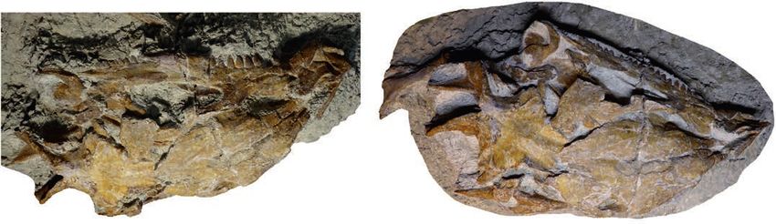
10                                                                                        ACTA PALAEONTOLOGICA POLONICA 66 (1), 2021

Fig. 7. A. Skull of Plateosaurus trossingensis (NAAG_00011238) from the Triassic of Frick, Switzerland. Photograph in lateral view (A1) (by Georg
Oleschinski, University of Bonn, Germany); ambient occlusion of photogrammetric model (A2); interpretative diagram (A3). B. Orthophoto of photo-
grammetric model of disarticulated skull material (NAAG_00011239). Abbreviations: d, dentary; f, frontal; fo, foramen magnum; j, jugal; mx, maxilla;
na, nasal; p, parietal; pf, prefrontal; pmx, premaxilla; po, postorbital; pt, pterygoid; q, quadrate; qj, quadratojugal; sa, surangular; scl, sclerotic ring;
sq, squamosal.

might be attributed to deformation. The orbit is subcircular                    MSF 12.3, MSF 1, MSF 15.8B, and probably MSF 15.4, while
and slightly longer than high (e.g., Fig. 4A4); it is as long as                MSF 23, MSF 16.1, and MSF 11.4 show six premaxillary
high on the right side of MSF 16.1 (Fig. 3A3, A4). The infra-                   tooth positions. The main body of the premaxilla is longer
temporal fenestra is dumbbell-shaped and anteroposteriorly                      than high in all skulls. In juvenile to subadult individuals of
constricted in the middle due to its convex posterior margin.                   Mussaurus and Massospondylus, the premaxilla is shorter
The anterior margin of the infratemporal fenestra can be                        relative to its height than in adults (Pol and Powell 2007). In
straight (e.g., MSF 15.4, Fig. 5A; MB.R.1937), or slightly                      the late-stage juvenile MSF 12.3, the left premaxilla is indeed
convex (e.g., Fig. 4A4) and contributing to the constriction.                   shorter than in any other of the examined skulls (Fig. 8A1,
The dorsal half of the infratemporal fenestra is more narrow                    A2), but this is at least partly the result of shortening due to
than the ventral half in MB.R.1937, while the opposite is the                   plastic deformation. The premaxilla is elongated in the ear-
case in NAAG_00011238 (Fig. 7A); these differences are                          ly-stage juvenile MSF 15.8B (Fig. 9A).
possibly due to plastic deformation. The extended postero-                          The ventral margin of the premaxilla is straight or
ventral corner of the infratemporal fenestra (Pol and Powell                    slightly concave in most specimens from Frick (MSF
2007) that is strongly pronounced in SMNS 13200 appears                         12.3, MSF 15.4, NAAG_00011238, SMA 09.1, MSF 16.1,
to be the result of a deformation of the quadratojugal (see                     MSF 1) and in the examined skulls from Trossingen and
section “quadratojugal” below). The ventral margin of the                       Halberstadt, but is strongly curved and sloping ventrally
infratemporal fenestra is substantially lower than that of                      in MSF 11.4 (Fig. 4A1–A4) and the juvenile specimen MSF
the orbit in all skulls. The supratemporal fenestra is subtri-                  15.8B (Fig. 9A), although the latter lacks the distal tip of the
angular in shape, with a posteromedially oriented anterior                      premaxilla. A ventrally sloping premaxilla was found to be
margin, a posterolaterally oriented posterior margin, and                       present in Massospondylus (Chapelle and Choiniere 2018).
a anterolaterally oriented lateral margin (e.g., MSF 12.3,                      The anterior margin of the main body of the premaxilla is
Fig. 8A1, A2; SMNS 13200; SMNS 12949; MB.R.1937).                               typically rounded, with the anteriormost point somewhat
    Premaxilla: The premaxilla consists of a main body as                       elevated above the level of the alveolar margin (e.g., MSF
well as a dorsal and a posterior process, which delimit the ex-                 11.4, Fig. 4A1–A4), but can be relatively straight (e.g., SMNS
ternal naris in its anterior, anterodorsal, and ventral extents.                52968), although not reaching the very straight condition
There are five premaxillary tooth positions in SMA 09.1, in                     seen in Massospondylus (Chapelle and Choiniere 2018;
NAAG_00011238, the two premaxillae of NAAG_00011239,                            McPhee et al. 2019). Plateosaurus has been coded as having
LALLENSACK ET AL.—DEFORMATIONS AND VARIABILITY IN TRIASSIC SAUROPODOMORPH 11
Fig. 8. A. Skull of the late-stage juvenile Plateosaurus trossingensis (MSF 12.3) from the Triassic of Frick, Switzerland. Ambient occlusion of pho-
togrammetric model in dorsal view (A1) and interpretative diagram (A2); photograph of the skull in oblique view (A3); photograph of the right lateral
view (A4) and interpretative diagram (A5). Abbreviations: at, atlas; d, dentary; f, frontal; j, jugal; l, lacrimal; ls, laterosphenoid; mx, maxilla; na, nasal;
oph, opisthotic; p, parietal; pf, prefrontal; pmx, premaxilla; po, postorbital; pra, prearticular; q, quadrate; qj, quadratojugal; sa, surangular; so, supraoccipi
tal; sq, squamosal; ud, undefined.
a convex anterodorsal margin of the premaxilla without an a greater variability: in ANMH FARB 6810, the anterior
inflection at the base of the dorsal process (Upchurch 1995; extent of the external naris is almost level with the posterior
Yates 2007). However, such an inflection is pronounced margin of the main body of the premaxilla (Prieto-Márquez
in NAAG_00011238 and the disarticulated premaxilla of and Norell 2011), while the external naris extends as far as
NAAG_00011239 (Fig. 7), and is also seen in MB.R.1937 30% on the right side of SMNS 13200. The narial fossa is
from Halberstadt and SMNS 52968 from Trossingen. It is continuous with the dorsal surface of the posterior process
only weakly pronounced in MSF 11.4 and the juvenile MSF and the ventral surface of the dorsal process, and delimited
15.8B, and absent in SMNS 13200 and AMNH FARB 6810 anteriorly, dorsally and ventrally by a rim, as best seen in
(Prieto-Márquez and Norell 2011). MSF 11.4 (Fig. 4A1, A3), but also in MSF 16.1 (Fig. 3), MSF
Dorsally, the main body of the premaxilla borders both 1, SMNS 52968, SMNS 13200, and MB.R.1936. In MSF 1,
the external naris and the narial fossa. The anterior extent the rim is broad and elevated above the lateral surface of
of the external naris is at approximately 20–25% of the the main body of the premaxilla; this could, however, be the
length of the main body of the premaxilla, as seen in MSF result of differential deformation of the central part of the
11.4, NAAG_00011238, MSF 16.1, and MSF 15.4. The ante- premaxilla. The extent of the narial fossa is variable. It is
rior extent is slightly larger in the juvenile specimen MSF smallest in SMA 09.1, where the distance between the ante-
15.8B (Fig. 9A). Plateosaurus skulls from Trossingen show rior extent of the external naris and that of the narial fossa12 ACTA PALAEONTOLOGICA POLONICA 66 (1), 2021 Fig. 9. Plateosaurus trossingensis material from the Triassic of Frick, Switzerland. A–D. Disarticulated skull of the early-stage juvenile (MSF 15.8B). A. MSF 15.8.935, premaxilla in medial view. B. MSF 15.8.1030, dentary in medial view. C. MSF 15.8.2029, left parietal in ventral (C1) and dorsal (C2) views. D. Left prefrontal (MSF 15.8.2028), in medial (D1) and lateral (D2) views. E, F. Isolated skull fragments pertaining to different individuals. E. Basisphenoid and parasphenoid (MSF 15.8.1043), in ventral (E1), dorsal (E2), and posterolateral (E3) views (photographs by Ursina Bachmann). F. Left squamosal (MSF 15.8.2030) in lateral view (photograph by Ursina Bachmann). G. Isolated dentary (MSF 33), in lateral (G1) and medial (G2) views. Abbreviations: boar, articular surface for basioccipital; bp, basipterygoid process; bt, basipterygoid tubera; mnpr, median process; ps, parasphenoid. is ca. 20% of the length of the main body of the premaxilla Ventral to this depression, the elevated portion of the lat- (Fig. 10A). It is largest in MSF 11.4, where this distance is eral surface of the premaxilla is confluent with the very 40% of the main body of the premaxilla, or nearly half of the low lateral surface of the maxilla, as seen in MSF 11.4 premaxillary tooth row; in this specimen, the narial fossa (Fig. 4A1–A4). The depression is also seen in SMNS 52968 is especially deep (Fig. 4A1, A3). The extent of the narial from Trossingen and MB.R.1936 from Halberstadt. The fossa is similarly variable in the skulls from Trossingen and right premaxilla of NAAG_00011239 has a large, elongated Halberstadt, being small in SMNS 52968 (22%) and large in foramen at the anterior edge of the base of the dorsal process SMNS 13200 and MB.R.1936 (33% and 35%, respectively). just outside the narial fossa. As best seen in MSF 11.4, the narial fossa itself is located The dorsal process is dorsoventrally high at the base, within a depression ventral and anterior to the narial fossa while its distal portion becomes dorsoventrally thin and that is delimited by a sickle-shaped margin (Fig. 4A1–A4). gradually broadens transversely in all skulls where this fea-
LALLENSACK ET AL.—DEFORMATIONS AND VARIABILITY IN TRIASSIC SAUROPODOMORPH                                                                            13

Fig. 10. A. Skull of Plateosaurus trossingensis (SMA 09.1) from the Triassic of Frick, Switzerland. The skull on exhibit at the Sauriermuseum Aathal
(SMA) (A1); note that parts posterior to the nasal, maxilla, and dentary are casted from NAAG_00011238; ambient occlusion of the museum exhibit (A2).
B. Photograph of the skull during preparation, showing additional elements in place (B1); photograph of the skull at an early stage of preparation (B2).
C. Right postorbital in lateral (C1) and medial (C2) views. D. Right squamosal in lateral (D1) and medial (D2) views. Abbreviations: bo, basioccipital;
bt, basipterygoid tubera; mx, maxilla; po, postorbital; ps, parasphenoid.

ture is observable. It extends distally between 80 and 90%                    and Upchurch (2004) argued that the maxillary tooth row
of the dorsal margin of the external naris, as seen in MSF                    extends posteriorly to a level below the middle of the orbit
15.4 (Fig. 5A) and SMA 09.1 (Fig. 10A). The dorsal process                    in Plateosaurus. This feature was also found to be present
is most anteriorly sloping in the juvenile specimen MSF                       in Coloradisaurus, Lufengosaurus, and Mussaurus, and
15.8B, where its proximal half is inclined at an angle of                     was argued to distinguish these forms from other basal
35° (Fig. 9A); the process is less steeply sloping in other                   sauropodomorphs such as Massospondylus (Galton and
skulls. The dorsal process is slightly curved along its length.               Upchurch 2004). However, in all examined skulls, the max-
Apaldetti et al. (2014) stated that the proximal part of the                  illary tooth row does only slightly overlap the orbit. Only
process is straight in massospondylids; this, however, also                   the left side of the Trossingen skull SMNS 13200 shows
applies to some skulls of Plateosaurus, most obviously in                     the tooth row extending until the middle of the orbit; this
MB.R.1937. The posterior process of the premaxilla extends                    is, however, probably the result of an anterior displacement
to the posteroventral corner of the external naris, where it                  of the prefrontal and surrounding bones due to plastic de-
contacts the ventral process of the nasal (e.g, Fig. 5).                      formation. In the right side of this skull, the tooth row only
    Maxilla: The maxilla consists of an elongated main                        extends for 23% of the diameter of the orbit, comparable to
body and a dorsal process that forms the anterior margin                      other Plateosaurus skulls.
of the antorbital fenestra and fossa. The maxilla contains                        In all specimens, the ventral margin of the maxilla is
23 tooth positions in NAAG_00011238; 28 on the left and                       slightly concave in its anterior half and convex in its poste-
a minimum of 25 on the right side in MSF 16.1; 24 in MSF                      rior half. Anterior to the dorsal process, the dorsal part of the
15.4; 22 in MSF 11.4; 24 in MSF 23; ca. 24 in the right                       main body is recessed and separated from the lateral surface
maxilla and at least 22 in the left maxilla of SMA 09.1;                      of the maxilla by a curved margin. Posteriorly, this border is
and a minimum of 22 in the sub-adult MSF 12.3. Galton                         continuous with the anterior edge of the dorsal process, and
14 ACTA PALAEONTOLOGICA POLONICA 66 (1), 2021
Fig. 11. Skull of Plateosaurus trossingensis (MSF 17.4) from the Triassic of Frick, Switzerland. Photograph of the skull in dorsolateral view at an early
stage of preparation (A1); photograph of the skull in oblique dorsolateral view at the final stage of preparation (A2), note the additionally exposed jugal;
interpretational diagram (A3); ambient occlusion of photogrammetric model of the preparation state shown in A1 (A4). Abbreviations: ect, ectopterygoid;
f, frontal; j, jugal; l, lacrimal; mx, maxilla; na, nasal; oph, opisthotic; p, parietal; pf, prefrontal; pmx, premaxilla; po, postorbital; ud, undefined.
anteriorly it continues up to the suture with the premaxilla, dorsal process, but rises again towards the posteroventral
strongly reducing the dorsoventral height of the lateral sur- corner of the antorbital fenestra, forming an apex near the
face of the maxilla in its anteriormost portion. This border is anteriormost extent of the jugal. This morphology, which
very pronounced in MSF 11.4 (Fig. 4A1–A4), but only weakly creates a concave floor of the antorbital fossa, is among the
developed in NAAG_00011238 and seemingly absent in Frick skulls best seen in MSF 11.4 (Fig. 4A1–A4), but also
SMA 09.1 (Fig. 10A), which is probably at least partly due to in MSF 17.4 (Fig. 11) and, less pronounced, in MSF 12.3
mediolateral plastic deformation. The part of the main body (Fig. 8) and NAAG_00011238 (Fig. 7A). Other skulls, how-
of the maxilla posterior to the dorsal process is very variable ever, show an aberrant morphology: In MSF 09.2, the main
in dorsoventral height in the different specimens. It is espe- body of the maxilla becomes dorsoventrally high already
cially high in MSF 23, MSF 12.3, and MSF 09.2, where the at mid-length of the maxilla, and the floor of the antorbital
level of the ventral margin of the antorbital fenestra is only fossa is slightly convex rather than concave (Fig. 12). In
slightly lower than that of the ventral margin of the exter- MSF 15.4, the lateral surface of the maxilla ventral to the
nal naris (Figs. 5A, 7A3, B, 10). In contrast, it is shallow in antorbital fossa is deepest just posterior to the anterior rim
NAAG_00011238 and especially in SMA 09.1, with the ven- of the dorsal process and decreases gradually to the postero-
tral margin of the antorbital fenestra located well below the ventral corner of the antorbital fenestra (Fig. 5A). Again,
ventral margin of the external naris (Figs. 7A, 10A). Similar similar variation is present in the examined skulls from
variation is present in the examined skulls from Trossingen: the German localities, with SMNS 13200, SMNS 52967,
in GPIT-PV-30784, the dorsoventral height of the maxilla AMNH FARB 6810, and MB.R.1937 showing a concave
anterior to the dorsal process is equal to the height posterior floor of the antorbital fossa while the floor is straight or
to the latter, while in SMNS 52968 the posterior part is only slightly convex in SMNS 12950.
about 65% of the height of the anterior part. A number of neurovascular foramina are distributed
Typically, the dorsal margin of the main body of the over the lateral surface of the maxilla, which, in the Frick
maxilla descends just posterior to the anterior rim of the material, are not always preserved. Several of these foram-LALLENSACK ET AL.—DEFORMATIONS AND VARIABILITY IN TRIASSIC SAUROPODOMORPH 15
the dorsal process, as best seen in NAAG_00011238, SMNS
52968, SMNS 13200, GPIT-PV-30784, and MB.R.1936.
The dorsal process of the maxilla comprises the anterior
rim, which is continuous with the lateral surface of the max-
illa, and the medial sheet, which is a recessed area posterior
to the rim that forms the medial wall of the antorbital fossa.
At its distal end, the dorsal process articulates with the ante-
rior process of the lacrimal; this articulation is hidden from
view by the overlapping nasal. The orientation and shape
of the dorsal process is highly variable, which can partly
be attributed to plastic deformation. The anterior rim is
oriented dorsally to posterodorsally at its base but often be-
comes more inclined distally. In MSF 11.4, the anterior rim
is steep and its posterior margin only slightly curved, rising
at 50° relative to the main body of the maxilla (Fig. 4A1–
A4). A distal extension is abruptly deflected posteriorly,
resulting in a trapezoidal shape of the anterior half of the
antorbital fossa. Similar morphologies are seen in MSF 16.1
(Fig. 3), MSF 23 (Fig. 6A), and SMA 09.1 (Fig. 10A). In the
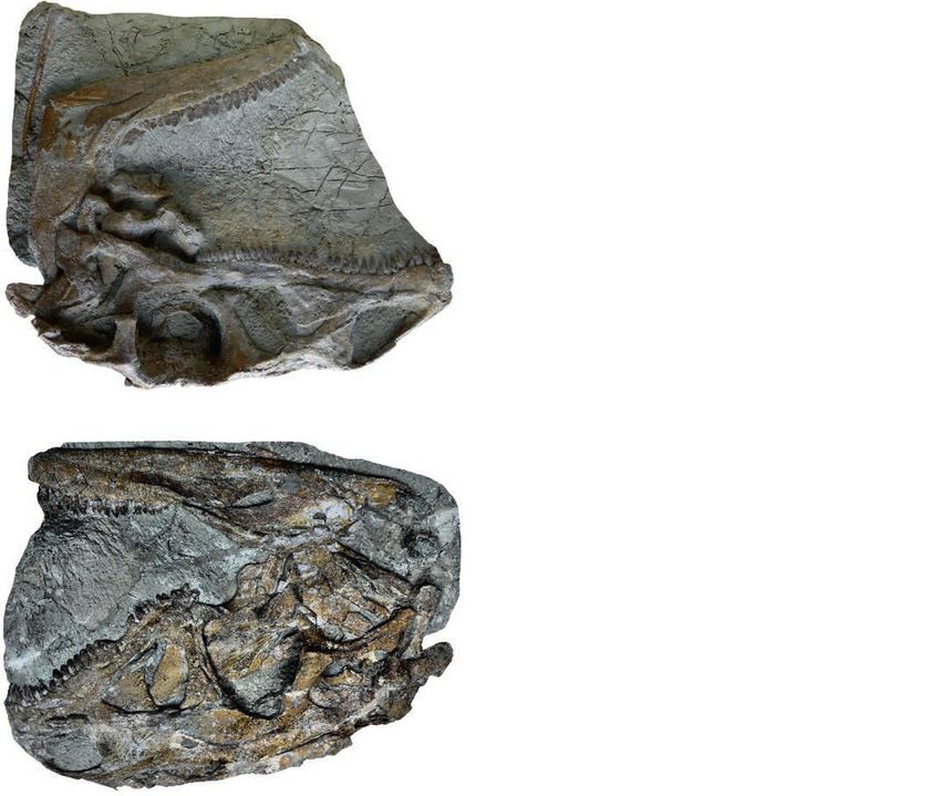
Fig. 12. Partly disarticulated skull of Plateosaurus trossingensis (MSF latter, the anterior rim terminates at the anterodorsal corner
09.2) from the Triassic of Frick, Switzerland. A. Main piece, showing the of the antorbital fossa, while the medial sheet continues to
left lateral side of parts of the skull. B. Partial right postorbital and jugal extend posteriorly after a sudden kink. In other skulls, the
in lateral view. C. Lower jaw in lateral view. Abbreviations: an, angular; anterior rim is curved along its length to varying degrees,
d, dentary; j, jugal; mx, maxilla; na, nasal; po, postorbital; sa, surangular. resulting in a more oval shape of the anterior half of the
ina are arranged in a row, where the posteriormost foramen antorbital fossa, as best seen in NAAG_00011238 (Fig. 7A).
is the largest. In NAAG_00011238, MSF 12-4, MSF 11.4, As seen in some skulls from Trossingen, the posterior edge
of the anterior rim can be straight while the anterior edge is
and MSF 23, the dorsoventral diameter of the posteriormost
strongly convex, which is most evident on the right side of
foramen is almost one-third of the height of the lateral sur-
SMNS 62968 but also seen in SMNS 13200. In some skulls,
face of the maxilla. This foramen is distinctively opened
such as MSF 15.4, the anterior rim rises at a much lower
posteriorly, forming a groove that gradually decreases in
angle (Fig. 5A). This angle as well as the curvature of the
depth posteriorly (e.g., Fig. 8A1). In the juvenile specimen
process may be exaggerated due to dorsoventral compres-
MSF 12.3, this groove is especially pronounced and long,
sion, and both features can vary between the left and right
extending for at least 15% of the total length of the laterally
sides of the same skull (e.g., MB.R.1937).
exposed surface of the maxilla (Fig. 8A1, A3). The more an- The medial sheet of the dorsal process of the maxilla is
terior foramina of the row open anteroventrally when their highly variable in both shape and extension. In MSF 11.4 and
morphology is readily preserved; this orientation is best seen MSF 16.1, the medial sheet occupies 50% of the antorbital
in SMNS 13200 from Trossingen. In NAAG_00011238, the fossa when measured along the greatest diameter of the latter
row comprises at least six foramina, all located ventral to the (Figs. 3, 4A1–A4). In MSF 15.4, on the other hand, the medial
antorbital fossa (Fig. 7A). As preserved in this skull, these sheet only extends to 35% (Fig. 5A). The posterior margin
foramina are comparatively small except for the penultimate of the medial sheet shows a pronounced crescent shape in
and especially the ultimate one; at least three additional the maxilla of NAAG_00011239, but is more or less straight
foramina are present on the dorsal process. In other skulls, in MSF 11.4. In NAAG_00011238, the posterior margin is
including MSF 11.4, MSF 23, MSF 15.4, and MSF 12.3, the straight along most of its length, but abruptly bends posteri-
row comprises fewer (between two and four) but larger fo- orly in its dorsalmost section (Fig. 7A). The posterior margin
ramina. In SMNS 13200, the row consists of eight foramina of the medial sheet is concave in SMA 09.1, but the dorsal
on the left and six on the right side; this discrepancy can be section again shows an abrupt bending. Pronounced left-right
either explained by single foramina not being preserved, or asymmetry in the shape of the medial sheet is seen in the
by asymmetry of the original foramina count. However, the skulls MB.R.1937 and SMNS 13200. In MB.R.1937, the me-
preserved foramina on both sides are large, and their posi- dial sheet of the right maxilla is crescent-shaped while that
tion and relative spacing is not compatible between the two of the left maxilla is much more straight. On the right side of
sides, indicating at least some degree of skull asymmetry, SMNS 13200, the posterior edge of the medial sheet is verti-
which also suggests that foramina count differs between in- cal in its dorsal half but abruptly becomes much less inclined
dividuals. Outside the row, additional foramina are typically in its ventral half; these features are much less pronounced on
present in the recessed region of the maxilla just anterior to the left side of the same skull. This asymmetry suggests that
the base of the dorsal process, and on the anterior border of the shape of the medial sheet is of limited use for taxonomy.16 ACTA PALAEONTOLOGICA POLONICA 66 (1), 2021
Nasal: The nasal is a thin bone roofing much of the mediolateral compression of MSF 11.4. The suture that sep-
anterior and anterolateral portions of the snout. The nasal arates the right and left nasal is located in a longitudinal
accounts for more than half the length of the skull roof in the depression in all skulls where it can be observed; this region
three specimens from Frick that allow precise measurement is crushed in all specimens.
of this character (MSF 11.4, MSF 12.3, MSF 15.4). These In the posterodorsal corner of the nasal, the lateral sur-
proportions are also found in skulls from Trossingen and face of the nasal extends posteriorly into a hook-like process
Halberstadt and are a distinguishing feature of Plateosaurus that extends onto the lateral surface of the anterior flange of
(Galton and Upchurch 2004). The nasal has two anteroven- the lacrimal. This process is preserved in MSF 11.4 and MSF
tral processes, the premaxillary and maxillary processes, 15.4 from Frick, MB.1927.19.1. from Halberstadt, and SMNS
which form the dorsal and posterior margins of the external 13200, SMNS 12949, and GPIT-PV-30784 from Trossingen.
naris, respectively. The premaxillary process is a thin and Typically, the process is rounded and broad, especially in MSF
rod-like element that is uniform, or slightly decreasing, in 11.4 (Fig. 4A1–A4). In SMNS 12949 and GPIT-PV-30784, in
dorsoventral height along its length. In dorsolateral view, contrast, it is narrow and pointed (Figs. 5B, 6B). The process
it may appear broader and thinning out distally to a greater is long and finger-like on the left side of SMNS 12949, but
degree. Variation of this feature is therefore likely the result short on the right side (Fig. 5B). As best seen in MSF 11.4
of different orientations of the main axis of deformation. and SMNS 12949, the hook-like process delimits a subrect-
The maxillary process contacts the posterior process angular embayment of the posterior margin of the nasal in
of the premaxilla ventrally, excluding the maxilla from the dorsal view (Figs. 4A1–A4, 5B). This embayment receives
external naris. This feature is present in Plateosaurus and the anterior processes of the lacrimal in SMNS 12949 and
Efraasia, while in other basal sauropodomorphs the maxilla in MSF 11.4, probably also the prefrontal. Medial to this
is not excluded from the external naris (Yates 2003; Galton embayment, the nasal greatly extends posteriorly to contact
and Upchurch 2004). In the skulls from Frick, this condition the frontal. This posterior extension of the nasal is best seen
is preserved in NAAG_00011238 and in MSF 15.4. The in GPIT-PV-30784 from Trossingen, where it reaches at least
maxillary process appears to not have reached the premax- 70% of the anteroposterior length of the prefrontal (Fig. 6B).
illa in GPIT-PV-30784, but this might be the result of dis- Prefrontal: The prefrontal forms the anterodorsal corner
articulation. The anterior margin of the maxillary process of the orbit and consists of an anterior flange, a posterior pro-
is sinuous, with a convexity just dorsal to its mid-height, cess, and a ventral process. The prefrontal is highly variable
as seen in MSF 11.4, MSF 15.4, MSF 16.1, SMNS 13200, in the examined skulls. It is well preserved on the right side of
SMNS 12949, SMNS 52968, GPIT-PV-30784, and AMNH MSF 11.4 (Fig. 4A1–A4) and on both sides of GPIT-PV-30784
FARB 6810 (e.g., Figs. 3–5). This convexity is not evident (Fig. 6B). In GPIT-PV-30784, its thin anterior lamina partly
in NAAG_00011238 and in MB.R.1937, which might be overlaps the posterodorsal part of the lacrimal, leaving the
due to an incomplete preservation of the anterior margin in anterior dorsal surface of the latter exposed in dorsal view.
these skulls. The lateral surface of the maxillary process is This overlap seems to be more extensive at least in AMNH
concave anterodorsally to posteroventrally. 6810 (Prieto-Márquez and Norell 2011). The overlap is more
A rounded shelf-like extension of the nasal over- extensive medially, as also seen in MSF 15.4, but does not
hangs the dorsal margin of the antorbital fenestra, result- reach as far anteriorly as the dorsally exposed surface of the
ing in a somewhat convex profile of the latter, as seen in lacrimal. MSF 11.4, in contrast, shows rectangular margin
NAAG_00011238, MSF 15.4, GPIT-PV-30784, the right side between the prefrontal and lacrimal in dorsal view, with an
of SMNS 13200, SMNS 12949, and MB.R.1937. This con- anterior process of the prefrontal located medial to the dor-
vex overhang is apparently absent in MSF 11.4 (Fig. 4A1– sally exposed surface of the lacrimal. This process extends
A3), but also missing on the left side of SMNS 13200, which anteriorly at least to a point above mid-length of the antor-
can possibly be attributed to deformation. Just posterior to bital fossa, and appears to fit, together with the lacrimal, into
the posterodorsal corner of the external naris, the nasal is an indentation formed by the posterior margin of the nasal.
swollen, which is most pronounced in MSF 15.4 (Fig. 5A). In both sides of GPIT-PV-30784, the anterior margin of
In lateral view, this swollen part forms an apex of the dorsal the anterior flange of the prefrontal shows a small but dis-
margin of the nasal, while the dorsal margin posterior to tinct notch (Fig. 6B), which is also seen in SMNS 13200. This
this apex is concave, as best seen, among the Frick skulls, in notch is absent in MSF 11.4; the region is not well-preserved
MSF 16.1, MSF 11.4, and MSF 15.4 (Figs. 3–5). The dorsal in other skulls. The anterior flange continues ventrally, over-
margin of the nasal may appear convex along its length as in lapping parts of the lateral surface of the lacrimal, before
NAAG_00011238, which is an effect of the direction of view terminating in a distinct step just dorsal to the lacrimal duct.
captured by the strongly compressed skull roof. In MSF This step is subrectangular in MSF 11.4, SMNS 13200, and
11.4, the concave part of the nasal posterior to the apex is SMNS 52968. In GPIT-PV-30784, MSF 15.8B, and MSF
sharply defined and separated from the lateral surface of the 15.4, in contrast, it forms a hook-like, ventrally directed tip
nasal by a pronounced rim (Fig. 4A1, A3). This depression (Figs. 5A, 6B, 9B). Ventral to the step, the ventral process of
and rim are not present or only very weakly pronounced in the prefrontal is twisted to articulate with the medial surface
the other skulls, and are probably exaggerated by the strong of the lacrimal, as seen in MSF 15.8B (Fig. 9D). In lateralYou can also read