Three new genera from South America and some taxonomic changes in Gyponini (Insecta: Hemiptera: Cicadellidae)
←
→
Page content transcription
If your browser does not render page correctly, please read the page content below
European Journal of Taxonomy 750: 70–93                                           ISSN 2118-9773
           https://doi.org/10.5852/ejt.2021.750.1363                        www.europeanjournaloftaxonomy.eu
                                                                                 2021 · Gonçalves C.C. et al.
This work is licensed under a Creative Commons Attribution License (CC BY 4.0).

                                             Research article
                   urn:lsid:zoobank.org:pub:511C6C89-69B0-468F-B121-1E4210D6E216

 Three new genera from South America and some taxonomic changes
          in Gyponini (Insecta: Hemiptera: Cicadellidae)

                    Clayton C. GONÇALVES 1,*, Alexandre C. DOMAHOVSKI 2,
                          Gabriel MEJDALANI 3 & Daniela M. TAKIYA 4
1,4
      Laboratório de Entomologia, Departamento de Zoologia, Instituto de Biologia, Universidade Federal
                do Rio de Janeiro, Caixa Postal 68044, Rio de Janeiro, 21941-971, RJ, Brazil.
             2
               Departamento de Zoologia, Universidade Federal do Paraná. Caixa Postal 19020,
                                      Curitiba, 81531-980, PR, Brazil.
         3
           Departamento de Entomologia, Museu Nacional, Universidade Federal do Rio de Janeiro,
                                    Rio de Janeiro, 20940-040, RJ, Brazil.
                       *
                           Corresponding author: clayton.correa.goncalves@gmail.com
                                    2
                                      Email: domahovskiac@yahoo.com.br
                                        3
                                          Email: mejdalan@acd.ufrj.br
                                          4
                                            Email: takiya@acd.ufrj.br
               1
                urn:lsid:zoobank.org:author:41E6BE64-C8CD-4BAF-86AA-272CF5BE88C5
               2
                 urn:lsid:zoobank.org:author:0B988BD5-599B-42CE-ADCB-50FC813E104E
                3
                  urn:lsid:zoobank.org:author:DE1C60FA-FB28-4F73-B316-18E901D5783D
               4
                 urn:lsid:zoobank.org:author:7E88BC1C-8D6A-411D-B97B-52E64EF5BA70

Abstract. Three new South American leafhopper genera of the tribe Gyponini are proposed: Beltrana
gen. nov. based on Beltrana reticulata gen. et sp. nov. from French Guiana, Fulana gen. nov. based on
Fulana brasiliensis gen. et sp. nov. from Brazil, and Sicrana gen. nov. based on Sicrana plana gen. et
sp. nov. from Brazil and Ecuador. Diagnoses, detailed descriptions, and illustrations are provided for
each taxon, as well as comparisons with closely related genera. In addition, the following synonyms are
proposed: Freytagana DeLong, 1975 as a junior synonym of Marganana DeLong, 1948 and F. gibsoni
DeLong, 1975 as a junior synonym of M. (Marganana) mexicana DeLong & Freytag, 1963. Chilella
DeLong & Freytag, 1967 is transferred from Gyponini to Selenomorphini Evans, 1974.

Keywords. Beltrana, Fulana, leafhoppers, Sicrana, taxonomy.

Gonçalves C.C., Domahovski A.C., Mejdalani G. & Takiya D.M. 2021. Three new genera from South America and
some taxonomic changes in Gyponini (Insecta: Hemiptera: Cicadellidae). European Journal of Taxonomy 750:
70–93. https://doi.org/10.5852/ejt.2021.750.1363

Introduction
Gyponini Stål, 1870 is the most diverse of the twelve tribes of Iassinae Walker, 1870 (Krishnankutty et al.
2016). DeLong & Freytag (1964), based on the diversity known at that time, suggested that Gyponini

                                                          70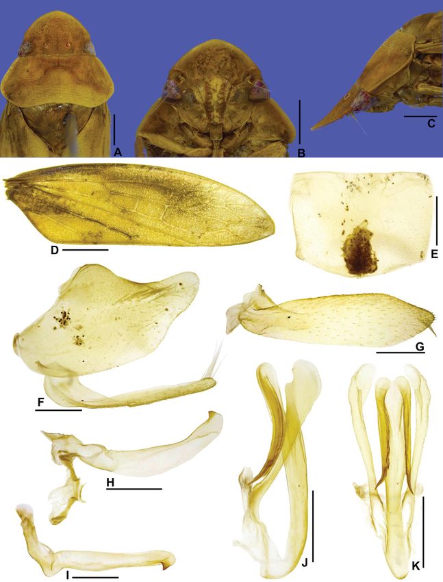
GONÇALVES C.C. et al., Three new genera of Gyponini from South America

could include more than 500 species. Currently, considering the recently described taxa, the tribe
comprises 1405 species in 71 genera (Domahovski & Cavichioli 2020, 2021; Domahovski et al. 2020;
Freytag 2021). Based on the large number of undescribed species deposited in Brazilian collections,
we believe that the diversity of Gyponini will exceed 2000 species. Specimens can be collected using
sweeping nets and sometimes yellow plates; however, light and Malaise traps are the most effective
methods for collecting these leafhoppers (Domahovski & Cavichioli 2018).

This paper proposes the establishment of three new Gyponini genera, with very peculiar morphological
features: Beltrana gen. nov. from French Guiana, Fulana gen. nov. from Brazil, and Sicrana gen. nov.
from Brazil and Ecuador. The names “Fulana, Beltrana, and Sicrana” form a Portuguese expression
equivalent to the English expression “Tom, Dick, and Harry”. This expression is used to refer to multiple
unspecified people. The names were chosen because this expression is very popular in Brazil and, also,
they end in ‘-ana’ which is a common suffix in most generic names of Gyponini.

In addition, the following synonyms are proposed: Freytagana DeLong, 1975 as a junior synonym
of Marganana DeLong, 1948 and F. gibsoni DeLong, 1975 as a junior synonym of M. (Marganana)
mexicana DeLong & Freytag, 1963. Chilella DeLong & Freytag, 1967 is transferred from Gyponini to
Selenomorphini Evans, 1974.

Material and methods
Specimens studied herein are deposited in the following collections:

CEIOC = Coleção Entomológica do Instituto Oswaldo Cruz, FIOCRUZ, Rio de Janeiro, Brazil
DZRJ = Coleção Entomológica Prof. José Alfredo Pinheiro Dutra, Universidade Federal do Rio de
        Janeiro, Rio de Janeiro, Brazil
DZUP = Coleção Entomológica Pe. Jesus Santiago Moure, Universidade Federal do Paraná, Curitiba,
        Brazil
EPNC = Escuela Politécnica Nacional, Quito, Ecuador
INPA = Instituto Nacional de Pesquisas da Amazônia, Manaus, Brazil
MNRJ = Departamento de Entomologia, Museu Nacional, Universidade Federal do Rio de Janeiro,
        Rio de Janeiro, Brazil
MZSP = Museu de Zoologia da Universidade de São Paulo, São Paulo, Brazil
OSUC = Insect Collection, Museum of Biological Diversity, Ohio State University, Columbus, USA
USNM = United States National Museum, Washington, D.C., USA

Terminology mainly follows Young (1968, 1977), except for head features (Hamilton 1981; Mejdalani
1993, 1998), wings (Dietrich 2005), and leg chaetotaxy (Rakitov 1997). Techniques used for dissection
of the male terminalia follow Oman (1949), with a few modifications described by Cavichioli & Takiya
(2012). Female terminalia were prepared according to the techniques of Zanol (1988) and Mejdalani
(1998). Square brackets [ ] in material label data were used to supplement abbreviated information.

                                                   71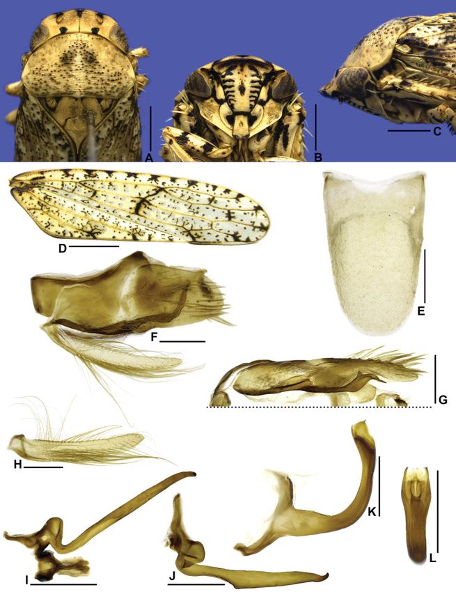
European Journal of Taxonomy 750: 70–93 (2021)

Results
Taxonomic treatments
                                    Class Insecta Linnaeus, 1758
                                  Order Hemiptera Linnaeus, 1758
                              Suborder Auchenorrhyncha Duméril, 1806
                                 Family Cicadellidae Latreille, 1825
                                  Subfamily Iassinae Walker, 1870
                                     Tribe Gyponini Stål, 1870

                                      Genus Beltrana gen. nov.
               urn:lsid:zoobank.org:act:4C0DFFC6-7F48-457C-97C6-361C182A69DC
                                           Figs 1, 6A–B

Type species
Beltrana reticulata gen. et sp. nov., by present designation and monotypy.

Diagnosis
Medium-sized leafhoppers (Fig. 6A–B). Head in dorsal view (Fig. 1A) moderately produced anteriorly,
median length of crown slightly less than interocular width; in lateral view (Fig. 1C), with crown-face
transition with anterior margin foliaceous and bicarinate. Forewing (Fig. 1D) with venation densely
reticulated, appendix very narrow. Male pygofer (Fig. 1F) without membranous apical digitiform
process. Subgenital plate (Fig. 1F, H) fully sclerotized. Aedeagus (Fig. 1K–L) simple, without apodemal
processes.

Etymology
The generic name is feminine. Beltrana, together with Fulana and Sicrana, forms a very popular
expression in Brazil (“Fulana, Sicrana e Beltrana”), which refers to unspecified people or people whose
actual names should not be mentioned.

Description
HEAD AND THORAX. Head in dorsal view (Fig. 1A) moderately produced anteriorly, median length of
crown slightly less than interocular width; transocular width about six-sevenths humeral width of
pronotum; crown with anterior margin broadly rounded, surface mostly flat, slightly concave medially,
with subtle oblique striae between ocelli and adjacent to anterior margin; ocellus medium-sized,
equidistant between median line and eye margin, closer to posterior than anterior margin of crown;
coronal suture distinct only on basal portion of crown. Head in ventral view (Fig. 1B) with face slightly
higher than wide; frontogenal suture distant from eye margin by slightly less than maximum width of
clypeus and extending to anterior margin of crown; antennal ledge carinated and adjacent to anterior
margin of crown, obliquely ascending and not extending over frons; frons approximately 1.7 times as
long as wide; epistomal suture indistinct; clypeus approximately 1.3 times as long as maximum width,
lateral margins parallel, apex slightly emarginated; maxillary plate produced ventrally, not reaching
clypeus apex; gena excavated just below eye, ventrolateral margin slightly excavated. Head in lateral
view (Fig. 1C) with crown-face transition distinct and foliaceous, with two carinae; frons concave
below anterior margin of crown. Pronotum (Fig. 1A) with transverse striae on disc and posterior half;
lateral margins straight, convergent anterad, and approximately as long as eye length; posterior margin
excavated; in lateral view (Fig. 1C), moderately declivous, continuous with head declivity. Mesonotum
(Fig. 1A) longer than wide; scutellum (Fig. 1C) flat. Forewing (Fig. 1D) hyaline, long, and narrow,
approximately 3.2 times as long as maximum width; venation distinct and densely reticulated; appendix

                                                   72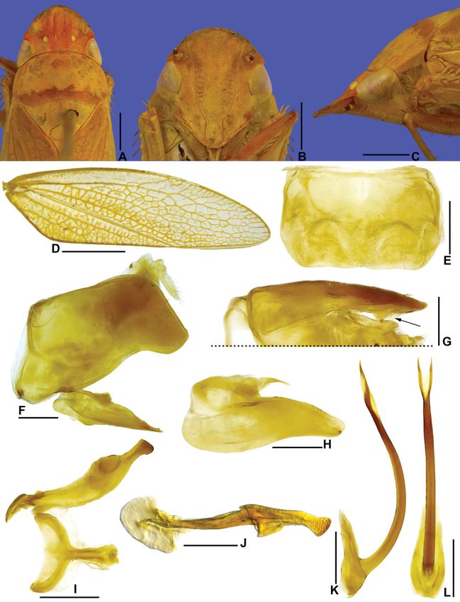
GONÇALVES C.C. et al., Three new genera of Gyponini from South America

very narrow. Profemur with AD, AM, and PD rows reduced and poorly defined, with exception of
apical setae AD1, AM1, and PD1, respectively; AV and PV rows formed by 4–5 setae; IC row formed by
slightly arched comb of fine setae, beginning at distal third of femur and extending to AM1. Protibia,
in cross-section, semi-circular; AV row formed by long setae, slightly longer and thicker towards apex;
AD and PD rows without differentiated setae; PV row with 5 setae. Metafemur with setal formula 2:2:1.
Metatibial AD row without intercalary setae between macrosetae; PD, AD, and AV rows with 26–28,
13–14, and 19–20 macrosetae, respectively. Metatarsomere I with outer setal row indistinct, apex with
5 platellae. Metatarsomere II apex with 4 platellae.

MALE TERMINALIA. Pygofer (Fig. 1F) without membranous apical digitiform process. Subgenital plate
(Fig. 1F, H) fully sclerotized. Connective (Fig. 1I) T-shaped. Aedeagus (Fig. 1K–L) simple, without
apodemal processes.

FEMALE TERMINALIA. Female unknown.

Distribution
French Guiana.

Remarks
An unpublished phylogenetic hypothesis based on 182 morphological characters combined with
sequence data (28S and 16S rDNA and cytochrome oxidase subunit I) recovered Beltrana gen. nov. as
the sister group of the clade Reticana DeLong & Freytag, 1964 + Chloronana DeLong & Freytag, 1964
(Gonçalves 2016). Beltrana gen. nov. shares the following morphological characteristics with Reticana
and Chloronana: (1) green coloration in life, yellow when preserved; (2) crown moderately produced
anteriorly; (3) crown-face transition distinct and foliaceous; (4) forewing with venation reticulated;
(5) metatibia without intercalary setae between macrosetae in AD row; (6) style short and with apical
denticles; and (7) aedeagus with shaft long, thin, and with apical processes. However, Beltrana gen. nov.
can easily be distinguished from Reticana and Chloronana by its more robust body (Fig. 6A); forewing
more densely reticulated (Fig. 1D); male pygofer without membranous apical digitiform process
(Fig. 1F); and subgenital plate fully sclerotized (Fig. 1F, H).

                                   Beltrana reticulata gen. et sp. nov.
                 urn:lsid:zoobank.org:act:2F33353D-4741-476E-B6A8-FA2F74B33A35
                                             Figs 1, 6A–B

Diagnosis
Pygofer (Fig. 1F) with apex distinctly truncated, ventral margin expanded posteroventrally, forming
subquadrate lobe; in dorsal view (Fig. 1G), with small preapical inner lobe. Subgenital plate (Fig. 1F, H)
with small sclerotized dorsal tooth near apex. Style (Fig. 1I–J) with knob-shaped apex bearing numerous
denticles. Aedeagus (Fig. 1K–L) with pair of short and acute apical processes.

Etymology
The specific epithet alludes to the reticulated venation of forewing (Fig. 1D).

Material examined
   Holotype
FRENCH GUIANA • ♂; French Guiana, Kaw Mountain Reserve, 0.5 km Rt. Rd. de Kaw, the hole; 1
Feb. 2008; H. Romack leg.; DNA voucher Entomologia, DZRJ ENT 2812; USNM.

                                                   73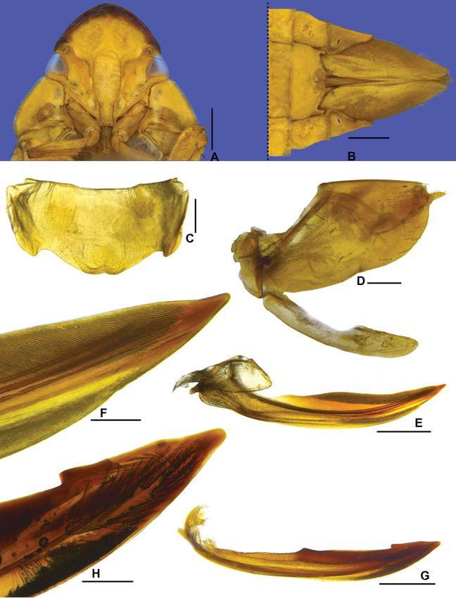
European Journal of Taxonomy 750: 70–93 (2021)

Fig. 1. Beltrana reticulata gen. et sp. nov., holotype, ♂ (USNM). A. Head and thorax, dorsal view.
B. Head, ventral view. C. Head and thorax, lateral view. D. Forewing. E. Sternite VIII. ventral view.
F. Pygofer, anal tube, valve, and subgenital plate, lateral view. G. Pygofer lobe and anal tube, dorsal
view (arrow indicates the inner lobe). H. Subgenital plate, ventral view (outer margin at top of figure).
I. Style and connective, dorsal view. J. Style, lateral view. K. Aedeagus, lateral view. L. Aedeagus,
posterior view. Scale bars: A–C = 1.0 mm; D = 2.0 mm; E–L = 0.5 mm.

                                                   74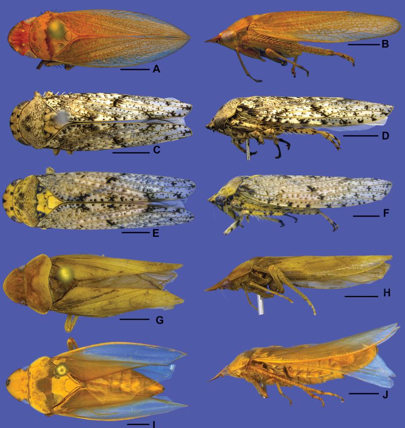
GONÇALVES C.C. et al., Three new genera of Gyponini from South America

   Paratype
FRENCH GUIANA • 1 ♂; same collection data as for holotype; DZRJ-AUCH #249; DZRJ.

Description
MEASUREMENTS. Total length: male 12.3–12.5 mm (n = 2).

COLORATION. Yellow (Fig. 6A–B). Crown (Fig. 1A) mostly orange, anterior and posterior margins yellow;
ocelli yellow. Pronotum (Fig. 1A) with anterior three-fourths yellow and posterior fourth dark yellow.
Forewing (Fig. 1D) yellow, translucent, with dark yellow venation. Legs (Fig. 6B) yellow.

HEAD AND THORAX. External morphological characters as in generic description.

MALE TERMINALIA. Sternite VIII (Fig. 1E) subrectangular, about 1.5 times as wide as long; lateral margins
slightly rounded; posterior margin rectilinear. Pygofer in lateral view (Fig. 1F) trapezoidal, length
approximately twice maximum height; apex distinctly truncated; macrosetae distributed only near apex,
presence of few short and robust setae near apex; ventral margin expanded posteroventrally, forming
subquadrate lobe; in dorsal view (Fig. 1G), with small preapical inner lobe. Subgenital plate in lateral
view (Fig. 1F) not produced posteriorly as far as pygofer apex; in ventral view (Fig. 1H), subtriangular,
basal half of external margin expanded laterally, bearing short setae on apical half; apical half narrowing
progressively towards apex; internal margin sinuous; dorsal surface with small, sclerotized tooth near
apex; apex rounded. Connective (Fig. 1I) approximately three-fifths of length of style; arms broad. Style
(Fig. 1I), in dorsal view, with apodeme very short, spatulate, and strongly flattened laterally; apophysis
elongated, flattened laterally; dorsal margin expanded laterally, forming preapical lamella; in lateral
view (Fig. 1J), outer lobe indistinct; apex sclerotized, knob-shaped, with numerous small lateral and
ventral denticles. Aedeagus (Fig. 1K–L) with preatrium reduced; dorsal apodeme developed dorsally,
not expanded laterally; shaft tubular and very elongated, curved anteriorly; apex with pair of short and
acute processes directed dorsally.

FEMALE TERMINALIA. Female unknown.

                                        Genus Fulana gen. nov.
               urn:lsid:zoobank.org:act:D3D2E937-9B40-42BB-AAC4-F821B4D22CB5
                                           Figs 2–3, 6C–F

Type species
Fulana brasiliensis gen. et sp. nov., by present designation and monotypy.

Diagnosis
Medium-sized leafhoppers (Fig. 6C–F). Head in dorsal view (Fig. 2A) short, median length of crown
about 2.5 times smaller than interocular width; crown transversely striated; ocellus large, closer to
anterior margin of crown; in lateral view (Fig. 2C), with crown-face transition distinct and narrow,
bicarinate. Pronotum (Fig. 2A) with punctures. Forewing (Fig. 2D) covered by numerous small brown
spots, appendix narrow. Male pygofer (Fig. 2F–G) with inner basiventral process; lateral lobe partly
separated from base by flexible membranous line. Subgenital plate (Fig. 2H) with ventral surface covered
by long filiform setae. Connective (Fig. 2I) longitudinally linear. Aedeagus (Fig. 2K–L) simple, without
apodemal processes. Second valvula of ovipositor (Fig. 3G–H) with few teeth restricted to apical fourth.

                                                    75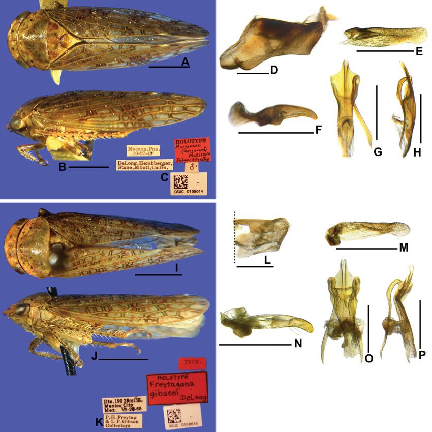
European Journal of Taxonomy 750: 70–93 (2021)

Etymology
The generic name is feminine. Fulana, together with Beltrana and Sicrana, forms a very popular
expression in Brazil (“Fulana, Sicrana e Beltrana”), which refers to unspecified people or people whose
actual names should not be mentioned.

Description
HEAD AND THORAX. Head in dorsal view (Fig. 2A) short; median length of crown about 2.5 times smaller
than interocular width; crown with anterior margin slightly sinuous; surface adjacent to ocelli slightly
concave between ocellus and eye; texture transversely striated; transocular width of head six-sevenths
of humeral width of pronotum; ocellus large, close to anterior margin of crown and slightly closer to eye
than to median line; coronal suture distinct, extending to near anterior margin of crown. Head in ventral
view (Figs 2B, 3A) with face wider than high; frontogenal suture distant from eye margin by basal width
of clypeus and surpassing antennal ledge, extending to anterior margin of crown; oriented obliquely
downward in relation to frons; frons approximately as wide as long, lateral margins strongly convergent
ventrally; epistomal suture distinct; clypeus approximately 1.2 times as long as maximum width, lateral
margins divergent apically, apex slightly emarginated; maxillary plate produced ventrally as far as
clypeus apex; gena with ventrolateral margin straight on mid-length and slightly excavated below eye
margin. Head in lateral view (Fig. 2C) with crown-face transition distinct and narrow, bicarinate; frons
and clypeus flattened. Pronotum (Fig. 2A) with conspicuous transverse striae and punctures on disc and
posterior half; lateral margins straight, convergent anterad, slightly shorter than eye length; posterior
margin straight; in lateral view (Fig. 2C), moderately declivous; head and pronotum in continuous
slope. Mesonotum (Fig. 2A) as long as wide; scutellum (Fig. 2C) slightly swollen. Forewing (Fig. 2D)
translucent, covered by numerous small brown spots; long and narrow, approximately 3.3 times as long
as wide; venation distinct; appendix very narrow and bordering first to second apical cells. Profemur
with AD, AM, and PD rows reduced and poorly defined, with exception of apical setae AD1, AM1, and
PD1, respectively; AV and PV rows formed by 2–3 setae; IC row formed by slightly arched comb of fine
setae, beginning at distal half of femur and extending to AM1. Protibia in cross-section semi-circular; AV
row formed by long setae, slightly longer and thicker towards apex; AD row without differentiated setae;
PD row with 7–8 setae; PV row with 6–7 setae. Metafemur with setal formula 2:2:1. Metatibia PD, AD,
and AV rows with 26–31, 13–14, and 16–19 macrosetae, respectively; AD row with 1–4 intercalary setae
between macrosetae; PV row with setae of apical half formed by sequence of a thicker and 2–4 thinner
setae. Metatarsomere I with two rows of setae, inner row consisting of larger and more robust setae than
those of outer row; apex with 6 platellae; metatarsomere II apex with 2–3 platellae.

MALE TERMINALIA. Sternite VIII (Fig. 2E) long, fully hiding subgenital plates. Pygofer (Fig. 2F–G)
with inner basiventral process adjacent to ventral margin; lobe partly separated from base by flexible
membranous cleft. Subgenital plate (Fig. 2H) with ventral surface covered by long filiform setae.
Connective (Fig. 2I) longitudinally linear. Aedeagus (Fig. 2K–L) simple, without apodemal processes.

FEMALE TERMINALIA. Second valvula of ovipositor (Fig. 3G) elongated, slightly higher along apical
fourth; apical portion (Fig. 3H) with dorsal margin bearing minute and uniform denticles.

Distribution
Brazil (Bahia, Minas Gerais, Paraná, Rio de Janeiro, Santa Catarina, and São Paulo states).

Remarks
An unpublished study based on 182 morphological characters combined with sequence data (28S and
16S rDNA and cytochrome oxidase subunit I) recovered Fulana gen. nov. as the sister group to all other
gyponines except Chilenana DeLong & Freytag, 1967 (Gonçalves 2016). In fact, the genera Fulana

                                                   76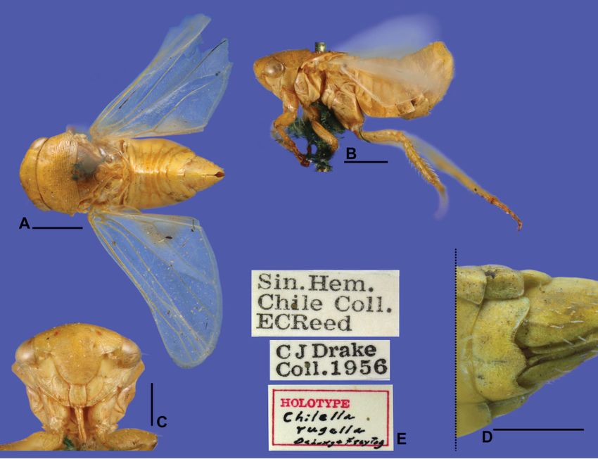
GONÇALVES C.C. et al., Three new genera of Gyponini from South America

gen. nov. and Chilenana share plesiomorphic characteristics with other tribes of Iassinae, which are
absent in other Gyponini genera: (1) male pygofer with lateral lobe partly separated from base by flexible
membranous cleft; and (2) presence of basiventral inner process on the male pygofer.

The new genus resembles Alapona DeLong, 1980, Ponana Ball, 1920, and Dumorpha DeLong &
Freytag, 1975 because of its slightly anteriorly produced and transversely striated crown; ocellus closest
to the anterior margin of the crown; forewing with dark spots; and metatibia with intercalary setae
between macrosetae of the AD row. However, Fulana gen. nov. differs from Alapona by the lack of flaps
in the aedeagus (Fig. 2K–L), from Ponana and Alapona by its thinner crown-face transition (Fig. 2C),
and from Ponana and Dumorpha by its aedeagus without apodemal processes (Fig. 2K).

                                  Fulana brasiliensis gen. et sp. nov.
               urn:lsid:zoobank.org:act:B1D1E77B-63FF-443C-8AEC-9952CA62AFAE
                                           Figs 2–3, 6C–F

Diagnosis
Male sternite VIII (Fig. 2E) with posterior margin rounded. Subgenital plate (Fig. 2F, H) narrow and
long. Style (Fig. 2J) broadened on apical two-thirds and tapering towards apex. Aedeagus (Fig. 2K–L)
with apex bilobed and with pair of small dorsal spines. Female sternite VII (Fig. 3B–C) with posterior
margin slightly excavated medially. First valvula of ovipositor (Fig. 3E) with apex acute.

Etymology
The species name is in reference to the country of origin of the studied specimens.

Material examined
   Holotype
BRAZIL • ♂; Brasil, RJ [Rio de Janeiro], P. [Parque] N. [Nacional] do Itatiaia, quiosque poço azul;
22°27′07″ S, 44°36′45″ W; 17–18 Nov. 2012; D.M. Takiya and A.P.M. Santos leg.; DNA voucher
Entomologia, DZRJ ENT 1889; DZRJ-AUCH #250; DZRJ.

     Paratypes
BRAZIL – Rio de Janeiro • 1 ♀; same collection data as for holotype; DZRJ-AUCH #251; DZRJ • 1 ♀;
Itatiaia; Sep. 1929; D. Mendes leg.; MNRJ • 1 ♀; Itatiaia; alt. 750 m; Jan. 1958; U. Barth leg.; MNRJ • 1 ♀; P.N.
Itatiaia, Casa do Pesquisador; 22.454° S, 44.608° W; alt. 800 m; light; 27–30 Oct. 2011; R.R. Cavichioli leg.;
DZUP-083577 • 1 ♀; Itatiaia; Oct. 1936; J.F. Zikan leg.; CEIOC • 1 ♀; same collection data as for
preceding; 11 Aug. 1938; CEIOC • 1 ♀; same collection data as for preceding; 23 Nov. 1938; CEIOC •
1 ♀; same collection data as for preceding; 12 Dec. 1939; CEIOC • 1 ♀; same collection data as for
preceding; 13 Aug. 1940; CEIOC. – Bahia • 1 ♂; Santa Terezinha, Serra Jibóia; 12°51′13″ S, 39°28′32″ W;
alt. 800 m; luz [light]; 8 Jun. 2007; J.A. Rafael and F.F. Xavier F° leg.; INPA • 1 ♂; Encruzilhada; Nov.
1975; M. Alvarenga leg.; DZUP-083578. – Minas Gerais • 1 ♂; Itamonte, P.N. do Itatiaia, Travessia
Serra Negra, Campo das Flores, segundo acampamento, em árvore; 22°19′55″ S, 44°04′42″ W; alt.
1960 m; 25–26 Nov. 2011; D.M. Takiya and A.P.M. Santos leg.; DZRJ-AUCH #252; DZRJ • 1 ♂;
Itabirito, Vale dos Tropeiros, Cachoeira das Carrancas; 20°12′28.3″ S, 43°38′26″ W; alt. 1046 m; 10 Oct.
2010; L. Dumas and N. Ferreira Jr. leg.; DZRJ-AUCH #253; DZRJ • 1 ♀; São Gonçalo, Rio Abaixo, Est.
[Estação] Amb. [Ambiental] Peti-Cemig; 7 Nov. 2003; A.F. Kumagai leg.; DZUP-083579. – Paraná •
1 ♀; Serra do Mar, Morro dos Perdidos; 25°51′52″ S, 48°53′14″ W; alt. 900 m; arm. [armadilha] de luz,
lençol; 17 Feb. 2007; J.A. Rafael and E. Grossi leg.; INPA • 1 ♂; S. [São] José [dos] Pinhais, BR 277,
km 54; luminosa; 23 Mar. 1985; C.I.I.F. [Centro de Identificação de Insetos Fitófagos]; DZUP-083580 •
1 ♀; same collection data as for preceding; 14 May 1985; DZUP-083581 • 1 ♂; same collection data

                                                       77
European Journal of Taxonomy 750: 70–93 (2021)
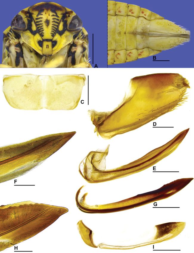
Fig. 2. Fulana brasiliensis gen. et sp. nov., holotype, ♂ (DZRJ). A. Head and thorax, dorsal view.
B. Head, ventral view. C. Head and thorax, lateral view. D. Forewing. E. Sternite VIII. ventral view.
F. Pygofer, anal tube, valve, and subgenital plate, lateral view. G. Pygofer lobe and anal tube, dorsal view.
H. Subgenital plate, ventral view (outer margin at top of figure). I. Style and connective, dorsal view.
J. Style, lateral view. K. Aedeagus, lateral view. L. Aedeagus, posterior view. Scale bars: A–C = 1.0 mm;
D = 2.0 mm; E–L = 0.5 mm.
78GONÇALVES C.C. et al., Three new genera of Gyponini from South America
Fig. 3. Fulana brasiliensis gen. et sp. nov., paratype, ♀ (DZRJ-AUCH #251). A. Head, ventral view.
B. Posterior portion of abdomen, ventral view. C. Sternite VII, ventral view. D. Pygofer and anal tube,
lateral view. E. First valvifer and first valvula of ovipositor, lateral view. F. Apex of first valvula, lateral
view. G. Second valvula of ovipositor, lateral view. H. Apex of second valvula, lateral view. I. Second
valvifer and gonoplac, lateral view. Scale bars: A–B = 1.0 mm; C–E, G, I = 0.5 mm; F, H = 0.3 mm.
79European Journal of Taxonomy 750: 70–93 (2021)
as for preceding; T.T. [Torre Telepar]; choque; 16–23 May 1985; DZUP-083582 • 1 ♂; same collection
data as for preceding; lâmpada; 3 Oct. 1986; Lev. [Levantamento] Ent. [Entomológico] Profaupar;
DZUP-083583 • 1 ♀; same collection data as for preceding; 1 Nov. 1986; DZUP-083584 • 1 ♀; same
collection data as for preceding; 4 Nov. 1986; DZUP-083585 • 1 ♂; same collection data as for preceding;
2 Dec. 1986; DZUP-083586 • 1 ♂; same collection data as for preceding; 31 Dec. 1986; DZUP-083587 •
1 ♀; same collection data as for preceding; 19 Jan. 1987; DZUP-083588 • 1 ♀; same collection data as
for preceding; 17 Jan. 1988; DZUP-083589 • 1 ♂; same collection data as for preceding; 19 Jan. 1988;
DZUP-083590 • 1 ♂; Colombo, Embrapa, BR 476, km 20; lâmpada; 18 Jan. 1988; Lev. Ent. Profaupar;
DZUP-083591 • 1 ♂; Campo Largo, Est. [Estrada] do Cerne, km 45.5; Feb. 1999; L. Chiamolera leg.;
DZUP-083592 • 2 ♂♂; Antonina, Res. Rio Cachoeira; 25.316° S, 46.696° W; alt. 50 m; luminosa; 20–
25 Nov. 2014; Entomologia UFPR; DZUP-083593, 083594 • 1 ♂; same collection data as for preceding;
luminosa solo, 5–10 Nov. 2015; MNRJ • 1 ♀; Tijucas do Sul, Voçoroca, Excursão Entomologia I,
UFPR; 4 Mar. 2015; A.C. Domahovski leg.; MNRJ • 1 ♂; Piraquara, Mananciais da Serra; luz; 4 Nov.
2008; P.C. Grossi leg.; MZSP • 1 ♂; same collection data as for preceding; 25°29′47″ S, 48°58′54″ W;
alt. 1021 m; Arm. [Armadilha] Lum. [Luminosa]; 28 Nov. 2018; A.P. Pinto, B.R. Araujo and A.C.
Domahovski leg.; MNRJ • 2 ♀♀; same collection data as for preceding; 25.4967° S, 48.9839° W; alt.
1010 m; malaise; 16 Feb.–11 Mar. 2019; G. Melo and A. Martins leg.; DZUP-083598, DZUP-083599.
– Santa Catarina • 1 ♂; Blumenau, Parque Spitzkopf, confluência dos Ribeirões Caeté e do Ouro,
churrasqueira; 20°00′21.60″ S, 49°06′45.70″ W; alt. 164 m; pano branco; 21 Jan. 2011; D.M. Takiya
and A.P.M. Santos leg.; DZRJ-AUCH #254, DZRJ. – São Paulo • 1 ♂; São Paulo, Alto da Serra; Mar.
1924; R. Spitz leg.; MZSP • 1 ♂, 1 ♀; 43 km a Nordeste de Miracatu; alt. 460 m; 8–9 Oct. 2005; Mielke
and Casagrande leg.; MZSP.
Description
MEASUREMENTS. Total length: male 10.2–11.5 mm (n = 21); female 12.3–14.5 mm (n = 20).
COLORATION. Pale yellow to yellow (Fig. 6C–F). Crown (Fig. 2A) pale yellow, with circular black spot
behind each ocellus, adjacent to posterior margin; anterior margin with three dark maculae, one medially
and two in front of ocelli. Face (Figs 2B, 3A) pale yellow to yellow; frons with about ten pairs of
dark bands over muscular impressions, dorsal portion with small dark punctuations; epistomal and
frontogenal sutures black; lorum with dark subtriangular macula adjacent to lateral margin of clypeus;
clypeus with central dark macula and apical margin black; gena with three black maculae that may be
interconnected, forming large spot in some specimens: one adjacent to antennal pit, one adjacent to inner
corner of compound eye, and one adjacent to lorum base. Pronotum (Fig. 2A) with small dark punctures
except near anterior margin, behind eyes. Mesonotum (Fig. 2A) with ringed black maculae near lateral
corners; in lateral view (Fig. 2C), lateral lobe of pronotum with large black spot, anepisternum with
small black spot. Forewing (Fig. 2D) pale yellow, translucent, with yellow-cream areas on basal two-
thirds; numerous small black spots throughout surface and several larger black spots on apices of anal
veins, along costal margin, over crossveins, and apical margin. Legs (Fig. 6D, F) pale yellow with
scattered dark maculae; tarsus of pro- and mesothoracic legs black.
HEAD AND THORAX. External morphological characters as in generic description.
MALE TERMINALIA. Sternite VIII (Fig. 2E) strongly produced posteriorly, about 1.6 times as long as wide;
lateral margins converging towards apex, posterior margin broadly rounded. Valve subrectangular, three
times as wide as long; posterior margin shallowly excavated medially. Pygofer in lateral view (Fig. 2F)
elongate, about 2.1 times as long as maximum height; apex rounded; macrosetae distributed only on
apical half; basiventral inner process (Fig. 2F) strongly sclerotized, long and thin, almost reaching
pygofer apex and contiguous to ventral margin, apical fourth curved dorsally; in ventral view (Fig. 2G),
process broadened preapically, with short preapical tooth on external side, apex acute. Subgenital plate
80GONÇALVES C.C. et al., Three new genera of Gyponini from South America
in lateral view (Fig. 2F) not produced posteriorly as far as pygofer apex; in ventral view (Fig. 2H)
long and narrow, about 6.4 times longer than wide; lateral margins parallel; apex abruptly rounded.
Connective (Fig. 2I) approximately one-fourth of length of style; stalk longer than wide; arms short,
slightly wider than stalk; dorsal keel developed. Style in dorsal view (Fig. 2I) with outer lobe developed
and rounded; in lateral view (Fig. 2J), broadened on apical two-thirds and tapering towards apex; ventral
margin with few denticles at median third; apex acute and curved dorsally. Aedeagus (Fig. 2K–L) with
preatrium long; dorsal apodeme projected dorsally; shaft elongated and tubular, curved posterodorsally,
expanded apically; apex bilobed, with pair of small apical spines.
FEMALE TERMINALIA. Sternite VII (Fig. 3B–C) 2.3 times as wide as long; posterolateral borders rounded,
posterior margin slightly excavated medially. Internal sternite VIII membranous. Pygofer (Fig. 3D)
elongate, about 1.8 times as long as maximum height; apex rounded; macrosetae distributed near apex
and ventral margin. First valvifer (Fig. 3E) subquadrate. First valvula of ovipositor (Fig. 3E) slightly
curved dorsally, about 9.6 times as long as high; with approximately same height along its entire length;
dorsal sculptured area beginning on basal third, its apical portion (Fig. 3F) strigate; blade apex acute.
Second valvifer (Fig. 3I) higher than long. Second valvula of ovipositor (Fig. 3G) slightly higher apically,
approximately 9.5 times longer than maximum height; apical portion (Fig. 3H) of dorsal margin with
about seven small and rounded teeth; blade apex tapered and acute. Gonoplac (Fig. 3I) 4.7 times as long
as high; dorsoapical and ventroapical margins convergent apically; dorsoapical margin short; ventral
margin with few short setae mainly near apex; apex subacute.
Remarks
Male specimens collected from different localities show a variation in the shape of the apical portion of
the ventral process of pygofer, which may be more curved or straighter, more dilated or slightly thinner.
In addition, some specimens have two conspicuous and rounded black spots over crossveins of discal
cells of forewing. However, remaining structures of the male terminalia and shape of the female sternite
VII are invariable and we thus consider these variations as intraspecific.
Specimens of Fulana brasiliensis gen. et sp. nov. were mainly collected in areas of Atlantic Forest, at
altitudes that vary from 50 to 1960 m above sea level. A putative second species of the new genus, known
only from female specimens from the Brazilian Amazon Rain Forest, State of Amazonas, Manaus, was
studied but will not be described until male specimens become available. This undescribed species is
similar to F. brasiliensis gen. et sp. nov. in all aspects of the external morphology and female terminalia
described herein. However, it can be distinguished by its larger size (15.5–15.8 mm); head in ventral
view with fewer black maculae on face (lorum with small spot adjacent to lateral margin of clypeus,
frons with punctuations and a semi-circular macula near anterior margin of crown, and antennal pits
black); and sternite VII more deeply excavated medially.
Genus Sicrana gen. nov.
urn:lsid:zoobank.org:act:5D4E6996-37CB-49B3-AF47-0F247BBFF9AA
Figs 4–5, 6G–J
Type species
Sicrana plana gen. et sp. nov., by present designation and monotypy.
Diagnosis
Large-sized, flattened leafhoppers (Fig. 6G–J). Head in dorsal view (Fig. 4A) with rugose surface,
strongly produced anteriorly, anterior margin parabolic; ocellus closer to median line than to adjacent
eye and closer to posterior than to anterior margin of crown; in lateral view (Fig. 4C), with crown-
81European Journal of Taxonomy 750: 70–93 (2021)
face transition distinct and strongly foliaceous, margined by two carinae. Face (Figs 4B, 5A) with
frons narrow; maxillary plate not reaching clypeus apex. Pronotum (Fig. 4A–C) with lateral margins
foliaceous, expanded laterally. Forewing (Fig. 4D) with appendix absent. Aedeagus (Fig. 4J–K) with
pair of apodemal processes. Second valvula of ovipositor with apical portion (Fig. 5G–H) with large
preapical tooth.
Etymology
The generic name is feminine. Sicrana, together with Beltrana and Fulana, forms a very popular
expression in Brazil (“Fulana, Sicrana e Beltrana’), which refers to unspecified people or people whose
actual names should not be mentioned.
Description
HEAD AND THORAX. Head in dorsal view (Fig. 4A) moderately produced anteriorly, median length of crown
almost as long as interocular width; transocular width three-fourths of humeral width of pronotum; crown
with anterior margin parabolic; surface flat; texture rugose; ocellus medium-sized, closer to median line
than to adjacent eye and closer to posterior than to anterior margin of crown; coronal suture distinct along
basal two-thirds of crown. Head in lateral view (Fig. 4C) with crown-face transition distinct and strongly
foliaceous, with two very close carinae; frons tumid. Head in ventral view (Figs 4B, 5A) approximately
as wide as high; frontogenal suture strongly sinuous, reaching antennal ledge and distant from eye
margin by twice maximum width of clypeus; antennal ledge carinated, strongly arched, adjacent to
anterior margin of crown and not extending over frons; frons narrow, approximately two times longer
than wide; epistomal suture indistinct; clypeus approximately 1.4 times as long as maximum width,
lateral margins parallel, apex slightly emarginated; maxillary plate very narrow, not reaching clypeus
apex; gena with ventrolateral margin slightly excavated, texture with several oblique striations parallel
to ventrolateral margin. Pronotum (Fig. 4A) rugose, except disc and posterior third with transverse
parallel striae; anterior margin almost straight; lateral margins convergent anterad, about two times
as long as eye length, rounded, carinated and foliaceous, expanded laterally; posterior margin slightly
excavated; in lateral view (Fig. 4C), pronotal surface declivous; head and pronotum in continuous slope.
Mesonotum (Fig. 4A) as long as wide; scutellum (Fig. 4C) flat. Forewing (Fig. 4D) long and narrow,
approximately 3.2 times as long as wide; venation with some additional crossveins located mainly
apically; appendix absent; apex subacute. Profemur with AD, AM, and PD rows reduced and poorly
defined, with exception of apical setae AD1, AM1, and PD1, respectively; AV and PV rows formed by
5–6 very short and thin setae; IC row formed by slightly arched comb of fine setae, beginning at distal
half of femur and extending to AM1. Protibia in cross-section semi-circular; AV row formed by short
setae, slightly longer and thicker towards apex, setae of apical portion shorter than diameter of tibia;
AD row without differentiated setae; PD row with three small setae and undifferentiated intercalary
setae; PV row with 4–5 small setae on apical half and undifferentiated intercalary setae. Metafemur with
setal formula 2:2:1. Metatibia PD, AD, and AV rows with 24–25, 12, and 16 macrosetae, respectively;
metatibia AD row without intercalary setae between macrosetae. Metatarsomere I with two rows of
cucullate setae, inner row formed by 5–7 setae, outer row reduced, with 0–2 median setae, apex with 5
platellae. Metatarsomere II apex with 2–3 apical platellae.
MALE TERMINALIA. Connective (Fig. 4H) transversely linear. Aedeagus (Fig. 4J–K) with pair of apodemal
processes.
FEMALE TERMINALIA. Second valvula of ovipositor (Fig. 5G) slightly higher preapically; apical portion
(Fig. 5H) with large preapical tooth.
82GONÇALVES C.C. et al., Three new genera of Gyponini from South America
Distribution
Brazil (Rondônia State) and Ecuador (Orellana Province).
Remarks
Sicrana gen. nov. shares similar characteristics of external morphology and male terminalia with
Clinonana Osborn, 1938, for instance: (1) large total length (> 13 mm); (2) head distinctly narrower than
pronotum; (3) crown with rugose texture; (4) transition crown-face distinct and foliaceous; (5) ocellus
closer to median line than to eye and closer to posterior than to anterior margin of crown; (6) pronotum
with lateral margins foliaceous, expanded laterally; (7) forewing with subacute apex; and (8) aedeagus
with apodemal processes and shaft with apical processes. However, Sicrana gen. nov. can easily be
distinguished from Clinonana by the following characteristics: (1) body flattened dorsoventrally (Fig. 6H,
J); (2) crown conspicuously more produced anteriorly (Fig. 4A); (3) frontogenal suture strongly sinuous
and not reaching the anterior margin of the crown (Figs 4B, 5A); (4) face as high as wide (Figs 4B,
5A); (5) maxillary plate not reaching the apex of the clypeus (Figs 4B, 5A); (6) pronotum with lateral
margins rounded (Fig. 4A); (7) forewing without appendix and maculae (Fig. 4D); (8) metafemur with
setal formula 2:2:1; and (9) male pygofer without processes (Fig. 4F).
Sicrana plana gen. et sp. nov.
urn:lsid:zoobank.org:act:7A4F2853-38B6-41C6-8EFD-DA2DDE7FB3CA
Figs 4–5, 6G–J
Diagnosis
Male pygofer (Fig. 4F) with dorsal margin strongly excavated; macrosetae located on apical half,
forming longitudinal row. Subgenital plate (Fig. 4F–G) ligulate, with tuft of filiform setae near apex
on dorsal surface. Style (Fig. 4H–I) with apex acute, more sclerotized and curved outwards. Aedeagus
(Fig. 4J–K) with dorsal apodeme with pair of long, claviform processes; shaft tubular with pair of long
apical processes directed anteroventrally. Female sternite VII (Fig. 5B–C) with posterior margin with
broad median lobe, rounded and emarginated medially.
Etymology
The species name refers to its flattened body (Fig. 6H, J).
Material examined
Holotype
ECUADOR • ♂; Ecuador, Orellana, Transect Ent. 1 km S of Onkonegare, Reserva Etnica Waorani,
Onkonegare Camp; 00°39′10″ S, 76°26′00″ W; alt. 220 m; fogging terre [sic! terra] firme forest; 2 Jul.
1995; T.L. Erwin et al. leg.; #1062; DNA voucher Entomologia, DZRJ ENT 2808; EPNC.
Paratypes
ECUADOR • 1 ♀; same collection data as for holotype but 9 Feb. 1995; #986; EPNC.
BRAZIL • 1 ♂; Brasil, Rondônia, Monte Negro, Reserva Legal, Loteamento; 10°16′35″ S, 63°20′40″ W;
alt. 187 m; light trap; 3–15 Dec. 2011; Amorim, Ament and Riccandi [sic! Riccardi] leg.; DZUP-083603.
Description
MEASUREMENTS. Total length: male 13.0–13.7 mm (n = 2); female 16.8 mm (n = 1).
83European Journal of Taxonomy 750: 70–93 (2021)
Fig. 4. Sicrana plana gen. et sp. nov., holotype, ♂ (EPNC). A. Head and thorax, dorsal view. B. Head,
ventral view. C. Head and thorax, lateral view. D. Forewing. E. Sternite VIII, ventral view. F. Pygofer,
valve, and subgenital plate, lateral view. G. Subgenital plate, ventral view (outer margin at top of figure).
H. Style and connective, dorsal view. I. Style, lateral view. J. Aedeagus, lateral view. K. Aedeagus,
posterior view. Scale bars: A–C = 1.0 mm; D = 2.0 mm; E–K = 0.5 mm.
84GONÇALVES C.C. et al., Three new genera of Gyponini from South America
Fig. 5. Sicrana plana gen. et sp. nov., paratype, ♀ (EPNC). A. Head, ventral view. B. Posterior portion
of abdomen, ventral view. C. Sternite VII, ventral view. D. Pygofer, second valvifer, gonoplac, and anal
tube, lateral view. E. First valvifer and first valvula of ovipositor, lateral view. F. Apex of first valvula,
lateral view. G. Second valvula of ovipositor, lateral view. H. Apex of second valvula, lateral view. Scale
bars: A–B = 1.0 mm; C–E, G = 0.5 mm; F, H = 0.3 mm.
85European Journal of Taxonomy 750: 70–93 (2021)
COLORATION. Male yellow (Fig. 6G–J) without markings. Female with face (Fig. 5A) with dark band
adjacent to margin of crown.
HEAD AND THORAX. External morphological characters as in generic description.
MALE TERMINALIA. Sternite VIII (Fig. 4E) subrectangular, about 1.3 times as wide as long; lateral
margins parallel, posterior margin slightly rounded. Valve subrectangular, 2.3 times as wide as long;
posterior margin convex. Pygofer in lateral view (Fig. 4F) moderately produced posteriorly, about 1.7
Fig. 6. Habitus of type specimens, dorsal and lateral view, respectively. A–B. Beltrana reticulata
gen. et sp. nov., holotype, ♂ (USNM). – C–D. Fulana brasiliensis gen. et sp. nov., holotype, ♂ (DZRJ).
– E–F. Fulana brasiliensis, paratype ♀ (DZRJ-AUCh #251). G–H. Sicrana plana gen. et sp. nov.,
holotype, ♂ (EPNC). – I–J. Sicrana plana gen. et sp. nov., paratype, ♀ (EPNC). Scale bars: 2.0 mm.
86GONÇALVES C.C. et al., Three new genera of Gyponini from South America
times as long as maximum height, without processes; dorsal margin strongly excavated; less than 10
macrosetae forming a longitudinal row, near dorsal margin of apical half; apex tapered and rounded.
Subgenital plate, in lateral view (Fig. 4F), produced posteriorly as far as pygofer apex; in ventral view
(Fig. 4G), ligulate, about 3.6 times as long as wide; ventral surface covered by small setae; dorsal
surface with tuft of moderately long filiform setae near apex; lateral margins divergent towards apex;
apex abruptly tapered and rounded. Connective (Fig. 4H) with anterior portion membranous, posterior
portion sclerotized; approximately one-seventh length of style; arms narrow, strongly divergent. Style in
dorsal view (Fig. 4H) with outer lobe poorly developed; in lateral view (Fig. 4I), with margins parallel;
ventral margin smooth; apex acute, more sclerotized and curved outwards. Aedeagus (Fig. 4J–K) with
preatrium absent; atrium expanded posteriorly; dorsal apodeme well developed; apodemal processes
claviform, extending to apex of shaft, subapical region of each process rounded and with preapical
excavation dorsally; shaft elongated and tubular, curved dorsally; apex with pair of processes parallel
to shaft, extending anteroventrally, half-length of shaft, each process with inner margin crenulate and
apex acute.
FEMALE TERMINALIA. Sternite VII (Fig. 5B–C) 2.3 times as wide as long; posterolateral angles developed
and subacute; posterior margin with broad median lobe, rounded and emarginated medially. Internal
sternite VIII membranous. Pygofer (Fig. 5D) elongate, about 1.5 times as long as maximum height; apex
rounded; few macrosetae distributed near apex and ventral margin. First valvifer (Fig. 5E) subtriangular.
First valvula of ovipositor (Fig. 5E) slightly curved dorsally; about seven times as long as high; slightly
higher on median third; dorsal sculptured area beginning at basal third; apex (Fig. 5F) with dorsal and
ventral sculptured areas strigate; blade apex acute. Second valvifer (Fig. 5D) higher than long. Second
valvula of ovipositor (Fig. 5G) slightly higher medially; approximately 7.5 times as long as maximum
height; median third with dorsal triangular protuberance; apical portion (Fig. 5H) with large preapical
tooth; blade apex tapered and acute. Gonoplac (Fig. 5D) 5.4 times as long as high; dorsoapical margin
long and straight; apex rounded.
Taxonomic notes
Genus Marganana DeLong, 1948
Margana DeLong, 1942: 109; pls 1, 24, 28, 32, 34 (type species: Ponana marginifrons var. suilla Ball,
1935). [Preoccupied name]
Marganana DeLong, 1948: 101. [New name for Margana].
Freytagana DeLong, 1975: 409; figs 1–8 (type species: Freytagana gibsoni DeLong, 1975). Syn. nov.
Marganana (Marganana) mexicana DeLong & Freytag, 1963
Fig. 7
Marganana (Marganana) mexicana DeLong & Freytag, 1963: 260, pl. I fig. 4, pl. II figs 6–11, 24.
Freytagana gibsoni DeLong, 1975: 409; figs 1–8. Syn. nov.
Material examined
Holotype
MEXICO • ♂; Mexico, Puebla, Necaxa; 27 Oct. 1945; DeLong, Hershberger, Stone and Elliot leg.;
OSUC-0169614.
Other material
MEXICO • ♂, holotype of Freytagana gibsoni; Mexico, Rte. 190, 28 mi. E of Mexico City; 28 Jul.
1965; P.H. Freytag and L.P. Gibson leg.; OSUC-0158615.
87European Journal of Taxonomy 750: 70–93 (2021)
Remarks
Margana was erected by DeLong (1942) to accomodate the type species, Ponana marginifrons var.
suilla Ball, 1935, from Arizona, USA. A few years later, DeLong (1948) proposed a new name for
the genus, Marganana, because the previous name had already been taken by a genus of Lepidoptera
Linnaeus, 1758. DeLong & Freytag (1963) erected a new subgenus, Declivana and two new species,
Marganana (Marganana) mexicana from Mexico and Marganana (Declivana) equata from Guyana.
Fig. 7. Marganana (Marganana) mexicana DeLong & Freytag, 1963. A–H. Holotype, ♂
(OSUC-0169614). A. Dorsal habitus. B. Lateral habitus. C. Labels. D. Pygofer, valve, and anal tube,
lateral view. E. Subgenital plate, ventral view. F. Style, lateral view. G. Aedeagus, posterior view.
H. Aedeagus, lateral view. – I–P. ♂, holotype of Freytagana gibsoni DeLong, 1975 (OSUC-0158615).
I. Dorsal habitus. J. Lateral habitus. K. Labels. L. Apical portion of pygofer, lateral view. M. Subgenital
plate, ventral view. N. Style, lateral view. O. Aedeagus, posterior view. P. Aedeagus, lateral view. Scale
bars: A–B, I–J = 2.0 mm; D–H, L–P = 0.5 mm.
88GONÇALVES C.C. et al., Three new genera of Gyponini from South America
According to DeLong (1942) and DeLong & Freytag (1963), Marganana differs from other Gyponini
genera by the following characteristics: (1) crown short and broad, with transversely striated surface;
(2) ocellus closer to median line than to adjacent eye; (3) transition crown-face with thickened margin
and carinate; (4) face short and broad; (5) pronotum broad and slightly wider than head; (6) forewing
long and narrow, with venation normal to reticulated, with veins wide, conspicuous, and dark margined,
and appendix developed; (7) hind tibia without outer secondary setae; and (8) aedeagus simple, without
basal processes.
The monotypic genus Freytagana was erected by DeLong (1975) to accomodate the type species,
Freytagana gibsoni DeLong, 1975, from Mexico. According to that author, Freytagana differs from
other gyponines by the following set of characteristics: (1) crown short and broad, with anterior and
posterior margins almost parallel; (2) ocellus closer to median line than to adjacent eye; (3) transition
crown-face distinct and thick; and (4) forewing long and narrow, with venation reticulated, and slight
appendix.
Based on the original descriptions of the genera and a detailed study of the holotypes of the two
included species, we conclude that there are no characters that distinguish Freytagana from Marganana.
Therefore, we suggest that Freytagana be treated as a junior synonym of Marganana. In addition,
considering the distribution data (the holotype of F. gibsoni and several paratypes of M. mexicana are
from the same locality – Mexico City), forewing venation, and male terminalia (Fig. 7A–H, I–P), we
concluded that holotypes of F. gibsoni and M. mexicana are conspecific, so that the former should be
treated as a junior synonym of the latter. We note that the holotype of F. gibsoni has its aedeagus shaft
damaged and twisted, besides being strongly bent ventrally at apical half. This may be the reason why
DeLong did not treat them as the same species.
Genus Chilella DeLong & Freytag, 1967
Chilella DeLong & Freytag, 1967: 112; pl. I fig. 2, pl. III fig. 45 (type species: Chilella rugella DeLong &
Freytag, 1967).
Chilella rugella DeLong & Freytag, 1967
Fig. 8
Material examined
Holotype
CHILE • ♀; Chile, Sin. Hem.; 1956; E.C. Reed and C.J. Drake leg.; USNM.
Remarks
The monotypic genus Chilella was erected by DeLong & Freytag (1967) based on a single female
specimen of Chilella rugella DeLong & Freytag, 1967 from Chile. According to the authors, Chilella
differs from other genera of Gyponini by the following set of characteristics: (1) crown short and broad;
(2) transition crown-face rounded, without defined margin; (3) ocelli distant from eyes, located on
anterior portion of crown; (4) forewing rugose, corrugated and without an appendix; and (5) foreleg
with metatibia intercalary setae between macrosetae.
Our study of the holotype of Chilella rugella (Fig. 8A–E) indicated that the position of the ocelli, which
are located in the crown-face transition, is incongruent with the diagnoses proposed for Gyponini by
DeLong (1942), Oman (1949), Linnavuori (1959), Dietrich (2005), and Krishnankutty et al. (2016).
According to those authors, the ocelli in Gyponini are located on the crown, away from the anterior
margin. Moreover, according to the key to the tribes of Iassinae proposed by Krishnankutty et al. (2016),
Chilella rugella is classified as Selenomorphini Evans, 1974, based on the following characteristics:
89European Journal of Taxonomy 750: 70–93 (2021)
(1) head with crown poorly delimited, rounded to face; (2) hind wing veins R4+5 and M1+2 separated
distally, not confluent at apex; and (3) body pigmentation primarily pale green or stramineous.
However, morphological characteristics of C. rugella are not congruent with any genera currently
included in Selenomorphini. Chilella differs from Selenomorphus Evans, 1974 and Linnavuoria Dai &
Dietrich, 2015 in having the crown-face transition indistinct, whereas in the last two genera the crown-
face transition is distinctly angulated in profile. Also, Chilella differs from Pachyopsis Uhler, 1877
and Uhleriana Domahovski, 2019 in having the metatibiae with intercalary setae. In addition, it differs
from Parapachyopsis Domahovski, 2019 in having the head narrower than the pronotum and from
Scaroidana Osborn, 1938 by its smaller size (9.0 mm) and position of the ocelli mesad to the antennal
pits (Fig. 8C), whereas in Scaroidana they are closer to the eyes and located above the antennal pits, in
ventral view.
Unfortunately, the holotype of C. rugella lost its forewings, which could have provided useful information
about its relationships with other genera of Selenomorphini. However, according to DeLong & Freytag
(1967), the forewings are rugose and corrugated, two features not found in any other genus of the tribe.
We propose the transfer of Chilella to Selenomorphini, maintaining it as a valid genus. However, the
discovery of male specimens as well as phylogenetic studies on the tribe are necessary to confirm our
proposal.
Fig. 8. Chilella rugella DeLong & Freytag, 1967, holotype, ♀ (USNM). A. Habitus, dorsal view.
B. Habitus, lateral view. C. Head, ventral view. D. Posterior portion of abdomen, ventrolateral view.
E. Labels. Scale bars: A–B = 2.0 mm; C–D = 1.0 mm.
90GONÇALVES C.C. et al., Three new genera of Gyponini from South America
Discussion
In this study, three new species of Gyponini from South America were described and illustrated. These
species have morphological characteristics that are clearly not in accordance with the diagnoses of
previously established genera of the tribe. Therefore, three new genera were erected to allocate them.
Holotypes of Marganana (Marganana) mexicana DeLong & Freytag, 1963 and Freytagana gibsoni
DeLong, 1975 were studied and we concluded that they are conspecific. Therefore, F. gibsoni is regarded
as a junior synonym of M. mexicana, and the monotypic genus Freytagana DeLong, 1975 is regarded as
a junior synonym of Marganana DeLong, 1948. In addition, our study of the holotype of Chilella rugella
DeLong & Freytag, 1967, the only species so far described for the genus, showed that the position of
the ocelli, which are located at the crown-face transition, is incongruent with the condition observed in
the Gyponini; in the latter group, the ocelli are consistently located on the crown. This feature, added to
the shape of the crown, the venation of the hind wings, and body color, supports the transfer of Chilella
to Selenomorphini. With the new genera described here and the proposed taxonomic changes, the
Gyponini now comprise 72 genera and 1406 valid species. Many species from the Neotropical region
remain undescribed, especially from poorly known and threatened biomes such as the Amazon Forest.
Of the 72 genera currently recognized in the Gyponini, 20 are monotypic. Approximately 70% of the
species of the tribe are included in only six genera, viz., Gypona Germar, 1821, Curtara DeLong &
Freytag, 1972, Polana DeLong, 1942, Ponana Ball, 1920, Gyponana Ball, 1920, and Darma Walker,
1858. The present work contributes to our knowledge of the morphological diversity within this
interesting leafhopper tribe.
Acknowledgments
The study of type specimens of previously described species was made possible through specimen loans
and lab equipment shared by Luciana Musetti and Hans Klompen (OSU), Stuart McKamey (USNM),
and Christopher Dietrich (INHS). Specimens described herein were made available through loans by
C. Dietrich, Jason Cryan (Natural History Museum of Utah), Márcio Felix (CEIOC), Marcio Oliveira
(INPA), and Rodney Cavichioli (DZUP). Terry Erwin, Michael Pogue, and Warren Steiner (USNM)
provided access to the Smithsonian Institution Ecuadorian rainforest canopy fogging samples to C.
Dietrich. We are grateful to James Zahniser (USDA) for producing images of the abdomen and labels
of the holotype of C. rugella. This study was mainly developed during CCG doctoral work at Programa
de Pós-graduação em Zoologia, Universidade Federal do Rio de Janeiro, which was supported by a
Coordenação de Aperfeiçoamento de Pessoal de Nível Superior (CAPES – finance code 001) stipend.
CCG is currently a post-doctoral fellow from Fundação Carlos Chagas Filho de Amparo à Pesquisa do
Estado do Rio de Janeiro (FAPERJ, proc. E-26/202.301/2019). ACD receives a doctoral stipend from
Conselho Nacional de Desenvolvimento Científico e Tecnológico (CNPq, proc. 140815/2017-1). DMT
is a research productivity fellow from CNPq (proc. 313677/2017-4) and a Cientista do Nosso Estado
fellow from FAPERJ (proc. E-26/202.672/2019). This work was partially supported by a grant from
FAPERJ to GM (proc. E-26/200.082/2019, Apoio Emergencial ao Museu Nacional). GM is a research
productivity fellow from CNPq (proc. 303229/2018-7).
References
Cavichioli R.R. & Takiya D.M. 2012. Description of a new species of Wolfniana and new records
of Rotigonalia (Hemiptera: Cicadellidae: Cicadellinae) from the state of Amazonas, Brazil. Zoologia
29 (1): 85–88. https://doi.org/10.1590/S1984-46702012000100011
DeLong D.M. 1942. A Monographic Study of the North American Species of the Subfamily Gyponinae
(Homoptera—Cicadellidae) Exclusive of Xerophloea. Ohio State University, Columbus.
91European Journal of Taxonomy 750: 70–93 (2021)
DeLong D.M. 1948. A proposed new genus name Marganana and the allotype description of Prairiana
hirsuta DeL. – Gyponinae (Homoptera: Cicadellidae). Ohio Journal of Science 48 (3): 101.
DeLong D.M. 1975. A new genus Freytagana, and new species of Mexican Gyponinae (Homoptera:
Cicadellidae). Ohio Journal of Science 48 (3): 409–410.
DeLong D.M. & Freytag P.H. 1963. Studies of the Gyponinae 1. The genus Marganana DeLong
(Homoptera: Cicadellidae). Ohio Journal of Science 63 (6): 258–262.
DeLong D.M. & Freytag P.H. 1964. Four genera of the World Gyponinae: a synopsis of the genera
Gypona, Gyponana, Rugosana and Reticana. Bulletin of the Ohio Biological Survey 2 (3): 1–227.
DeLong D.M. & Freytag P.H. 1967. Studies of the Gyponinae: two new genera, Chilenana and Chilella.
Ohio Journal of Science 67 (2): 105–112.
Dietrich C.H. 2005. Keys to the families of Cicadomorpha and subfamilies and tribes of Cicadellidae
(Hemiptera: Auchenorrhyncha). Florida Entomologist 88: 502–517.
https://doi.org/10.1653/0015-4040(2005)88[502:KTTFOC]2.0.CO;2
Domahovski A.C. & Cavichioli R.R. 2018. Two new remarkable genera and two new species of Gyponini
(Hemiptera: Cicadellidae: Iassinae) from Brazil. European Journal of Taxonomy 476: 1–17.
https://doi.org/10.5852/ejt.2018.476
Domahovski A.C. & Cavichioli R.R. 2020. Three new species of Polana (Varpulana) (Hemiptera:
Cicadellidae: Gyponini) from Brazil, with updated key to males and taxonomic notes on the female
genitalia. Zootaxa 4767 (4): 501–517. https://doi.org/10.11646/zootaxa.4767.4.1
Domahovski A.C. & Cavichioli R.R. 2021. Magnana, a new genus of Gyponini from Southeastern
Brazil (Hemiptera: Cicadellidae: Iassinae). Papéis Avulsos de Zoologia 61: e20216140: 1–5.
https://doi.org/10.11606/1807-0205/2021.61.40
Domahovski A.C., Gonçalves C.C., Takiya D.M. & Cavichioli R.R. 2020. Description of two new species
of Regalana and a new genus of Gyponini (Hemiptera: Cicadellidae: Iassinae). Zootaxa 4731 (1): 89–
102. https://doi.org/10.11646/zootaxa.4731.1.6
Freytag P.H. 2020. A new species of Acusana from French Guiana (Hemiptera: Cicadellidae: Gyponinae).
Entomological News 129 (1): 1–5. https://doi.org/10.3157/021.129.0101
Gonçalves C.C. 2016. Análise filogenética de Gyponini Stål, 1870 (Insecta: Hemiptera: Cicadellidae:
Iassinae). PhD Thesis, Museu Nacional, Universidade Federal do Rio de Janeiro, Rio de Janeiro.
Hamilton K.G.A. 1981. Morphology and evolution of the rhynchotan head (Insecta: Hemiptera,
Homoptera). Canadian Entomologist 113 (11): 953–974. https://doi.org/10.4039/Ent113953-11
Krishnankutty S.M., Dietrich C.H., Dai W. & Siddappaji M.H. 2016. Phylogeny and historical
biogeography of leafhopper subfamily Iassinae (Hemiptera: Cicadellidae) with a revised tribal
classification based on morphological and molecular data. Systematic Entomology 41 (3): 580–595.
https://doi.org/10.1111/syen.12175
Linnavuori R. 1959. Revision of the Neotropical Deltocephalinae and some related subfamilies
(Homoptera). Annales Zoologici Societatis Zoologicae Botanicae ‘Vanamo’ 20: 1–370.
Mejdalani G. 1993. Morfologia da cabeça de Versigonalia ruficauda (Walker, 1851), com notas sobre
a terminologia (Homoptera, Cicadellidae, Cicadellinae). Revista Brasileira de Entomologia 37 (2):
279–288.
92You can also read