Neural Activity During Audiovisual Speech Processing: Protocol For a Functional Neuroimaging Study
←
→
Page content transcription
If your browser does not render page correctly, please read the page content below
JMIR RESEARCH PROTOCOLS                                                                                                                      Bálint et al

     Protocol

     Neural Activity During Audiovisual Speech Processing: Protocol
     For a Functional Neuroimaging Study

     András Bálint1,2, MSc; Wilhelm Wimmer1,2, PD, PhD; Marco Caversaccio1,2, Prof Dr; Stefan Weder1,2, PD, MD
     1
      Department of Otorhinolaryngology, Head and Neck Surgery, Inselspital, Bern University Hospital, University of Bern, Bern, Switzerland
     2
      Hearing Research Laboratory, ARTORG Center for Biomedical Engineering Research, University of Bern, Bern, Switzerland

     Corresponding Author:
     Stefan Weder, PD, MD
     Department of Otorhinolaryngology, Head and Neck Surgery
     Inselspital, Bern University Hospital, University of Bern
     Freiburgstrasse 18
     Bern, 3010
     Switzerland
     Phone: 41 31 632 33 47
     Email: stefan.weder@insel.ch

     Related Article:
     This is a corrected version. See correction statement in: https://www.researchprotocols.org/2022/6/e40527

     Abstract
     Background: Functional near-infrared spectroscopy (fNIRS) studies have demonstrated associations between hearing outcomes
     after cochlear implantation and plastic brain changes. However, inconsistent results make it difficult to draw conclusions. A major
     problem is that many variables need to be controlled. To gain further understanding, a careful preparation and planning of such
     a functional neuroimaging task is key.
     Objective: Using fNIRS, our main objective is to develop a well-controlled audiovisual speech comprehension task to study
     brain activation in individuals with normal hearing and hearing impairment (including cochlear implant users). The task should
     be deductible from clinically established tests, induce maximal cortical activation, use optimal coverage of relevant brain regions,
     and be reproducible by other research groups.
     Methods: The protocol will consist of a 5-minute resting state and 2 stimulation periods that are 12 minutes each. During the
     stimulation periods, 13-second video recordings of the clinically established Oldenburg Sentence Test (OLSA) will be presented.
     Stimuli will be presented in 4 different modalities: (1) speech in quiet, (2) speech in noise, (3) visual only (ie, lipreading), and
     (4) audiovisual speech. Each stimulus type will be repeated 10 times in a counterbalanced block design. Interactive question
     windows will monitor speech comprehension during the task. After the measurement, we will perform a 3D scan to digitize
     optode positions and verify the covered anatomical locations.
     Results: This paper reports the study protocol. Enrollment for the study started in August 2021. We expect to publish our first
     results by the end of 2022.
     Conclusions: The proposed audiovisual speech comprehension task will help elucidate neural correlates to speech understanding.
     The comprehensive study will have the potential to provide additional information beyond the conventional clinical standards
     about the underlying plastic brain changes of a hearing-impaired person. It will facilitate more precise indication criteria for
     cochlear implantation and better planning of rehabilitation.
     International Registered Report Identifier (IRRID): DERR1-10.2196/38407

     (JMIR Res Protoc 2022;11(6):e38407) doi: 10.2196/38407

     KEYWORDS
     hearing loss; brain plasticity; functional near-infrared spectroscopy (fNIRS); cochlear implant; neuroimaging; speech understanding;
     comprehension; speech; brain activation; brain activity; hearing impairment; cortical activation; neural; brain; protocol;
     spectroscopy; cochlear; hearing

     https://www.researchprotocols.org/2022/6/e38407                                                        JMIR Res Protoc 2022 | vol. 11 | iss. 6 | e38407 | p. 1
                                                                                                                       (page number not for citation purposes)
XSL• FO
RenderX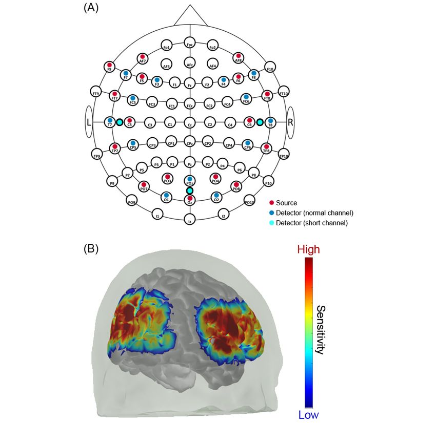
JMIR RESEARCH PROTOCOLS                                                                                                               Bálint et al

                                                                          do not interfere with the CI, are quiet (which is important in
     Introduction                                                         auditory tasks), are noninvasive, suitable for all ages, and enable
     Background                                                           the evaluation of responses to spoken words and whole
                                                                          sentences.
     Disabling hearing loss is a major communication and health
     problem that affects over 6% of the overall population and over      Previous fNIRS studies with implanted adults showed evidence
     50% of adults above the age of 65. For adults, deafness leads        of cortical reorganization. However, when comparing study
     to social isolation, unemployment, and reliance on social            findings, there are contradictory results. For example, some
     services. This problem will increase with demographic change.        studies suggest that strong activation of the auditory cortex
     It is estimated that by 2050, 10% of the global population will      during lipreading tasks is a negative predictor of speech
     be living with disabling hearing loss [1]. In patients with severe   understanding with the implant [22,23]. Other publications
     to profound hearing loss, a cochlear implant (CI) offers an          describe an opposite effect or no effect [24,25].
     effective treatment [2]. A CI is a neuroprosthetic device that       According to a recent review on fNIRS measurements in CI
     electrically stimulates the auditory nerve in response to acoustic   patients, at the current stage, it is difficult to draw a general
     stimulation. CIs enable deaf patients to regain their speech         conclusion about the potential positive or negative effects of
     understanding [3,4], improve sound localization [5], and increase    cortical reorganization. Instead, methodological aspects must
     their quality of life [6]. However, hearing outcomes after           first be clarified [26]. The effect of cross-modal plasticity may
     implantation surgery vary widely in both prelingually and            be more complex than suggested in previous studies. One
     postlingually deafened patients. About 20%-30% of                    problem with measuring functional brain activation is that many
     postlingually deafened patients who receive a CI do not gain         variables need to be controlled. For example, it makes a
     the expected benefit from the implant. Nowadays, over 75% of         remarkable difference how patients are selected (pre- or
     the variance in CI outcomes remains unclear [7-9].                   postlingually deafened) [24], whether a study participant is
     Consequently, it is not possible to predict preoperatively how       actively engaged in the experiment (otherwise mind wandering
     well a CI candidate will perform with the implant. Therefore,        might occur) [27], how the stimuli are presented, and whether
     there is an urgent need to better understand this variability and    the task performance is monitored [28]. Poorly controlled
     find ways to improve outcomes for people with poor language          variables during an fNIRS experiment can lead to
     comprehension.                                                       misinterpretations and mistakes in data analysis.
     In the absence of auditory input, the sensory deprivation induces    The aim of our study protocol is to develop a well-controlled
     reallocation of cortical areas (so-called brain plasticity). This    and reproducible fNIRS task to evaluate brain activation in
     leads to functional reorganization within the auditory and           response to speech comprehension in individuals with normal
     auditory-related brain cortex, with new functions being assigned     hearing, those with hearing impairments, and CI users. Our
     to these brain regions [10]. As an example, the visual takeover      hypothesis is that through such a task, we can identify cortical
     (also referred as cross-modal reorganization) in the impaired        networks that are clearly correlated to hearing performance with
     auditory brain areas has been demonstrated. It means that visual     the implant. Identified brain activation patterns may later be
     information, for instance, during a lipreading task, can be          used preoperatively as biomarkers of speech understanding with
     processed partially in former auditory associated brain areas        the implant.
     [11-14]. A CI can counteract these hearing loss–induced plastic
     changes, and the success of the rehabilitation depends on them.      Objectives
     It has been shown that different hearing outcomes after              Using fNIRS, our main objective is to develop an audiovisual
     implantation correlate with these reorganization processes           speech comprehension task to measure functional brain activity
     [3,15-17].                                                           regarding speech understanding. The task should comply with
     We use functional imaging to study these described plastic brain     the following criteria: it should (1) be deducible from clinically
     changes. However, in CI recipients, there are important              established hearing tests; (2) induce maximal cortical activation
     considerations to make. Despite the efforts of CI manufacturers      (and thus allow reproducible recognition of activation patterns);
     to allow structural magnetic resonance imaging (MRI) with a          (3) align with the international 10-10 system of electrode
     surgically implanted device, the technique has limitations. The      placement, using optimally spaced optode positions with
     outer speech processor cannot be worn during MRI scanning            maximal coverage over the responsible brain regions. Short
     and thus cannot be used to assess evoked auditory responses          separation channels should allow noise reduction; (4) be
     associated with functional MRI. Furthermore, the implanted           time-efficient (to avoid fatigue due to experiment duration; (5)
     magnet and electrode array of the CI cause imaging artifacts in      be suitable for normal hearing, hearing impaired, and cochlear
     MRI and stimulation artifacts in electroencephalography (EEG)        implant users; and (6) be reproducible by other research groups.
     [18-20].                                                             We will correlate the fNIRS recordings with (1) data from
     Functional near-infrared spectroscopy (fNIRS), on the other          patients’ history, (2) clinically validated questionnaires, and (3)
     hand, is ideal for this patient population [21]. The technique       performance during the fNIRS measurements (eg, speech
     uses near-infrared light to measure the blood oxygen saturation      comprehension during the fNIRS task).
     of the cerebral cortex. This allows indirect conclusions to be
     drawn about neuronal activation. Other advantages of fNIRS
     are that the measurements are not affected by electrical pulses,

     https://www.researchprotocols.org/2022/6/e38407                                                 JMIR Res Protoc 2022 | vol. 11 | iss. 6 | e38407 | p. 2
                                                                                                                (page number not for citation purposes)
XSL• FO
RenderX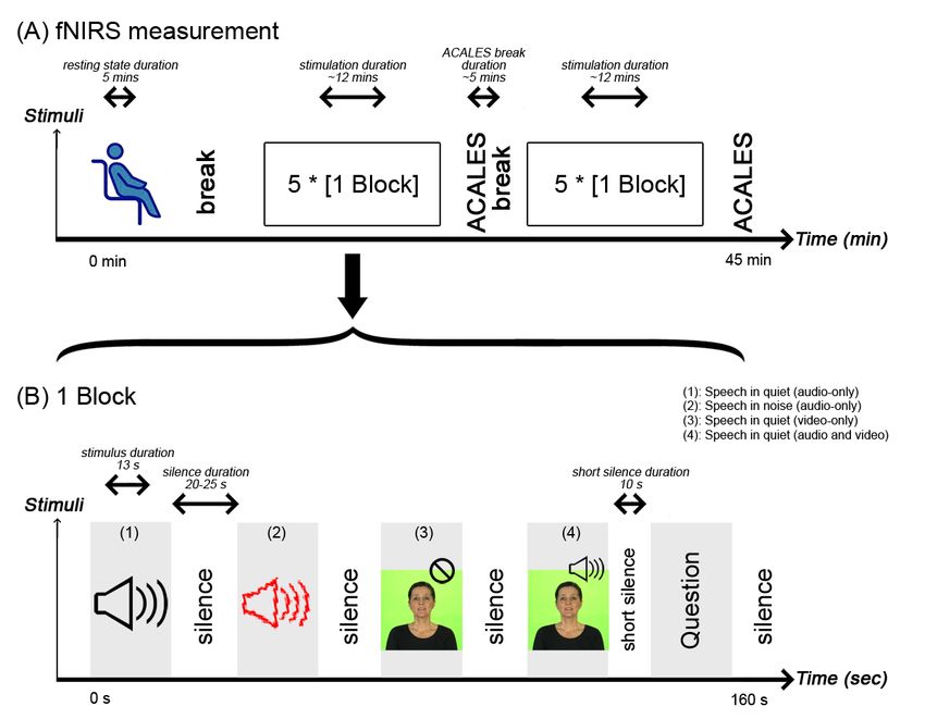
JMIR RESEARCH PROTOCOLS                                                                                                                            Bálint et al

                                                                                    hair [29,30]. Participants with a severe cardiac, psychiatric, or
     Methods                                                                        neurological disease (eg, epilepsy) or brain injury will be
     Study Design                                                                   excluded from the study (refer to Multimedia Appendix 1 for
                                                                                    details). CI users must be bilaterally and postlingually deafened,
     This research project is a prospective single-center study and                 with an unaided pure-tone average (PTA) hearing threshold
     will be conducted at the Department of Otolaryngology, Head                    exceeding a hearing level (HL) of 80 dB.
     and Neck Surgery at the Bern University Hospital, Inselspital,
     Bern, Switzerland.                                                             The ear through which the acoustic stimulation will be presented
                                                                                    needs to be implanted for at least 1 year. This will ensure that
     Ethics Approval                                                                hearing rehabilitation after implantation is completed.
     The protocol was designed in accordance with the ethical
                                                                                    Participants will be allocated to one of 3 groups: (1) normal
     principles of the Declaration of Helsinki. The study setup was
                                                                                    hearing “control” cohort, (2) CI users with good speech
     approved by the local ethical committee (reference number
                                                                                    understanding (“overperformer”), or (3) CI users with poor
     2020-02978) and fulfils all the patient data regulations of
                                                                                    speech understanding (“underperformer”). CI users with
     Switzerland.
                                                                                    moderate speech perception (ie, between 40% and 70% aided
     Participants and Eligibility Criteria                                          monosyllabic word recognition score) will not be recruited
     All study participants must (1) be at least 18 years old, (2) be               because we want to investigate the functional mechanisms
     native German speakers, and (3) have preferably light and thin                 specifically for good and poor outcomes. Table 1 provides an
                                                                                    overview of the categorization criteria for each subgroup.

     Table 1. Overview of categorization according to participants’ hearing performancea.

         Criterion                                       Normal hearing               CIb “overperformer”                    CI “underperformer”

         Unaided PTAc hearing threshold                  ≤20 dB HLd                   ≥80 dB HL                              ≥80 dB HL

         Word recognition score                          100%                         ≥70%                                   ≤40%

     a
         Word recognition score will be measured using Freiburg monosyllabic test lists at a 65 dB sound pressure level.
     b
         CI: cochlear implant.
     c
         PTA: pure-tone average.
     d
         HL: hearing level.

                                                                                    study (20 listeners with normal hearing, 20 CI overperformers,
     Sample Size                                                                    and 20 CI underperformers).
     Pilot measurements were performed on 10 participants to
     estimate an appropriate sample size. We compared the median                    Recruitment
     relative change in the concentration of oxygenated hemoglobin                  Recruitment will be done through the CI center of our
     in the auditory cortex. After acoustic stimulation (speech in                  department. Potential study candidates will be screened based
     quiet), an increase of 1.315 (SD 1.275) µMolar*cm was                          on their medical records and will be subsequently informed
     measured, while during the resting state, the value fluctuated                 verbally or in writing about the study procedure. Candidates
     close to 0. A power analysis to test a 2-sided hypothesis at 95%               who are willing to participate and able to complete all tests
     significance and 80% power showed that we need at least 15                     required for the study will be asked to sign an informed consent
     participants with normal hearing to detect auditory activations.               form.
     In addition, we considered previous findings from auditory
     fNIRS studies [26,28,31,32]. We compared the size of their
                                                                                    Study Procedure
     study cohorts, the fNIRS systems used, the optode arrangements                 Table 2 shows the time schedule for participants. The enrollment
     used, and the reliability of their results. To allow for a possibly            and the data collection sessions are described in more detail in
     larger variation, we propose including 60 individuals in this                  the subsequent subsections.

     https://www.researchprotocols.org/2022/6/e38407                                                              JMIR Res Protoc 2022 | vol. 11 | iss. 6 | e38407 | p. 3
                                                                                                                             (page number not for citation purposes)
XSL• FO
RenderX
JMIR RESEARCH PROTOCOLS Bálint et al
Table 2. Overview of the study procedure.
Item Enrollment session Data collection session
Information sheet √
Medical history √
Questionnaires √
Hearing tests √
fNIRSa recording √
Behavioral assessment √
Optode position registration √
Total duration 30 min 90-120 min
a
fNIRS: functional near-infrared spectroscopy.
(SPL) of 65 dB [39]. Additionally, we will perform the widely
Enrollment Session used Oldenburg Sentence Test (OLSA) [40-42]. The sentences
Potential study candidates will be invited to an enrollment will be played with 65 dB SPL background noise, using an
session. First, we will hand out the information sheet and answer adaptive version of the female OLSA test [43-45]. The OLSA
any questions the candidates may have. To assess full eligibility, sentences will also be used as a stimulus during the fNIRS
the candidates will have to fill in questionnaires and perform measurement. Speech material will be presented from a
additional hearing tests before data collection. Bilateral CI users loudspeaker (Control 1 Pro) placed in front of the participants
will be asked to turn off and remove the audio processor of the at a distance of 1 m.
worse hearing ear to limit acoustic stimulation exclusively to
the better ear. The worse hearing ear will be covered using an Data Collection Session
ear plug. The full enrollment session will take a maximum of Experimental Setup
30 minutes.
During fNIRS recording, each study participant will sit in a
Questionnaires comfortable chair with an armrest, headrest, and lumbar support
Questions on medical history will target the candidates’ (Figure 1). A desk will be placed in front of the participant with
handedness (Edinburgh Handedness Inventory) and the presence the electrical equipment. Visual stimuli will be presented
of diseases, which are among the exclusion criteria [33-35]. through a computer screen (P2210, Dell) placed on the table at
Additional questions on health status will inquire about the a distance of 120 cm in front of the participant. The acoustic
presence of influences that could alter the brain activity of stimuli will be played through a loudspeaker (8040B, Genelec)
interest, such as the use of stimulants [36]. CI users will receive placed above the monitor at a distance of 130 cm from the ears.
questions about the duration of their hearing loss, and if they The loudspeaker will receive input from an external ASIO sound
have tinnitus, about the objectivity and laterality of their tinnitus card (Scarlett 2i2, FocusRite) connected to the control laptop
[37]. The Hearing Ability Questionnaires will investigate (XPS 13, Dell) via USB. The system will be calibrated to 65
lipreading experience and hearing-associated factors, including dB SPL with the OLSA calibration noise and an acoustic
the Speech, Spatial, and Qualities (SSQ-12) questions [38]. The analyzer (XL2, NTi Audio).
question sheet should cover the subjective assessment of hearing The stimulation protocol will be controlled by a custom-written
ability in the last 6 months. script (Python 3.8.8) using Tkinter and python-vlc libraries.
Hearing Tests The script will send triggers via the serial interface to a
trigger-box (MMBT-S Interface Box, NEUROSPEC AG), which
The audiometric measurements and the fNIRS recordings will
converts the signals to transistor-transistor Logic (TTL) levels.
take place in an acoustic chamber (6 m × 4 m × 2 m) with a
The TTL-encoded signals will then be received by the fNIRS
separate ventilation system and electromagnetic shielding. The
machine (FOIRE-3000, Shimadzu).
broadband reverberation time is ~200 ms.
Participants will interact with the control laptop using the
In normal hearing participants, we will assess pure tone
buttons of a mouse (WM527, Dell). The pointing function of
air-conduction hearing thresholds with a clinical audiometer
the mouse will be disabled to ensure that participants control
(GSI 61, Grason-Stadler). The findings must confirm that
the experiment only by clicking and rolling. During the fNIRS
participants have no hidden or undetected hearing loss (Table
measurement, the participants will be able to press an alarm
1). For CI users, audiograms are available from clinical routine
button (Switchbox, Delock) positioned in a reachable distance
measurements.
on the table.
In all participants, we will measure the word recognition score
for Freiburg monosyllabic word lists at a sound pressure level
https://www.researchprotocols.org/2022/6/e38407 JMIR Res Protoc 2022 | vol. 11 | iss. 6 | e38407 | p. 4
(page number not for citation purposes)
XSL• FO
RenderXJMIR RESEARCH PROTOCOLS Bálint et al
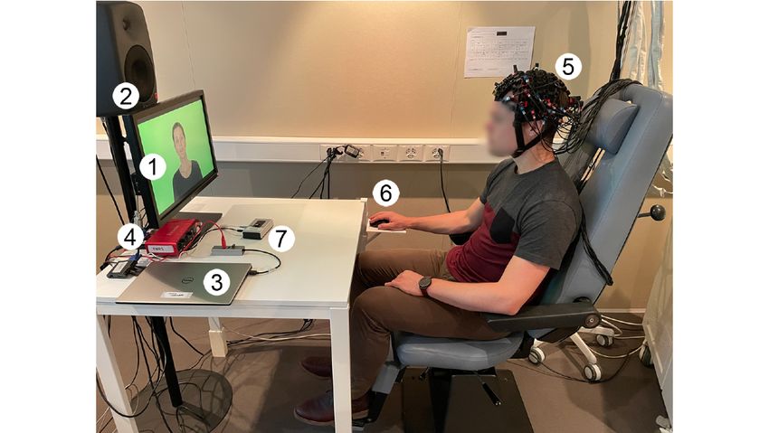
Figure 1. Experimental setup during functional near-infrared spectroscopy (fNIRS) recording. The participant will receive the stimulation via the
computer screen (1) and the loudspeaker (2). The loudspeaker will be connected to the control laptop (3) via an external soundcard (4). The fNIRS cap
(5) will be fitted on the participant's head, and the subject will interact using a response mouse (6). The alarm button (7) will be positioned in front of
the subject.
shorter channels (15 mm) provide information about the
Optode Placement
interfering systemic signals in the outer cortex and longer
We will select the regions of interest (ROIs) for the placement channels (36+ mm) about brain activation in deep regions
of the optodes considering previous studies. We expect [55,56]. Practically, however, the signal-to-noise ratio may be
responses related to audiovisual speech comprehension in the poor in long distances, so in many cases we will not be able to
auditory and visual cortex, more specifically in the following use those channels. The Monte Carlo sensitivity simulation of
ROIs: superior temporal gyrus (STG), primary visual cortex all source-detector pairs is shown in Figure 2B and indicates a
(V1), and visual association cortex (V2) [28,31,46-50]. uniform sensitivity profile across the temporal, visual, and
Additionally, during similar conditions, the left inferior frontal prefrontal cortical regions [57]. The sampling rate will be set
gyrus (LIFG) has been associated with effortful listening to 14 Hz.
[27,51], and elevated cortical responses have been reported in
the middle temporal gyrus (MTG) and middle frontal gyrus The optode holder cap will be assembled using the
(MFG) [52]. Based on the defined ROIs, we will determine the manufacturer's components (Holder kit, Shimadzu) and custom
optimal selection of EEG coordinates using the fNIRS optode's 3D printed parts (colored optode markers and stabilizers for
location decider (fOLD) toolbox [53]. We will further consider different head sizes). The parts will be designed in a solid
the position of the audio processor and receiver coil in CI modelling software (SolidWorks 2019, Dassault Systemes) and
participants to avoid interference with optodes. printed using a 3D printer (Prusa i3 MK3S+, Prusa Research).
The montage will consist of 16 sources and 16 detectors placed At the end of the experiment, we will digitize the position of
on the surface of the skull according to the international 10-10 all optodes with a depth sensing camera (Structure Sensor Pro,
system of electrode placement (Figure 2A) [54]. The Occipital Inc) connected to an iPad (iPad Pro 2020, Apple Inc).
source-detector pairs will result in a total of 43 channels in a The depth sensing camera will be set up for optimized scanning
multidistance setup: 3 of them are short-separation channels of dark objects with low ambient infrared light. The infrared
with a 15-mm interoptode distance, 4 are extra-long channels exposure time, gain, and depth resolution will be set to the
with a distance of 36-37 mm, and 36 are normal length channels highest available settings so that the colored optode markers
that are approximately 30 mm apart. In a multidistance approach, can be easily identified on the 3D scan.
https://www.researchprotocols.org/2022/6/e38407 JMIR Res Protoc 2022 | vol. 11 | iss. 6 | e38407 | p. 5
(page number not for citation purposes)
XSL• FO
RenderXJMIR RESEARCH PROTOCOLS Bálint et al
Figure 2. Functional near-infrared spectroscopy (fNIRS) montage. (A) Optode arrangement on the head. Sixteen sources (red circles) and 16 detectors
(blue and cyan circles) will be placed on the scalp, forming a total of 43 channels. Three of the detectors (cyan circles) will be forming short-separation
channels. (B) Sensitivity map of the optode arrangement.
stimulations. Once the participant confirms that the task is
Functional Near-Infrared Spectroscopy
understood, we will start the definitive recording. The functional
During fNIRS recordings, we will instruct the participants to recordings will begin with a 5-minute resting state period (Figure
concentrate on the screen, follow the instructions, and reduce 3A). We will instruct the participant to sit still, close their eyes,
head movements. If the participants feel uncomfortable, an and relax but try not to fall asleep. Then 2 stimulation sessions
emergency button in front of them will be made available to approximately 12 minutes each will follow. Between the 3
stop the experiment. We will give all instructions both verbally sessions (ie, the resting and the 2 stimulation sessions), the
and in writing. Before the recordings, the participants will participants can take a break of their chosen duration.
conduct a short familiarization session with 4 example
https://www.researchprotocols.org/2022/6/e38407 JMIR Res Protoc 2022 | vol. 11 | iss. 6 | e38407 | p. 6
(page number not for citation purposes)
XSL• FO
RenderXJMIR RESEARCH PROTOCOLS Bálint et al
Figure 3. Functional near-infrared spectroscopy (fNIRS) measurement overview. (A) Following the resting state measurement, 2 x 5 counterbalanced
blocks will be presented, with breaks in between. (A) A single block consists of a (1) speech in quiet (audio only), (2) speech in noise (audio only), (3)
speech in quiet (video only), (4) speech in quiet (audio and video) stimulation, and an additional question.
example, if the sentence is “Nina gives 12 red flowers,” the
Stimuli
question is either ”How many red flowers?“ or ”Who gives 12
As stimulus material, we will use a video version of the female red flowers?“ To answer the question, the participant will have
OLSA test [40,58]. A single stimulus will consist of one to select 1 of 4 choices: 2 randomly selected numbers/names
sentence (eg, “Nina gives twelve red flowers”) which will be from the OLSA sentence matrix (wrong answers), an option if
repeated 3 times. The duration of one stimulus will be 13 the respondent is not sure of the answer (skipped answer), and
seconds, comparable to hemodynamic responses [59]. the correct answer. For the previous question, a possible
A single stimulation block will contain 4 different stimuli, combination could be (1) ”Britta,” (2) “Nina,” (3) “Peter,” and
presented in one of the following modalities in a (4) “I cannot decide.” The participant will select an option with
counterbalanced order (Figure 3B): (1) speech in quiet (audio the roller on the computer mouse and confirm the answer with
only), (2) speech in noise (audio only), (3) speech in quiet (video a double click. In the previous example, the participant must
only, ie, lipreading), and (4) speech in quiet (audio and video). select the second option (”Nina“). The questions and the answers
The stimulation will be followed by 20-25 seconds of will be randomly generated, and the position of the question
nonstimulus interval, during which a white fixation point will within the blocks will also be randomly chosen.
be presented on a black screen. During the audio-only The shortened nonstimulus interval of 10 seconds prior to the
conditions, the same black screen will be displayed so that the question window will allow us to evaluate the fNIRS responses.
participant will have no indication other than hearing whether Therefore, the interleaved questions will not harm the overall
the stimulation has already started or not. effectiveness of the measurement. After the question is
At random points, participants will be asked to answer questions answered, the regular 20-25 second relaxation time will be
to ensure attention and monitor speech comprehension during applied to ensure that the brain responses return to baseline.
the test. The questions will be displayed in the nonstimulus Overall, 2 questions per modality will be asked, resulting in 8
epoch, for which the nonstimulus interval will be shortened to questions throughout the entire fNIRS measurement.
10 seconds. The questions will ask to repeat the correct name
or number of the last sentence from 4 possible answers. For
https://www.researchprotocols.org/2022/6/e38407 JMIR Res Protoc 2022 | vol. 11 | iss. 6 | e38407 | p. 7
(page number not for citation purposes)
XSL• FO
RenderXJMIR RESEARCH PROTOCOLS Bálint et al
Following the breaks, before the first stimulation, there will be oxygenated and deoxygenated hemoglobin should be negatively
a stimulus-free interval of 20 seconds. This will ensure correlated [74].
homogeneity of responses, meaning that all stimuli are perceived
Optode Positions
under similar circumstances. Overall, 10 blocks will be
presented, resulting in 10 responses per stimulation modality, We will perform the postprocessing of the scans with a 3D mesh
and the total fNIRS measurement time will be around 45 processing tool (MeshLab) and custom-written scripts
minutes. At the beginning of every event (start/stop of a block, (MathWorks) [75].
resting state, stimulation, question, answer), a trigger will be We will manually select the coordinates of the optodes and
sent from the control computer to the fNIRS machine through anatomical landmarks with MeshLab on the obtained 3D scans.
the trigger-box and stored as an extra channel in the fNIRS raw The list of coordinates will then be exported and projected into
data. Montreal Neurological Institute (MNI) space. The MNI
Listening Effort coordinates will be displayed on the preoperative MRI scan of
every CI-user participant, and the exact source of measured
Following 5 stimulation blocks, we will ask every participant
hemodynamic activation will be determined. Additionally, the
to rate their listening effort to the different stimuli, their rating
mean and the standard deviation of optode coordinates will be
of fatigue and their level of mind-wandering (Figure 3A)
calculated and reported as quality measure for optode fittings
[60-64]. To evaluate the listening effort, we will use Adaptive
[55,76].
Categorical Listening Effort Scaling (ACALES) [65].
fNIRS Recordings
Data Management
Data analysis will be performed in Python using the
All written source documents will be completed in a neat, legible
MNE-Toolbox [77] and MNE-NIRS package [78]. Individual
manner to ensure accurate interpretation of data. For each
epochs will be subtracted from the channel data, from t=0
participant, a case report form (CRF) will be maintained,
seconds to t=24 seconds relative to the stimulus onset. The
including the participant number. In CRFs and other
epochs will be baseline-corrected by subtracting the mean of
project-specific documents, participants are only identified by
the signal between t =−5 seconds and t=0 seconds. Using the
a unique participant number. fNIRS measurements will be stored
Glover canonical hemodynamic response function [79] a design
in a closed research environment (REDCap, Vanderbilt
matrix for the general linear model (GLM) will be constructed
University, Nashville, United States). The secure web
[80,81]. After GLM fitting, the regression results will be stored.
application is running on a local server maintained and backed
Following this, temporal and spatial features will be extracted
up by the University of Bern. All documents related to the study,
from each epoch (amplitude, area under curve, peak latency,
including the CRFs will be considered as source data, and these
laterality, power). The regression results and the extracted
will be stored at the measurement site in accordance with
features will be weight-averaged over ROIs by taking the inverse
relevant standards.
of the standard error of the GLM fit for each channel [67]. The
Data Analysis data will be averaged over the participants, and group-level
statistics will be calculated using correlation analysis and linear
fNIRS Preprocessing mixed-effects models.
Data preprocessing will be performed in MATLAB
(MathWorks) using the Homer2 (v2.3) [66] and NIRS [67] Behavioral Data
toolboxes. The signal quality will be checked based on the heart The answers from the questionnaires will be digitized, and
rate content of the signal, using a sliding window approach correlation analysis will be performed to reveal relations
[68-71]. Channels and time points with insufficient signal quality between the measured brain activation patterns and the evaluated
will be removed. Short channel correction will be applied to questionnaires. Additionally, further behavioral data will be
the absorbance data, using short separation regression [56,72]. obtained from the triggers, such as reaction time to questions
The motion artifacts will be removed with a WaveletFilter across the measurement as a measure of fatigue or response
module of the NIRS toolbox [67]. The signal will be bandpass accuracy for each stimulation type as a measure of speech
filtered between 0.01 and 0.12 Hz with the BandpassFilter understanding.
function from the Homer toolbox [66]. Then, the absorbance
data will be converted to concentration changes of oxygenated Results
hemoglobin (HbO) and deoxygenated hemoglobin (HbR) in
mMolar*cm using the following equations, as specified by the The enrollment for the study described in this protocol started
manufacturer based on the modified Beer-Lambert law [73]: in August 2021. The first results are expected at the end of 2022.
ΔHbO = (−1.4887) * Abs[780nm] + 0.5970 * Discussion
Abs[805nm] + 1.4878 * Abs[830nm]
ΔHbR = 1.8545 * Abs[780nm] - 0.2394 * The postoperative adaptive or maladaptive effect of existing
Abs[805nm] − 1.0947 * Abs[830nm] cross-modal reorganization in CI candidates is a complex
question. The available studies show contradictory findings. A
A further correction step will be performed to reduce noise
recent review states that it is important to discuss the
based on the principle that the concentration changes of
methodological aspects of such functional neuroimaging
examinations [22-26,46,47,50]. One problem with measuring
https://www.researchprotocols.org/2022/6/e38407 JMIR Res Protoc 2022 | vol. 11 | iss. 6 | e38407 | p. 8
(page number not for citation purposes)
XSL• FO
RenderXJMIR RESEARCH PROTOCOLS Bálint et al
functional brain activation is that many variables must be This allows us to study not only audiovisual activations but also
considered. To better control these variables, we present hereby speech perception in noise, the effects of fatigue, and activity
an audiovisual speech comprehension task that fulfills the 6 related to higher-order cortical processing. Many other studies
points outlined below. have not had the opportunity to cover such a wide range of
cortical regions, mostly due to hardware limitations [22-25,50].
First, the test should be deducible from clinically established
We use the Edinburgh Handedness Inventory to control for
hearing tests. We used the video version of a widely used
handedness, which might affect the laterality of brain activation
clinical test (Oldenburg Sentence Test) [58]. Functional brain
[33-35]. We also perform a spatial registration of optode
activation patterns can therefore be correlated with clinical
positions to increase reproducibility. Furthermore, these
findings. These results are easier to interpret than custom-made
measured positions can be projected into MNI space and
speech materials [23,46,47,50]. Our employed stimulation
displayed on MRI images. In the diagnostic workup, MRI are
design consists of complete sentences, which reflect everyday
routinely performed prior to CI surgery. The method will allow
life and real language comprehension much better than
a more accurate localization of hemodynamic responses
nonspeech auditory stimuli or speech snippets
compared to atlas-based approaches [55,76].
[13,23,25,28,46,47]. Before conducting the fNIRS experiment,
we will repeat clinical speech comprehension tests (ie, Freiburg Additionally, we use a multidistance channel setup. The optodes
monosyllabic test, Oldenburg Sentence Test). This enables a of the regular channels are ~30 mm apart from each other.
clear grouping of the CI participants into good and poor Additional short channels with a 15-mm interoptode distance
performers. During the fNIRS experiment, we will continue to over the auditory and visual cortex provide extracerebral
assess speech comprehension in 4 different situations (ie, speech information to remove confounding systemic signals. It is
in quiet, speech in noise, visual-speech, audiovisual speech) recommended to use a systemic physiology controlled fNIRS
with interleaved comprehension questions. This allows us to approach, although this has rarely been applied in previous
maintain attention and monitor speech comprehension while studies [55].
measuring brain activity. This advantage has only been applied
Fourth, it should be time efficient to avoid fatigue due to
by one research group [22,24]. To assess listening effort during
experiment duration. The longest task the participants will be
the fNIRS task, we will use a validated questionnaire (ie,
required to complete will last 12 minutes, and the total
ACALES) [65]. Listening effort in CI users is an active topic
measurement time will be around 45 minutes. Regular breaks
of discussion [82]; its possible influence on the measured
will be provided, and the total duration of the experiment is
cortical activation, to the best of our knowledge, has never been
expected to be around 120-150 minutes.
reported before. To describe the subjective hearing perception
in their daily lives, participants will complete validated Fifth, it should be suitable for participants with normal hearing,
questionnaires (ie, SSQ-12) on the day of the test [38]. We will hearing impairments, and those using CIs. The audio material
conduct our tests in a validated audio chamber (as used in is presented through a loudspeaker, so the task is suitable for
clinically performed hearing tests). people with normal hearing as well as for hearing aid and CI
users. Alternatively, an insert earphone or a direct CI audio
Second, the task should induce maximal cortical activation (and
input simulation would be feasible. However, these 2 approaches
thus allow reproducible recognition of activation patterns). We
have the disadvantage that the 3 aforementioned groups cannot
use an optimized counterbalanced block design. The duration
not be stimulated identically. Our optode placement was chosen
of 1 stimulus will be 13 seconds, and the interstimulus break
to allow for easy attachment of the implant coil.
will be between 20 and 25 seconds, comparable to hemodynamic
responses [49,59]. Our task requires the active participation of Sixth, it should be reproducible by other research groups. The
the participants. Previous studies have shown that this can audiovisual version of the OLSA was published in 2021 and is
significantly increase brain activation [63,64]. Furthermore, we now accessible [58]. Moreover, we are happy to share our setup
mitigate mind wandering and fatigue by filling out validated upon request.
questionnaires [60-62]. As far as we know, in persons with
In summary, the proposed audiovisual speech comprehension
hearing impairments, this has never been reported in the context
task will help us understand neural correlates to speech
of fNIRS measurements. To avoid fatigue (which can lead to
understanding. In the first stage, we will perform these
decreased brain activation), we keep the fNIRS task as short as
measurements postoperatively to better understand the
possible. Additionally, participants can take 2 breaks of
corresponding neuronal networks with an activated implant. In
self-selected duration.
the subsequent stage, we will perform the measurements pre-
Third, it should be in alignment with the international 10-10 and postoperatively to make prognostic calculations. The
system of electrode placement, using optimally spaced optode comprehensive study will have the potential to provide
positions with maximal coverage over the responsible brain additional prognostic information beyond the conventional
regions. Short separation channels should allow noise reduction. clinical standards regarding the underlying plastic brain changes
Our optode placement covers the following brain regions: of a person with hearing impairment. Our study will facilitate
superior temporal gyrus (STG), primary visual cortex (V1), more precise indication criteria for cochlear implantation and
visual association cortex (V2), left inferior frontal gyrus (LIFG), a better planning of rehabilitation.
middle temporal gyrus (MTG), and middle frontal gyrus (MFG).
https://www.researchprotocols.org/2022/6/e38407 JMIR Res Protoc 2022 | vol. 11 | iss. 6 | e38407 | p. 9
(page number not for citation purposes)
XSL• FO
RenderXJMIR RESEARCH PROTOCOLS Bálint et al
Acknowledgments
The study is funded by the Wonderland Foundation, the Gottfried und Julia Bangerter-Rhyner Foundation, and the UniBern
Research Foundation.
Conflicts of Interest
None declared.
Multimedia Appendix 1
Neurological, cardiac, psychiatric, or other major diseases used as exclusion criteria.
[PDF File (Adobe PDF File), 50 KB-Multimedia Appendix 1]
References
1. World Health Organization. Addressing the rising prevalence of hearing loss. Geneve, Switzerland: World Health
Organization; 2018. URL: https://www.who.int/
2. Bond M, Mealing S, Anderson R, Elston J, Weiner G, Taylor R, et al. The effectiveness and cost-effectiveness of cochlear
implants for severe to profound deafness in children and adults: a systematic review and economic model. Health Technol
Assess 2009 Sep;13(44):1-330 [FREE Full text] [doi: 10.3310/hta13440] [Medline: 19799825]
3. Strelnikov K, Marx M, Lagleyre S, Fraysse B, Deguine O, Barone P. PET-imaging of brain plasticity after cochlear
implantation. Hear Res 2015 Apr;322:180-187. [doi: 10.1016/j.heares.2014.10.001] [Medline: 25448166]
4. Wimmer W, Weder S, Caversaccio M, Kompis M. Speech intelligibility in noise with a pinna effect imitating cochlear
implant processor. Otol Neurotol 2016 Jan;37(1):19-23. [doi: 10.1097/MAO.0000000000000866] [Medline: 26427637]
5. Fischer T, Schmid C, Kompis M, Mantokoudis G, Caversaccio M, Wimmer W. Pinna-imitating microphone directionality
improves sound localization and discrimination in bilateral cochlear implant users. Ear Hear 2021;42(1):214-222 [FREE
Full text] [doi: 10.1097/AUD.0000000000000912] [Medline: 32701730]
6. McRackan TR, Bauschard M, Hatch JL, Franko-Tobin E, Droghini HR, Nguyen SA, et al. Meta-analysis of quality-of-life
improvement after cochlear implantation and associations with speech recognition abilities. Laryngoscope 2018 Apr
21;128(4):982-990 [FREE Full text] [doi: 10.1002/lary.26738] [Medline: 28731538]
7. Geers AE, Nicholas J, Tobey E, Davidson L. Persistent language delay versus late language emergence in children with
early cochlear implantation. J Speech Lang Hear Res 2016 Feb;59(1):155-170 [FREE Full text] [doi:
10.1044/2015_JSLHR-H-14-0173] [Medline: 26501740]
8. Blamey P, Artieres F, Başkent D, Bergeron F, Beynon A, Burke E, et al. Factors affecting auditory performance of
postlinguistically deaf adults using cochlear implants: an update with 2251 patients. Audiol Neurootol 2013;18(1):36-47.
[doi: 10.1159/000343189] [Medline: 23095305]
9. Lazard DS, Vincent C, Venail F, Van de Heyning P, Truy E, Sterkers O, et al. Pre-, per- and postoperative factors affecting
performance of postlinguistically deaf adults using cochlear implants: a new conceptual model over time. PLoS One 2012
Nov 9;7(11):e48739 [FREE Full text] [doi: 10.1371/journal.pone.0048739] [Medline: 23152797]
10. Bavelier D, Neville HJ. Cross-modal plasticity: where and how? Nat Rev Neurosci 2002 Jun;3(6):443-452. [doi:
10.1038/nrn848] [Medline: 12042879]
11. Lazard DS, Lee H, Truy E, Giraud A. Bilateral reorganization of posterior temporal cortices in post-lingual deafness and
its relation to cochlear implant outcome. Hum Brain Mapp 2013 May 30;34(5):1208-1219 [FREE Full text] [doi:
10.1002/hbm.21504] [Medline: 22287085]
12. Karns CM, Dow MW, Neville HJ. Altered Cross-Modal Processing in the Primary Auditory Cortex of Congenitally Deaf
Adults: A Visual-Somatosensory fMRI Study with a Double-Flash Illusion. J Neurosci 2012 Jul 11;32(28):9626-9638.
[doi: 10.1523/jneurosci.6488-11.2012]
13. Stropahl M, Chen L, Debener S. Cortical reorganization in postlingually deaf cochlear implant users: Intra-modal and
cross-modal considerations. Hear Res 2017 Jan;343:128-137 [FREE Full text] [doi: 10.1016/j.heares.2016.07.005] [Medline:
27473503]
14. Lee H, Truy E, Mamou G, Sappey-Marinier D, Giraud A. Visual speech circuits in profound acquired deafness: a possible
role for latent multimodal connectivity. Brain 2007 Nov 05;130(11):2929-2941. [doi: 10.1093/brain/awm230] [Medline:
17906328]
15. McKay CM. Brain plasticity and rehabilitation with a cochlear implant. Adv Otorhinolaryngol 2018;81:57-65. [doi:
10.1159/000485586] [Medline: 29794427]
16. Sharma A, Campbell J, Cardon G. Developmental and cross-modal plasticity in deafness: evidence from the P1 and N1
event related potentials in cochlear implanted children. Int J Psychophysiol 2015 Feb;95(2):135-144 [FREE Full text] [doi:
10.1016/j.ijpsycho.2014.04.007] [Medline: 24780192]
https://www.researchprotocols.org/2022/6/e38407 JMIR Res Protoc 2022 | vol. 11 | iss. 6 | e38407 | p. 10
(page number not for citation purposes)
XSL• FO
RenderXJMIR RESEARCH PROTOCOLS Bálint et al
17. Rouger J, Lagleyre S, Démonet JF, Fraysse B, Deguine O, Barone P. Evolution of crossmodal reorganization of the voice
area in cochlear-implanted deaf patients. Hum Brain Mapp 2012 Aug 06;33(8):1929-1940 [FREE Full text] [doi:
10.1002/hbm.21331] [Medline: 21557388]
18. Srinivasan R, So C, Amin N, Jaikaransingh D, D'Arco F, Nash R. A review of the safety of MRI in cochlear implant patients
with retained magnets. Clin Radiol 2019 Dec;74(12):972.e9-972.e16. [doi: 10.1016/j.crad.2019.06.011] [Medline: 31324337]
19. Shew M, Wichova H, Lin J, Ledbetter LN, Staecker H. Magnetic resonance imaging with cochlear implants and auditory
brainstem implants: Are we truly practicing MRI safety? Laryngoscope 2019 Feb 09;129(2):482-489. [doi:
10.1002/lary.27516] [Medline: 30412276]
20. Wagner L, Maurits N, Maat B, Baskent D, Wagner AE. The cochlear implant EEG artifact recorded from an artificial brain
for complex acoustic stimuli. IEEE Trans Neural Syst Rehabil Eng 2018 Feb;26(2):392-399. [doi:
10.1109/tnsre.2018.2789780]
21. Saliba J, Bortfeld H, Levitin DJ, Oghalai JS. Functional near-infrared spectroscopy for neuroimaging in cochlear implant
recipients. Hear Res 2016 Aug;338:64-75 [FREE Full text] [doi: 10.1016/j.heares.2016.02.005] [Medline: 26883143]
22. Anderson CA, Wiggins IM, Kitterick PT, Hartley DEH. Pre-operative brain imaging using functional near-infrared
spectroscopy helps predict cochlear implant outcome in deaf adults. J Assoc Res Otolaryngol 2019 Oct 8;20(5):511-528
[FREE Full text] [doi: 10.1007/s10162-019-00729-z] [Medline: 31286300]
23. Zhou X, Seghouane A, Shah A, Innes-Brown H, Cross W, Litovsky R, et al. Cortical speech processing in postlingually
deaf adult cochlear implant users, as revealed by functional near-infrared spectroscopy. Trends Hear 2018 Jul
19;22:2331216518786850 [FREE Full text] [doi: 10.1177/2331216518786850] [Medline: 30022732]
24. Anderson CA, Wiggins IM, Kitterick PT, Hartley DEH. Adaptive benefit of cross-modal plasticity following cochlear
implantation in deaf adults. Proc Natl Acad Sci U S A 2017 Sep 19;114(38):10256-10261 [FREE Full text] [doi:
10.1073/pnas.1704785114] [Medline: 28808014]
25. Mushtaq F, Wiggins IM, Kitterick PT, Anderson CA, Hartley DEH. The benefit of cross-modal reorganization on speech
perception in pediatric cochlear implant recipients revealed using functional near-infrared spectroscopy. Front Hum Neurosci
2020 Aug 14;14:308 [FREE Full text] [doi: 10.3389/fnhum.2020.00308] [Medline: 32922273]
26. Harrison SC, Lawrence R, Hoare DJ, Wiggins IM, Hartley DEH. Use of functional near-infrared spectroscopy to predict
and measure cochlear implant outcomes: a scoping review. Brain Sci 2021 Oct 28;11(11):1439 [FREE Full text] [doi:
10.3390/brainsci11111439] [Medline: 34827438]
27. Wild CJ, Yusuf A, Wilson DE, Peelle JE, Davis MH, Johnsrude IS. Effortful listening: the processing of degraded speech
depends critically on attention. J Neurosci 2012 Oct 03;32(40):14010-14021. [doi: 10.1523/jneurosci.1528-12.2012]
28. Weder S, Shoushtarian M, Olivares V, Zhou X, Innes-Brown H, McKay C. Cortical fNIRS Responses Can Be Better
Explained by Loudness Percept than Sound Intensity. Ear Hear 2020;41(5):1187-1195. [doi:
10.1097/AUD.0000000000000836] [Medline: 31985534]
29. Orihuela-Espina F, Leff DR, James DRC, Darzi AW, Yang GZ. Quality control and assurance in functional near infrared
spectroscopy (fNIRS) experimentation. Phys Med Biol 2010 Jul 07;55(13):3701-3724. [doi: 10.1088/0031-9155/55/13/009]
[Medline: 20530852]
30. Wyser DG, Kanzler CM, Salzmann L, Lambercy O, Wolf M, Scholkmann F, et al. Characterizing reproducibility of cerebral
hemodynamic responses when applying short-channel regression in functional near-infrared spectroscopy. Neurophoton
2022 Jan 1;9(01):15004. [doi: 10.1117/1.nph.9.1.015004]
31. Shoushtarian M, Alizadehsani R, Khosravi A, Acevedo N, McKay CM, Nahavandi S, et al. Objective measurement of
tinnitus using functional near-infrared spectroscopy and machine learning. PLoS One 2020 Nov 18;15(11):e0241695 [FREE
Full text] [doi: 10.1371/journal.pone.0241695] [Medline: 33206675]
32. Shoushtarian M, Weder S, Innes-Brown H, McKay CM. Assessing hearing by measuring heartbeat: The effect of sound
level. PLoS One 2019 Feb 28;14(2):e0212940 [FREE Full text] [doi: 10.1371/journal.pone.0212940] [Medline: 30817808]
33. Oldfield R. The assessment and analysis of handedness: The Edinburgh inventory. Neuropsychologia 1971 Mar;9(1):97-113.
[doi: 10.1016/0028-3932(71)90067-4]
34. Khedr E, Hamed E, Said A, Basahi J. Handedness and language cerebral lateralization. Eur J Appl Physiol 2002 Aug
1;87(4-5):469-473. [doi: 10.1007/s00421-002-0652-y] [Medline: 12172889]
35. Watson NF, Dodrill C, Farrell D, Holmes MD, Miller JW. Determination of language dominance with near-infrared
spectroscopy: comparison with the intracarotid amobarbital procedure. Seizure 2004 Sep;13(6):399-402 [FREE Full text]
[doi: 10.1016/j.seizure.2003.09.008] [Medline: 15276143]
36. Sargent A, Watson J, Topoglu Y, Ye H, Suri R, Ayaz H. Impact of tea and coffee consumption on cognitive performance:
An fNIRS and EDA Study. Appl Sci 2020 Apr 01;10(7):2390. [doi: 10.3390/app10072390]
37. Hu S, Anschuetz L, Huth ME, Sznitman R, Blaser D, Kompis M, et al. Association between residual inhibition and neural
activity in patients with tinnitus: protocol for a controlled within- and between-subject comparison study. JMIR Res Protoc
2019 Jan 09;8(1):e12270 [FREE Full text] [doi: 10.2196/12270] [Medline: 30626571]
38. Noble W, Jensen NS, Naylor G, Bhullar N, Akeroyd MA. A short form of the Speech, Spatial and Qualities of Hearing
scale suitable for clinical use: the SSQ12. Int J Audiol 2013 Jun 08;52(6):409-412 [FREE Full text] [doi:
10.3109/14992027.2013.781278] [Medline: 23651462]
https://www.researchprotocols.org/2022/6/e38407 JMIR Res Protoc 2022 | vol. 11 | iss. 6 | e38407 | p. 11
(page number not for citation purposes)
XSL• FO
RenderXJMIR RESEARCH PROTOCOLS Bálint et al
39. Hahlbrock KH. Sprachaudiometrie: Grundlagen und Praktische Anwendung Einer Sprachaudiometrie für das deutsche
Sprachgebiet; 157 Abbildungen in 305 Einzeldarstellungen 9 Tabellen. Teningen, Germany: Thieme; 1970.
40. Wagener K, Brand T, Kollmeier B. Entwicklung und Evaluation eines Satztests für die deutsche Sprache Teil III: Evaluation
des Oldenburger Satztests. Zeitschrift für Audiologie 1999:38 [FREE Full text]
41. Ahrlich M. Optimierung und Evaluation des Oldenburger Satztests mit Weiblicher Sprecherin und Untersuchung des Effekts
des Sprechers auf die Sprachverständlichkeit. Oldenburg, Germany: Carl von Ossietzky Universität Oldenburg; 2013.
42. Wagener K, Hochmuth S, Ahrlich M, Zokoll-v. d. Laan M, Kollmeier B. Der weibliche oldenburger satztest. The female
version of the Oldenburg sentence test. In: Proceedings of the 17th Jahrestagung der Deutschen Gesellschaft für Audiologie.
2014 Mar 13 Presented at: Jahrestagung der Deutschen Gesellschaft für Audiologie; 2014; Oldenburg, Germany URL:
http://www.uzh.ch/orl/dga2014/programm/wissprog/Wagener.pdf
43. Wimmer W, Kompis M, Stieger C, Caversaccio M, Weder S. Directional microphone contralateral routing of signals in
cochlear implant users. Ear Hear 2017;38(3):368-373. [doi: 10.1097/aud.0000000000000412]
44. Gawliczek T, Wimmer W, Munzinger F, Caversaccio M, Kompis M. Speech understanding and sound localization with a
new nonimplantable wearing option for Baha. Biomed Res Int 2018 Sep 25;2018:5264124 [FREE Full text] [doi:
10.1155/2018/5264124] [Medline: 30356363]
45. Wardenga N, Batsoulis C, Wagener KC, Brand T, Lenarz T, Maier H. Do you hear the noise? The German matrix sentence
test with a fixed noise level in subjects with normal hearing and hearing impairment. Int J Audiol 2015 Nov 10;54 Suppl
2(sup2):71-79. [doi: 10.3109/14992027.2015.1079929] [Medline: 26555195]
46. Chen L, Puschmann S, Debener S. Increased cross-modal functional connectivity in cochlear implant users. Sci Rep 2017
Aug 30;7(1):10043 [FREE Full text] [doi: 10.1038/s41598-017-10792-2] [Medline: 28855675]
47. Chen L, Sandmann P, Thorne JD, Bleichner MG, Debener S. Cross-modal functional reorganization of visual and auditory
cortex in adult cochlear implant users identified with fNIRS. Neural Plast 2016;2016:4382656-4382613 [FREE Full text]
[doi: 10.1155/2016/4382656] [Medline: 26819766]
48. Weder S, Zhou X, Shoushtarian M, Innes-Brown H, McKay C. Cortical processing related to intensity of a modulated noise
stimulus-a functional near-infrared study. J Assoc Res Otolaryngol 2018 Jun 9;19(3):273-286 [FREE Full text] [doi:
10.1007/s10162-018-0661-0] [Medline: 29633049]
49. Wiggins IM, Anderson CA, Kitterick PT, Hartley DE. Speech-evoked activation in adult temporal cortex measured using
functional near-infrared spectroscopy (fNIRS): Are the measurements reliable? Hear Res 2016 Sep;339:142-154 [FREE
Full text] [doi: 10.1016/j.heares.2016.07.007] [Medline: 27451015]
50. Olds C, Pollonini L, Abaya H, Larky J, Loy M, Bortfeld H, et al. Cortical activation patterns correlate with speech
understanding after cochlear implantation. Ear Hear 2016;37(3):e160-e172 [FREE Full text] [doi:
10.1097/AUD.0000000000000258] [Medline: 26709749]
51. Lawrence RJ, Wiggins IM, Anderson CA, Davies-Thompson J, Hartley DE. Cortical correlates of speech intelligibility
measured using functional near-infrared spectroscopy (fNIRS). Hear Res 2018 Dec;370:53-64 [FREE Full text] [doi:
10.1016/j.heares.2018.09.005] [Medline: 30292959]
52. Defenderfer J, Forbes S, Wijeakumar S, Hedrick M, Plyler P, Buss AT. Frontotemporal activation differs between perception
of simulated cochlear implant speech and speech in background noise: An image-based fNIRS study. Neuroimage 2021
Oct 15;240:118385 [FREE Full text] [doi: 10.1016/j.neuroimage.2021.118385] [Medline: 34256138]
53. Zimeo Morais GA, Balardin JB, Sato JR. fNIRS Optodes' Location Decider (fOLD): a toolbox for probe arrangement
guided by brain regions-of-interest. Sci Rep 2018 Feb 20;8(1):3341 [FREE Full text] [doi: 10.1038/s41598-018-21716-z]
[Medline: 29463928]
54. Chatrian GE, Lettich E, Nelson PL. Ten percent electrode system for topographic studies of spontaneous and evoked EEG
activities. Am J EEG Technol 2015 Feb 10;25(2):83-92. [doi: 10.1080/00029238.1985.11080163]
55. Yücel MA, Lühmann AV, Scholkmann F, Gervain J, Dan I, Ayaz H, et al. Best practices for fNIRS publications. Neurophoton
2021 Jan 1;8(01):12101. [doi: 10.1117/1.nph.8.1.012101]
56. Scholkmann F, Kleiser S, Metz AJ, Zimmermann R, Mata Pavia J, Wolf U, et al. A review on continuous wave functional
near-infrared spectroscopy and imaging instrumentation and methodology. Neuroimage 2014 Jan 15;85 Pt 1:6-27. [doi:
10.1016/j.neuroimage.2013.05.004] [Medline: 23684868]
57. Zhang Q, Brown EN, Strangman GE. Adaptive filtering for global interference cancellation and real-time recovery of
evoked brain activity: a Monte Carlo simulation study. J Biomed Opt 2007;12(4):044014 [FREE Full text] [doi:
10.1117/1.2754714] [Medline: 17867818]
58. Llorach G, Kirschner F, Grimm G, Zokoll MA, Wagener KC, Hohmann V. Development and evaluation of video recordings
for the OLSA matrix sentence test. Int J Audiol 2022 Apr 10;61(4):311-321. [doi: 10.1080/14992027.2021.1930205]
[Medline: 34109902]
59. Handwerker DA, Ollinger JM, D'Esposito M. Variation of BOLD hemodynamic responses across subjects and brain regions
and their effects on statistical analyses. Neuroimage 2004 Apr;21(4):1639-1651. [doi: 10.1016/j.neuroimage.2003.11.029]
[Medline: 15050587]
60. Micklewright D, St Clair Gibson A, Gladwell V, Al Salman A. Development and Validity of the Rating-of-Fatigue Scale.
Sports Med 2017 Mar 10;47(11):2375-2393. [doi: 10.1007/s40279-017-0711-5]
https://www.researchprotocols.org/2022/6/e38407 JMIR Res Protoc 2022 | vol. 11 | iss. 6 | e38407 | p. 12
(page number not for citation purposes)
XSL• FO
RenderXJMIR RESEARCH PROTOCOLS Bálint et al
61. Unsworth N, McMillan BD. Similarities and differences between mind-wandering and external distraction: a latent variable
analysis of lapses of attention and their relation to cognitive abilities. Acta Psychol (Amst) 2014 Jul;150:14-25. [doi:
10.1016/j.actpsy.2014.04.001] [Medline: 24793128]
62. Stawarczyk D, Majerus S, Maj M, Van der Linden M, D'Argembeau A. Mind-wandering: phenomenology and function as
assessed with a novel experience sampling method. Acta Psychol (Amst) 2011 Mar;136(3):370-381. [doi:
10.1016/j.actpsy.2011.01.002] [Medline: 21349473]
63. Woods DL, Stecker GC, Rinne T, Herron TJ, Cate AD, Yund EW, et al. Functional maps of human auditory cortex: effects
of acoustic features and attention. PLoS One 2009 Apr 13;4(4):e5183 [FREE Full text] [doi: 10.1371/journal.pone.0005183]
[Medline: 19365552]
64. Jäncke L, Mirzazade S, Joni Shah N. Attention modulates activity in the primary and the secondary auditory cortex: a
functional magnetic resonance imaging study in human subjects. Neurosci 1999 May;266(2):125-128. [doi:
10.1016/s0304-3940(99)00288-8]
65. Krueger M, Schulte M, Zokoll MA, Wagener KC, Meis M, Brand T, et al. Relation between listening effort and speech
intelligibility in noise. Am J Audiol 2017 Oct 12;26(3S):378-392. [doi: 10.1044/2017_aja-16-0136]
66. Huppert TJ, Diamond SG, Franceschini MA, Boas DA. HomER: a review of time-series analysis methods for near-infrared
spectroscopy of the brain. Appl Opt 2009 Apr 01;48(10):D280-D298 [FREE Full text] [doi: 10.1364/ao.48.00d280] [Medline:
19340120]
67. Santosa H, Zhai X, Fishburn F, Huppert T. The NIRS Brain AnalyzIR Toolbox. Algorithms 2018 May 16;11(5):73. [doi:
10.3390/a11050073]
68. Pollonini L, Bortfeld H, Oghalai JS. PHOEBE: a method for real time mapping of optodes-scalp coupling in functional
near-infrared spectroscopy. Biomed Opt Express 2016 Nov 15;7(12):5104. [doi: 10.1364/boe.7.005104]
69. Wyser D, Mattille M, Wolf M, Lambercy O, Scholkmann F, Gassert R. Short-channel regression in functional near-infrared
spectroscopy is more effective when considering heterogeneous scalp hemodynamics. Neurophoton 2020 Jul 1;7(03). [doi:
10.1117/1.nph.7.3.035011]
70. Sappia MS, Hakimi N, Colier WNJM, Horschig JM. Signal quality index: an algorithm for quantitative assessment of
functional near infrared spectroscopy signal quality. Biomed Opt Express 2020 Oct 27;11(11):6732. [doi: 10.1364/boe.409317]
71. Perdue KL, Westerlund A, McCormick SA, Nelson CA. Extraction of heart rate from functional near-infrared spectroscopy
in infants. J Biomed Opt 2014 Jun 01;19(6):067010. [doi: 10.1117/1.jbo.19.6.067010]
72. Saager RB, Berger AJ. Direct characterization and removal of interfering absorption trends in two-layer turbid media. J
Opt Soc Am A Opt Image Sci Vis 2005 Sep 01;22(9):1874-1882. [doi: 10.1364/josaa.22.001874] [Medline: 16211814]
73. Baker WB, Parthasarathy AB, Busch DR, Mesquita RC, Greenberg JH, Yodh AG. Modified Beer-Lambert law for blood
flow. Biomed Opt Express 2014 Oct 28;5(11):4053. [doi: 10.1364/boe.5.004053]
74. Cui X, Bray S, Reiss AL. Functional near infrared spectroscopy (NIRS) signal improvement based on negative correlation
between oxygenated and deoxygenated hemoglobin dynamics. Neuroimage 2010 Feb 15;49(4):3039-3046 [FREE Full text]
[doi: 10.1016/j.neuroimage.2009.11.050] [Medline: 19945536]
75. Cignoni P, Callieri M, Corsini M, Dellepiane M, Ganovelli F, Ranzuglia G. MeshLab: an Open-Source Mesh Processing
Tool. In: Eurographics Italian Chapter Conference. Geneva, Switzerland: The Eurographics Association; 2008:129-136.
76. Novi SL, Forero EJ, Rubianes Silva JAI, de Souza NGSR, Martins GG, Quiroga A, et al. Integration of spatial information
increases reproducibility in functional near-infrared spectroscopy. Front Neurosci 2020 Jul 28;14:746 [FREE Full text]
[doi: 10.3389/fnins.2020.00746] [Medline: 32848543]
77. Gramfort A, Luessi M, Larson E, Engemann DA, Strohmeier D, Brodbeck C, et al. MNE software for processing MEG
and EEG data. Neuroimage 2014 Feb 01;86:446-460 [FREE Full text] [doi: 10.1016/j.neuroimage.2013.10.027] [Medline:
24161808]
78. Luke R, Larson E, Shader M, Innes-Brown H, Van Yper L, Lee A, et al. Analysis methods for measuring passive auditory
fNIRS responses generated by a block-design paradigm. Neurophoton 2021 Apr 1;8(02):2020. [doi:
10.1117/1.nph.8.2.025008]
79. Glover GH. Deconvolution of impulse response in event-related BOLD fMRI. Neuroimage 1999 Apr;9(4):416-429. [doi:
10.1006/nimg.1998.0419] [Medline: 10191170]
80. Abraham A, Pedregosa F, Eickenberg M, Gervais P, Mueller A, Kossaifi J, et al. Machine learning for neuroimaging with
scikit-learn. Front Neuroinform 2014;8:14 [FREE Full text] [doi: 10.3389/fninf.2014.00014] [Medline: 24600388]
81. Kamran MA, Jeong MY, Mannan MMN. Optimal hemodynamic response model for functional near-infrared spectroscopy.
Front Behav Neurosci 2015 Jun 16;9:151 [FREE Full text] [doi: 10.3389/fnbeh.2015.00151] [Medline: 26136668]
82. Abdel-Latif KHA, Meister H. Speech recognition and listening effort in cochlear implant recipients and normal-hearing
listeners. Front Neurosci 2021 Feb 10;15:725412 [FREE Full text] [doi: 10.3389/fnins.2021.725412] [Medline: 35221883]
Abbreviations
ACALES: Adaptive Categorical Listening Effort Scaling
CI: cochlear implant
https://www.researchprotocols.org/2022/6/e38407 JMIR Res Protoc 2022 | vol. 11 | iss. 6 | e38407 | p. 13
(page number not for citation purposes)
XSL• FO
RenderXYou can also read