Morphometric and gross anatomy studies of gonads in day-old Arabic chick (Gallus turcicus) lead to gonadal asymmetry
←
→
Page content transcription
If your browser does not render page correctly, please read the page content below
E3S Web of Conferences 335, 00030 (2022)                                 https://doi.org/10.1051/e3sconf/202233500030
The 2nd ICESAI 2021

      Morphometric and gross anatomy studies of
      gonads in day-old Arabic chick (Gallus turcicus)
      lead to gonadal asymmetry
      Iswati Iswati1,* and Gatot Ciptadi2
      1Politeknik   Pembangunan Pertanian Malang, 65200, Indonesia
      2Faculty   of Animal Science, Universitas Brawijaya, Malang, 65145, Indonesia

                     Abstract. The differentiation of avian gonads occurred since the
                     embryonic stage, resulted in asymmetric morphology. This study aimed to
                     analyse the morphometric and gross anatomy of gonads in Day old Arabic
                     chick. This study utilized 116 Day old Arabic chick. After necropsy, they
                     were divided into 61 male and 55 female. The variables studies were
                     length, width, volume, location, colour, and shape of the gonads. Data
                     analysis used descriptive analysis and independent T test. The results
                     showed a significant difference (P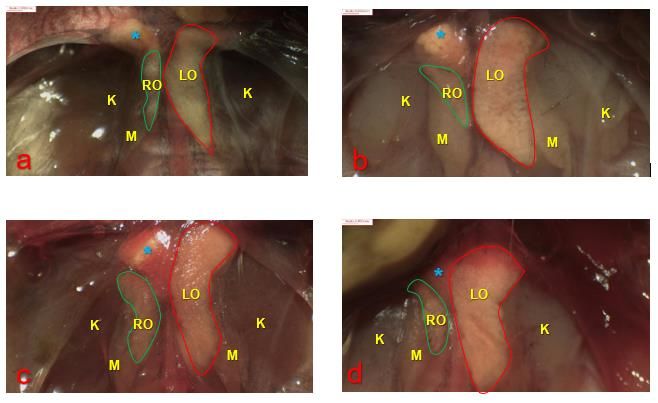
E3S Web of Conferences 335, 00030 (2022) https://doi.org/10.1051/e3sconf/202233500030
The 2nd ICESAI 2021
proof of sex as soon as possible. One of the proofs is through gonad identification (post-
mortem) in day-old chicks because they are relatively more available than molecular sexing
[4]. Gonads are reproductive organs, ovaries to produce ova in females, and testes to
produce spermatozoa in males [5]. Because the size of the gonads of freshly hatched
chickens is relatively small, it is necessary to observe their morphology to determine the
testes or ovaries.
The avian gonads have location and morphology differences from mammals; birds'
testes are in the abdominal cavity, normally, the right ovary regresses. The left ovaries have
been identified as functional gonad, and asymmetric development of the right and left
ovaries has been reported since the embryonic phase [6]. The male, both gonads develop
into testes [7]. Testicular asymmetry is also common in poultry, especially in passerines,
and the degree of asymmetry decreases with the increasing age of chickens. The right and
left testes are functional, although the left is larger in most species [8]. The size of the testes
is often associated with their reproductive ability [9]. Testicular morphometric studies have
been carried out on adult Nigerian local chickens under physiological conditions to predict
the reproductive ability of the testes [10]. Left-Right (L:R) asymmetry could be detected in
both sexes in chickens [11].
Based on the description, it is necessary to reveal the shape and size of the ovary and
testes of Day-old Arabic chick. This study aimed to analyse the morphometric and gross
anatomy of the testes and ovaries of Day-old Arabic chick. Descriptions and information
about their morphological characters are essential to confirm gonadal observations for
purposes of proving to sex, to predict male quality through post-hatching development, and
detect the gonadal abnormalities in Day-old chick (DOC).
2 Materials and methods
2.1 Day-old-chick Arabic chicken
This study used 116 Day-old Arabic chick, resulting in mating between hen and Arabic
gold rooster in previous studies [4]. Day-old chick obtained from the chicken breeding unit
in Politeknik Pembangunan Pertanian Malang. The research was conducted at the Poultry
Unit and the Reproduction Laboratory of the Politeknik Pembangunan Pertanian Malang.
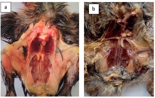
The chicks have been estimated for their sex by feather sexing, then were euthanized by
cervical dislocation. The necropsy procedures incise the ventral abdomen until the
peritoneal cavity opens and removes the viscera organs, so the gonads are visible. The
testes and ovaries were not removed from the abdominal cavity and were not weighed
because relatively very small. The chicken was fixed in a dorsal lying position with the
gonads still attached to the abdominal cavity, observed with direct eyes and then clarified
under a trinocular stereo microscope connected to a computer to identify the gonads is
easier.
2.2 Research variable
The gonads were examined for colour, shape, and location. Ovarian morphometrics
includes the length and width of the right and left ovaries, while the testes include length,
width, and volume. Data collection on the shape, colour, and location of the gonads was
carried out under a stereo microscope with 10x magnification. The stereo microscope used
is the Euromax SB 1903-P series produced by Holland with a DC 5000P camera.
Measurement of the length and width of the gonads using the micrometre software from the
2
E3S Web of Conferences 335, 00030 (2022)                       https://doi.org/10.1051/e3sconf/202233500030
The 2nd ICESAI 2021

     camera. Quantitative data on testicular length and width were tabulated to calculate
     testicular volume.
         Length of testes and ovaries was measured from the anterior end to the posterior end of
     the gonad, and gonad width was measured at the broadest part of the gonad. Testis’s shape
     is ellipsoid [9], so the testicular volume was calculated using formula = (4/3) x π x (a) 2 (b).
     Where (a) is: half of the width and (b) is half of the length [12] or with formula (simplified)
     namely testicular volume = 0.523 x(Width)2(Length) [8].

     2.3 Statistical analyses

     The results of the gross anatomy identification of testes and ovaries were shown with
     explanation descriptions. The morphometric data were analysed using descriptive analysis.
     Differences in size between the right and left gonads were analysed by independent T-test
     at significance P
E3S Web of Conferences 335, 00030 (2022) https://doi.org/10.1051/e3sconf/202233500030
The 2nd ICESAI 2021
3.2 Morphometric and Gross Anatomy of Day-old Arabic Chick Ovaries
Gross anatomy observation of the gonads showed that the shape of the ovary is irregular,
and the left ovary has a more defined shape than the right ovary. The ovaries are close to
the adrenal glands, where the right side is caudolateral of the adrenal gland, while the left
side covers the ventral adrenal gland. The ovaries are located in the ventromedial
mesonephros although the mesonephros is sometimes not visible due to regression.
In general, the ovaries of Day-old Arabic chick have an elongated shape with slightly
curved laterally or towards the kidneys. The left ovary shows a curvature towards the right
kidney with various shapes and uneven edges. The right ovary is smaller with an elongated
and irregular shape. Almost all of the left adrenal glands are covered by the ovaries, and the
right adrenal gland is more visible. The colour of the ovaries is creamy yellow, transparent
pink, and sometimes grey. The surface of the left ovary appears smoother, slightly compact.
This ovarian character is still similar to the morphology of the ovaries of 1-week old
chickens, as described by [6]. The gross anatomy of the ovary of Day-old Arabic chick was
shown in Figure 2.
Fig. 2. Gross anatomy of ovaries Day-old Arabic chick. RO=Right ovary; LO=Left ovary;
K=kidney; M=mesonephros; Blue (*) = Adrenal Gland. The images were taken using a
Euromax SB 1903-P stereo microscope and a DC 5000P camera, magnification: 10x,
scale:0.655 mm.
Figure 2, part (a) the shape of the right ovary: small elongated, the right mesonephros
appears small, and the left mesonephros is not visible; (b). the left ovary is much larger, the
right and left mesonephros are still visible clearly; (c) the right ovary is like a bean arch, the
right, and left mesonephros are still visual even though they are smaller; (d). The right
ovary is small, like a bean arch and the adrenal gland and the right mesonephros are not
visible. The difference between the right and left ovaries: the left ovary is more elongated
than the right ovary, anterior end of the left ovary is wider than the posterior end. The left
ovary appears thinner than the right ovary.
4E3S Web of Conferences 335, 00030 (2022) https://doi.org/10.1051/e3sconf/202233500030
The 2nd ICESAI 2021
Table 1. Ovaries Morphometric of the Day-old Arabic chick (Average ± SEM)
and T-test result.
Characteristics Right ovary Left ovary
Length of ovary (mm) 1.67±0.08 3.69±0.05
Width of ovary (mm) 0.64±0.03 1.18±0.03
Longest ovary size (mm) 3.34 4.55
Shortest ovary size (mm) 0.66 2.23
Difference in length (left-right) of the ovary (mm) 0.97±0.09
Difference in width (left-right) of the ovary (mm) 0.58±0.06
The T-test between right and left ovary length P-valueE3S Web of Conferences 335, 00030 (2022) https://doi.org/10.1051/e3sconf/202233500030
The 2nd ICESAI 2021
bright yellow, still have to be confirmed histologically to support the determination of its
quality.
The description of the location, shape, and colour of the Day-old Arabic chick testes is
presented in Figure 3.
Fig. 3. Gross anatomy of Day-old Arabic chick testes. R= Right testes; LT= Left testes;
K=kidney; M=mesonephros; Blue (*): Adrenal Gland. The images were taken using a
Euromax SB 1903-P stereo microscope and a DC 5000P camera, magnification: 10x,
scale:0.655 mm.
The testes of birds in this study are bean-shaped and elongated ovals, generally
belonging to the category of an ellipsoid or prolate spheroid shape [8]. The form of the
testes tends to be straight, lengthwise cranial and caudal, or slightly curved towards the
kidney. Figure 3 shows that (a) the left testis is larger than the right testis, the right and left
sides show clear mesonephros; (b) the size of the right testis is larger than the left testis; (c).
The length of the right testis is almost the same as that of the left. However, the left testis is
bigger and grey with a wider anterior end and black colour (d). the left testis is longer than
the right testis, most of the left testis is darker in colour, and the anterior end of the right
testis is grey. The results on Day-old Arabic chick, the number of detected black colour on
the testes was 22.95%. Melanoblasts cause dark colour in the interstitium, so the testes are
melanistic. Bicoloured testes are not considered gonads abnormal. The testes with bicolour
do not show any histologic abnormalities in African flamingos [16]. In this study, the
position of the right and left testes tends to be symmetrical, but there are differences in size.
The morphometrics of Day-old Arabic chick is presented in Table 2.
Table 2 showed the size (length, width and volume) of left testis is larger than right
testis. The results of the independent T-test analysis showed that the length and volume of
the testes were significantly different between the right and left testes (PE3S Web of Conferences 335, 00030 (2022) https://doi.org/10.1051/e3sconf/202233500030
The 2nd ICESAI 2021
The right testis is larger than the left testis, only 26.5%, while both testes are the same in
8.2% of birds [8].
Table 2. Testes Morphometric of Day-old Arabic chick (Average ± SEM)
Characteristics Right testis Left testis
Length of the testis (mm) 2.47±0.08 3.29±0.07
Width of testis (mm) 0.98±0.03 1.04±0.02
The volume of the testis (mm³) 1.46±0.12 2.03±0.13
Difference in length (left-right) of the ovar (mm) 0.82±0.08
Difference in width (left-right) of the ovar (mm) 0.05±0.03
Volume total of testis (mm³) 3.49±0.22
T-test between right and left testis length P-value 0.001 **
T-test between right and left testis width P-value 0.194*
T -test between the volume of right and left testis P-value 0.002**
Note: * = not significant difference; **= a significant difference (PE3S Web of Conferences 335, 00030 (2022) https://doi.org/10.1051/e3sconf/202233500030
The 2nd ICESAI 2021
pemanfaatannya, (Balai Peneliti Ternak, Sukabumi, Indonesia, 2007).
3. G. Indra, Achmanu;, A. Nurgiartiningsih, Junal Ternak Tropika. 14, 8–14 (2013).
4. Iswati, M.H. Natsir, G. Ciptadi, T. Susilawati, Journal of Animal & Plant Sciences, 44,
7708–7716 (2020).
5. S. Guioli, S. Nandi, D. Zhao, J. Burgess-Shannon, R. Lovell-Badge, M. Clinton, Sex.
Dev. 8, 227–242 (2014).
6. J.D.L. Mfoundou, Y.J. Guo, M.M. Liu, X.R. Ran, D.H. Fu, Z.Q. Yan, M.N. Li, X.R.
Wang, Poult. Sci. 100, 101191 (2021).
7. R. Tóth, B. Lázár, Á. Südy, A. Nagy, A. Kidane, M. Anand, E. Gócza, N. Biotechnol.
33, S212 (2016).
8. G.R. Graves, The Auk. 121, 473 (2004).
9. N.A. Bachmid, F.Y. Purba, A.M.S. Apada, D.K. Sari, IOP Conf. Ser. Earth Environ.
Sci. 343 (2019).
10. A.K. Okpe Godwin Chidozie, Nwatu Ugochukwu, Anim. Res. Int. 7, 1163–1168
(2010)
11. S. Intarapat, O. Satayalai, Anat. Res. Int. 2014, 1–9 (2014).
12. A. Dawson, J. Avian Biol. 34, 19–123 (2003).
13. Y. Ishimaru, T. Komatsu, M. Kasahara, Y. Katoh-Fukui, H. Ogawa, Y. Toyama, M.
Maekawa, K. Toshimori, R.A.S. Chandraratna, K.I. Morohashi, H. Yoshioka,
Development. 135 (2008) 677–685.
14. B.G.M. Jamieson, Reproductive Biology and Phylogeny of Birds (CRC Press, Boca
Raton, Florida, United States, 2011).
15. C.G. Scanes, Sturkie ’ s Avian Physiology (Academic Press, San Diego, United States,
2015).
16. L. Crosta, H. Gerlach, M. Bürkle, L. Timossi, Exot. Anim. Pract. 6, 57–83 (2003).
17. S. Intarapat, C.D. Stern, J. Poult. Sci. 51, 352–358 (2014).
18. T.K. TriBudi, T. Kostaman, Majalah Ilmiah Peternakan, 23, 107–112 (2020).
19. T.R. Birkhead, K.L. Buchanan, T.J. Devoogd, E.J. Pellatt, T. Székely, C.K. Catchpole,
Song, Anim. Behav. 53 965–971 (1997).
20. P. Dharani, S. Ushakumary, V. Sundaram, C. Joseph, G. Ramesh. Int. J. Morphol. 36,
909-914 (2018).
8You can also read


