MicroRNA-dependent control of neuroplasticity in affective disorders - Nature
←
→
Page content transcription
If your browser does not render page correctly, please read the page content below
Martins and Schratt Translational Psychiatry (2021)11:263
                                    https://doi.org/10.1038/s41398-021-01379-7                                                                                                     Translational Psychiatry

                                     REVIEW ARTICLE                                                                                                                                             Open Access

                                    MicroRNA-dependent control of neuroplasticity
                                    in affective disorders
                                    Helena Caria Martins1 and Gerhard Schratt                                  1

                                      Abstract
                                      Affective disorders are a group of neuropsychiatric disorders characterized by severe mood dysregulations
                                      accompanied by sleep, eating, cognitive, and attention disturbances, as well as recurring thoughts of suicide. Clinical
                                      studies consistently show that affective disorders are associated with reduced size of brain regions critical for mood
                                      and cognition, neuronal atrophy, and synaptic loss in these regions. However, the molecular mechanisms that mediate
                                      these changes and thereby increase the susceptibility to develop affective disorders remain poorly understood.
                                      MicroRNAs (miRNAs or miRs) are small regulatory RNAs that repress gene expression by binding to the 3ʹUTR of
                                      mRNAs. They have the ability to bind to hundreds of target mRNAs and to regulate entire gene networks and cellular
                                      pathways implicated in brain function and plasticity, many of them conserved in humans and other animals. In
                                      rodents, miRNAs regulate synaptic plasticity by controlling the morphology of dendrites and spines and the expression
                                      of neurotransmitter receptors. Furthermore, dysregulated miRNA expression is frequently observed in patients
                                      suffering from affective disorders. Together, multiple lines of evidence suggest a link between miRNA dysfunction and
                                      affective disorder pathology, providing a rationale to consider miRNAs as therapeutic tools or molecular biomarkers.
                                      This review aims to highlight the most recent and functionally relevant studies that contributed to a better
                  1234567890():,;
1234567890():,;
                  1234567890():,;
1234567890():,;

                                      understanding of miRNA function in the development and pathogenesis of affective disorders. We focused on in vivo
                                      functional studies, which demonstrate that miRNAs control higher brain functions, including mood and cognition, in
                                      rodents, and that their dysregulation causes disease-related behaviors.

                                    Introduction                                                                                        unknown and the available pharmacotherapies are not
                                      Affective disorders are a group of neuropsychiatric                                               fully efficacious6.
                                    diseases characterized by a complex set of symptoms that                                               It is now accepted that the full manifestation of affective
                                    include disturbances in mood, sleep, cognition, attention,                                          disorders requires an interaction between genetic pre-
                                    and recurring thoughts of suicide1. The two most pre-                                               disposition (heritability estimates 37% for MDD7, higher
                                    valent disease presentations are major depressive disorder                                          than 60% for BD8) and environmental risk factors, such as
                                    (MDD) characterized by severe and chronically lower                                                 childhood maltreatment or social isolation. Recent tech-
                                    mood and bipolar disorder (BD) with periods of depres-                                              nical advances in the neuroscience field uncovered
                                    sion, mania, hypomania, and mixed episodes. Both con-                                               marked dysregulations of neuroplasticity in the brains of
                                    ditions are leading causes of disability2,3 and increased                                           MDD patients. Particularly, neuronal atrophy, synaptic
                                    mortality4,5. Over the last 50 years, significant efforts were                                       loss, and reduction in the volume of brain regions critical
                                    made into decoding the neurobiology of mood disorders.                                              for mood regulation such as the hippocampus and medial
                                    However, the pathological mechanisms are still largely                                              prefrontal cortex (mPFC)9–11. The molecular mechanisms
                                                                                                                                        linking gene–environment interactions to defects in
                                                                                                                                        neuroplasticity, however, are still elusive.
                                                                                                                                           MicroRNAs (miRNAs or miRs) are single-stranded,
                                    Correspondence: Gerhard Schratt (gerhard.schratt@hest.ethz.ch)
                                    1                                                                                                   usually 19–22 nucleotide long RNAs, which belong to the
                                     Lab of Systems Neuroscience, Institute for Neuroscience, Department of
                                    Health Science and Technology, Swiss Federal Institute of Technology ETH,                           class of small non-coding RNAs. They bind to the 3ʹUTR
                                    8057 Zurich, Switzerland

                                    © The Author(s) 2021
                                                        Open Access This article is licensed under a Creative Commons Attribution 4.0 International License, which permits use, sharing, adaptation, distribution and reproduction
                                                        in any medium or format, as long as you give appropriate credit to the original author(s) and the source, provide a link to the Creative Commons license, and indicate if
                                    changes were made. The images or other third party material in this article are included in the article’s Creative Commons license, unless indicated otherwise in a credit line to the material. If
                                    material is not included in the article’s Creative Commons license and your intended use is not permitted by statutory regulation or exceeds the permitted use, you will need to obtain
                                    permission directly from the copyright holder. To view a copy of this license, visit http://creativecommons.org/licenses/by/4.0/.
Martins and Schratt Translational Psychiatry (2021)11:263 Page 2 of 16
of one or multiple target transcripts and repress gene weight, sucrose preference, and locomotor activity
expression at the post-transcriptional level, either by induced by chronic stress to the same extent as fluox-
promoting mRNA decay or by reducing mRNA transla- etine22. Chronic antidepressant administration acted
tion levels12. Remarkably, miRNAs regulate up to 60% of beyond the raphe by promoting the release of signaling
all protein-coding genes13 and are often expressed in a molecules that antagonized the levels of miR-16 in the
brain region- or developmental stage-specific manner14. locus coeruleus and hippocampus23. Specifically in the
In this way, they control many biological processes within hippocampus, reduced levels of miR-16 unlocked the
the developing and adult brain, which have been pre- translation of the anti-apoptotic and neurotrophic factor
viously implicated in affective disorders. In rodents, Bcl-2, which resulted in increased neurogenesis and
miRNAs regulate the morphology of dendrites and spines, facilitation of the antidepressant effect of fluoxetine23 (Fig.
expression of neurotransmitter receptors, and synaptic 1). Follow-up studies, however, were not able to fully
plasticity15. In human studies, miRNA expression is often recapitulate these initial findings. In humans, serum
altered in MDD16 and BD17 patients. Altogether, miRNAs samples of MDD patients presented lower levels of miR-
are very exciting targets for future therapeutic interven- 16, and subsequent neutralization of miR-16 by intra-
tions or as prognostic and diagnostic biomarkers in cerebroventricular (i.c.v.) injection in rats produced
affective disorders. depressive-like behaviors in the sucrose preference test
This review highlights the most relevant and recent (SPT) and forced swim test (FST)24. These discrepancies
advances in rodent model systems, which established are found also in the expression studies where chronic
miRNAs known to regulate neuroplasticity as potential stress was shown to both up-regulate25–27 and/or down-
mechanistic links between environmental stressors and regulate28,29 miR-16. These inconsistencies are most likely
gene expression changes that lead to the development of the result of studying different stress models, at different
behavioral phenotypes of MDD and BD. Notably, miR- time points during and after the stress, in different brain
NAs that modulate affective-like behavior control the regions, and with different miRNA detection/normal-
expression of one or several genes primarily involved in ization methods. Therefore, although the role of miR-16
signaling pathways activated by serotonin, glucocorti- as a readout for antidepressant treatment appears well
coids, neurotrophic factors, and Wnt (Table 1). established, it is still rather unclear whether miR-16 is
causally involved in the pathophysiology of affective
MicroRNAs that modulate serotonergic neurotransmission disorders.
Impaired neurotransmission mediated by serotonin (5-
hydroxytryptamine/serotonin, 5-HT) is the most studied miR-34 family
vulnerability factor of affective illnesses. Several lines of The miR-34 family (composed of miR-34a, b, and c) is
evidence suggest that a reduction in the 5-hydro- one of the miRNAs for which differential expression is
xytryptamine/serotonin receptor 1A (5-HTR1A) binding most consistently observed across genetic profiling,
causes the serotonin deficit observed in the cortex and in vitro and in vivo studies of mood disorders. Genetic
hippocampus of MDD patients18–20. Consequently, the variation in miR-34b/c was significantly associated with
first treatment strategies focused on elevating the levels of higher susceptibility to MDD and correlated with negative
monoamines in the brain. A classic example are selective life events and cognitive dysfunction30,31. Circulating
serotonin reuptake inhibitors (SSRIs), like fluoxetine, levels of miR-34a and miR-34b/c were up-regulated in
which reduce presynaptic serotonin transporters’ (SERT) MDD patients when compared with healthy controls32,33,
binding and protein levels, thereby preventing the reup- and higher expression levels of miR-34a were also mea-
take of the neurotransmitter from the synaptic cleft and sured in postmortem cerebellar tissue of BD patients34. In
increasing its levels at the synapse21. accordance with these results, the expression of miR-34a
dropped significantly with the administration of anti-
miR-16 depressants and mood stabilizers35,36. Its central role in
The Kellermann lab published two very comprehensive the regulation of the stress response has been firmly
studies demonstrating that fluoxetine interferes with established37–41, but direct evidence related to its func-
SERT translation via miR-16 (Fig. 1). They showed that tional role in affective disorders became available just
chronic injection of fluoxetine into the mouse ser- recently. Lo Iacono et al. showed that chronic stress
otonergic raphe nuclei increases serotonin by promoting induces expression of miR-34a, particularly in the mouse
the maturation of miR-16, which in turn decreases SERT raphe nuclei, which was required to develop a depressive-
expression by direct interaction with the SERT mRNA. like response42. Notably, raphe nuclei conditional
Importantly, injection of miR-16 into the raphe and knockout (cKO) of the three miR-34s (TKO) reversed the
injection of anti-miR-16 into the locus coeruleus ame- increase in immobility induced by chronic stress in the
liorated the deterioration of the coat, reduction in body FST42. Interestingly, Andolina et al. showed that theTable 1 List of miRNAs implicated in affective-like behavior through functional studies in animal models.
Animal model Behavioral phenotype Molecular mechanism Ref
Serotonin
neurotransmission
22
miR-16 KD of miR-16 in the rat hippocampus with an Antidepressant-like effect in the FST and SPT. Targets SERT and Bcl-2. Increases hippocampal
anti-miR-16. neurogenesis.
23
KD of miR-16 by i.c.v. injection of anti-miR-16 Depressive-like phenotype in the FST and SPT. Targets SERT. Decreased serotonin in the CSF.
in rats.
42
miR-34 family TKO mouse. Antidepressant-like phenotype in the FST. Prevents stress-induced release of serotonin in
the mPFC.
43
TKO mouse. Antidepressant-like phenotype in the FST. Targets CRFR1. Prevents stress-induced release of
serotonin in the mPFC and GABA release in
the BLA.
44
miR-135a Overexpression of miR-135a in in serotonergic Antidepressant-like phenotype in the FST. Targets 5-HTR1A and SERT. Increases synaptic
Martins and Schratt Translational Psychiatry (2021)11:263
neurons of the raphe nuclei of mice. serotonin levels.
Glucocorticoid signaling
58
miR-17–92 cluster cKO mouse of miR-17–92 cluster in adult Depressive-like phenotype in the FST, TST, and SPT. Targets SGK1. Decreases the number of proliferative
neural progenitors of the hippocampus. progenitors, and newborn neurons in the
hippocampal DG.
59
miR-15 KD of miR-15a in BLA with a sponge lentivirus. Anxiety-like phenotype in the elevated plus maze. Regulated by glucocorticoids and targets FKBP51.
Neurotrophic factors
67
miR-101 Overexpression of miR-101 in the VLO with Antidepressant-like phenotype in the FST and SPT. Targets DUSP1.
a mimic
73
miR-182 Overexpression of miR-182 in the Depressive-like phenotype in the FST, SPT, and NSFT. Targets BDNF.
hippocampus with a lentivirus
77
miR-323 KD of miR-323 in the mouse Cg1/2 with a Antidepressant-like phenotype in the TST. Targets ErbB4.
sponge AAV.
Cytoskeletal-regulatory proteins
79
miR-134 Overexpression in the rat mPFC with an AAV- Depressive-like phenotype in the FST and SPT. Targets Limk1. Decreases dendritic spine density
miR-134. and synapse number in the vmPFC.
102
miR-212/132 tTA::miR-132 transgenic mice. Moderate overexpression of miR-132 enhanced spatial memory Supra-physiological levels of miR-132 increase
and cognitive capacity in the Barnes maze task and NORT. dendritic spine density in hippocampal neurons.
Supra-physiological level of miR-132 impaired cognition.
Page 3 of 16Table 1 continued
Animal model Behavioral phenotype Molecular mechanism Ref
Serotonin
neurotransmission
103
AChE-R overexpressing TgR mice. Cognitive impairments in the two-unit serial maze. Targets AChE. Cholinergic hyper-excitation when
exposed to pilocarpine.
104
cKO of miR-132/-212 in excitatory neurons of Cognitive deficits in spatial memory, recognition memory, and miR-212 targets Stx1a. miR-132 targets Mash1.
the mouse forebrain. in the NORT, Barnes maze, and contextual fear-
conditioning tests.
105
miR-132 transgenic and miR-132/212 Anxiety-like phenotype in the elevated plus maze and open Targets SIRT1 and PTEN.
knockout mice. field assay.
107
miR-218 Overexpression in the mouse mPFC with an Antidepressant-like phenotype in the FST and SIT. Targets DCC. Increases density of dendritic spines
AAV-miR-218. the mPFC.
Wnt signaling
Martins and Schratt Translational Psychiatry (2021)11:263
118
miR-124 KD of miR-124 in the rat hippocampus using Antidepressant-like phenotype in the FST and SPT. Targets BDNF.
lentivirus.
119
KD of miR-124 in the rat PFC using lentivirus. Antidepressant-like phenotype in the FST, SPT, and NSFT. Targets SIRT1.
120
KD of miR-124 by i.c.v. injection of an Antidepressant-like phenotype in the TST and SPT. Targets GR. Increases hippocampal neuron
antagomir in mice. proliferation.
123
Overexpression of miR-124 in excitatory Antidepressant-like phenotype in the SIT, SPT, and NSFT. Targets HDAC4/5, and GSK3β. Increases spine
hippocampal neurons with an AAV-miR-124 density in the DG neurons.
124
Overexpression of miR-124 in the mouse Antidepressant-like phenotype in the FST, TST, SPT, and SIT. Targets STAT3. Decreases microglia activation.
hippocampus with a lentivirus.
125
miR-214 KD of miR-214 by i.c.v. injection of an Antidepressant-like phenotype in the TST and SIT. Targets β-catenin. Decreases amplitude of mEPSC,
antagomir in mice. and number of dendritic spines in hippocampal
neurons.
127
miR-221 KD of miR-221 by i.c.v. injection of an Antidepressant-like phenotype in the FST, TST, and SPT. Targets Wnt-2. Decreases neuronal proliferation
antagomir in mice. and promotes neuronal apoptosis in the
hippocampus.
New insights
131
miR-9 Overexpression of CircDYM in the Antidepressant-like phenotype in the FST and TST. Targets HECTD1. CircDYM overexpression
hippocampus. CircDYM acts as an prevented miR-9-induced microglia activation.
endogenous sponge of miR-9.
Page 4 of 16Martins and Schratt Translational Psychiatry (2021)11:263 Page 5 of 16
Ref
cerebrospinal fluid, PTEN phosphatase and tensin homolog, DCC deleted in colorectal cancer, DG dentate gyrus, DUSP1 dual-specific phosphatase 1, FKBP51 FK506-binding protein 51, FST forced swim test, GABA γ-
Limk1 LIM motif-containing protein kinase 1, miRNA or miR microRNA, mPFC medial prefrontal cortex, NORT novel object recognition test, NSFT novelty-suppressed feeding test, SERT serotonin transporter, SGK1 serum- and
KD knockdown, 5-HTR1A 5-hydroxytryptamine/serotonin receptor 1A, BDNF brain-derived neurotrophic factor, BLA basolateral amygdala, cKO conditional knockout, CRFR1 corticotropin-releasing factor receptor 1, CSF
glucocorticoid-inducible protein kinase-1, SIRT1 sirtuin 1, SIT social interaction test, mEPSC mini excitatory postsynaptic currents, SPT sucrose preference test, STAT3 signal transducer and activator of transcription 3, TKO triple
aminobutyric acid, GR glucocorticoid receptor, GSK3β glycogen synthase kinase 3 beta, HDAC4/5 histone deacetylase 4/5, HECTD1 HECT domain E3 ubiquitin protein ligase 1, i.c.v. intracerebroventricular, KD knockdown,
135
constitutive deletion of miR-34s also reduces the immo-
bility of mice in the FST by preventing the activation of
Increases the number of immature neurons and
the raphe nuclei and hence the release of serotonin and γ-
aminobutyric acid (GABA) in the mPFC and basolateral
newborn mature neurons in the mouse amygdala (BLA), respectively43. Similarly, Lo Iacono et al.
observed that in the absence of miR-34, stressed mice
lacked the induction of serotonin production in the mPFC
normally present in wild-type (WT) mice42. Altogether,
these findings support the role of miR-34 in modulating
Molecular mechanism
neurotransmission between brain regions important for
cognitive and emotional responses. Furthermore, the
hippocampal DG.
prospect that by regulating miR-34 levels one can mod-
ulate an individual’s susceptibility to stress, makes miR-34
a strong downstream target for gene–environment
interactions responsible for the development of affective
disorders. Thus far, the function of miR-34 in the ser-
otonergic raphe nuclei was linked to its target
corticotropin-releasing factor receptor 1 (CRFR1)43 (Fig.
KD of miR-139 via intranasal administration of Antidepressant-like phenotype in the FST, TST, and NSFT.
1). Nevertheless, it would be very interesting to investigate
other possible targets more directly associated with neu-
ronal transmission and plasticity, such as HTR2C or
GRM739. Another interesting finding was that although
miR-34a is predominantly found in the raphe nuclei, miR-
34c is highly expressed in the hippocampus42. Thus, it is
very appealing to postulate that miR-34a, b, and c regulate
constitutive knockout, TST tail suspension test, VLO ventrolateral orbital cortex, vmPFC ventromedial prefrontal cortex.
different sets of target genes in a context-dependent
Behavioral phenotype
manner, which in turn could be highly informative for the
development of specific antidepressant therapies.
miR-135
miR-135 is a great example of the pleiotropic functions
that can be exerted by miRNAs. Depending on the cell
type, expression levels, and degree of miRNA activity, a
miRNA will bind preferentially to a specific target pro-
ducing different disease symptoms. Issler et al. demon-
strated that in the raphe nuclei, miR-135a controls the
expression of the serotonin autoreceptor HTR1A and
transporter SERT, and in doing so, it mediates anti-
depressant response44 (Fig. 1). Although chronic social
defeat stress (CSDS) did not alter the levels of miR-135a,
an antagomir in mice.
acute and chronic SSRI antidepressant treatment robustly
increased miR-135a expression levels in the raphe nuclei
Animal model
of both unstressed and stressed mice. In line with this
observation, miR-135a overexpression specifically in ser-
otonergic neurons did not change the baseline behavior of
mice, however, it conferred resilience against chronic
stress in tests of anxiety- and depressive-like behaviors. In
the FST, after chronic stress, mice overexpressing
Table 1 continued
miR135a spent significantly less time immobile than
neurotransmission
controls. On the other hand, miR-135a lentiviral knock-
down (KD) in the raphe nuclei significantly attenuated the
miR-139
Serotonin
response to antidepressants resulting in higher immobility
times after SSRI administration. Furthermore, mice
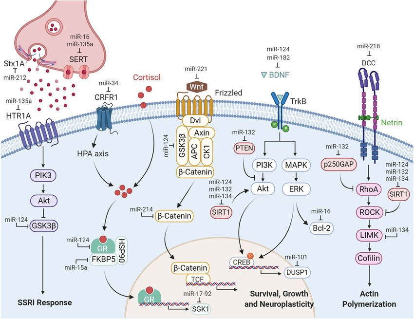
overexpressing miR-135a had lower tissue concentrationMartins and Schratt Translational Psychiatry (2021)11:263 Page 6 of 16 Fig. 1 miRNAs affect animal behavior related to MDD and BD by regulating the expression of genes involved in serotonergic neurotransmission and signaling pathways activated by glucocorticoids, BDNF, Wnt, and guidance cues. Typical antidepressants, such as SSRIs, block monoamine reuptake by the SERT and, when chronically administered, lead to the desensitization of serotonin receptors (HTR)21. Signaling by glucocorticoids, BDNF, and Wnt controls multiple physiological functions such as neuronal survival, growth, and plasticity61,110,149. During development, the Netrin-1/DCC pathway plays a critical role in axonal outgrowth toward the prefrontal cortex and the organization of mPFC connectivity in adulthood150. Created with BioRender.com. Abbreviations: Stx1A, syntaxin 1A; SERT, serotonin transporter; HTR1A, serotonin receptor 1A; PIK3, phosphoinositide 3-kinase; Akt, protein kinase B; GSK3β, glycogen synthase kinase 3 beta; SSRI, selective serotonin reuptake inhibitor; CRFR1, corticotropin-releasing factor receptor 1; HPA axis, hypothalamus–pituitary–adrenal axis; GR, glucocorticoid receptor; FKBP5, FK506-binding protein 5; HSP90, heat shock protein 90; SGK1, serum- and glucocorticoid-inducible protein kinase-1; Wnt, Wingless-related integration site; Dvl, dishevelled protein; APC, adenomatous polyposis coli protein; CK1, casein kinase 1; TCF, T cell factor; BDNF, brain-derived neurotrophic factor; TrkB, neurotrophic receptor tyrosine kinase 2; PTEN phosphatase and tensin homolog; SIRT1, sirtuin 1; CREB, cAMP response element-binding protein; DUSP1, dual- specific phosphatase 1; MAPK, mitogen-activated protein kinase; ERK, extracellular signal-regulated kinase; Bcl-2, B-cell lymphoma 2; DCC, deleted in colorectal carcinoma; p250GAP, Rho GTPase activating protein 32; RhoA, Ras homolog family, member A; ROCK, Rho-associated coiled-coil kinases; LIMK, LIM motif-containing protein kinase. of serotonin and increased serotonin metabolism indi- beneficial in one brain region but detrimental in a dif- cating that miR-135 decreases depressive symptoms by ferent region or tissue. Moreover, their ability to target inducing the degradation of HTR1A and SERT, thereby multiple genes may also lead to the silencing of unin- increasing the synaptic levels of serotonin in the brain44. tended targets not related to the disease. Other side effects Intriguingly, Mannironi et al. showed that KD of miR-135 related to the miRNA itself or the delivery method may in the mouse amygdala led to increased spontaneous include immune system activation, cellular toxicity, and excitatory postsynaptic currents (EPSCs) and anxiety-like saturation of the miRNA biogenesis machinery46. On the behavior. This effect was likely due to increased levels of other hand, upon careful monitoring of unwanted side the complexin-1 and complexin-2 target genes, both effects, chemical modifications and small changes to the regulators of synaptic vesicle function45. In the Issler et al. miRNA sequence can help avoid off-target effects and study, the authors also measured lower levels of miR-135a optimize gene-silencing efficiency47. in the blood of depressed patients44. Whilst the work on miR-135a paved the way for a better understanding MicroRNAs that modulate glucocorticoid signaling of the serotonergic regulation in psychopathologies and Chronic stress is a well-known risk factor for the devel- advanced miR-135a as a potential biomarker for depres- opment of affective disorders. Depressed patients consistently sion, it also raised new questions about potential off- show hyperactivity of the hypothalamus–pituitary–adrenal target effects of miRNA manipulation. Manipulating the (HPA) axis and increased levels of the glucocorticoid hor- expression and/or activity of a given miRNA may be mones, which are thought to be a consequence of impaired
Martins and Schratt Translational Psychiatry (2021)11:263                                                    Page 7 of 16

glucocorticoid receptor (GR) function. Additionally,          behavioral response to chronic stress and its down-
antidepressant treatment ameliorates depressive symp-         regulation specifically impairs the recovery process.
toms by normalizing the HPA axis hyperactivity48,49.          Similarly, elevations in miR-15a were observed in the
Therefore, miRNAs that target components of the GR            peripheral blood of subjects either treated with dex-
signaling pathway are candidates to modulate the activity     amethasone or experiencing childhood trauma. Interest-
of the HPA axis and antidepressant action.                    ingly, miR-15a targets FKBP51, a known inhibitor of the
                                                              GR, which was previously implicated in affective dis-
miR-17–92 cluster                                             orders60 (Fig. 1). Therefore, it might be worth investi-
  The polycistronic miR-17–92 cluster is located on           gating whether enhanced FKBP51 expression is
human chromosome 13 and codes for six miRNAs (miR-            responsible for the anxiolytic effect of miR-15a KD.
17, miR-18a, miR-19a, miR-20a, miR-19b-1, and miR-92a)        Besides, miR-15a was reduced in postmortem samples of
with well-established roles as oncogenes50. Individual        PFC from BD patients, whereas miR-15b was up-
members of this cluster and its two mammalian paralogs        regulated in the blood of individuals with high familial
(miR-106b~25 and miR-106a~363 clusters) were more             risk to develop BD. Taken together, these findings support
recently associated with psychiatric disorders since they     the need for additional studies on miR-15 in the context
were shown to regulate heart51,52 and immune53 function,      of stress-induced psychiatric disorders.
both of which are often compromised in mental ill-
ness54,55. In BD specifically, dysregulation of miR-106a       MicroRNAs that modulate neurotrophic factors
and miR-106b has been consistently reported in profiling         The brain-derived neurotrophic factor (BDNF), is a key
studies performed in blood56 and postmortem PFC17,57 of       regulator of neurogenesis and development, synaptic
manic patients. The most relevant study concerning the        formation, maturation, and plasticity61 and was very early
causal role of miR-17–92 in behaviors related to affective    on connected with affective disorders. Observations that
disorders was published by Jin et al.58. They showed that a   antidepressants induce the expression of BDNF in rodents
cKO of the miR-17–92 cluster in adult neural progenitor       and depressed patients led to the hypothesis that
cells of the dentate gyrus (DG) decreased the number of       depression is associated with lower levels of BDNF and
proliferative progenitors and newborn neurons, possibly       antidepressants act by restoring its levels62. Other neu-
by regulating serum- and glucocorticoid-inducible protein     rotrophic factors, such as neuregulins (NRGs), bind to
kinase-1 (SGK1) (Fig. 1). miR-17–92 cKO mice spent            ErbB receptors and also promote neuronal development
significantly more time immobile in the FST and tail           and plasticity63,64. Polymorphisms in the genes encoding
suspension test (TST) and consumed less sucrose in the        NRG1 and ErbB4 were first associated with schizo-
SPT than control mice, all of which indicate enhanced         phrenia65, but since then accumulating evidence suggest
depressive-like behavior in the absence of miR-17–92.         that NRG1-ErbB4 signaling is associated with affective
Remarkably, overexpression of miR-17–92 had the               disorders, although the mechanism is still unkown64.
opposite effect on both neurogenesis and depressive-like      Therefore, miRNAs that regulate BDNF and NRGs and/or
behaviors58. Taken together, this study showed in a very      downstream signaling genes are promising therapeutic
in-depth and complete manner that down-regulation of          targets.
the miR-17–92 cluster upon chronic stress elicited a
depressive phenotype in mice by impairing hippocampal         miR-101
neurogenesis. Thus, increasing the levels of miR-17–92          miR-101 dysregulation was first associated with mood
cluster miRNAs could represent a novel promising anti-        disorders in the PFC of depressed individuals that com-
depressant strategy.                                          mitted suicide16. Later, it was shown that miR-101 is also
                                                              down-regulated in the PFC of a rat model of MDD, the
miR-15                                                        Flinders Sensitive Line while its target glutamate trans-
  Volk et al. showed that KD of miR-15a in the mouse          porter SLC1A1 mRNA and protein levels were
BLA produces strong anxiolytic effects under CSDS59.          increased66. More recently, the functional relevance of
After observing that miR-15a levels were elevated in the      miR-101 was assessed in vivo in the rat ventrolateral
amygdala of stressed mice, the authors searched for           orbital cortex (VLO)67. Zhao et al. demonstrated that
behavioral changes upon overexpression of miR-15a. The        chronic unpredictable mild stress (CUMS) decreased the
elevation of miR-15a in the BLA was not sufficient to          levels of miR-101 in the VLO in comparison with control
produce any changes. However, miR-15a KD animals              animals while intra-VLO microinjection with a miR-101
showed a tendency to spend less time and travel less in       mimic had an antidepressant effect. miR-101 over-
the open arms of the elevated plus maze, which was sig-       expression in chronically stressed rats increased their
nificant following chronic social defeat. Volk et al. con-     sucrose preference in the SPT, to the same extent as
cluded that miR-15a is essential to form the appropriate      fluoxetine, and decreased their immobility time in the
Martins and Schratt Translational Psychiatry (2021)11:263 Page 8 of 16
FST. Chronic mild stress also increased the levels of dual- miR-323
specific phosphatase 1 (DUSP1), a validated target of miR- Up-regulation of miR-323 levels was first reported in
10168 (Fig. 1), which has previously been implicated in the the frontal cortex of newborn rats after maternal stress74
pathophysiology of MDD69. Stress-induced elevation of and in the plasma of mild cognitive impairment
DUSP1 expression in turn resulted in lower levels of patients75,76. However, very recently miR-323 was for the
phosphorylated extracellular signal-regulated kinase first time shown to be involved in depression77. Fiori et al.
(ERK) and BDNF. Importantly, miR-101 overexpression showed that miR-323a together with miR-204, miR-320b,
abolished the differential regulation of DUSP1, phospho- and miR-331 was up-regulated in the dorsal anterior
ERK, and BDNF expression between CUMS-exposed and cingulate cortex (ACC) and the lateral habenula of indi-
control rats, suggesting that the ERK/BDNF pathway viduals that committed suicide during an episode of
could be an important mediator of the antidepressant MDD, compared to age-matched controls. miR-323
effect of miR-101 (Fig. 1). In future studies, exploring if regulated the levels of its target ErbB4, which showed
miR-101 modulates ERK/BDNF-related processes like reduced levels in both the ACC and the habenula of
neuronal survival and synaptic plasticity70,71 in the con- depressed patients, and in HEK293T cells transfected with
text of affective disorders are warranted. a miR-323 mimic. Behavioral phenotyping was carried out
after bilateral injection in the mouse Cg1/2 region of
AAVs bearing a miR-323 precursor or sponge to over-
miR-182 express or KD miR-323, respectively. Mice with over-
The relationship between genetic variation in miRNA expression exhibited an anxiety-like phenotype in the
and susceptibility to depression was studied for the first open field and elevated plus maze test while inhibition of
time two decades ago72. The authors performed a miR-323 resulted in less anxiety- and depressive-like
mutational screening study and discovered a significant behaviors, in particular higher mobility time in the TST.
association between the T allele of the rs76481776 The levels of ErbB4 were also measured in the mouse
polymorphism in the pre-miR-182 and late insomnia in ACC. KD mice showed significantly higher levels of ErbB4
MDD patients. This likely represents a gain-of-function while miR-323 overexpression resulted in a trend for
mutation since increased levels of mature miR-182 were decreased expression of Erbb477. This data nicely corre-
produced from a pre-miR-182 carrying the rs76481776 lates with a recent study by Wang et al.78. Here the
compared to the WT allele. miR-182 overexpression in authors showed that CSDS in mice decreased the protein
turn caused a significant down-regulation of the direct levels of NRG1 and ErbB4 in the mPFC and hippocampus,
target gene CLOCK, which is involved in sleep/awake and NRG1 lateral ventricle administration rescued the
cycle regulation72. Consistent with these early findings, depression-like behavior of the CSDS mice78. Taken
a more recent study showed that miR-182 depletion has together, both studies implicate the NRG1-ErbB4 signal-
an antidepressant effect in rats73. Li et al. observed an ing pathway in the pathology of depression and the KD of
up-regulation of miR-182 and reduction of BDNF levels miR-323 as a possible therapeutic target. Therefore, future
in the hippocampus of rats exposed to CUMS. Further studies should explore a possible neuronal function of
overexpression of miR-182 in the hippocampus of miR-323 potentially underlying anxiety- and depression-
stressed rats with a lentiviral injection significantly like behaviors and whether miR-323 function is mediated
exacerbated the expression of depressive-like behaviors by its target ErbB4.
induced by CUMS, which was associated with a decrease
in BDNF and CREB1 levels. Chronically stressed rats microRNAs that modulate cytoskeletal-regulatory proteins
injected with the miR-182 lentivirus showed a sig- Structural brain changes are a core manifestation of
nificant increase in the latency to approach the food affective disorders. Brain-imaging techniques and post-
pellet in the novelty-suppressed feeding test (NSFT), mortem studies consistently show a reduction in the
lower sucrose preference in the SPT, and higher volume, neuronal size, and loss of synapses in the mPFC
immobility time in the FST. Interestingly, both silencing and hippocampus of MDD patients9. Many of these
of miR-182 and injection of BDNF in the hippocampus changes are mimicked by chronic stress exposure in
of stressed rats rescued the depressive-like phenotype in rodents9. The following miRNAs target cytoskeletal-
all tested behaviors. Behavioral rescue was accompanied regulatory proteins and are dysregulated upon stress,
by higher BDNF, CREB1, and pCREB1 protein levels. providing a potential mechanistic link between stress and
The authors further confirmed BDNF is a direct affective disorder-associated neuromorphological changes.
target of miR-182 and propose that miR-182-dependent
inhibition of BDNF could be partially responsible miR-134
for depression-associated phenotypes caused by Fan et al. showed that CUMS significantly induced the
stress73 (Fig. 1). expression of miR-134 in the rat ventromedial PFCMartins and Schratt Translational Psychiatry (2021)11:263 Page 9 of 16
(vmPFC) and that overexpression of miR-134 was suffi- was very early on associated with affective disorders,
cient to produce depressive-like behaviors79. Both chronic especially depression. miR-132 is increased in the serum92
stress and injection of an AAV overexpressing miR-134 in and peripheral blood of MDD patients93–95 as well as in
the vmPFC of unstressed rats resulted in increased rodent models of depression93,96. Additionally, treatment
immobility times in the FST and anhedonia in the SPT. with SSRI antidepressant drugs induced both miR-21297
Abnormal behavior was accompanied by a reduction in and miR-13298–100, whereas electroconvulsive stimulation
dendritic spine density and synapse number. Infusion of only induced the expression of miR-212101. Direct evi-
an AAV-miR-134 sponge into the vmPFC blocked miR- dence from behavioral experiments that miR-132 mod-
134 function and significantly ameliorated changes in ulates depressive- or manic-like behaviors is unfortunately
behavior and neuronal structure induced by CUMS. The lacking. However, higher levels of miR-132 were positively
authors went on to show that both chronic stress and correlated with deficits in visual memory94 and poorer
miR-134 injection decreased the levels of the target gene cognitive performance in attention and executive func-
LIM motif-containing protein kinase 1 (Limk1) and tion95 in depressed patients. Therefore, miR-132 could
phosphorylation of the Limk1 substrate cofilin within the specifically affect learning- and memory-related aspects of
vmPFC79 (Fig. 1). Interestingly, Limk1 regulates actin affective disorders. Hansen et al. were the first to suggest
dynamics by phosphorylating cofilin80 and Schratt et al. that miR-132 is an important regulator of cognition
showed that miR-134 reduces dendritic spine size by within a critical range of expression102. Endogenous miR-
inhibiting Limk1 translation in hippocampal neurons81. 132 expression was induced in the hippocampus of mice
Thus, structural abnormalities induced by chronic stress undergoing a Barnes maze spatial memory task by ~1.5-
in the brain could arise from the dysregulated expression fold. Induction to this level of expression using transgenic
of miRNAs that control actin dynamics. In further sup- mice in the presence of doxycycline enhanced memory
port of a “pro-depressive” role of miR-134, Shen et al. capacity. However, in the absence of doxycycline, miR-
recently reported that environmental enrichment (EE) 132 was elevated >3-fold over control levels and produced
reversed the CUMS-induced depressive-like behavior via severe cognitive effects102. Shaltiel et al. showed that acute
a miR-134/SIRT1 pathway. EE reduced the levels of miR- stress-induced deficits in cognition were a result of
134 and induced SIRT1 protein expression leading to chronically high levels of miR-132 levels in the mouse
increased dendritic spine density, dendrite arborization, hippocampus and the resulting suppression of its target
and expression of synaptic proteins in the rat hippo- gene acetylcholinesterase103. On the other hand, the
campus82 (Fig. 1). Apparently at odds with this functional conditional deletion of miR-212−132 in excitatory fore-
data, lower levels of miR-134 were measured in the brain neurons using the cre/lox system led to cognitive
plasma of MDD and bipolar mania patients compared to deficits in spatial memory, recognition memory, and novel
healthy individuals as well as in the plasma, hippocampus, object recognition104. All the above studies provide evi-
PFC, and olfactory bulb of rats exposed to CUMS79,83. dence that, within a critical window of expression, miR-
Although miRNAs can be robustly detected in peripheral 132 supports cognitive capacity. Deviation from this
fluids, alterations in levels of circulating miRNAs and physiological expression level in either direction, e.g.,
those from tissues are often inconsistent, suggesting the caused by stress, would be expected to disturb cognitive
existence of tissue-specific mechanisms that control homeostasis. In this regard, Aten et al. showed recently
miRNA abundance. Future work exploring the correlation that chronic stress-induced miR-132 and miR-212
between miR-134 expression in the plasma and brain of expression in the amygdala and that both over-
healthy individuals and patients should help to evaluate expression and KO of miR-132 produced anxiety-related
the potential of miR-134 as a therapeutic target and/or a behaviors105. It would be very interesting to test whether
novel disease biomarker in affective disorders. stress-induced miR-132 also produces depressive-like
behaviors or related cognitive impairments.
miR-212/132
The miR-212/132 cluster contains four miRNAs (miR- miR-218
212, miR-212*, miR-132, and miR-132*) which are derived The role of miR-218 in the development of mood dis-
from the same intronic primary transcript. In the brain, orders and the brain, in general, was only very recently
the functional contribution of miR-132 was thoroughly uncovered. Torres-Berrío and Lopez et al. first reported a
researched in comparison to the other members of the strong reduction in miR-218 in the PFC of two separate
cluster. It promotes neurite outgrowth and dendritic spine and independent cohorts of depressed patients who com-
formation in an activity-dependent manner, primarily by mitted suicide in comparison with healthy sudden death
targeting p250GAP (Fig. 1)84–88, and it is involved in brain controls. These findings were mirrored in mice, where
plasticity89,90. Since BDNF is a known upstream regulator miR-218 levels were reduced in mice susceptible to CSDS
of miR-212/132 expression91, the miR-212/132 cluster compared to those that were resilient106. Torres-BerríoMartins and Schratt Translational Psychiatry (2021)11:263 Page 10 of 16 et al. further assessed the involvement of miR-218 in the MicroRNAs that modulate Wnt signaling development of stress vulnerability following CSDS in Wnt signaling is a highly conserved signal transduction mice107. Here, Torres-Berrío used the single social defeat pathway important for the survival, growth, and plasticity (SSD) paradigm to show that microinfusion of an antag- of neurons110. It was first linked to affective disorders by omir against miR-218 in the mPFC and intranasal infusion the observation that the BD drug lithium modulated the of the antagomir promoted stress susceptibility of mice in activity of two important components of the pathway, the social interaction test (SIT). Importantly, both methods GSK3β and β-catenin111. Later, Wnt2 expression was resulted in increased immobility duration in the FST, shown to be induced by different classes of anti- showing that miR-218 KD by itself was sufficient to induce depressants and to produce antidepressant-like pheno- depressive-like behaviors. On the contrary, overexpression types when overexpressed in the rat hippocampus112. of miR-218 in pyramidal neurons of the mPFC prevented Below, we further discuss how specific miRNAs regulate the development of social avoidance and alterations in the Wnt pathway at different levels to influence spine morphology following CSDS, without any effect in depressive-like behaviors. control mice. miR-218 overexpression prevented the stress-induced decrease in the density of dendritic spines in the mPFC of socially defeated mice, more specifically the miR-124 reduction in the density of thin spines. Torres-Berrío and miR-124 is abundantly expressed in the brain where it Lopez et al. established the Netrin-1 guidance cue receptor drives neuronal differentiation and neurogenesis113,114. In DCC (deleted in colorectal cancer) as a critical miR-218 rodents, miR-124 expression is induced by early-life target by showing that increased DCC expression in PFC stress115–117, chronic stress118,119, as well as in the hip- neurons caused susceptibility to stress-induced depressive- pocampus of mice chronically treated with corticosterone like behaviors, whereas DCC deletion was protective (Fig. (CORT)120. Likewise, in MDD patients, miR-124 is up- 1). Taken together, these studies provide rather compelling regulated in both blood and postmortem PFC100,121,122. At evidence that a reduction in miR-218 levels in the mPFC the behavioral level, manipulation of miR-124 produced induces depressive-like behaviors in mice by rendering conflicting effects. On one hand, suppression of miR-124 them more susceptible to chronic stress. This work raised using lentiviruses ameliorated the depressive-like pheno- many interesting questions, which were in part addressed type of chronically stressed mice in the SPT and in follow-up studies. Rocchi et al. showed recently that FST118,119. In addition, the administration of a miR-124 miR-218 overexpression up-regulates the GluA2 subunit antagomir had beneficial effects on depression-related and increases glutamatergic synaptic transmission at both behaviors and partly reversed the deficits in hippocampal single-neuron and network levels108, which is consistent dendritic spine density and neuronal proliferation with a previous observation that miR-218 rescued the induced by CORT116. On the other hand, Higuchi et al. stress-induced loss of dendritic spines107. Moreover, observed that chronic stress reduced the levels of miR- Manitt et al. showed that during adolescence DCC 124, an effect that was blocked by antidepressant treat- expression in dopaminergic neurons is required for ment123. Also, overexpression of miR-124 specifically in appropriate organization of synaptic connectivity within excitatory hippocampal neurons using AAV injection the mPFC109. Therefore, dysregulations in miR-218 and blocked the development of depressive-like behaviors DCC expression during adolescence, a critical period for induced by chronic stress in the SPT. In the same study, stress-related disorders, may hinder mPFC wiring and inhibition of miR-124 increased the behavioral suscept- glutamatergic neurotransmission in mood-related circuits ibility to mild repeated restraint stress (MRRS). Interest- that will increase the predisposition to develop affective ingly, Higuchi also observed that miR-124 modulates disorders. Along these lines, it would be very interesting to neuronal morphology under stress. However, contrary to explore whether changes in miR-218 in susceptible mice the results of Wang et al.120, hippocampal inhibition of are already present before chronic stress or are rather a miR-124 enhanced the stress-induced loss of dendritic consequence of stress, and whether the protective effect of spines and decreased dendritic length of DG neurons, miR-218 against stress is indeed a result of enhanced both of which were rescued by overexpression of miR- synaptic connectivity, particularly related to the glutama- 124. Moreover, pharmacological inhibition of the targets tergic system. Torres-Berrío et al. also showed that mice GSK3 and HDAC4/5 resulted in antidepressant-like susceptible to CSDS exhibit reduced levels of miR-218 in behavioral effects123. Overall, this study suggests that blood107. Thus, it is very appealing to pursue miR-218 as a down-regulation of miR-124 and the concomitant up- biomarker and assess whether miR-218 is altered in the regulation of GSK3 and HDAC4/5 are causally involved in blood of MDD patients and possibly correlates with disease the atrophy of hippocampal neurons induced by chronic symptom severity and antidepressant treatment. stress (Fig. 1). In further support of a protecting effect of
Martins and Schratt Translational Psychiatry (2021)11:263                                                       Page 11 of 16

miR-124, Lou et al. showed recently that restoring miR-         miR-214 agomir125. Therefore, the intranasal adminis-
124 levels in conditions of chronic stress alleviated the       tration of short oligonucleotide targeting miRNAs might
increased immobility in the TST and FST and inhibited           represent a particularly promising novel strategy for
microglial activation, possibly by degrading STAT3              miRNA therapeutics in affective disorders.
mRNA124. Discrepancies within in vivo studies may arise
from different sources. In the case of miR-124, different       miR-221
genetic backgrounds of the animal models, stress para-             miR-221 has been consistently found elevated in plasma
digms, cell type, and developmental stage, likely resulted      and cerebrospinal fluid (CSF) samples of MDD
in different behavioral outcomes. Further studies are           patients32,127,128. Furthermore, in vitro treatment of
required to clarify whether the primary function of miR-        human cell lines with the SSRI paroxetine significantly
124 is “pro- or anti-depressant”.                               decreased the levels of miR-221 while increasing the levels
                                                                of its target integrin subunit beta 3 (ITGB3)129,130.
miR-214                                                         Building upon these findings, Lian et al. showed that miR-
  Until very recently the role of miR-214 in neuronal           221 is up-regulated in the hippocampus of mice subjected
processes and the etiology of affective disorders was very      to CUMS and further characterized its relevance
much unknown. However, Deng et al. uncovered a novel            in vivo128. Intracerebroventricular injection of an antag-
function of miR-214 in synaptic plasticity and showed that      omir to silence miR-221 prevented the decrease in sucrose
miR-214 is involved in the pathophysiology of MDD               preference in the SPT and decreased the immobility times
induced by CSDS in mice by targeting the Wnt pathway            in the FST and TST of stressed mice. It also reversed the
component β-catenin both in vitro and in vivo. Two              decrease of its target Wnt2 as well as phospho-CREB and
models of depression, CUMS and CSDS, elevated the               BDNF protein levels that were induced by CUMS. On the
expression of miR-214 and decreased β-catenin protein           other hand, overexpression of miR-221 by injection of an
levels in the mPFC of depressed mice. KD of miR-214 by i.       agomir reversed the treatment of fluoxetine on CUMS
c.v. injection of an antagomir increased the levels of          mice in the SPT and FST behavioral tests, and the
β-catenin and improved the depressive-like phenotype of         induction of Wnt2, phospho-CREB, and BDNF. Using
socially defeated mice. Compared to defeated mice, miR-         primary mouse hippocampal neurons, the authors showed
214 antagomir-treated animals spent more time socially          that miR-221 is a negative regulator of neuronal pro-
interacting with an unfamiliar target in the SIT and spent      liferation and promoted neuronal apoptosis by modulat-
less time immobile in the TST, without any significant           ing the expression of Wnt2. Together, these findings
effect in the SPT. Lentiviral overexpression of β-catenin       suggest that chronic stress inhibits hippocampal neuronal
produced the same results, showing that miR-214/                proliferation and promotes apoptosis by down-regulating
β-catenin is important for depressive-like behavior             the Wnt2/CREB/BDNF axis via miR-221 (Fig. 1). Alto-
expression but not anhedonia. Interestingly, the silencing      gether, miR-221 might represent a promising candidate
of miR-214 prevented the decrease in amplitude of               target for future therapeutic strategies.
mEPSC and the number of dendritic spines in hippo-
campal neurons of depressed mice125. This result suggests       New insights
that miR-214 antagomir acts as an antidepressant by               Despite the considerable efforts mentioned in the pre-
elevating the levels of β-catenin in the mPFC and               sent review, the pursuit of better diagnostic and ther-
improving the maladaptive synaptic plasticity induced by        apeutic strategies for affective disorders has yet to advance
chronic stress in mice (Fig. 1). Dias et al. also showed that   suitable targets. Therefore, we wanted to highlight recent
overexpression of β-catenin in the mouse NAc had a pro-         studies that, due to their interest and novelty, break new
resilient, anxiolytic, and antidepressant-like effect126.       ground for future research avenues in the field.
Additionally, using β-catenin ChIP-seq and qPCR, the              Zhang et al. published the first in vivo evidence of a
authors validated the miRNA biogenesis gene Dicer 1 as a        circular RNA (circRNA)-miRNA-mRNA regulatory net-
direct transcriptional target of β-catenin126. The findings      work important for the development of depressive-like
by Deng et al. and Dias et al. established a novel con-         behaviors131. CircDYM, which has one target site for miR-
nection between Wnt signaling and miRNAs in the brain,          9, was down-regulated in the blood of MDD patients as
whereby β-catenin promotes resilience by enhancing the          well as in the plasma and hippocampus of mice exposed
biogenesis of specific miRNAs, such as miR-214125,126.           to chronic unpredictable stress (CUS). Concomitantly,
Finally, Deng et al. pointed to a potential non-invasive        miR-9 expression was significantly increased in both
method of therapeutically delivering the antagomir to the       cases. Overexpression of CircDYM in the hippocampus
brain. Intranasal delivery of antagomir-214 produced the        prevented the CUS-induced depressive-like behaviors in
same antidepressant effect as i.c.v. injection in the SIT and   the FST and TST. At the molecular level, the authors
TST, which was abolished by intracranial injection of a         demonstrated that CircDYM, acting as a miR-9 sponge,
Martins and Schratt Translational Psychiatry (2021)11:263 Page 12 of 16 led to an up-regulation of miR-9 target gene HECT Conclusions domain E3 ubiquitin protein ligase 1 (HECTD1), ubiqui- Major depression and bipolar disorder are leading tination of heat shock protein 90 (HSP90), and reduced causes of disability worldwide and represent a global microglia activation. The findings open a new avenue burden for society. Genetic studies made significant to study circRNAs as potential biomarkers or an alter- advances in identifying genetic variants that increase the native therapeutic strategy to regulate miRNA levels in susceptibility to develop mood disorders. We now affective disorders. understand that mood disorders arise from a complex Another promising research field studies exosomal interaction of multiple genes with the environment. As a miRNAs as potential biomarkers for neuropsychiatric result, the traditional approach of manipulating a single disorders132,133. In a very interesting and innovative study, gene in rodent models have yielded very little success Wei et al. showed that miR-139 is strongly up-regulated in simulating the clinical setting. blood-derived exosomes of 33 MDD patients134. In mice, MicroRNAs are negative regulators of gene expression, intranasal administration of a miR-139 antagomir ame- with known biological functions critical for the develop- liorated depressive-like behaviors by preventing the ment and physiology of mammals. Notably, each miRNA CUMS-mediated increase in miR-139 expression. These can regulate hundreds of targets, and often one mRNA behavioral effects of miR-139 inhibition were paralleled by contains many conserved sites for the same or different a decrease in hippocampal neurogenesis. Interestingly, miRNAs136. In the brain, a subset of miRNAs localize at mice injected in the tail vein with exosomes isolated from synapses and control the expression of genes locally the blood of MDD patients developed depressive-like during synapse development and plasticity15. Therefore, behaviors and showed significantly reduced neurogenesis. their potential to control higher brain functions through On the contrary, exosomes derived from healthy indivi- the regulation of entire gene networks puts them at the duals and peripherally injected into chronically stressed forefront of preclinical research for psychiatric disorders. mice had an antidepressant effect134. In further support In this review, we highlighted the most promising for the role of exosomal miRNAs in the pathophysiology miRNAs that regulate neuroplasticity and animal behavior of affective disorders, Amoah et al. uncovered an related to affective disorders. Interestingly, miRNAs that astrocyte-enriched and exosome-secreted miRNA sig- modulate affective-like behavior regulate the expression nificantly increased in the orbitofrontal cortex (OFC) of of one or several genes primarily involved in signaling BD and schizophrenia patients with a positive history of pathways activated by serotonin, glucocorticoids, neuro- psychosis at the time of death135. This up-regulation trophic factors, and Wnt. Moreover, they respond to correlated with lower expression levels of its targets glu- stress, neuronal activity, and antidepressant treatments. tamate ionotropic receptor NMDA-type subunit 2B Therefore, directly targeting these miRNAs has great (GRIN2B) and glutamate ionotropic receptor AMPA-type potential for the treatment of affective disorders. Mole- subunit 2 (GRIA2). Antipsychotic treatment reduced cular tools such as antagomirs and “sponges” or miRNA miR-223 synthesis in neurons but increased miR-223 mimics and precursors can be delivered to silence or exosomal secretion in astrocytes. Notably, the addition of overexpress miRNAs, respectively137. Additionally, the astrocytic exosomes containing miR-223 to cortical neu- introduction of mutations and single nucleotide poly- ronal cultures resulted in increased neuronal miR-223 morphisms (SNPs) in genes encoding for miRNAs, expression and reductions in neuronal Grin2b, which miRNA promoters, seed sequences, or 3ʹUTRs of target were reversed by inhibition of miR-223 in astrocytes. In genes via CRISPR/Cas9 or AAV/lentiviral approaches this context, it would be very interesting to investigate if in vivo will be very useful to understand miRNA function astrocytic miR-223, which is transferred to neurons via and related molecular pathways. Combining these tools exosomes, could also regulate synaptic plasticity and with viral vectors, light activation or brain region or cell- behavior. Taken together, both studies show that type-specific Cre-driver transgenic mouse lines should exosome-enriched miRNAs are dysregulated in affective further increase spatial and temporal resolution137,138. disorders and that their secretion can regulate gene Treatment with antidepressants and antipsychotics often expression in neurons and ultimately animal behavior. shows undesired side effects, such as weight gain and However, it should be considered that exosomes could metabolic changes139,140. In this regard, cell-type-specific target other molecules in the periphery that indirectly manipulation of miRNA activity represents an advantage regulate gene expression in the brain, a concern also over the more conventional psychopharmacological acknowledged by the authors. Future studies will need to therapies as it could spare structures important for other characterize the full spectrum of mRNAs, proteins, and functions, such as appetite regulation. miRNAs present in exosomes and to demonstrate a causal Throughout this review, we also discussed some relationship between altered exosomal miRNAs and the important challenges that we believe need to be met deregulation of their target genes in the brain. to successfully expand the field of miRNA-based
You can also read