MicroRNA 204 plays a role as a tumor suppressor in Newcastle disease virus induced oncolysis in lung cancer A549 cells
←
→
Page content transcription
If your browser does not render page correctly, please read the page content below
ONCOLOGY LETTERS 21: 482, 2021

      MicroRNA‑204 plays a role as a tumor suppressor in Newcastle
         disease virus‑induced oncolysis in lung cancer A549 cells
              YING LIANG1*, WEN‑YU TIAN2*, JUAN‑JUAN HUANG1*, LING‑XI GAO1 and XIAO‑HUI FAN1

                1
                 Department of Microbiology, School of Preclinical Medicine, Guangxi Medical University, Nanning,
                        Guangxi Zhuang Autonomous Region 530021; 2Department of Clinical Laboratory,
                                     Tianjin Children's Hospital, Tianjin 300000, P.R. China

                                        Received July 3, 2020; Accepted March 26, 2021

                                                   DOI: 10.3892/ol.2021.12743

Abstract. Tumor development and progression are closely           during recent years (2). However, the prognosis of patients
associated with various microRNAs (miRNAs/miRs). We               with advanced lung cancer remains poor (2,3). Viral therapy,
have previously shown that Newcastle disease virus (NDV)          using oncolytic viruses, eliminating neoplasms through cell
strain 7793 induces oncolysis in lung cancer. However, how        lysis and death mechanisms, is urgently required as one of
NDV exerts its oncolytic effect on lung cancer remains to be      the novel treatment strategies. The avian paramyxovirus
investigated. The present study assessed the role of miR‑204      Newcastle disease virus (NDV) possesses antitumor activity
in the NDV‑induced oncolysis of lung cancer A549 cells by         and is safe for the host (4,5), as demonstrated in animal models
oncolysis induction in vitro. miR‑204 was significantly upregu‑   and several phase II clinical trials (6‑8). Numerous investiga‑
lated in NDV‑treated A549 cells. Overexpression or inhibition     tions have been conducted into the antitumor mechanisms of
of miR‑204 was significantly associated with NDV‑induced          NDV, including its ability to stimulate immunoreactive cells
oncolysis in A549 cells. Caspase‑3 and Bax, major regulators      and to induce tumor cell death (9‑13). The latter has been
of the apoptosis pathway, were regulated by miR‑204, and the      called the oncolytic effect of NDV, and involves multiple
association between caspase‑3‑related apoptosis and miR‑204       tumor cell death mechanisms (14). However, how NDV exerts
was identified in NDV‑mediated oncolysis. These data demon‑       its oncolytic effect on lung cancer remains to be investigated.
strated that miR‑204 as a tumor suppressor played a role in           MicroRNAs (miRNAs/miRs) are non‑coding small RNA
NDV‑induced oncolysis in lung cancer cells. The present study     molecules that regulate gene expression after transcription,
demonstrates the potential of strategies using miRs to improve    and thus affect numerous biological processes, including cell
oncolytic NDV potency, and highlights miR‑204 as a tumor          proliferation, differentiation, development and apoptosis. In
suppressor in NDV‑induced oncolysis of lung cancer cells.         recent years, the involvement of miRNA in the regulation of
                                                                  tumor cell apoptosis through post‑transcriptional regulation of
Introduction                                                      cascade signaling pathways has been revealed (15). Therefore,
                                                                  combined with NDV‑induced tumor cell death, we hypothesized
Lung cancer is a common malignancy, ranking first among           that miRNAs play a regulatory role as tumor suppressors in
all causes of cancer‑related mortality worldwide, with a          the NDV‑induced oncolysis of tumor cells. miR‑204 as a tumor
5‑year survival rate of
2 LIANG et al: miR-204 PLAYS A ROLE IN NDV-INDUCED APOPTOSIS IN A549 CELLS
NDV strain 7793 was obtained from the laboratory in the One Step TUNEL Apoptosis assay kit (Beyotime Institute
Department of Microbiology of Guangxi Medical University of Biotechnology) was used according to the manufac‑
(Nanning, China). A stock of infectious virus was propagated turer's instructions to measure the apoptosis of cells. Briefly,
in embryonated chicken eggs. The allantoic fluid was collected 1x10⁵ A549 cells were immobilized with 4% paraformal‑
from the eggs and centrifuged (300‑400 x g for 30 min at 4˚C), dehyde for 30 min at room temperature and permeabilized
and then subjected to ultracentrifugation (50,000 x g for 60 min using 0.2% Triton X‑100. The TdT reaction mixture (100 µl)
at 4˚C). The pellet was resuspended in phosphate‑buffered was prepared according to the manufacturer's instructions
saline (PBS) and purified twice using a 35% sucrose gradient and applied to cells for 1 h at 37˚C in the dark. After sealing
and ultracentrifugation (97,000 x g for 60 min at 4˚C). Purified with anti‑fluorescence quenching sealing solution, the films
virus was resuspended in PBS containing 0.1% EDTA. NDV were observed under a fluorescence microscope (EVOS FL;
titers were determined using hemagglutination tests, with Thermo Fisher Scientific, Inc.). The cells in five random fields
a single hemagglutination unit (HU) defined as the lowest (magnification, x100) were counted.
virus concentration leading to visible agglutination of chicken
erythrocytes. The ultraviolet (UV)‑NDV 7793 was produced Cytopathic assays. In brief, for cell cytopathic assays, 1x10⁵
by inactivating NDV 7793 with UV light for 30 min (254 nm; A549 cells were seeded in 6‑well plates until reaching 80‑90%
2 mW/cm2; 30 cm). confluency in triplicate. Prior to culture with virus, the cells
were washed twice with PBS. The A549 cells were cultured
A549 cell culture with NDV or UV‑NDV in vitro. A549 with NDV 7793 (25 HU/105 cells) at 37˚C with 5% CO2 for
cells (1x10⁵) were seeded in 6‑well plates until reaching 24, 48 and 72 h. For the negative control (NC), A549 cells
80‑90% confluency in triplicate. Prior to culture with virus, were treated with PBS in parallel and processed in the same
the cells were washed twice with PBS. The A549 cells were way. Finally, cells in each group were captured using a light
cultured with NDV 7793 (25 HU/105 cells) or UV‑NDV 7793 microscope with a green filter (magnification, x100) and six
(25 HU/105 cells) at 37˚C with 5% CO2 and were collected areas were randomly selected to be captured.
at 24, 48 and 72 h. As the positive control, A549 cells were
cultured with 20 mg/l cisplatin (Sigma‑Aldrich; Merck KGaA). Transfection and RT‑quantitative PCR analysis. A549 cells
As the negative control (NC), A549 cells were mock‑cultured were seeded in 6‑well plates at 1x10⁵ cells/ml/well 1 day
in parallel and processed in the same way. Cells were collected before the transfection. On the following day, transfection
by centrifugation (300‑400 x g for 10 min at 4˚C), washed was performed when the cells had reached ~70% conflu‑
twice in PBS and used in western blotting, reverse transcrip‑ ence. miR‑204 mimics and inhibitors were purchased from
tion‑ quantitative PCR, apoptosis and cytotoxicity assays. GenePharma (Shanghai, China) and the sequences for these
All experiments using living virus and cell culture were are shown in Table I. The NCs used for the mimic and inhib‑
conducted in biosafety level‑2 containment of the Department itor were non‑targeting sequences. The final concentration of
of Microbiology of Guangxi Medical University and were miRNA used was 50 nM. Transfections were conducted with
approved by Guangxi Nanning Health Committee (protocol Lipofectamine™ 3000 (Invitrogen; Thermo Fisher Scientific,
no. 00003). Inc.) according to the manufacturer's instructions. The trans‑
fection medium was replaced 4‑6 h after transfection. Cells
Cytotoxicity assays. Cell Counting Kit‑8 (CCK‑8) (Dojindo were cultured at 37˚C and collected 48 h post‑transfection for
Molecular Technologies, Inc.) was used according to the manu‑ subsequent experimentations.
facturer's instructions to measure the viability and proliferation Total RNA was extracted using a MiniBEST Universal
of cells. A549 cells (1x10⁵) were treated with various titers of RNA Isolation kit (Takara Biotechnology Co., Ltd.) and
NDV (64, 128, 256, 512, 1024 and 2048 HU) and incubated quantified by the NanoDrop ND‑2000 (Thermo Scientific,
for 24, 48 and 72 h at 37˚C. Subsequently, 10 µl CCK‑8 solu‑ USA). cDNA was obtained using PrimeScript™ RT reagent
tion was added to each well and incubated for 2 h at 37˚C. The kit (Takara Biotechnology Co., Ltd.) according to the
absorbance was measured at 450 nm using a microplate reader. manufacturer's instructions. The cDNA samples were used
Cell viability was calculated using the following formula: Cell for quantitative PCR analysis in triplicate to determine the
viability = (OD of control ‑ OD of treatment)/(OD of control ‑ expression levels of miR‑204, caspase‑3 and Bax using a
OD of blank) x 100. The assay was repeated 3 times. QuantiFast SYBR‑Green PCR kit (Qiagen, Inc.) and an ABI
NDV‑triggered oncolysis in tumor cells was analyzed 7500 Real‑Time PCR instrument. Primers were purchased
using an Annexin V‑fluorescein isothiocyanate (FITC) from Sangon Biotech, Co., Ltd., and sequences are shown
Apoptosis Detection kit containing PI (Becton‑Dickinson in Table II. The thermocycling conditions were as follows:
and Company) in accordance with the manufacturer's instruc‑ 95˚C for 10 min, followed by 40 cycles at 95˚C for 10 sec, 60˚C
tions. Briefly, 1x10⁶ cells were washed with cold PBS twice, for 30 sec and 72˚C for 30 sec. miRNA expression levels were
and re‑suspended in 300 µl binding buffer. After incubation normalized to U6 expression levels and the mRNA levels of
with 3 µl Annexin V‑FITC at room temperature for 10 min caspase‑3 and Bax were normalized to β‑actin levels using the
in the dark, the cells were mixed with 200 µl binding buffer 2‑∆∆Cq method (18).
prior to flow cytometry using a FACSVerse flow cytometer
(BD Pharmingen; BD Biosciences). The data were analyzed Western blotting analysis. The cells were treated with 10 µl/mg
using ModFit LT (version 2.0; Verity Software House, Inc.) RIPA lysis buffer (cat. no. P0013B; Beyotime Institute of
to determine cell distribution. Results are presented as means Biotechnology) and centrifuged at 4˚C at 25,000 x g for
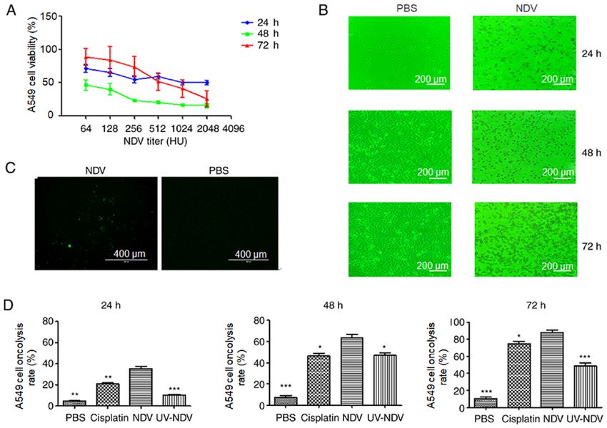
from triplicate cultures. 10 min. The protein concentration was determined using aONCOLOGY LETTERS 21: 482, 2021 3 Table I. Sequences of miR-204 mimics and inhibitors. miRNA Sense Antisense miR-204 mimics 5'-UUCCCUUUGUCAUCCUAUGCCU-3' 5'-AAGGGAAACAGUAGGAUUCGGA-3' Mimic-NC 5'-UUUGUACUACACAAAAGUACUG-3' 5'-AAACAUGAUGUGUUUUCAUGAC-3' miR-204 inhibitor 5'-AGGCUUAGGAUGACAAAGGGAA-3' Inhibitor-NC 5'-CAGUACUUUUGUGUAGUACAAA-3' NC, negative control; miR/miRNA, microRNA. Table II. Primers used to detect miRNA, caspase-3 and BAX expression levels with reverse transcription-quantitative PCR. Gene Forward primer Reverse primer miR-204-5p 5'-TCCCTTTGTCATCCTATGCCTAA-3' 5'-AAGGGAAACAGUAGGAUUCGGA-3' U6 5'-GGAACGATACAGAGAAGATTAGC-3' 5'-TGGAACGCTTCACGAATTTGCG-3' Caspase-3 5'-AAGGCAGAGCCATGGACCAC-3' 5'-CTGGCAGCATCATCCACACATAC-3' BAX 5'-AGATGTGGTCTATAATGCGTTTTCC-3' 5'-CAGAAGGCACTAATCAAGTCAAGGT-3' β-actin 5'-TGGCACCCAGCACAATGAA -3' 5'-CTAAGTCATAGTCCGCCTAGAAGCA-3' miR/miRNA, microRNA. BCA Protein Assay kit (cat. no. P0009; Beyotime Institute with NDV decreased A549 cell viability in a dose‑dependent of Biotechnology). The protein samples (50 µg/lane) were manner. The apoptotic effect of A549 cells after treatment with separated by 10% SDS‑PAGE (Bio‑Rad Laboratories, Inc.) NDV was observed. There was a marked and rapid decrease and transferred to a PVDF membrane (EMD Millipore). The in the viability of A549 cells 48 h after treatment with NDV in membrane was blocked with a 5% non‑fat milk solution at comparison with 24 or 72 h of treatment, and the viability of the room temperature for 1 h and subsequently incubated with A549 cells after treatment with NDV at 1:2048 for 48 h was the the following primary antibodies at 4˚C overnight: Mouse lowest (Fig. 1A). The effects of NDV‑induced cytopathy in A549 antibodies against full‑length caspase‑3 (cat. no. 9668; cells were also observed (Fig. 1B), including the appearance of 1:1,000; Cell Signaling Technology, Inc.), mouse anti‑Bax A549 cell lesions after stimulation with NDV for 24 h, a clear (cat. no. 89477S; 1:1,000; Cell Signaling Technology, Inc.) and cytopathic effect at 48 h and a large number of dead cells at 72 h. mouse anti‑β ‑actin (cat. no. 3700T; 1:2,000; Cell Signaling Observation under TUNEL fluorescence microscopy showed Technology, Inc.; used as an internal control). The next day, the apoptotic A549 cells after exposure to NDV‑7793 (Fig. 1C). blots were incubated at room temperature with HRP‑conjugated After treatment with NDV, the oncolysis rate was increased anti‑mouse IgG secondary antibody (cat. no. 7076; 1:2,500; compared with that in the PBS group (P
4                             LIANG et al: miR-204 PLAYS A ROLE IN NDV-INDUCED APOPTOSIS IN A549 CELLS

Figure 1. Oncolysis induction in NDV‑stimulated human lung cancer A549 cells in vitro. (A) A549 cell viability after NDV stimulation at 24, 48 and 72 h,
respectively, was detected by Cell Counting Kit‑8 test. (B) NDV‑induced cytopathic effect was observed through light microscopy with a green filter (mag‑
nification, x100; scale bar, 200 µm). (C) Observation under TUNEL fluorescence microscopy showed that apoptotic cells appeared when A549 cells were
exposed to NDV‑7793 (magnification, x100; scale bar, 400 µm). (D) Oncolysis induction in NDV‑stimulated human lung cancer A549 cells in vitro was
analyzed using Annexin V‑fluorescein isothiocyanate. *P
ONCOLOGY LETTERS 21: 482, 2021                                                                      5

Figure 3. Overexpression and inhibition of miR‑204 are significantly associated with NDV‑inducted oncolysis capacity in A549 cells. (A) A549 cells were incu‑
bated with miR‑204 inhibitors or mimics. Total RNA was extracted and subjected to quantitative PCR. Unstimulated A549 cells were used as NCs. miR‑204
expression level was significantly increased or decreased after transfection of miR‑204 inhibitors or mimics to A549 cells, respectively. **P
6                              LIANG et al: miR-204 PLAYS A ROLE IN NDV-INDUCED APOPTOSIS IN A549 CELLS

Figure 4. miR‑204‑regulated triggered oncolysis pathway is dependent on caspase‑3 and Bax activity. A549 cells were pre‑incubated with miR‑204 inhibitors
or mimics following stimulation with Newcastle disease virus. The expression of apoptosis‑related proteins full‑length caspase‑3 and Bax were detected by
(A) quantitative PCR and (B) western blotting analysis. The expression levels of Bax and full‑length caspase‑3 were upregulated. *P
ONCOLOGY LETTERS 21: 482, 2021                                                             7

the miRNA regulation activity of NDV upon oncolytic activity         Authors' contributions
should be demonstrated further in animal models.
    Although the level of miR‑204 was significantly associated       YL contributed significantly to the statistical analysis and
with the NDV oncolytic effect, it was also found that miR‑204        writing the manuscript. JJH performed the PCR and transfec‑
inhibitors or mimics had a limited effect on oncolysis. Cells        tion experiments. WYT performed the western blotting and the
have very strong compensatory mechanisms (36). Each gene             apoptosis analysis. LXG performed the virus purification and
may be targeted by hundreds of miRNAs, and each miRNA                cell cultures. XHF contributed to the study design and provided
may regulate hundreds of genes (37). Therefore, this limited         crucial experiment materials. YL, WYT, JJH, LXG and XHF
effect might be due to the existence of a large number of            confirm the authenticity of the raw data. All authors have read
cellular regulatory factors, which can offset or mask the effect     and approved the final manuscript.
of inhibiting apoptosis.
    Apoptosis consists of death receptor ligand/extrinsic and        Ethics approval and consent to participate
mitochondrial/intrinsic pathways. It has been confirmed that
NDV induces tumor cell apoptosis through both of these               Not applicable.
pathways (38). The intrinsic and extrinsic pathways converge
downstream at regulatory factor caspase‑3, which is required         Patient consent for publication
to participate in tumor cell apoptosis induced by NDV (39). In
the present study, miR‑204 overexpression resulted in upregu‑        Not applicable.
lation of full‑length caspase‑3, while inhibition via an miR‑204
inhibitor decreased the level of full‑length caspase‑3. Therefore,   Competing interests
miR‑204 may regulate NDV oncolysis in A549 cells through the
expression of full‑length caspase‑3. Activated initiator caspases    The authors declare that they have no competing interests.
subsequently free full‑length caspase‑3 of their short inhibi‑
tory prodomain, allowing them to cleave a large set of cellular      References
substrates (40). Therefore, not only full‑length caspase‑3, but
also the levels of cleaved caspase‑3 should be considered in          1. Sandler A, Gray R, Perry MC, Brahmer J, Schiller JH, Dowlati A,
future studies in order to demonstrate the role of caspase‑3 in          Lilenbaum R and Johnson DH: Paclitaxel‑carboplatin alone
                                                                         or with bevacizumab for non‑small‑cell lung cancer. N Engl J
the process of apoptosis. Bax, a member of the Bcl‑2 family,             Med 355: 2542‑2550, 2006.
is another signaling protein involved in apoptosis regulation         2. Hirsch FR, Scagliotti GV, Mulshine JL, Kwon R, Curran WJ Jr,
by binding to the permeability transition pore complex, which            Wu YL and Paz‑Ares L: Lung cancer: Current therapies and new
                                                                         targeted treatments. Lancet 389: 299‑311, 2017.
is responsible for the regulation of mitochondrial membrane           3. Adam V, Dooms C and Vansteenkiste J: Lung cancer at the intensive
permeability. The present study also demonstrated that phar‑             care unit: The era of targeted therapy. Lung Cancer 89: 218‑221, 2015.
                                                                      4. Fournier P and Schirrmacher V: Oncolytic Newcastle disease
macological inhibitors and mimics of miR‑204 decreased and               virus as cutting edge between tumor and host. Biology (Basel) 2:
promoted the expression of Bax, respectively, in A549 cells,             936‑975, 2013.
indicating that miR‑204 regulates NDV oncolysis in A549 cells.        5. Lam HY, Yeap SK, Pirozyan MR, Omar AR, Yusoff K, Abd‑Aziz S and
                                                                         Alitheen NB: Corrigendum to ‘Safety and clinical usage of Newcastle
    In summary, the present study provides preliminary                   disease virus in cancer therapy’. BioMed Res Int 2017: 4529437, 2017.
evidence that NDV induces apoptosis in tumor cells via an             6. Batliwalla FM, Bateman BA, Serrano D, Murray D, Macphail S,
miR‑204‑dependent pathway. miR‑204 plays a role as a tumor               Maino VC, Ansel JC, Gregersen PK and Armstrong CA: A
                                                                         15‑year follow‑up of AJCC stage III malignant melanoma
suppressor in NDV‑induced apoptosis in tumor cells. These                patients treated postsurgically with Newcastle disease virus
findings indicate that the regulation of apoptosis signaling             (NDV) oncolysate and determination of alterations in the CD8
by miR‑204 influences tumor survival in NDV‑treated lung                 T cell repertoire. Mol Med 4: 783‑794, 1998.
                                                                      7. Cassel WA and Murray DR: A ten‑year follow‑up on stage II malignant
cancer. These results highlight the miRNA‑regulatory activity            melanoma patients treated postsurgically with Newcastle disease virus
of NDV upon oncolytic activity.                                          oncolysate. Med Oncol Tumor Pharmacother 9: 169‑171, 1992.
                                                                      8. Schirrmacher V, van Gool S and Stuecker W: Breaking therapy
                                                                         resistance: An update on oncolytic Newcastle disease virus for
Acknowledgements                                                         improvements of cancer therapy. Biomedicines 7: 66, 2019.
                                                                      9. Cuadrado‑Castano S, Sanchez‑Aparicio MT, García‑Sastre A
                                                                         and Villar E: The therapeutic effect of death: Newcastle disease
Not applicable.                                                          virus and its antitumor potential. Virus Res 209: 56‑66, 2015.
                                                                     10. Keshavarz M, Nejad ASM, Esghaei M, Bokharaei‑Salim F,
Funding                                                                  Dianat‑Moghadam H, Keyvani H and Ghaemi A: Oncolytic
                                                                         Newcastle disease virus reduces growth of cervical cancer cell
                                                                         by inducing apoptosis. Saudi J Biol Sci 27: 47‑52, 2020.
This study was supported by a grant from the Natural Science         11. Xu Q, Rangaswamy US, Wang W, Robbins SH, Harper J, Jin H
Foundation of Guangxi, China (no. 2018GXNSFDA281043),                    and Cheng X: Evaluation of Newcastle disease virus mediated
                                                                         dendritic cell activation and cross‑priming tumor‑specific
the National Natural Science Foundation of China                         immune responses ex vivo. Int J Cancer 146: 531‑541, 2020.
(no. 81960511) and the Guangxi First‑class Discipline Project        12. Jarahian M, Watzl C, Fournier P, Arnold A, Djandji D, Zahedi S,
for Basic and Medicine Sciences (no. GXFCDP‑BMS‑2018).                   Cerwenka A, Paschen A, Schirrmacher V and Momburg F:
                                                                         Activation of natural killer cells by Newcastle disease virus
                                                                         hemagglutinin‑neuraminidase. J Virol 83: 8108‑8121, 2009.
Availability of data and materials                                   13. Washburn B, Weigand MA, Grosse‑Wilde A, Janke M,
                                                                         Stahl H, Rieser E, Sprick MR, Schirrmacher V and Walczak H:
                                                                         TNF‑related apoptosis‑inducing ligand mediates tumoricidal
The datasets used and/or analyzed during the current study are           activity of human monocytes stimulated by Newcastle disease
available from the corresponding author on reasonable request.           virus. J Immunol 170: 1814‑1821, 2003.
8 LIANG et al: miR-204 PLAYS A ROLE IN NDV-INDUCED APOPTOSIS IN A549 CELLS
14. Koks CA, Garg AD, Ehrhardt M, Riva M, Vandenberk L, Boon L, 28. Zhang CX, Ye LW, Liu Y, Xu XY, Li DR, Yang YQ, Sun LL
De Vleeschouwer S, Agostinis P, Graf N and Van Gool SW: and Yuan J: Antineoplastic activity of Newcastle disease virus
Newcastle disease virotherapy induces long‑term survival and strain D90 in oral squamous cell carcinoma. Tumour Biol 36:
tumor‑specific immune memory in orthotopic glioma through 7121‑7131, 2015.
the induction of immunogenic cell death. Int J Cancer 136: 29. Zhang S, Gao L, Thakur A, Shi P, Liu F, Feng J, Wang T, Liang Y,
E313‑E325, 2015. Liu JJ, Chen M, et al: miRNA‑204 suppresses human non‑small
15. Ors‑Kumoglu G, Gulce‑Iz S and Biray‑Avci C: Therapeutic cell lung cancer by targeting ATF2. Tumour Biol 37: 11177‑11186,
microRNAs in human cancer. Cytotechnology 71: 411‑425, 2019. 2016.
16. Li P, Wang Q and Wang H: MicroRNA‑204 inhibits the prolif‑ 30. Jiang D, Li M, Yu Y, Shi H and Chen R: microRNA‑34a aggravates
eration, migration and invasion of human lung cancer cells by coxsackievirus B3‑induced apoptosis of cardiomyocytes through
targeting PCNA‑1 and inhibits tumor growth in vivo. Int J Mol the SIRT1‑p53 pathway. J Med Virol 91: 1643‑1651, 2019.
Med 43: 1149‑1156, 2019. 31. Geekiyanage H and Galanis E: MiR‑31 and miR‑128 regulates
17. Guo W, Zhang Y, Zhang Y, Shi Y, Xi J, Fan H and Xu S: poliovirus receptor‑related 4 mediated measles virus infectivity
Decreased expression of miR‑204 in plasma is associated with a in tumors. Mol Oncol 10: 1387‑1403, 2016.
poor prognosis in patients with non‑small cell lung cancer. Int J 32. Rovira‑Rigau M, Raimondi G, Marín MA, Gironella M,
Mol Med 36: 1720‑1726, 2015. Alemany R and Fillat C: Bioselection reveals miR‑99b and
18. Livak KJ and Schmittgen TD: Analysis of relative gene expression miR‑485 as enhancers of adenoviral oncolysis in pancreatic
data using real‑time quantitative PCR and the 2(‑Delta Delta cancer. Mol Ther 27: 230‑243, 2019.
C(T)) method. Methods 25: 402‑408, 2001. 33. Li T, Pan H and Li R: The dual regulatory role of miR‑204 in
19. Ghrici M, El Zowalaty M, Omar AR and Ideris A: Induction of cancer. Tumour Biol 37: 11667‑11677, 2016.
apoptosis in MCF‑7 cells by the hemagglutinin‑neuraminidase 34. Santoni G, Morelli MB, Santoni M, Nabissi M, Marinelli O and
glycoprotein of Newcastle disease virus Malaysian strain Amantini C: Targeting transient receptor potential channels by
AF2240. Oncol Rep 30: 1035‑1044, 2013. MicroRNAs drives tumor development and progression. Adv
20. Li N, Guo X, Liu L, Wang L and Cheng R: Molecular mechanism Exp Med Biol 1131: 605‑623, 2020.
of miR‑204 regulates proliferation, apoptosis and autophagy of 35. Tajbakhsh A, Mokhtari‑Zaer A, Rezaee M, Afzaljavan F,
cervical cancer cells by targeting ATF2. Artif Cells Nanomed Rivandi M, Hassanian SM, Ferns GA, Pasdar A and Avan A:
Biotechnol 47: 2529‑2535, 2019. Therapeutic potentials of BDNF/TrkB in breast cancer; current
21. Raihan J, Ahmad U, Yong YK, Eshak Z, Othman F and Ideris A: status and perspectives. J Cell Biochem 118: 2502‑2515, 2017.
Regression of solid breast tumours in mice by Newcastle disease 36. Wang X, Jia Y, Wang X, Wang C, Lv C, Li X, Chu Z, Han Q,
virus is associated with production of apoptosis related‑cytokines. Xiao S, Zhang S, et al: MiR‑375 has contrasting effects on
BMC Cancer 19: 315, 2019. Newcastle disease virus growth depending on the target gene. Int
22. Ghosh S: Cisplatin: The first metal based anticancer drug. Bioorg J Biol Sci 15: 44‑57, 2019.
Chem 88: 102925, 2019. 37. Trobaugh DW and Klimstra WB: MicroRNA regulation of RNA
23. Dimitrov KM, Afonso CL, Yu Q and Miller PJ: Newcastle virus replication and pathogenesis. Trends Mol Med 23: 80‑93,
disease vaccines ‑ A solved problem or a continuous challenge? 2017.
Vet Microbiol 206: 126‑136, 2017. 38. Bian J, Wang K, Kong X, Liu H, Chen F, Hu M, Zhang X,
24. Yurchenko KS, Zhou P, Kovner AV, Zavjalov EL, Shestopalova LV Jiao X, Ge B, Wu Y, et al: Caspase‑ and p38‑MAPK‑dependent
and Shestopalov AM: Oncolytic effect of wild‑type Newcastle induction of apoptosis in A549 lung cancer cells by Newcastle
disease virus isolates in cancer cell lines in vitro and in vivo on disease virus. Arch Virol 156: 1335‑1344, 2011.
xenograft model. PLoS One 13: e0195425, 2018. 39. Yan Y, Liang B, Zhang J, Liu Y and Bu X: Apoptotic induction of
25. Lazar I, Yaacov B, Shiloach T, Eliahoo E, Kadouri L, Lotem M, lung adenocarcinoma A549 cells infected by recombinant RVG
Perlman R, Zakay‑Rones Z, Panet A and Ben‑Yehuda D: The Newcastle disease virus (rL‑RVG) in vitro. Mol Med Rep 11:
oncolytic activity of Newcastle disease virus NDV‑HUJ on 317‑326, 2015.
chemoresistant primary melanoma cells is dependent on the 40. Lamkanfi M and Kanneganti TD: Caspase‑7: A protease involved
proapoptotic activity of the inhibitor of apoptosis protein Livin. J in apoptosis and inflammation. Int J Biochem Cell Biol 42: 21‑24,
Virol 84: 639‑646, 2010. 2010.
26. Al‑Shammari AM, Salman MI, Saihood YD, Yaseen NY,
Raed K, Shaker HK, Ahmed A, Khalid A and Duiach A: In vitro This work is licensed under a Creative Commons
synergistic enhancement of Newcastle disease virus to 5‑fluoro‑ Attribution-NonCommercial-NoDerivatives 4.0
uracil cytotoxicity against tumor cells. Biomedicines 4: 3, 2016. International (CC BY-NC-ND 4.0) License.
27. Kalyanasundram J, Hamid A, Yusoff K and Chia SL: Newcastle
disease virus strain AF2240 as an oncolytic virus: A review.
Acta Trop 183: 126‑133, 2018.You can also read