Medical Physics Letter - In vivo functional chronic imaging of a small animal model using optical-resolution photoacoustic microscopy
←
→
Page content transcription
If your browser does not render page correctly, please read the page content below
Medical Physics Letter

In vivo functional chronic imaging of a small animal model
using optical-resolution photoacoustic microscopy
         Song Hu, Konstantin Maslov, and Lihong V. Wanga兲
         Department of Biomedical Engineering, Optical Imaging Laboratory, Washington University in St. Louis,
         St. Louis, Missouri 63130-4899
         共Received 27 February 2009; revised 16 April 2009; accepted for publication 26 April 2009;
         published 27 May 2009兲
         Optical-resolution photoacoustic microscopy 共OR-PAM兲 has been validated as a valuable tool for
         label-free volumetric microvascular imaging. More importantly, the advantages of noninvasiveness
         and measurement consistency suggest the use of OR-PAM for chronic imaging of intact microcir-
         culation. Here, such chronic imaging is demonstrated for the first time by monitoring the healing
         process of laser-induced microvascular lesions in a small animal model in vivo. The central part of
         a 1 mm by 1 mm region in a nude mouse ear was treated under a continuous-wave laser to create
         a microvascular lesion for chronic study. The region of interest was imaged before the laser treat-
         ment, immediately after the treatment, and throughout the healing process using both the authors’
         OR-PAM system and a commercial transmission-mode optical microscope. Three-dimensional mi-
         crovascular morphology and blood oxygenation information were imaged simultaneously at
         capillary-level resolution. Transmission-mode optical microscopic images were acquired for com-
         parison. OR-PAM has potential important applications in microcirculatory physiology or patho-
         physiology, tumor angiogenesis, laser microsurgery, and neuroscience. © 2009 American Associa-
         tion of Physicists in Medicine. 关DOI: 10.1118/1.3137572兴

         Key words: optical-resolution photoacoustic microscopy, chronic imaging, label-free, noninvasive,
         wound healing, hemoglobin oxygen saturation

I. INTRODUCTION                                                     II. METHODS
Chronic imaging of microcirculation in vivo permits direct
                                                                       In our OR-PAM system 共Fig. 1兲, the animal tissue is irra-
visualization of long-term microhemodynamics, which is              diated with a short-pulsed laser beam generated by a
closely associated with disease progression,1,2 neural              wavelength-tunable laser set, consisting of an Nd:YLF pump
dynamics,3 and functional recovery from pathological                laser 共INNOSLAB, Edgewave, Wuerselen, Germany兲 and a
states.4 However, existing high-resolution microvascular im-        dye laser 共CBR-D, Sirah, Kaarst, Germany兲. Wideband ultra-
aging techniques suffer in different regards when employed          sonic waves—referred to as photoacoustic waves—are in-
for chronic studies. For example, invasive procedures5 and          duced as a result of transient thermoelastic expansion due to
fluorescence labeling6,7 generally disturb the normal physiol-      the laser excitation, collected via an acoustic lens, and then
ogy of the microcirculation, discourage longitudinal studies,       detected by a high-frequency ultrasonic transducer 关V2022
and impede clinical translations.                                   共BC兲, Olympus NDT, Kennewick, WA兴. To maximize the
   To overcome these difficulties, we have developed a              sensitivity for in vivo capillary imaging, the optical illumina-
bright-field photoacoustic microscopy with optically                tion and the ultrasonic detection in our system are configured
diffraction-limited lateral resolution, called OR-PAM, for in       confocally by an acoustic-optical beam splitter. In this simple
                                                                    design, two right-angle prisms form a cube with a thin sili-
vivo microvascular imaging down to the capillary level.8 OR-
                                                                    cone oil layer in between. The glass and the silicone oil have
PAM is highly sensitive to the physiologically specific opti-
                                                                    similar optical refractive indices 共1.1 in ratio兲 but very dis-
cal absorption of hemoglobin, thereby bypassing the limita-
                                                                    similar acoustic impedances 共12.7 in ratio兲, which makes the
tions of invasiveness and phototoxicity. By keeping the             layer optically transmissive but acoustically reflective. The
microcirculation intact, OR-PAM is ideal for chronic studies.       lateral resolution of this acoustic-optical confocal configura-
In this Letter, we report on the first demonstration of using       tion is determined by the product of the two point-spread
OR-PAM for functional chronic imaging. The healing pro-             functions of the optical illumination and the acoustic detec-
cess of a laser-induced microvascular lesion was monitored          tion. Since acoustic resolution is difficult to achieve down to
over a period of 12 days. Chronic OR-PAM imaging permits            the capillary level due to the frequency-dependent ultrasonic
direct visualization of the morphological and functional re-        absorption, a microscopic objective lens 共RMS4X, Thorlabs,
covery of microcirculation after laser destruction.                 Newton, NJ兲 is employed to achieve diffraction-limited op-

2320   Med. Phys. 36 „6…, June 2009            0094-2405/2009/36„6…/2320/4/$25.00            © 2009 Am. Assoc. Phys. Med.      2320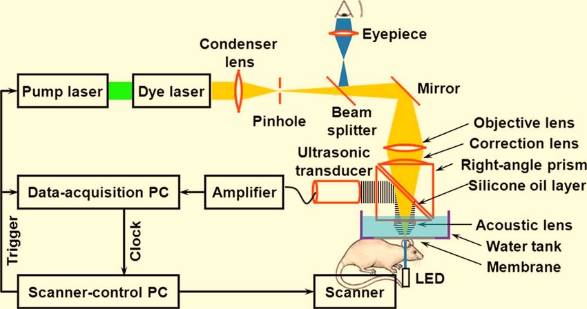
2321 Hu, Maslov, and Wang: In vivo functional chronic imaging using OR-PAM 2321
Foxn 1NU, Harlan Co., Indianapolis, IN ⬃25 g兲 was gently
removed with human hair-removing lotion 共Surgi cream,
Ardell International, Los Angeles, CA兲. For daily monitor-
ing, a dose of 87 mg/kg ketamine and 13 mg/kg xylazine was
administered intraperitoneally to anesthetize the animal right
before transferring it to a stereotaxic imaging stage. During
the experiment, a dual-wavelength 共570 and 578 nm兲 OR-
PAM measurement was utilized to extract the total hemoglo-
bin concentration 共HbT兲 and the hemoglobin oxygen satura-
tion 共sO2兲. 570 nm is an isosbestic point, and the
photoacoustic signal acquired at this wavelength reflects
FIG. 1. Schematic of the OR-PAM system.
HbT. 578 nm is a local absorption peak of oxyhemoglobin,
which helps differentiate it from deoxyhemoglobin. A de-
tailed description of the method used for computing sO2 can
tical resolution. For chronic study, a transmission-mode op- be found in a previously published paper.9 Throughout each
tical microscope is integrated into our system by adding a experiment, anesthesia was maintained using vaporized isof-
light-emitting diode beneath the animal holder. Utilizing the lurane 共1.0%–1.5% isoflurane with an airflow rate of
reverse optical path of the OR-PAM illumination, the imag- 1 l/min兲, and the body temperature of the animal was main-
ing region can be viewed under an eyepiece. This addition tained at 37 ° C with a temperature controlled heating pad.
helps us quickly target the same region of interest 共ROI兲 At the end of the chronic study, the animal was euthanized
during multiday monitoring. The detailed system description by an intraperitoneal administration of pentobarbital at a dos-
and performance can be found in our previously published age of 100 mg/kg.
paper.8 At the beginning of the chronic imaging, we selected and
The advantages of OR-PAM include the following: 共i兲 En- photographed a 1 mm by 1 mm region in the mouse ear
dogenous optical absorption contrast enables direct imaging under a commercial transmission-mode optical microscope.
of microvessels down to capillaries, with a high signal-to- Then we imaged the ROI using OR-PAM 关Fig. 2共a兲兴, after
noise ratio 共SNR兲;8 共ii兲 multiwavelength measurement per- which we switched the excitation source to a continuous-
mits vessel-by-vessel mapping of blood oxygenation,9–11 wave 共cw兲 laser 共output power: 150 mW; wavelength:
holding the potential for functional studies such as brain 532 nm兲 and removed the pinhole to enable the laser to cre-
function mapping;12 共iii兲 time-resolved ultrasonic detection ate a microvascular lesion for study. The central part of the
provides depth information without depth scanning; 共iv兲 ROI 共0.25 mm by 0.25 mm兲 was then scanned with the fo-
working in reflection mode noninvasively makes the tech- cused cw laser beam 共diameter: ⬃30 m兲 for ⬃10 min.
nique applicable to more anatomical sites in vivo, without The ROI was imaged immediately after the cw laser treat-
disturbing the microcirculatory function. ment 关Fig. 2共b兲兴 and in the subsequent 12 days 关Fig. 2共c兲,
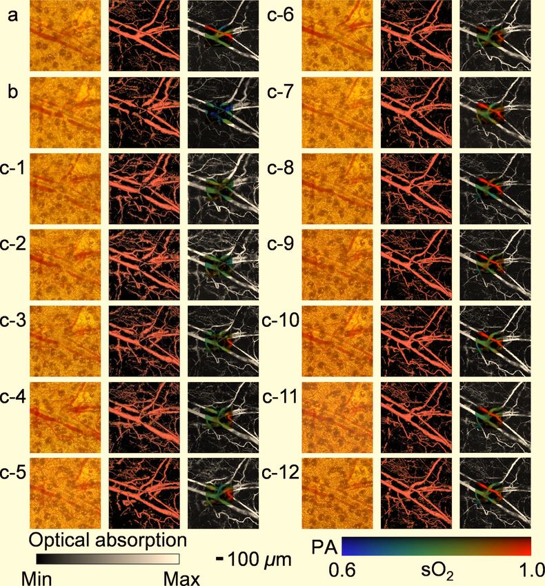
共c-1兲–共c-12兲兴 using both our OR-PAM system and the com-
mercial transmission-mode optical microscope. Our results
III. RESULTS clearly show a four-step wound healing process:14 共i兲 Vessel
The above advantages, along with measurement consis- regression and hemostasis occurred right after the laser de-
tency, permit OR-PAM to monitor long-term microhemody- struction 关Fig. 2共b兲兴; 共ii兲 inflammation, the second phase of
namics. One promising application lies in the realm of laser wound healing, was exhibited in the form of vasodilation 24
microsurgery, where OR-PAM can provide fruitful structural h after the injury and lasted for about 5 days 关Fig. 2共c兲,
and functional information about the targeted microvascular 共c-1兲–共c-5兲兴. Hypoxia facilitated the synthesis of the vascular
lesions, such as their morphology, precise location, and endothelial growth factor to trigger angiogenesis 关Figs. 2共b兲
blood oxygenation, which will facilitate accurate diagnosis and 2共c兲, 共c-1兲–共c-5兲兴; 共iii兲 about 3 days after the wound
and proper treatment. More importantly, OR-PAM enables occurred, the ingrowth of new capillaries started to restore
noninvasive monitoring of the healing process of the mi- the microcirculation 关Fig. 2共c兲, 共c-3兲兴, which was previously
crovascular lesions after laser microsurgery, which has been supplied by the damaged arteriole; 共iv兲 after 12 days, the
desired for a long time.13 damaged arteriole-venule pair was almost completely recov-
A mouse ear model was chosen to demonstrate this ability ered to normal status 关Fig. 2共c兲, 共c-12兲兴. The microvascular
in vivo because it is among the few anatomical sites that are morphology imaged by OR-PAM is partially validated by the
readily imaged by transmission-mode optical microscopy, commercial transmission-mode optical microscope; however,
which can be used to partially validate OR-PAM. All experi- capillary networks are only visible to OR-PAM. The consis-
mental animal procedures were carried out in conformance tency in microvascular morphology during the multiday
with the laboratory animal protocol approved by the School monitoring implies the robustness of our technique.
of Medicine Animal Studies Committee of Washington Uni-
versity in St. Louis. IV. CONCLUSION AND DISCUSSION
One day before the beginning of the chronic study, the In this work, we demonstrated the capability of OR-PAM
hair on the left ear of a nude mouse 共Hsd:Athymic Nude- for functional chronic imaging of microhemodynamics in
Medical Physics, Vol. 36, No. 6, June 20092322 Hu, Maslov, and Wang: In vivo functional chronic imaging using OR-PAM 2322 FIG. 2. OR-PAM monitoring of the healing process of a laser-induced microvascular lesion. 共a兲 Before laser destruction. 共b兲 Immediately after laser destruction. 共c兲 On each of the subsequent 12 days. The left image in each part 关共a兲–共c-12兲兴 is a photograph taken by a commercial transmission-mode optical microscope; the middle one is the front view of the 3D microvascular morphology acquired by OR-PAM at 570 nm; the right one is the maximum-amplitude projection 共MAP兲 image overlaid by the sO2 mapping of the laser-damaged region. vivo noninvasively. However, this technique has potentially chronic studies of cortical plasticity15 as well as neurovascu- broader applications. In cancer research, OR-PAM can moni- lar coupling16 at the capillary level. tor tumor angiogenesis and evaluate tumor therapy.1 In drug development for microvascular dysfunctions, such as stroke, OR-PAM can trace drug functioning and evaluate drug effi- cacy. In the physiological study of angiogenesis, OR-PAM ACKNOWLEDGMENTS can help understand the signal transduction pathway by per- The authors appreciate Professor James Ballard’s close turbing it and detecting the difference before and after per- reading of the manuscript. This work was sponsored by Na- turbation. In laser microsurgery, surgical lasers can be tional Institutes of Health Grant Nos. R01 EB000712, R01 readily integrated into our OR-PAM system to perform on- NS46214 共Bioengineering Research Partnerships兲, R01 site high-precision microsurgery with presurgery diagnosis EB008085, and U54 CA136398 共Network for Translational and postsurgery evaluation. In neuroscience, the noninvasive Research兲. L.V.W. has a financial interest in Endra, Inc., feature and fine imaging resolution make OR-PAM ideal for which, however, did not support this work. Medical Physics, Vol. 36, No. 6, June 2009
2323       Hu, Maslov, and Wang: In vivo functional chronic imaging using OR-PAM                                                                     2323

 a兲
   Author to whom correspondence should be addressed. Electronic mail:            Lett. 33, 929–931 共2008兲.
                                                                               9
   lhwang@biomed.wustl.edu                                                        H. F. Zhang, K. Maslov, M. Sivaramakrishnan, G. Stoica, and L. V.
 1
  R. K. Jain, L. L. Munn, and D. Fukumura, “Dissecting tumour patho-              Wang, “Imaging of hemoglobin oxygen saturation variations in single
  physiology using intravital microscopy,” Nat. Rev. Cancer 2, 266–276            vessels in vivo using photoacoustic microscopy,” Appl. Phys. Lett. 90,
  共2002兲.                                                                         053901 共2007兲.
 2                                                                             10
  D. Fukumura and R. K. Jain, “Imaging angiogenesis and the microenvi-            H. F. Zhang, K. Maslov, G. Stoica, and L. V. Wang, “Functional photoa-
  ronment,” APMIS 116, 695–715 共2008兲.                                            coustic microscopy for high-resolution and noninvasive in vivo imaging,”
 3
  K. Deisseroth, G. Feng, A. K. Majewska, G. Miesenbock, A. Ting, and             Nat. Biotechnol. 24, 848–851 共2006兲.
                                                                               11
  M. J. Schnitzer, “Next-generation optical technologies for illuminating        H. F. Zhang, K. Maslov, and L. V. Wang, “In vivo imaging of subcuta-
  genetically targeted brain circuits,” J. Neurosci. 26, 10380–10386 共2006兲.     neous structures using functional photoacoustic microscopy,” Nature Pro-
 4
  A. Zepeda, C. Arias, and F. Sengpiel, “Optical imaging of intrinsic sig-       tocols 2, 797–804 共2007兲.
                                                                               12
  nals: Recent developments in the methodology and its applications,” J.          D. Malonek and A. Grinvald, “Interactions between electrical activity and
  Neurosci. Methods 136, 1–21 共2004兲.                                             cortical microcirculation revealed by imaging spectroscopy: Implications
 5
  A. M. Iga, S. Sarkar, K. M. Sales, M. C. Winslet, and A. M. Seifalian,          for functional brain mapping,” Science 272, 551–554 共1996兲.
                                                                               13
  “Quantitating therapeutic disruption of tumor blood flow with intravital        R. R. Anderson and J. A. Parrish, “Selective photothermolysis: Precise
  video microscopy,” Cancer Res. 66, 11517–11519 共2006兲.                          microsurgery by selective absorption of pulsed radiation,” Science 220,
 6
  E. Laemmel, M. Genet, G. Le Goualher, A. Perchant, J. F. Le Gargasson,          524–527 共1983兲.
  and E. Vicaut, “Fibered confocal fluorescence microscopy 共Cell-viZio兲        14
                                                                                  G. A. Chin, R. F. Diegelmann, and G. S. Schultz, in Wound Healing,
  facilitates extended imaging in the field of microcirculation. A compari-       edited by A. F. Falabella and R. S. Kirsner 共Talyor & Francis, Boca
  son with intravital microscopy,” J. Vasc. Res. 41, 400–411 共2004兲.              Raton, 2005兲, pp. 17–37.
 7                                                                             15
  D. Kleinfeld, P. P. Mitra, F. Helmchen, and W. Denk, “Fluctuations and          S. A. Masino and R. D. Frostig, “Quantitative long-term imaging of the
  stimulus-induced changes in blood flow observed in individual capillaries       functional representation of a whisker in rat barrel cortex,” Proc. Natl.
  in layers 2 through 4 of rat neocortex,” Proc. Natl. Acad. Sci. U.S.A. 95,      Acad. Sci. U.S.A. 93, 4942–4947 共1996兲.
  15741–15746 共1998兲.                                                          16
                                                                                  X. Wang, Y. Pang, G. Ku, X. Xie, G. Stoica, and L. V. Wang, “Noninva-
 8
  K. Maslov, H. F. Zhang, S. Hu, and L. V. Wang, “Optical-resolution              sive laser-induced photoacoustic tomography for structural and functional
  photoacoustic microscopy for in vivo imaging of single capillaries,” Opt.       in vivo imaging of the brain,” Nat. Biotechnol. 21, 803–806 共2003兲.

Medical Physics, Vol. 36, No. 6, June 2009
You can also read


