Management of Unilateral Maxillary Canine Impaction
←
→
Page content transcription
If your browser does not render page correctly, please read the page content below
Volume 33       Issue 1                                                                                  Article 4

2021

Management of Unilateral Maxillary Canine Impaction
Maureen Antolis
Universitas Indonesia

Krisnawati E. Tarman
universitas indonesia

Follow this and additional works at: https://www.tjo.org.tw/tjo

   Part of the Orthodontics and Orthodontology Commons

Recommended Citation
Antolis, Maureen and Tarman, Krisnawati E. (2021) "Management of Unilateral Maxillary Canine
Impaction," Taiwanese Journal of Orthodontics: Vol. 33 : Iss. 1 , Article 4.
DOI: 10.38209/2708-2636.1094
Available at: https://www.tjo.org.tw/tjo/vol33/iss1/4

This Case Report is brought to you for free and open access by Taiwanese Journal of Orthodontics. It has been
accepted for inclusion in Taiwanese Journal of Orthodontics by an authorized editor of Taiwanese Journal of
Orthodontics.
CASE REPORT

Management of Unilateral Maxillary
Canine Impaction

Maureen Antolis*, Krisnawati E. Tarman

Department of Orthodontics Faculty of Dentistry, Universitas Indonesia, Jakarta, Indonesia

ABSTRACT

   Maxillary canine is an essential part of teeth for smile esthetic and function. Orthodontic treatment for patient with
impacted maxillary canine is considered to be more complex and require good biomechanical control during traction.
This case report describes an orthodontic treatment of a 17-year old female patient with unilateral maxillary canine
impaction on the left side. Orthodontic traction was used to guide the canine into normal occlusion. The orthodontic
treatment had been completed within 19 months and successfully obtained favourable esthetic and stable occlusion.
Taiwanese Journal of Orthodontics 2021;33(1):26e32

Keywords: Tooth impacted; Maxillary canine; Orthodontic traction

1. INTRODUCTION                                                                  and resorption of the affected tooth.4,7 However, the
                                                                                 maxillary canine plays an important role of esthetic
                                                                                 and guidance of occlusion, thus the treatment pri-
B    eing the last tooth to be developed through
     the longest path of eruption, the maxillary                                 ority is to preserve canine into its position.8 In this
                                                                                 case report, we present a case of palatally impacted
canine has the highest incidence of impaction
                                                                                 canine in a 17-year old female patient and was
after third molar.1 The incidence has been re-                                   treated using a preadjusted edgewise system.
ported to have range between 0.92 e 2.2%.2,3
Furthermore, impacted maxillary canine normally
occurs two times more in women than man.4                                        2. CASE REPORT
Reports stated that majority of maxillary canine                                 2.1. Clinical Examination
impaction occurs unilaterally2 at which most of
the cases (50% - 85%) are palatally displaced.5                                     A 17-year old female patient came to orthodontic
   The etiologies of impacted canine are believed                                clinic with chief complaint of missing canine on her
due to multifactorial and idiopathic origins.6 How-                              upper left side and irregularly arranged upper and
ever, there are several theories proposed as poten-                              lower front teeth.
tial etiological factors; for example, prolonged                                    Extra oral examination revealed a good facial
retention or early loss of deciduous canine, absence                             profile (Figure 1). The intraoral examination pre-
or anomaly of upper lateral incisors, local obstruc-                             sented full permanent dentition except upper left
tion, pathology, and genetic factor.1,7 Early diagnosis                          maxillary canine. Clinical examination showed
and timely interception provide better prognosis to                              bulging on palatal between upper left lateral incisor
manage the impacted canine.                                                      and first premolar. The upper and lower teeth are
   The treatment is considered extremely challenging                             mildly displaced with maxillary dental midline
due to the potential adverse sequelae of impacted                                deviated 1 mm to the left side and mandibular
canines, including resorption of adjacent roots,                                 dental midline deviated 0.5 mm to the right side of
referred pain, infection, dentigerous cyst formation,                            her facial midline. There was also mild incisor
                                                                                 proclination with an overjet of 4 mm. The canine

Received 2 March 2021; revised 1 April 2021; accepted 17 April 2021.
Available online 16 June 2021.

* Corresponding author at: Department of Orthodontics, University of Indonesia, Jl. Salemba Raya No. 4, Jakarta Pusat, Jakarta, 10430 Indonesia.
E-mail address: maureen_antolis@hotmail.com (M. Antolis).

https://doi.org/10.38209/2708-2636.1094
2708-2636/© 2021 Taiwan Association of Orthodontist. This is an open access article under the CC-BY-NC-ND license
(http://creativecommons.org/licenses/by-nc-nd/4.0/).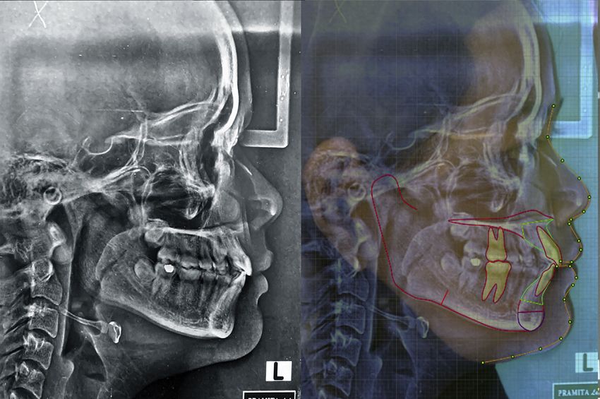
Taiwanese Journal of Orthodontics                                                                     M. ANTOLIS, K.E. TARMAN
2021;33(1):26e32                                                                      UNILATERAL MAXILLARY CANINE IMPACTION

                                    Figure 1. The extra oral photographs before treatment.

relationship was Class I on the right side and the                  The cephalometric analysis presented Class I
molar relationship was Class I on both sides                      skeletal relationship with mild bimaxillary progna-
(Figure 2).                                                       thism and proclination (Figure 5 and Table 1).
  Based on vertical parallax technique, the loca-
tion of impacted tooth was confirmed on the                        2.2. Diagnosis
palatal side (Figure 3). The prognosis of the
impacted tooth was also assessed according to the                   The 17 years old female patient was diagnosed as
reference of previous study by Mc Sherry and                      Class I malocclusion with upper left canine palatally
Counihan et al.9,10 Panoramic radiograph showed                   impacted.
the canine was angulated 40 to midline, left
canine was not overlapping with the adjacent                      2.3. Treatment Objectives
lateral incisor, crown of the canine was located in
the cervical of lateral incisor and the apical of the               To achieve alignment of the impacted canine
canine was located between lateral incisor and                       into the upper arch
first premolar. In brief, the impacted canine had a                  To correct maxillary and mandibular midline in
good prognosis of traction (Figure 4).                               line with facial midline

                                    Figure 2. The intraoral photographs before treatment.

                                                             27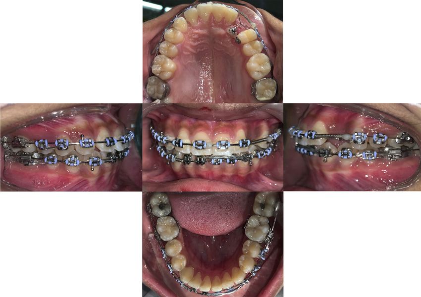
M. ANTOLIS, K.E. TARMAN Taiwanese Journal of Orthodontics
UNILATERAL MAXILLARY CANINE IMPACTION 2021;33(1):26e32
2.4. Treatment plan
Levelling and aligning upper and lower arch
Midline correction and opening space for the
impacted canine
Traction of the impacted canine
Releveling and realigning the upper arch
Finishing
Retention
2.5. Treatment Progress
An active orthodontic treatment was begun with
preadjusted edgewise appliance with .022” MBT
Figure 3. Periapical and occlusal radiograph before treatment. bracket. Levelling and aligning were performed on
both arches. After 6 months treatment, consolida-
To establish Class I canine and molar relationship tion of upper arch was finally done, followed by
To achieve orthodontic functional occlusion placing an open coil spring between left premolar
Figure 4. The panoramic radiography before treatment.
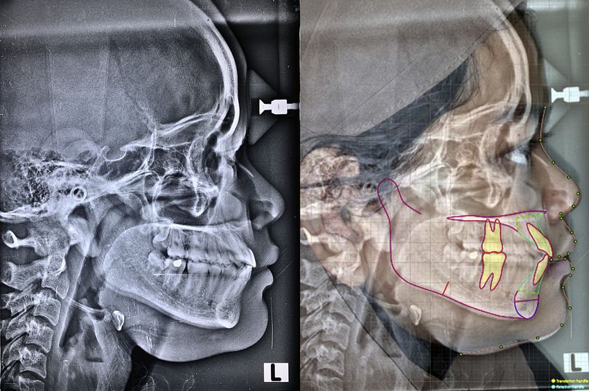
Figure 5. Lateral cephalometric radiograph before treatment.
28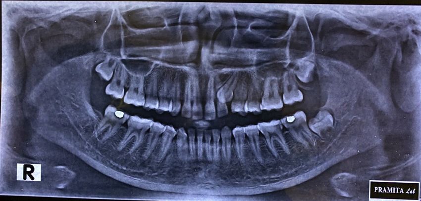
Taiwanese Journal of Orthodontics                                                                                M. ANTOLIS, K.E. TARMAN
2021;33(1):26e32                                                                                 UNILATERAL MAXILLARY CANINE IMPACTION

Table 1. Cephalometric measurements in before and after treatment.          phases were performed (Figure 7). Finishing and
                      Mean          Pre-treatment     Post-treatment        detailing were carried out with bending and settling
SNA                   81 ± 3       87               87                   elastics. The active treatment had successfully been
SNB                   78 ± 3       85               85                   completed within 19 months.
ANB                   3 ± 2        2                2
Angle of Co           0 ± 10       6                5
SN-MP                 32 ± 3       30               33                   2.6. Treatment Result
MMPA                  27 ± 4       23               24
Interincisal angle    135 ± 10     122              120                    The palatally impacted canine was properly
UI-SN                 104 ± 6      114              113                  aligned in the maxillary arch by orthodontic trac-
UI-PP                 109 ± 6      120              120
                                                                            tion. Ideal overjet and overbite were also achieved.
LI-MP                 90 ± 4       95               96
Upper lip - E line    1 ± 2 mm      0 mm              0 mm                  Class I canine and molar relationship with a
Lower lip - E line    0 ± 2 mm        1 mm            0 mm                  functional occlusion were established (Figures 8
                                                                            and 9). The panoramic film demonstrated root
                                                                            parallelism was achieved with minimal root
and lateral incisor in order to give space for the                          blunting (Figure 10). The cephalometric radio-
impacted canine. Once an adequate space had been                            graph analysis and superimposition have shown
achieved, attachment was bonded on the canine tip,                          no significant change in skeletal and dental
and traction with piggyback archwire was conduct-                           parameter (Figures 11 and 12). In addition, the
ed (Figure 6). After 8 months, the upper left canine                        patient was suggested to use essix retainers for
was bonded, and both releveling and realignment                             retention phase.

                                                    Figure 6. Traction of the impacted canine.

                                                 Figure 7. Piggyback of the impacted canine.

                                                                       29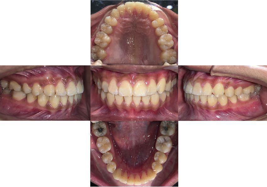
M. ANTOLIS, K.E. TARMAN Taiwanese Journal of Orthodontics
UNILATERAL MAXILLARY CANINE IMPACTION 2021;33(1):26e32
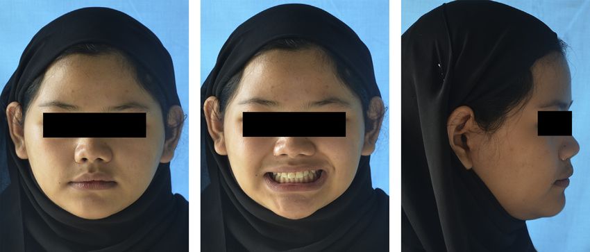
Figure 8. The extraoral photographs after treatment.
Figure 9. The intraoral photographs after treatment.
Figure 10. Panoramic radiograph after treatment.
30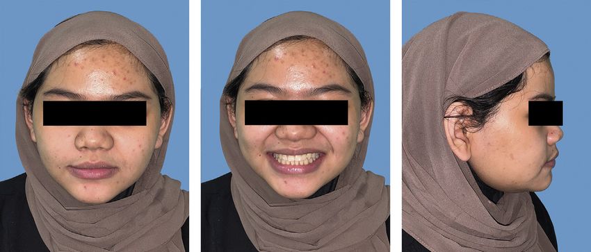
Taiwanese Journal of Orthodontics                                                                                M. ANTOLIS, K.E. TARMAN
2021;33(1):26e32                                                                                 UNILATERAL MAXILLARY CANINE IMPACTION

                                         Figure 11. The lateral cephalometric radiograph after treatment.

              Figure 12. The cephalometric superimposition before and after treatment. (red: before treatment; blue: after treatment).

3. DISCUSSION                                                                environmental factors, such as local obstruction,
                                                                             early loss of primary canine, displacement of the
  The etiology of palatally displaced canine is
                                                                             permanent tooth.7,12,13 In this case, premature loss
obscure. Several theories have been proposed to
                                                                             of deciduous canine could be the reason of inade-
explain the etiology of palatally impacted canine at
                                                                             quate space for canine eruption and the canine be-
which guidance and genetic theories are the most
                                                                             comes palatally displaced.
commonly accepted reasons.11 Guidance theory
                                                                               There are three diagnostic methods for impacted
states that insufficient distal aspect of the root on
                                                                             canine: inspection, palpation, and radiography.14
lateral incisor or agenesis of lateral incisor will result
                                                                             Whether it locates buccally or palatally, the canine
to a lack of guidance for permanent canine to erupt.
                                                                             bulge should be seen between the lateral incisor and
On the other hand, genetic theory highlights the
                                                                             first premolar. Abnormalities or agenesis of lateral
multiple genetic factors which could be responsible
                                                                             incisor could also indicate a higher risk of canine
for palatal impaction. Other possible etiologies
                                                                             impaction. In addition, the deciduous canine
which are related for palatal canine impaction are
                                                                             mobility should be observed to prevent any pro-
systemic conditions, including cleft lip and palate,
                                                                             longed retention of the tooth. Clinical palpation of
Pierre Robin syndrome, endocrine deficiency, and
                                                                             the canine bulge from age of 8 years old is

                                                                        31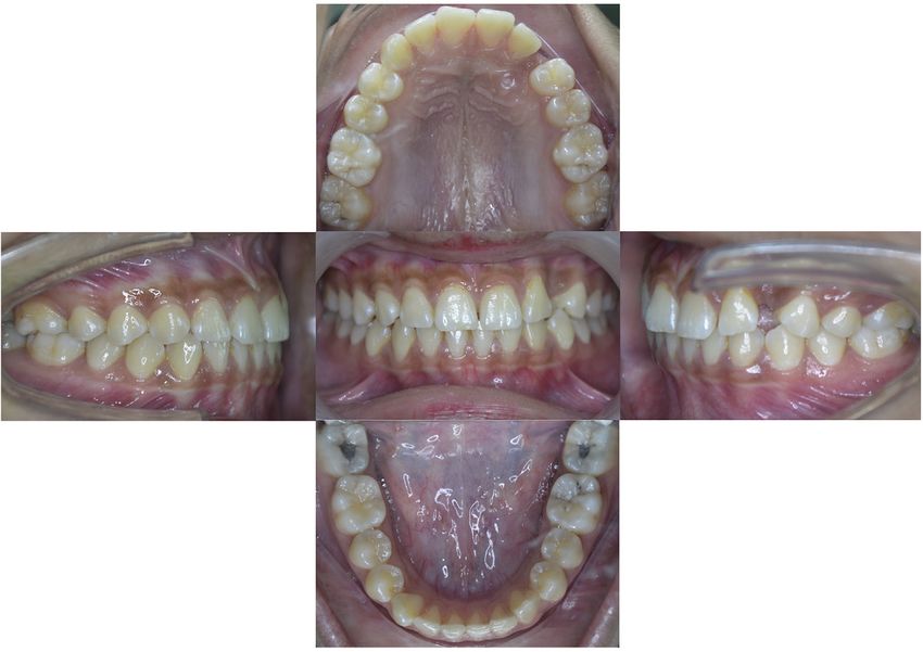
M. ANTOLIS, K.E. TARMAN                                                                              Taiwanese Journal of Orthodontics
UNILATERAL MAXILLARY CANINE IMPACTION                                                                                 2021;33(1):26e32

recommended by previous study as it was proven to               ETHICAL APPROVAL
bring significant benefit for determining canine
                                                                  This study was approved by the Institutional Re-
position.15 In addition, radiographs are required to
                                                                view Board of Faculty of Dentistry Universitas
locate the impacted canine in three dimensions and
                                                                Indonesia.
detect any root resorption. Parallax radiographic
techniques or cone bean computed tomography
have been commonly recommended in the diag-                     PATIENT CONSENT
nosis of impacted canine case.10                                  Provided.
   Assessment towards the position of impacted
canine to determine its prognosis should be per-                Conflict of Interest Statement
formed before starting the orthodontic treatment.14
There are four conditions that should be examined                 The authors declare no conflicts of interest.
which are horizontal overlap of canine towards
lateral incisor, vertical height of canine tip, angula-         REFERENCES
tion of impacted canine, and apical origin of
impacted canine.10 In this case, those four criteria             1. Litsas G, Ahu A. A review of early displaced maxillary ca-
assessments have shown a good prognosis of canine                   nines: etiology, diagnosis and interceptive treatment. Open
position. Considering this finding, we decided to                    Dent J 2011;5(1):39e47.
                                                                 2. Dachi SF, Howell FV. A survey of 3,874 routine full-mouth
choose orthodontic traction of the impacted tooth.                  radiographs. I. A study of retained roots and teeth. Oral Surg
   Prior to traction, adequate and enough space                     Oral Med Oral Pathol 1961;14(8):916e24.
to accommodate canine eruption must present.16                   3. Bishara SE. Impacted maxillary canines: a review. Am J Orthod
                                                                    Dentofacial Orthop 1992;101(2):159e71.
Since palatally impacted canine seldom erupts                    4. Ericson S, Kurol J. Radiographic assessment of maxillary
without surgical intervention, orthodontic treatment                cacine eruption in children with clinical signs of erution
usually includes surgical exposure.7,17 However in                  disturbance. Eur J Orthod 1986;8(3):133e40.
                                                                 5. Jacoby H. The etiology of maxillary canine impactions. Am J
our case, the canine tip emerged through the                        Orthod 1983;84(2):125e32.
gingiva after there was a sufficient space, therefore             6. Becker A. Palatal displacement of canine is genetic and related
the attachment could be bonded on impacted teeth                    to congenital absence of teeth. J Dent Res 1997;76(9):1526.
                                                                 7. Graber LW, Vanarsdall RL, Vig KW, Huang GJ. Orthodontics
without any surgical intervention.                                  current principles and techniques. 6th ed. St. Louis, MO: Elsevier;
   Both anchorage planning and biomechanics dur-                    2017.
ing orthodontic treatment hold an important role in              8. Pokorny PH, Wiens JP, Litvak H. Occlusion for fixed pros-
                                                                    thodontics: a historical perspective of the gnathological in-
treating impaction cases. In this case, the palatally               fluence. J Prosthet Dent 2008;99(4):299e313.
impacted canine was ligated piggyback on a                       9. McSherry PF. The ectopic maxillary canine: a review. Br J
.017  .025 inch SS arch wire. The attachment in its                Orthod 1998;25(3):209e16.
                                                                10. Counihan K, AI-Awadhi EA, Butler J. Guidelines for the
labial surface was repositioned into more cervical                  assessment of the impacted maxillary canine. Dent Update
for several times to apply more vertical force line                 2013;40(9):770e7.
that encouraged the canine erupted labially and                 11. Richardson G, Russell KA. A review of impacted permanent
                                                                    maxillary cuspids ddiagnosis and prevention. J CanDent
occlusally. The force should be light, less than 2 oz.18            Assoc 2000;66(9):497e501.
According to the treatment result, the cephalometric            12. Becker A, Chaushu S. Etiology of maxillary canine impaction:
analysis revealed only a small proclination that is                 a review. Am J Orthod Dentofacial Orthop 2015;148(4):557e67.
                                                                13. Manne R, Gandikota C, Juvvadi SR, Rama HR, Anche S.
insignificant in skeletal and dental parameters. The                 Impacted canines: etiology, diagnosis, and orthodontic man-
orthodontic treatment was completed in 19 month                     agement. J Pharm Bioallied Sci 2012;4(Suppl 2):S234e8.
and successfully obtained favourable esthetic and               14. Hsu YC, Kao CT, Chou CC, Tai WK, Yang PY. Diagnosis and
                                                                    management of impacted maxillary canines. Taiwan J Orthod
stable occlusion.                                                   2020;31(1):4e11.
                                                                15. Ngan P, Hornbrook R, Weaver B. Early timely management of
                                                                    ectopically erupting maxillary canines. Semin Orthod 2005;
4. CONCLUSION                                                       11(3):152e63.
                                                                16. Bedoya MM, Park JH. A review of the diagnosis and man-
   Maxillary canine impaction is a common case                      agement of impacted maxillary canines. J Am Dent Assoc 2009;
in orthodontics. Through a combination of clinical                  140(12):1485e93.
and radiographic diagnosis and careful planning of              17. Kokich VG. Surgical and orthodontic management of
                                                                    impacted maxillary canines. Am J Orthod Dentofacial Orthop
orthodontic biomechanics, the palatally impacted                    2004;126(3):278e83.
canine treatment will give an esthetic and func-                18. Bishara SE. Clinical management of impacted maxillary ca-
tionally stable results.                                            nines. Semin Orthod 1998;4(2):87e98.

                                                           32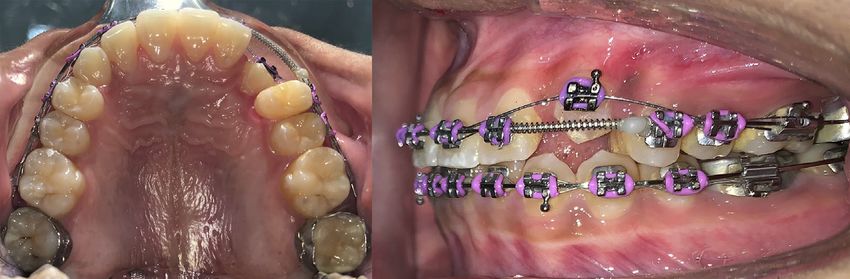
You can also read