Management of Two Cases of Spina Bifida and Neonatal Genital Prolapse at the University Hospital of Parakou and Review of the Literature
←
→
Page content transcription
If your browser does not render page correctly, please read the page content below
Open Journal of Modern Neurosurgery, 2022, 12, 216-221
                                                                                            https://www.scirp.org/journal/ojmn
                                                                                                       ISSN Online: 2163-0585
                                                                                                         ISSN Print: 2163-0569

Management of Two Cases of Spina Bifida and
Neonatal Genital Prolapse at the University
Hospital of Parakou and Review of the
Literature

Kouassi Jean Marie Maurin Kisito Quenum1*, Ayaovi Armel Hadonou2,
Toyifia Eudoxie Bernice Quenum Hountondji3, Sophonie Dokpe1, Zachée Agbo1,
Matine Balagoun1, Olatundji Holden Fatigba1
1
 Service de Neurochirurgie du Centre hospitalo universitaire départemental/Borgou Alibori (CHUD/BA), Parakou, Benin
2
 Service de chirurgie du Centre hospitalo universitaire départemental/Borgou Alibori (CHUD/BA), Parakou, Benin
3
 Centre de prise en charge médicale intégrée des nourrissons et femmes enceintes atteints de la drépanocytose (CPMI-NFED),
Parakou, Benin

How to cite this paper: Quenum,               Abstract
K.J.M.M.K., Hadonou, A.A., Quenum
Hountondji, T.E.B., Dokpe, S., Agbo, Z.,      Spina bifida, or spinal dysraphism, is a malformative pathology related to an
Balagoun, M. and Fatigba, O.H. (2022)         anomaly in the development of the nervous system, occurring during embryo-
Management of Two Cases of Spina Bifida       genesis. The neural tube does not close properly around the 28th day of life
and Neonatal Genital Prolapse at the Uni-
versity Hospital of Parakou and Review of
                                              and affects the development of the spinal column and spinal cord. Spina bifi-
the Literature. Open Journal of Modern        da is characterised by damage to the nervous system and will generate handi-
Neurosurgery, 12, 216-221.                    caps and damage of varying degrees: neurological motor, sensory, cognitive,
https://doi.org/10.4236/ojmn.2022.124023      genito-phincter (bladder and anorectal) deficits with consequences for the
                                              quality of life of these people. The literature describes the association between
Received: June 23, 2022
Accepted: October 17, 2022
                                              spinal dysraphism and genital prolapse. However, genital prolapse is an ex-
Published: October 20, 2022                   ceptional and rare entity in newborns. We report the observations of two
                                              newborns: the first case of a newborn born at term, at 7 days of age, who pre-
Copyright © 2022 by author(s) and             sented a prolapse of the uterine cervix in association with myelomeningocele,
Scientific Research Publishing Inc.
                                              without any neuromuscular repercussions, and the second case of a newborn
This work is licensed under the Creative
Commons Attribution International             at 10 days of age, presenting with a lumbosacral spina bifida and a uterine
License (CC BY 4.0).                          prolapse. They benefited from conservative medical treatment characterised
http://creativecommons.org/licenses/by/4.0/   by manual reduction of the prolapse in both cases with a favourable evolu-
               Open Access                    tion. In the case of spina bifida, a cure of myelomeningocele was performed
                                              surgically with simple postoperative course.

                                              Keywords
                                              Spina Bifida, Genital Prolapse, Neonate, Complications, CHUD/BA

DOI: 10.4236/ojmn.2022.124023         Oct. 20, 2022             216                        Open Journal of Modern Neurosurgery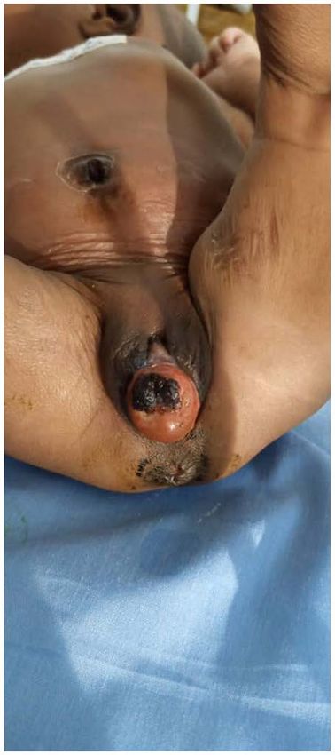
K. J. M. M. K. Quenum et al.
1. Introduction
Spina bifida, or spinal dysraphism, is a malformative disorder of the nervous sys-
tem resulting from an abnormality in the development of the nervous system, oc-
curring at an early stage of pregnancy. The neural tube (tubular outline of the
central nervous system) does not close properly and affects the development of
the spinal column and spinal cord. A neural tube closure defect is characterised
by damage to the nervous system and will generate so-called “associated” han-
dicaps, with consequences of varying degrees: neurological motor, sensory, in-
tellectual, bladder and bowel deficits with a significant impact on the quality of
life of these people. The exact cause is not known but the involvement of vitamin
B9 (or folic acid) taken in sufficient quantities reduces the risk. Certain medica-
tions during pregnancy increase the risk of dysraphism.
Genital prolapse is a rare entity in the newborn. An association with congenit-
al malformations of the central nervous system has been reported in the litera-
ture. We report the clinical observations of two full term newborns with cervical
prolapse and uterine prolapse in association with lumbar myelomeningocele,
without neuromuscular repercussions and treated at the Centre Hospital Un-
iversitaire et Départemental de Borgou Alibori (CHUD/BA) of Parakou.
OBJECTIVES: To report the clinical and therapeutic particularities of this rare
association through these two observations.
2. Patients and Method
CASE 1
This is a female newborn from a non-consanguineous marriage, in a primi-
parous, primigravida patient, 22 years old, without any particular pathological
history and having carried a pregnancy to term, irregularly monitored. The baby
was born by vaginal delivery, with a birth weight of 2420 g, and had an APGAR
score of 10/10. She was admitted to our facility on the fourth day of life with a
neonatal fever of 39 degrees. The physical examination revealed a dorsolumbar
swelling with ulceration of the skin in the centre and a tuft of hair on the cranial
part of the swelling. No sensory-motor deficits in the pelvic limbs were observed.
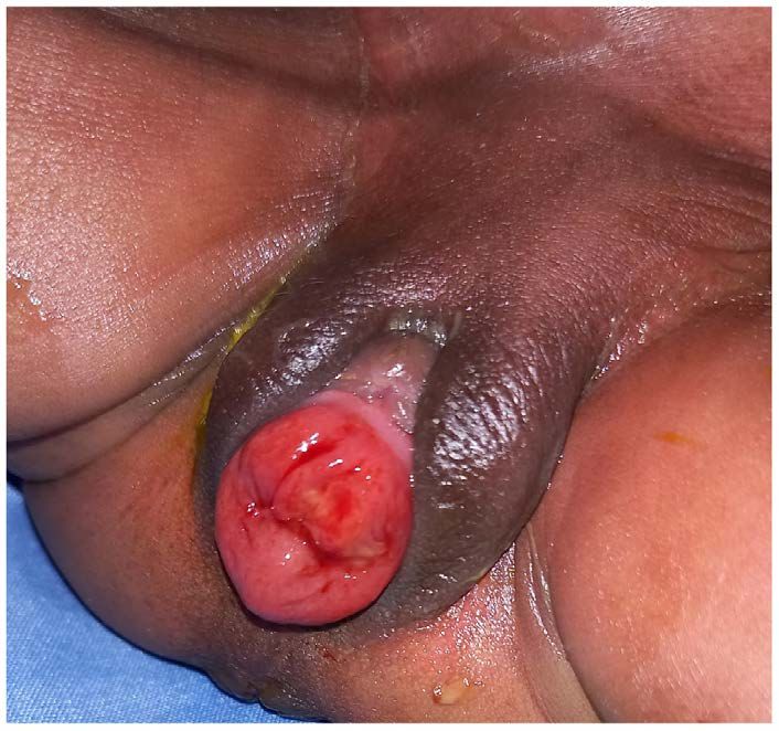
Examination of the perineum revealed a prolapse of the uterine cervix through
the vaginal orifice (Figure 1). The ureteral orifice was normal and the anal sphinc-
ter tone was preserved. The clinical malformative work-up concluded that the
baby had an incipient macrocrania with bulging anterior and posterior fonta-
nelles indicating incipient hydrocephalus. The archaic reflexes were preserved
with an axial tone present. Abdominal and pelvic ultrasound did not reveal any
abnormality apart from the absence of visualization of the uterus. In view of the
infectious clinical and biological syndrome with a positive cytobacteriological
examination of the cerebral spinal fluid, the diagnosis of meningitis caused by
staphylococcus was retained and a bi-antibiotic therapy was started. The CT
scan of the lumbar spine confirmed spina bifida on 2 levels T12 and L1. In addi-
tion to antibiotic therapy, a cure of the myelomeningocele (Figure 2) was per-
DOI: 10.4236/ojmn.2022.124023 217 Open Journal of Modern Neurosurgery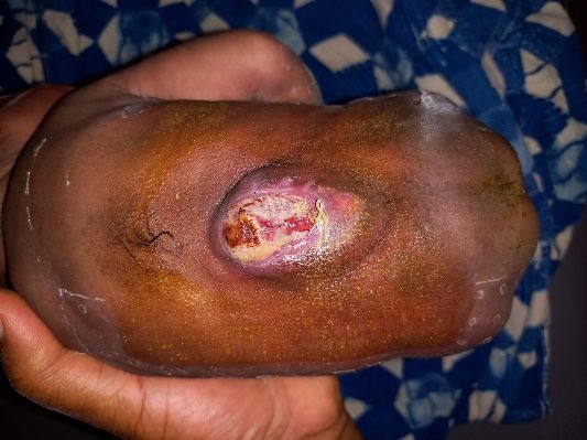
K. J. M. M. K. Quenum et al.
formed 3 weeks later. In emergency, a digital reduction of the prolapse uterin
was performed, with placement of a sterile pad for 48 hours. The evolution was
favourable without recurrence over a follow-up period of six months.
For the hydrocephalus, he underwent 1 month later to a ventriculo-peritoneal
shunt when the cerebral spinal fluid was steril with favourable results.
(a) (b)
Figure 1. A genital uterine prolapse (a) and after manual reduction, con-
trol after 48 hours (b).
(a) (b)
(c) (d)
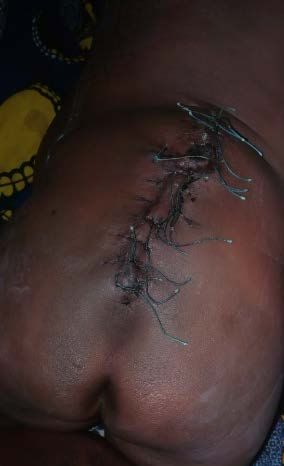
Figure 2. Thoraco lumbar spina bifida (a) with per operative dissection of
myelomeningocele (b). Post operative aspect (c). Aesthetic result after 3
months (d).
DOI: 10.4236/ojmn.2022.124023 218 Open Journal of Modern Neurosurgery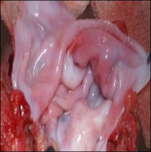
K. J. M. M. K. Quenum et al.

                                  CASE 2
                                  This is a female newborn, admitted at 2 days of age, born after an ongoing
                                pregnancy, for a genital anomaly associated with spina bifida. The perineal ex-
                                amination found a genital mass suggestive of uterine prolapse with the presence
                                of a crust on the cervix, a sign of mucosal dryness. No associated rectal prolapse
                                (Figure 3). The neurological examination did not reveal any motor deficit in the
                                pelvic limbs. The lumbar CT confirmed L1 spina bifida and the brain CT re-
                                turned normal. Additional CT scans of the abdomen did not reveal any in-
                                tra-abdominal malformations. She underwent emergency manual reduction of
                                the genital prolapse after moistening the cervix and disinfection to remove the
                                crust. A sterile tampon was kept in place for 48 hours. The evolution was favora-
                                ble, without recurrence of the genital prolapse. She was operated on for her spi-
                                na bifida with a favourable evolution and a satisfactory aesthetic result.

                                3. Discussion
                                Spina bifida is a congenital malformation with incomplete closure of the neural
                                tube (at the end of the first month of embryogenesis) followed by incomplete
                                closure of the last vertebrae. The frequency is currently 1/2000 births in France.
                                The highest frequency is found in English-speaking countries (up to 6/1000).
                                The favourable factors are now known are: genetic (PAX 3 gene) [1]; deficiency
                                (lack of vitamin B9 and zinc during the first month of pregnancy); metabolic
                                (insulin-dependent maternal diabetes); thermal (fever and baths that are too hot
                                during the first month of pregnancy). Antenatal detection by ultrasound is only
                                69% reliable. Significant progress is expected with three-dimensional images.
                                The consequences vary in severity. Some of them are underlying the malforma-
                                tion: sensory-motor paralysis of the lower limbs (from S2 to D12); orthopaedic
                                deformities (talus, knee flessum, hip dysplasia with a high risk of dislocation for

                                             (a)                  (b)                          (c)

                                   Figure 3. (a) Uterine prolapse with dryness crust. (b) Image after manual reduc-
                                   tion. (c) Thoraco lumbar spina bifida.

DOI: 10.4236/ojmn.2022.124023                        219                        Open Journal of Modern Neurosurgery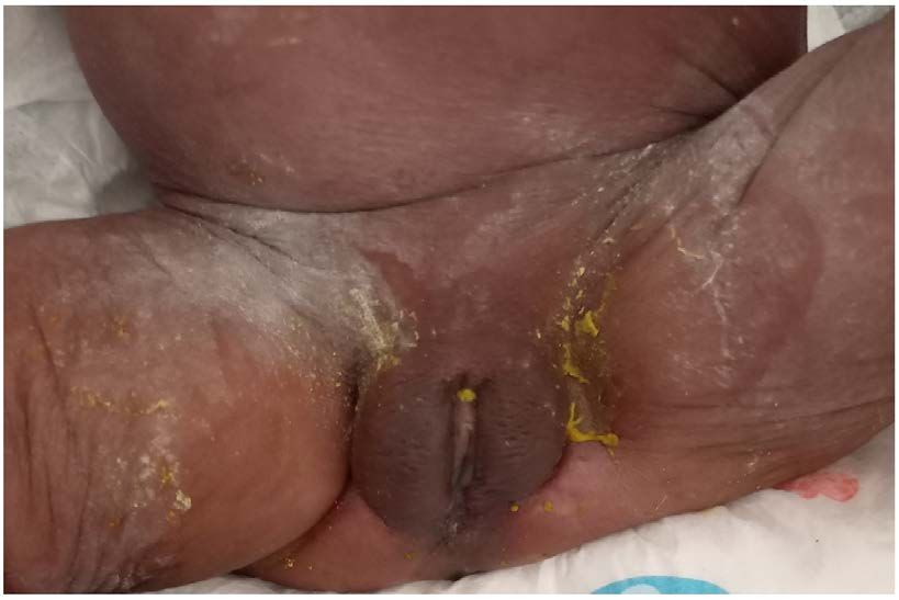
K. J. M. M. K. Quenum et al.

                                the L4 level, hyperlordosis, scoliosis) [1] [2]; osteoporosis with a risk of sponta-
                                neous fracture of the lower limbs; bladder and bowel disorders (incontinence
                                and retention); genito-sexual disorders (especially in men). Others are overlying
                                the malformation: hydrocephalus (often requiring early shunting) [3] [4]; Ar-
                                nold-Chiari type II malformation (often affecting higher functions and vision).
                                The others are of a general nature: multiple allergies (particularly to latex); en-
                                docrine disorders (ectopic testicle, advanced puberty in girls); overweight. In the
                                two cases reported, the newborns presented genital prolapses with the first child
                                developing early hydrocephalus and an infectious complication requiring ap-
                                propriate antibiotic therapy; for the hydrocephalus, she benefited from a ventri-
                                culoperitoneal shunt and a cure for myelomeningocele [5].
                                  The management of these “congenital hydrocephalic paraplegics” is complex,
                                multidisciplinary and long-term [5] [6] [7]. There is a need for regular follow-up
                                in neurosurgery [5], orthopaedics and urology, and for rehabilitation sessions
                                [1] [8]. We insist on the quality of the management of vesico-renal problems,
                                whatever the motor level [9] [10] [11], because they condition the functional and
                                then vital prognosis of these patients in the long term. For the second newborn,
                                after manual reduction of the prolapse and surgical cure of her myelomeningo-
                                cele, the outcome was favourable without recurrence of the prolapse.

                                4. Conclusion
                                Newborn uterine prolapse is an exceptional complication associated with spina
                                bifida. The treatment is manual reduction of the prolapse structure with good
                                outcome, followed by myelomeningocele surgery. Acid folic taking during the
                                period before and the first month of pregnancy must be an important recom-
                                mendation for spina bifida prevention.

                                Ethical Aspect
                                Parental consent has obtained for the use of patient data, and no image allows
                                identification.

                                Conflicts of Interest
                                The authors declare no conflicts of interest regarding the publication of this pa-
                                per.

                                References
                                [1]    Barber, M.D. and Maher, C. (2013) Epidemiology and Outcome Assessment of Pel-
                                       vic Organ Prolapse. International Urogynecology Journal, 24, 1783-1790.
                                       https://doi.org/10.1007/s00192-013-2169-9
                                [2]    Mukenge, T., Balde, F., Benmassaoud, Z., Oualili, I., Alaoui, O., Mahmoudi, A.,
                                       Khattala, K. and Bouabdallah, Y. (2021) Uterine Prolapse: The Other Exceptional
                                       Complication of Spina Bifida in Newborns. Open Journal of Pediatrics, 11, 50-54.
                                       https://doi.org/10.4236/ojped.2021.111005
                                [3]    Swift, S., Woodman, P., O’Boyle, A., Kahn, M., Valley, M., Bland, D., et al. (2005)

DOI: 10.4236/ojmn.2022.124023                          220                         Open Journal of Modern Neurosurgery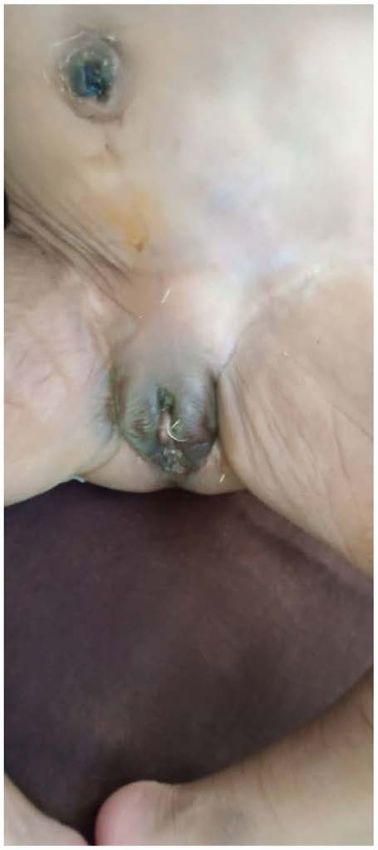
K. J. M. M. K. Quenum et al.

                                       Pelvic Organ Support Study (POSST): The Distribution, Clinical Definition, and
                                       Epidemiologic Condition of Pelvic Organ Support Defects. American Journal of
                                       Obstetrics & Gynecology, 192, 795-806. https://doi.org/10.1016/j.ajog.2004.10.602
                                 [4]   Baskaran, D. and Mohan, P. and Nazeeb (2012) Purse String Suturing in a Neonatal
                                       Prolapsed Uterus. Indian Journal of Surgery, 74, 143-145.
                                       https://doi.org/10.1007/s12262-011-0361-z
                                 [5]   McCarty, D.J., Sheinberg, D.L., Luther, E. and McCrea, H.J. (2019) Myelomeningo-
                                       cele Associated Hydrocephalus: Nationwide Analysis and Systematic Review. Jour-
                                       nal of Neurosurgery, 47, E5. https://doi.org/10.3171/2019.7.FOCUS19469
                                 [6]   Abdelsalam, S.E.A., Desouki, N.M. and Alaal, N.A.A. (2006) Use of Foley Catheter
                                       for Management of Neonatal Genital Prolapse: Case Report and Review of the Lite-
                                       rature. Journal of Pediatric Surgery, 41, 449-452.
                                       https://doi.org/10.1016/j.jpedsurg.2005.11.031
                                 [7]   Jijo, Z.W., Betele, M.T. and Ali, A.S. (2018) Congenital Uterovaginal Prolapse in a
                                       Newborn. Case Reports in Obstetrics and Gynecology, 2018, Article ID: 1425953.
                                       https://doi.org/10.1155/2018/1425953
                                 [8]   Choudhary, S.V., Bisati, S. and Koley, S. (2011) Congenital Cutis Laxa with Rectal
                                       and Uterovaginal Prolapsed. Indian Journal of Dermatology, Venereology and Le-
                                       prology, 77, 321-324. https://doi.org/10.4103/0378-6323.79706
                                 [9]   Hyginus, E.O. and John, C.O. (2013) Congenital Uterovaginal Prolapse Present at
                                       Birth. Journal of Surgical Technique and Case Report, 5, 89-91.
                                       https://doi.org/10.4103/2006-8808.128741
                                [10]   Fathi, A.P.K. (2008) Semiconservative Management of Neonatal Vaginal Prolapse.
                                       Journal of Pediatric Surgical Specialties, 2 p.
                                [11]   McGlone, L. and Patole, S. (2004) Neonatal Genital Prolapse. Journal of Paediatrics
                                       and Child Health, 40, 156-157. https://doi.org/10.1111/j.1440-1754.2004.00321.x

DOI: 10.4236/ojmn.2022.124023                          221                         Open Journal of Modern Neurosurgery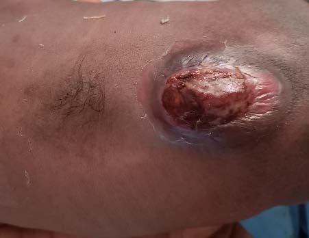
You can also read