Machine Learning for Cataract Classification/Grading on Ophthalmic Imaging Modalities: A Survey - arXiv
←
→
Page content transcription
If your browser does not render page correctly, please read the page content below
Noname manuscript No.
                                              (will be inserted by the editor)

                                          Machine Learning for Cataract Classification/Grading on Ophthalmic
                                          Imaging Modalities: A Survey
                                          Xiaoqing Zhang · Yan Hu · Zunjie Xiao · Jiansheng Fang · Risa Higashita · Jiang
                                          Liu ∗
arXiv:2012.04830v4 [eess.IV] 2 Apr 2022

                                          Received: date / Accepted: date

                                          Abstract Cataracts are the leading cause of visual impair-             Clinically, cataracts are the loss of crystalline lens trans-
                                          ment and blindness globally. Over the years, researchers have      parency, which occur when the protein inside the lens clumps
                                          achieved significant progress in developing state-of-the-art       together [3]. They are associated with many factors [103],
                                          machine learning techniques for automatic cataract classi-         such as developmental abnormalities, trauma, metabolic dis-
                                          fication and grading, aiming to prevent cataracts early and        orders, genetics, drug-induced changes, ages, etc. Genetics
                                          improve clinicians’ diagnosis efficiency. This survey pro-         and aging are two of the most important factors for cataracts.
                                          vides a comprehensive survey of recent advances in ma-             According to the causes of cataracts, they can be catego-
                                          chine learning techniques for cataract classification/grading      rized as age-related cataract, pediatrics cataract (PC), and
                                          based on ophthalmic images. We summarize existing liter-           secondary cataract [3, 103]. According to the location of the
                                          ature from two research directions: conventional machine           crystalline lens opacity, they can be grouped into nuclear
                                          learning methods and deep learning methods. This survey            cataract (NC), cortical cataract (CC), and posterior subcap-
                                          also provides insights into existing works of both merits          sular cataract (PSC) [94, 96]. NC denotes the gradual cloud-
                                          and limitations. In addition, we discuss several challenges of     ing and the progressive hardening in the nuclear region. CC
                                          automatic cataract classification/grading based on machine         is the form of white wedged-shaped and radially oriented
                                          learning techniques and present possible solutions to these        opacities, and it develops from the outside edge of the lens
                                          challenges for future research.                                    toward the center in a spoke-like fashion [103, 19]. PSC is
                                                                                                             granular opacities, and its symptom includes small bread-
                                          Keywords Cataract, classification and grading, ophthalmic
                                                                                                             crumbs or sand particles, which are sprinkled beneath the
                                          image, machine learning, deep learning
                                                                                                             lens capsule [96].
                                                                                                                 Over the past years, ophthalmologists have used several
                                          1 Introduction                                                     ophthalmic images to diagnose cataract based on their expe-
                                                                                                             rience and clinical training. This manual diagnosis mode is
                                          According to World Health Organization (WHO) [5, 113], it          error-prone, time-consuming, subjective, and costly, which
                                          is estimated that approximately 2.2 billion people suffer vi-      is a great challenge in developing countries or rural com-
                                          sual impairment. Cataract accounts for about 33% of visual         munities, where experienced clinicians are scarce. To pre-
                                          impairment and is the number one cause of blindness (over          vent cataract early and improve the precision and efficiency
                                          50%) worldwide. Cataract patients can improve life quality         of cataract diagnosis, researchers have made great efforts in
                                          and vision through early intervention and cataract surgery,        developing computer-aided diagnosis (CAD) techniques for
                                          which are efficient methods to reduce blindness ratio and          automatic cataract classification/grading [104] on different
                                          cataract-blindness burden for society simultaneously.              ophthalmic images, including conventional machine learn-
                                          ∗   denotes corresponding author                                   ing methods and deep learning methods. The conventional
                                                                                                             machine learning method is a combination of feature ex-
                                          XQ Zhang, ZJ Xiao, Y Hu, JS Fang, R Higashita · J Liu
                                          Research Institute of Trustworthy Autonomous Systems and Depart-
                                                                                                             traction and classification/grading. In the feature extraction
                                          ment of Computer Science and Engineering, Southern University of   stage, a variety of image processing methods have been pro-
                                          Science and Technology, Shenzhen, China                            posed to obtain visual features of cataract according to dif-
                                          E-mail: liuj@sustech.edu.cn                                        ferent ophthalmic images, such as density-based statistics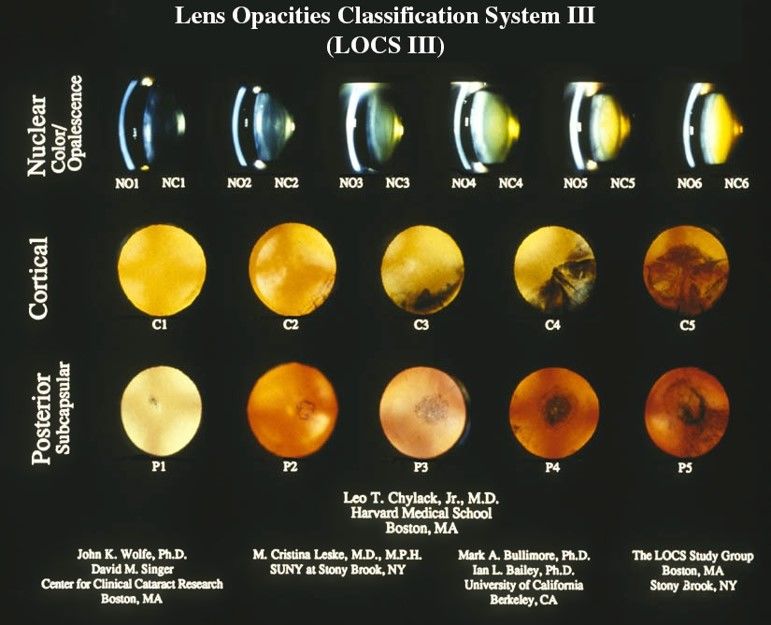
2                                                                                                                                                                                                 Short form of author list

                                                                                Cataract Classification and Grading on Ophthalmic Images

                                    Ophthalmic image                                  Grading System                                               Methodological tasks                           Cataract types

                                                                                                                                                                                            NC                     CC
              Slit lamp image                           Fundus image      LOCS                              WGS               Methodologies

                                                                                                                                                                                            PSC                    PCO
          Retroillumination image                      Ultrasonic image   JHS                            OCCCGS

                                                                                                                                 Classification/Grading             Feature extration

           Digital Cameral image                       AS-OCT image       FCS                              WHO

                                                                                                                                     Segmentation                     Localization

                                                                                                                               Clinical Tasks

                                                                                                                               Computer-aided diagnosis               Intervention

                                                                                                                                       Screaning                Cataract surgery planning

Fig. 1 Overall organization framework of this survey.

method, density histogram method, bag-of-features (BOF)                                                                 cording to collected papers, our summary, and discussion
method, Gabor Wavelet transform, Gray level Cooccurrence                                                                with experienced ophthalmologists. To understand this sur-
Matrix (GLCM), Haar wavelet transform, etc [71, 168, 96,                                                                vey easily, we also review ophthalmic imaging modalities,
167, 42, 136, 115, 39, 114]. In the classification/grading                                                              cataract grading systems, and commonly-used evaluation mea-
stage, strong classification methods are applied to recog-                                                              sures in brief. Then we introduce ML techniques step by
nize different cataract severity levels, e.g., support vector                                                           step. We hope this survey can provide a valuable summary
machine (SVM) [87, 170, 122]. Over the past ten years,                                                                  of current works and present potential research directions of
deep learning has achieved great success in various fields,                                                             ML-based cataract classification/grading in the future.
including medical image analysis, which can be viewed as
a representation learning approach. It can learn low-, mid-,
and high-level feature representations from raw data in an                                                              2 Ophthalmic imaging modalities for cataract
end-to-end manner (e.g., ophthalmic images). In the recent,                                                             classification/grading
various deep neural networks have been utilized to tackle
                                                                                                                        To our best understanding, this survey introduces six differ-
cataract classification/grading tasks like convolutional neu-
                                                                                                                        ent eye images used for cataract classification/grading for
ral networks (CNNs), attention-based networks, Faster-RCNN
                                                                                                                        the first time: slit-lamp image, retroillumination image, ul-
and multilayer perceptron neural networks (MLP). E.g., Zhang
                                                                                                                        trasonic image, fundus image, digital camera image, and an-
et al. [185] proposed a multi-region fusion attention network
                                                                                                                        terior segment optical coherence tomography (AS-OCT) im-
to recognize nuclear cataract severity levels.
                                                                                                                        age, as shown in Fig. 2. In the following section, we will
     Previous surveys had summarized cataract types, cataract
                                                                                                                        introduce each ophthalmic image type step by step and then
classification/grading systems, and ophthalmic imaging modal-
                                                                                                                        discuss their advantages and disadvantages.
ities, respectively [186, 103, 129, 105, 174, 41, 45]; how-
ever, none had summarized ML techniques based on oph-
thalmic imaging modalities for automatic cataract classifica-                                                           2.1 Slit lamp image
tion/ grading systematically. To the best of our knowledge,
this is the first survey that systematically summarizes recent                                                          The slit lamp camera [35, 154] is a high-intensity light source
advances in ML techniques for automatic cataract classifi-                                                              instrument, which is comprised of the corneal microscope
cation/ grading. This survey mainly focuses on surveying                                                                and the slit lamp. Silt lamp image can be accessed through
ML techniques in cataract classification/grading, comprised                                                             slit lamp camera, which is usually used to examine the an-
of conventional ML methods and deep learning methods.                                                                   terior segment and posterior segment structure of the hu-
We survey these published papers through Web of Science                                                                 man eye eyelid, sclera, conjunctiva, iris, crystalline lens, and
(WoS), Scopus, and Google Scholar databases. Fig. 1 pro-                                                                cornea. Fig. 3 offers four representative slit lamp images for
vides a general organization framework for this survey ac-                                                              four different cataract severity levels.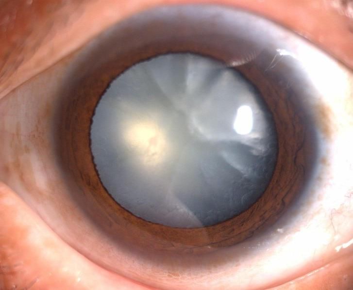
Machine Learning for Cataract Classification/Grading on Ophthalmic Imaging Modalities: A Survey 3
(a) (b) (c)
Fig. 4 Four cataract severity levels on fundus images [10]. (a) Normal;
(b) Immature; (c) Mature; (d) Hypermature
(d) (e) (f) 2.4 Fundus image
Fig. 2 Six different ophthalmic images. (a) Slit lamp image; (b) The fundus camera [117, 118] is a unique camera in con-
Retroillumination image; (c) Ultrasonic image; (d) Fundus image (e) junction with a low power microscope, which is usually used
Digital camera image (f) Anterior segment optical coherence tomogra- to capture fundus images operated by ophthalmologists or
phy image.
professional operators. Fundus image is a highly specialized
form of eye imaging and can capture the eye’s inner lining
and the structures of the back of the eye. Fig. 4 shows four
fundus images of different cataract severity levels.
2.5 Digital camera image
Fig. 3 Slit lamp images with four nuclear cataract severity levels.
Commonly used digital cameras can access digital camera
images like smartphone cameras. Compared with the fun-
2.2 Retroillumination image
dus camera and slit lamp device, the digital camera is eas-
ily available and easily used. Hence, using digital cameras
Retroillumination image is a non-stereoscopic medical im-
for cataract screening has great potential in the future, espe-
age, which is accessed through the crystalline lens camera
cially for developing countries and rural areas, where people
[153, 44]. It can be used to diagnose CC and PSC in the
have limitations to access expensive ophthalmology equip-
crystalline lens region. Two types of retroillumination im-
ment and experienced ophthalmologists.
ages through the crystalline lens camera can be obtained: an
anterior image focused on the iris, which corresponds to the
anterior cortex of the lens, and a posterior image focused on
2.6 Anterior segment optical coherence tomography image
3-5mm more posteriorly, which intends to image the opacity
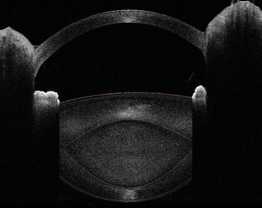
of PSC. Anterior segment optical coherence tomography (AS-OCT)
[2] imaging technique is one of optical coherence tomog-
raphy (OCT) imaging techniques. It can be used to visual-
ize and assess anterior segment ocular features, such as the
2.3 Ultrasonic image tear film, cornea, conjunctiva, sclera, rectus muscles, ante-
rior chamber angle structures, and lens [157, 61, 169, 62].
In clinical cataract diagnosis, Ultrasound image is a commonly- AS-OCT image can provide high-resolution visualization of
used ophthalmic image modality to evaluate the hardness the crystalline lens in vivo in the eyes of people in real-time
of cataract lens objectively [69]. Frequently applied Ultra- without impacting the tissue, which can help ophthalmolo-
sound imaging techniques usually are developed based on gists get different information of the crystalline lens through
measuring ultrasonic attenuation and sound speed, which the circumferential scanning mode. Recent works have sug-
may increase the hardness of the cataract lens [68]. High- gested that the AS-OCT images can be used to locate the
frequency Ultrasound B-mode imaging can be used to mon- lens region and accurately characterize opacities of different
itor local cataract formation, but it cannot measure the lens cataract types quantitatively [49, 116]. Fig. 5 offers an AS-
hardness accurately [148]. To make up for the B-scan de- OCT image, which can quickly help us know the crystalline
ficiency, the Ultrasound imaging technique built on Nak- lens structure.
agami statistical model called Ultrasonic Nakagami imag- Discussion: Though six different ophthalmic images are
ing [150, 149, 151] was developed, which can be used for used for cataract diagnosis, slit lamp images and fundus im-
the visualization of local scatterer concentrations in biolog- ages are the most commonly-used ophthalmic images for
ical tissues. clinical cataract diagnosis and scientific research purposes.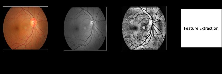
4                                                                                                                         Short form of author list

                                                                         tems for clinical practice and scientific research purposes.
                                                                         This section briefly introduces six existing cataract classifi-
                                                                         cation/grading systems.

                                                                         3.1 Lens opacity classification system

                             Cortical region                             Lens opacity classification system (LOCS) was first intro-
                                                                         duced in 1988, which has developed from LOCS I to LOCS
                                                                         III [24, 22, 23]. LOCS III is widely used for clinical di-
                             Nuclear region
                                                                         agnosis and scientific research. In the LOCS III, as shown
                                                                         in Fig. 6, six representative slit lamp images for nuclear
                                                Posterior subcapsular    cataract grading based on nuclear color and nuclear opales-
                                                       region            cence; five representative retroillumination images for cor-
                                                                         tical cataract grading; five representative retroillumination
Fig. 5 AS-OCT image. nuclear region is used for NC diagnosis; cor-       images for grading posterior subcapsular cataract. The cataract
tical region is used for CC diagnosis; posterior subcapsular region is
used for PSC diagnosis.                                                  severity level is graded on a decimal scale by spacing inter-
                                                                         vals regularly.

This is because existing cataract classification/grading sys-
tems are built on them. Slit lamp images can capture the
lens region but cannot distinguish the boundaries between
nuclear, cortical, and capsular regions. Hence, it is difficult
for clinicians to diagnose different cataract types accurately
based on slit lamp images. Fundus images only contain opac-
ity information of cataract and do not contain location in-
formation of cataract, which is mainly applied to cataract
screening.
    Retroillumination images are usually used to diagnose
CC and PSC clinically, which have not been widely stud-
ied. Digital camera images are ideal ophthalmic images for
cataract screening because they can be collected through
mobile phones, which are easy and cheap for most peo-
ple. Like fundus images, digital camera images only have
opacity information of cataract but do not contain location
information of different cataract types. Ultrasonic images
can capture the lens region and evaluate the hardness of                 Fig. 6 Lens opacity classification system III.
the cataract lens, but they cannot distinguish different sub-
regions, e.g., cortex region. AS-OCT image is a new oph-
thalmic image that can distinguish different sub-regions, e.g.,
cortex and nucleus regions, significant for cataract surgery             3.2 Wisconsin grading System
planning and cataract diagnosis. However, there is no cataract
classification/grading system built on AS-OCTs; thus, it is              Wisconsin grading system was proposed by the Wisconsin
urgent to develop a clinical cataract classification/grading             Survey Research Laboratory in 1990 [88, 48, 31]. It con-
system based on AS-OCT images. Moreover, existing auto-                  tains four standard photographs for grading cataract severity
matic AS-OCT image-based cataract classification has been                levels. The grades for cataract severity levels are as follows:
rarely studied.                                                          grade 1, as clear or clearer than Standard 1; grade 2, not as
                                                                         clear as Standard 1 but as clear or clearer than Standard 2;
                                                                         grade 3, not as clear as Standard 2 but as clear or clearer
3 Cataract classification/grading systems                                than Standard 3; grade 4, not as clear as Standard 3 but as
                                                                         clear or clearer than Standard 4; grade 5, at least as severe as
To classify or grade the severity levels of cataract (lens opac-         Standard 4; and grade 6, 7 and 8, cannot grade due to severe
ities) accurately and quantitatively, it is crucial and neces-           opacities of the lens (please see detail introduction of Wis-
sary to build standard/gold cataract classification/grading sys-         consin grading system in literature [88]). Wisconsin grading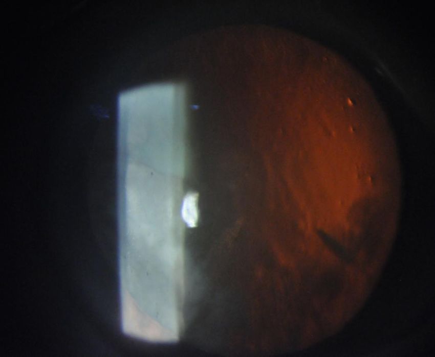
Machine Learning for Cataract Classification/Grading on Ophthalmic Imaging Modalities: A Survey                                    5

system also uses a decimal grade for cataract grading with             blurred; grade 2: the larger branches of retinal vein or artery
0.1-unit interval space, and the range of the decimal grade is         were blurred; grade 3: the optic disc region was blurred;
from 0.1 to 4.9.                                                       grade 4: The whole fundus image was blurred.
                                                                           Discussion: From the above-mentioned six existing cataract
                                                                       classification/grading systems, we can conclude that five cataract
3.3 Oxford clinical cataract classification and grading                classification systems are built on slit lamp images, and one
system                                                                 is built on fundus images, which can explain that most exist-
                                                                       ing cataract works based on these two ophthalmic imaging
Oxford Clinical Cataract Classification and Grading System             modalities. However, these cataract classification/grading sys-
(OCCCGS) is also a slit-lamp image-based cataract grad-                tems are subjective due to the limitations of these two imag-
ing system [135, 57]. Different to the LOCS III uses photo-            ing devices. Furthermore, to improve the precision of cataract
graphic transparencies of the lens as cataract grading stan-           diagnosis and the efficiency of cataract surgery, it is neces-
dards, it adopts standard diagrams and Munsell color sam-              sary to develop new and objective cataract classification sys-
ples to grade the severity of cortical, posterior subcapsu-            tems on other ophthalmic image modalities, e.g., AS-OCT
lar, and nuclear cataract [57]. In the OCCCGS, five stan-              images.
dard grading levels are used for evaluating the severity level
of cataract based on cataract features, such as cortical fea-
tures, nuclear features, morphological features, etc. [135].
E.g., the severity levels of nuclear cataract are graded as            4 Datasets
follows: Grade 0: No yellow detectable; Grade 1: yellow
just detectable; Grade 2: definate yellow; Grade 3: orange             In this section, we introduce ophthalmic image datasets used
yellow; Grade 4: reddish brown; Grade 5: blackish brown                for cataract classification/grading, which can be grouped in
[135].                                                                 private datasets and public datasets.

3.4 Johns Hopkins system
                                                                       4.1 Private datasets
Johns Hopkins system (JHS) was first proposed in 1980s
                                                                       ACHIKO-NC dataset [101]: ACHIKO-NC is the slit-lamp
[158]. It has four standard silt lamp images, which denotes
                                                                       lens images dataset selected from the SiMES I database,
the severity level of cataract based on the opalescence of the
                                                                       used to grade nuclear cataracts. It comprised 5378 images
lens. For nuclear cataract, Grade 1: opacities that are defi-
                                                                       with decimal grading scores (0.3 to 5.0). Professional clin-
nitely present but not thought to reduce visual acuity; Grade
                                                                       icians determine the grading score of each slit lamp image.
2: opacities are consistent with visual acuity between 20/20
                                                                       ACHIKO-NC is a widely used dataset for automatic nuclear
and 20/30; Grade 3 opacities are consistent with vision be-
                                                                       cataract grading according to existing works.
tween 20/40 and 20/100; Grade 4: opacities are consistent
                                                                           ACHIKO-Retro dataset [101]: ACHIKO-Retro is the
with the vision of 20/200 or less.
                                                                       retro-illumination lens image dataset selected from SiMES
                                                                       I database, used to grade CC and PSC. Each lens has two
3.5 WHO cataract grading system                                        eye image types: anterior image and posterior image. The
                                                                       anterior image focuses on the plane centered in the anterior
WHO cataract grading system was developed by a group                   cortex region, and the posterior image focuses on the pos-
of experts in WHO [144, 4]. The target to develop it is to             terior capsule region. Most previous CC and PSC grading
enable relatively inexperienced observers to grade the most            works were conducted on the ACHIKO-Retro dataset.
common types of cataracts reliably and efficiently. It uses                CC-Cruiser dataset [80]: CC-Cruiser is the slit lamp
four severity levels for grading NC, CC, and PSC based on              image dataset collected from Zhongshan Ophthalmic Center
four standard images accordingly.                                      (ZOC) of Sun Yat-Sen University, which is used for cataract
                                                                       screening. It is comprised of 476 normal images and 410
                                                                       infantile cataract images.
3.6 Fundus image-based cataract classification system                      Multicenter dataset [80]: Multicenter is the slit lamp
                                                                       image dataset, which is comprised of 336 normal images
Xu et al. [165] proposed a fundus image-based cataract clas-           and 421 infantile cataract images. It was collected from four
sification system (FCS) through observing the blur level.              clinical institutions: the Central Hospital of Wuhan, Shen-
They used five levels to evaluate the blur levels on fundus            zhen Eye Hospital, Kaifeng Eye Hospital, and the Second
images: grade 0: clear; grade 1: the small vessel region was           Affiliated Hospital of Fujian Medical University.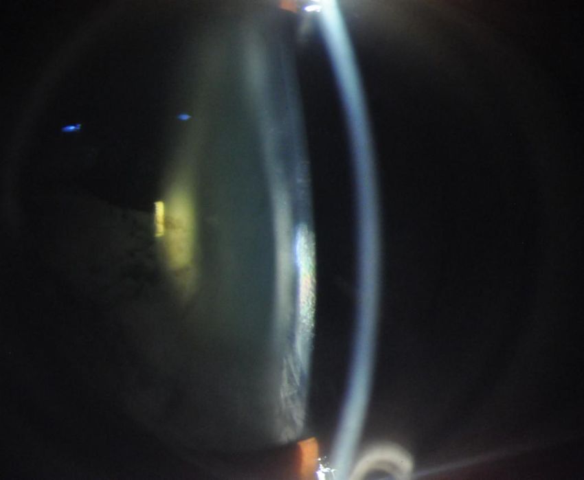
6                                                                                                                Short form of author list

4.2 Public datasets                                                      Slit lamp image: The procedures to extract features from
                                                                    slit lamp images are comprised of the lens structure detec-
EyePACS dataset [25]: EyePACS is the fundus image dataset           tion and feature extraction.
collected from EyePACS, LLC , a free platform for retinopa-
thy screening, used to classify different levels of cataract. It
is made available by California Healthcare Foundation. The
dataset comprises 88,702 fundus retinal images in which
1000 non-cataract images and 1441 cataract images are pro-
vided.
    HRF dataset [120]: The high-resolution fundus (HRF)
image database is selected from different open-access datasets:
structured analysis of the retina (STARE) [63], standard di-
abetic retinopathy database (DIARETDB0) [86], e-ophtha              Fig. 8 The procedures to extract image features from slit lamp images
[26], methods to evaluate segmentation and indexing tech-           for automatic nuclear cataract classification/grading.
niques in the field of retinal ophthalmology (MESSIDOR)
database [27], digital retinal images for vessel extraction
                                                                            Fig. 8 offers a representative slit lamp image-based fea-
(DRIVE) database [137], fundus image registration (FIRE)
                                                                       ture extraction flowchart. Firstly, according to the histogram
[60] dataset, digital retinal images for optic nerve segmenta-
                                                                       analysis results of the lens, the foreground of the lens is de-
tion database (DRIONS-DB) [12], Indian diabetic retinopa-
                                                                       tected by setting the pixel thresholding, and the background
thy image dataset (IDRiD) [119], available datasets released
                                                                       of the lens is even based on slit lamp images. Afterward, we
by Dr. Hossein Rabbani [108], and other Internet sources.
                                                                       analyze the profile on the horizontal median line of the im-
                                                                       age. The largest cluster on the line is detected as the lens,
                                                                       and the centroid of the cluster is detected as the horizontal
5 Machine learning techniques
                                                                       coordinate of the lens center. Then, we get the profile on
This section mainly investigates on ML techniques for cataract the vertical line through the point. Finally, the center of the
classification/grading over the years, which is comprised of           lens is estimated, and the lens can be further estimated as an
conventional ML methods and deep learning methods.                     ellipse with the semi-major axis radius estimated from the
                                                                       horizontal and vertical profile.
                                                                            The lens contour or shape needs to be captured by fol-
5.1 Conventional machine learning methods                              lowing   the lens location. Researchers commonly used the
                                                                       active shape model (ASM) method for the lens contour de-
Over the past years, scholars have developed massive state-            tection [95, 96, 93] and achieved 95% accuracy of the lens
of-the-art conventional ML methods to automatically clas-              structure detection. The ASM can describe the object shape
sify/grade cataract severity levels, aiming to assist clinicians       through an iterative refinement procedure to fit an example
in diagnosing cataract efficiently and accurately. These meth-         of the object into a new image based on the statistical mod-
ods consist of feature extraction and classification /grading,         els [91]. Based on the detected lens contour, many feature
as shown in Fig. 7. Table 1 summarizes conventional ML                 extraction methods have been proposed to extract informa-
methods for cataract classification/grading based on differ-           tive features, such as bag-of-features (BOF) method, grad-
ent ophthalmic images.                                                 ing protocol-based method, semantic reconstruction-based
                                                                       method, statistical texture analysis [71, 168, 96, 167, 42,
                                                                       136], etc.
                                                                            Retroillumination image: It also consists of two stages
                                                                       for feature extraction based on retroillumination images: pupil
Fig. 7 Flowchart of conventional machine learning based classification
                                                                       detection and opacity detection [93, 96, 21, 42], as shown in
and grading.                                                           Fig. 9. Researchers usually use a combination of the Canny
                                                                       edge detection method, the Laplacian method, and the con-
                                                                       vex hull method to detect the edge pixel in the pupil detec-
                                                                       tion stage. The non-linear least-square fitting method is used
5.1.1 Feature extraction                                               to fit an ellipse based on the detected pixels. In the opacity
                                                                       detection stage, the input image is transformed into the po-
Considering the characteristics of different imaging tech-             lar coordinate at first. Based on the polar coordinate, classi-
niques and cataract types, we introduce feature extraction             cal image processing methods are applied to detect opacity
methods based on ophthalmic image modalities.                          such as global threshold, local threshold, edge detection, and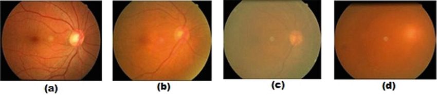
Machine Learning for Cataract Classification/Grading on Ophthalmic Imaging Modalities: A Survey                               7

Table 1 Conventional ML methods for cataract classification/grading based on ophthalmic images.
                   Literature            Method                     Image Type         Year    Application Cataract Type
                      [94]             ASM + SVR                 Slit Lamp Image       2009     Grading         NC
                      [71]           ASM + Ranking               Slit Lamp Image       2009     Grading         NC
                      [95]             ASM + SVR                 Slit Lamp Image       2010     Grading         NC
                      [72]           ASM + Ranking               Slit Lamp Image       2010     Grading         NC
                      [91]              ASM + LR                 Slit Lamp Image       2007     Grading         NC
                     [168]             SF + SWLR                 Slit Lamp Image       2016     Grading         NC
                     [167]             BOF + GSR                 Slit Lamp Image       2013     Grading         NC
                       [8]                SVM                    Slit Lamp Image       2016     Grading         NC
                      [78]                SVM                    Slit Lamp Image       2017     Grading         NC
                     [155]                SVM                    Slit Lamp Image       2017     Grading         NC
                     [136]                 IGB                   Slit Lamp Image       2014     Grading         NC
                      [78]                  RF                   Slit Lamp Image       2017   Classification    NC
                      [16]                SRCL                   Slit Lamp Image       2018     Grading         NC
                      [76]      Hough Circular Transform         Slit Lamp Image       2019   Classification    NC
                      [85]                  RF                   Slit Lamp Image       2019   Classification    PC
                      [85]                  NB                   Slit Lamp Image       2019   Classification    PC
                      [96]        Canny + Spatial Filter     Retroillumination Image   2010   Classification   PSC
                      [93]              EF + PCT             Retroillumination Image   2008   Classification   PSC
                      [21]                Canny              Retroillumination Image   2011   Classification   PSC
                     [179]                MRF                Retroillumination Image   2017   Classification   PSC
                      [42]                 LDA               Retroillumination Image   2011   Classification PSC & CC
                      [92]    Radial-edge & Region-growing   Retroillumination Image   2008   Classification    CC
                      [13]                 PCA                  Ultrasonic Image       2015   Classification  Cataract
                      [13]                Bayes                 Ultrasonic Image       2015   Classification  Cataract
                      [13]                KNN                   Ultrasonic Image       2015   Classification  Cataract
                      [13]                SVM                   Ultrasonic Image       2015   Classification  Cataract
                      [13]                 FLD                  Ultrasonic Image       2015   Classification  Cataract
                       [8]                SVM                   Ultrasonic Image       2016   Classification  Cataract
                       [8]                  RF                  Ultrasonic Image       2016   Classification  Cataract
                       [8]            Bayes Network             Ultrasonic Image       2016   Classification  Cataract
                       [7]             PCA + SVM                Ultrasonic Image       2014   Classification  Cataract
                       [6]     Nakagami Distribution +CRT       Ultrasonic Image       2014   Classification  Cataract
                      [77]                SVM                   Ultrasonic Image       2013   Classification  Cataract
                      [39]            GLCM + KNN              Digital Camera Image     2015   Classification  Cataract
                      [40]            GLCM + KNN              Digital Camera Image     2015   Classification  Cataract
                     [114]               K-Means              Digital Camera Image     2016   Classification  Cataract
                     [115]                 IMF                Digital Camera Image     2016   Classification  Cataract
                      [87]                SVM                 Digital Camera Image     2016   Classification  Cataract
                      [53]             WT + MDA                   Fundus Image         2015   Classification  Cataract
                     [170]            Wavelet-SVM                 Fundus Image         2015   Classification  Cataract
                     [170]            Texture-SVM                 Fundus Image         2015   Classification  Cataract
                     [170]               Stacking                 Fundus Image         2015   Classification  Cataract
                      [32]           PCA + Bagging                Fundus Image         2015   Classification  Cataract
                      [32]              PCA + RF                  Fundus Image         2015   Classification  Cataract
                     [175] Multi-feature Fusion&Stacking          Fundus Image         2019   Classification  Cataract
                      [32]            PCA + GBDT                  Fundus Image         2015   Classification  Cataract
                      [32]             PCA + SVM                  Fundus Image         2015   Classification  Cataract
                      [10]        Haar Wavelet + Voting           Fundus Image         2019   Classification  Cataract
                     [122]             SVM + GA                   Fundus Image         2017   Classification  Cataract
                      [74]            AWM + SVM                   Fundus Image         2019   Classification  Cataract
                     [133]                  DT                    Fundus Image         2016   Classification  Cataract
                     [133]          Bayesian Network              Fundus Image         2016   Classification  Cataract
                     [134]             DWT+SVM                    Fundus Image         2019   Classification  Cataract
                     [134]                 SSL                    Fundus Image         2019   Classification  Cataract
                     [183]                  RF                   AS-OCT image          2021   Classification    NC
                     [183]                SVM                    AS-OCT image          2021   Classification    NC

region growing. Apart from the above methods, literature                  Ultrasound Images & Digital Camera Images & AS-
[179] uses Watershed and Markov random fields (MRF) to                 OCT Images: For ultrasound images, researchers adopt the
detect the lens opacity, and results showed that the proposed          Fourier Transform (FT) method, textural analysis method,
framework got competitive results of PSC detection.                    and probability density to extract features [8, 7, 6].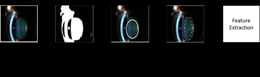
8                                                                                                              Short form of author list

                                                                     5.1.2 Classification & grading

                                                                         In this section, we mainly introduce conventional machine
                                                                         learning methods for cataract classification/grading.
                                                                              Support vector machine: Support vector machine (SVM)
                                                                         is a classical supervised machine learning technique, which
                                                                         has been widely be used for classification and linear regres-
                                                                         sion tasks. It is a popular and efficient learning method for
                                                                         medical imaging applications. For the cataract grading task,
                                                                         Li et al. [94, 95] utilized support vector machine regression
                                                                         (SVR) to grade the severity level of cataract and achieved
                                                                         good grading results based on slit lamp images. The SVM
                                                                         classifier is widely used in different ophthalmic images for
                                                                         the cataract classification task. E.g., literature [78, 155] ap-
                                                                         plies SVM to classify cataract severity levels based on slit-
                                                                         lamp images. For other ophthalmic image types, SVM also
                                                                         achieves good results based on extracted features [87, 170,
                                                                         170, 122].
                                                                              Linear regression: Linear regression (LR) is one of the
Fig. 9 The procedures to extract features from retroillumination im-     most well-known ML methods and has been used to address
ages for cortical cataract (CC) and posterior subcapsular cataract (PSC)
                                                                         different learning tasks. The concept of LR is still a basis
diagnosis.
                                                                         for other advanced techniques, like deep neural networks.
                                                                         Linear functions determine its model in LR, whose param-
                                                                         eters are learned from data by training. Literature [91] first
    The procedures to extract features from digital camera               studies automatic cataract grading with LR on slit lamp im-
images is the same to slit lamp images, but different image              ages and achieves good grading results. Followed by litera-
processing methods are used, such as Gabor Wavelet trans-                ture [91], Xu et al. [167, 168] proposed the group sparsity
form, Gray level Co-occurrence Matrix (GLCM), morpho-                    regression (GSR) and similarity weighted linear reconstruc-
logical image feature, and Gaussian filter [115, 39, 114, 40].           tion (SWLR) for cataract grading and achieved better grad-
                                                                         ing results.
    The steps to detect lens region for AS-OCT images are
                                                                              K-nearest neighbors: K-nearest neighbors (KNN) method
also similar to slit lamp images. Literature [183] uses intensity-
                                                                         is a simple, easy-to-implement supervised machine learning
based statistics method and intensity histogram method to
                                                                         method used for classification and regression tasks. It uses
extract image features from AS-OCT images.
                                                                         similarity measures to classify new cases based on stored
    Fundus image: Over the years, researchers have devel-                instances. Y.N. Fuadah et al.[39] used the KNN to detect
oped various wavelet transform methods to preprocess fun-                cataract on digital camera images and achieved 97.2% ac-
dus images for extracting valuable features, as shown in Fig. 10, curacy. Literature [13] also uses KNN for cataract classifi-
such as discrete wavelet transform (DWT), discrete cosine                cation on Ultrasonic images, which were collected from the
transform (DCT), Haar wavelet transform, and top-bottom                  animal model.
hat transformation [191, 53, 10].                                             Ensemble learning method: Ensemble learning method
                                                                         uses multiple machine learning methods to solve the same
                                                                         problem and usually obtain better classification performance.
                                                                         Researchers [170, 175, 10] have used several ensemble learn-
                                                                         ing methods for cataract classification, such as Stacking,
                                                                         Bagging, and Voting. Ensemble learning methods achieved
                                                                         better cataract grading results than single machine learning
                                                                         methods.
                                                                              Ranking: Ranking denotes a relationship within the list
                                                                         in a/an descending/ascending order. Researchers [71, 72] ap-
Fig. 10 The procedures to extract features from fundus images for        plied the ranking strategy to automatic cataract grading by
cataract classification or screening.                                    computing the score of each image from the learned ranking
                                                                         function such as RankBoost and Ranking SVM and achieved
                                                                         competitive performance.
Machine Learning for Cataract Classification/Grading on Ophthalmic Imaging Modalities: A Survey                            9

    Other machine learning methods: Apart from the above-              5.2.1 Multilayer perceptron neural networks
mentioned conventional ML methods, other advanced ML
methods are also proposed for automatic cataract classifica-     Multilayer perceptron (MLP) neural network is one type of
tion/grading, such as Markov random field (MRF), random          artificial neural network (ANNs) composed of multiple hid-
forest (RF), Bayesian network, linear discriminant analysis      den layers. Researchers often combined the MLP with hand-
(LDA), k-means, and decision tree (DT), etc. E.g., litera-       engineered feature extraction methods for cataract classifi-
ture [179] applies Markov random field (MRF) to automatic        cation to get expected performance. Zhou et al. [191] com-
CC classification and achieves good results. [134] uses semi-    bined a shallow MLP model with feature extraction methods
supervised learning (SSL) framework for cataract classifica-     to classify cataract severity levels automatically. Caixinha et
tion based on fundus images and achieves competitive per-        al. [6] utilized the MLP model for the cataract classification
formance.                                                        and achieved 96.7% of accuracy.
    Furthermore, we can draw the following conclusions:               Deep neural networks (DNNs) comprise many hidden
                                                                 layers, and they are capable of capturing more informative
 – For feature extraction, despite the characteristics of oph-
                                                                 feature representations from ophthalmic images through com-
    thalmic images, various image processing techniques are
                                                                 parisons to MLP. DNN usually is used as dense (fully-connected)
    developed to extract useful features, like edge detection
                                                                 layers of CNN models. Literature [177, 29, 175] uses DNN
    method, wavelet transform method, texture extraction meth-
                                                                 models for cataract classification and achieves accuracy over
    ods, etc. However, no previous works systematically com-
                                                                 90% on fundus images.
    pare these feature extraction methods based on the same
                                                                      Recently, several works [146, 97, 110, 147, 188] have
    ophthalmic images, providing a standard benchmark for
                                                                 demonstrated that constructing deep network architectures
    other researchers. Furthermore, existing works have not
                                                                 purely on multi-layer perceptrons (MLPs) can get competi-
    verified the effectiveness of a classical feature extraction
                                                                 tive performance on ImageNet classification task with spa-
    method on different ophthalmic images, which is signif-
                                                                 tial and channel-wise token mixing, to aggregate the spatial
    icant for the generalization ability of a feature extraction
                                                                 information and build dependencies among visual features.
    method and building commonly-used feature extraction
                                                                 These MLP-based models have promising results in classi-
    baselines on cataract classification/grading tasks.
                                                                 cal computer vision tasks like image classification, seman-
 – For classification/grading, researchers have made great
                                                                 tic segmentation, and image reconstruction. However, to the
    efforts in developing state-of-the-art ML methods in rec-
                                                                 best of our knowledge, MLP-based models have not been
    ognizing cataract severity levels on ophthalmic images
                                                                 used to tackle ocular diseases tasks include cataract based
    and demonstrated that ML methods can achieve compet-
                                                                 on different ophthalmic images, which can be an emerging
    itive performance on extracted features. We found that
                                                                 research direction for cataract classification/grading in the
    no existing research has made a comparison between
                                                                 future.
    ML methods comprehensively based on the same oph-
    thalmic image or different ophthalmic image types; thus,
    it is necessary to build conventional ML baselines for       5.2.2 Convolutional neural networks
    cataract classification/grading, which can help researchers
    reproduce previous works and prompt the development          Convolutional neural network (CNN) has been widely used
    of cataract classification/grading tasks.                    in the ophthalmic image processing field and achieved sur-
                                                                 prisingly promising performance [140, 90, 14, 38, 1, 100,
                                                                 109, 163, 141, 70, 180]. CNN consists of an input Layer,
5.2 Deep learning methods                                        multiple convolutional layers, multiple pooling layers, mul-
                                                                 tiple fully-connected layers, and an output layer. The func-
In recent years, with the rapid development of deep learn-       tion of convolutional layers is to learn low-, middle, and
ing techniques, many deep learning methods ranging from          high-level feature representations from the input images through
the artificial neural network (ANN), multilayer perceptron       massive convolution operations in different stages.
(MLP) neural network, backpropagation neural network (BPNN), For slit lamp images, previous works usually used classi-
convolutional neural network (CNN), recurrent neural net-        cal image processing methods to localize the region of inter-
work (RNN), attention mechanism, to Transformer-based            est (ROI) of the lens, and the lens ROI is used as the inputs
methods, which have been applied to solve different learning     features of CNN models [102, 46, 164]. E.g., Liu et al.[102]
tasks such as image classification and medical image seg-        proposed a CNN model to detect and grade the severe levels
mentation. In this survey, we mainly pay attention to deep       of posterior capsular opacification (PCO), which used the
learning methods in cataract classification tasks, and Table 2   lens ROI as the inputs of CNN. Literature [164] uses origi-
provides a summary of deep learning methods for cataract         nal images as inputs for CNN models to detect cataract auto-
classification/ grading based on different ophthalmic images.    matically through Faster R-CNN. Literature [104] develops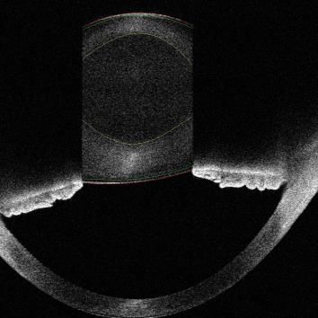
10                                                                                                                    Short form of author list

Table 2 Deep learning methods for cataract classification/grading on different ophthalmic images.
                        Literature      Method                Image Type          Year     Application     Cataract Type
                           [43]       CNN+RNN              Slit Lamp Image        2015      Grading             NC
                          [102]          CNN               Slit Lamp Image        2017    Classification       PCO
                           [78]          CNN               Slit Lamp Image        2017    Classification       PCO
                          [104]          CNN               Slit Lamp Image        2017    Classification     Cataract
                          [176]          CNN               Slit Lamp Image        2018    Classification     Cataract
                           [79]       CNN+RNN              Slit Lamp Image        2018    Classification       PCO
                          [164]      Faster R-CNN          Slit Lamp Image        2019      Grading             NC
                          [145]          CNN               Slit Lamp Image        2019    Classification        NC
                           [83]          CNN               Slit Lamp Image        2019    Classification        NC
                          [160]          CNN               Slit Lamp Image        2019    Classification        NC
                           [65]          CNN               Slit Lamp Image        2020    Classification        NC
                           [81]          CNN               Slit Lamp Image        2021    Classification     Cataract
                           [80]         R-CNN               slit lamp Image       2021    Classification       PCO
                           [66]          CNN                slit lamp Image       2021    Classification     cataract
                          [142]      Wavelet + ANN       Digital Camera Image     2016    Classification     Cataract
                            [8]          MLP               Ultrasonic Image       2016    Classification     Cataract
                          [181]        Attention           Ultrasonic Image       2020    Classification     Cataract
                          [159]          CNN               Ultrasonic Image       2021    Classification     Cataract
                          [170]          BPNN                Fundus Image         2015    Classification     Cataract
                           [99]          CNN                 Fundus Image         2017    Classification     Cataract
                           [29]          CNN                 Fundus Image         2017    Classification     Cataract
                          [175]          CNN                 Fundus Image         2019    Classification     Cataract
                          [191]          MLP                 Fundus Image         2019    Classification     Cataract
                          [191]          CNN                 Fundus Image         2019    Classification     Cataract
                          [177]          CNN                 Fundus Image         2019    Classification     Cataract
                          [120]          CNN                 Fundus Image         2019    Classification     Cataract
                          [166]          CNN                 Fundus Image         2020    Classification     Cataract
                          [121]          CNN                 Fundus Image         2021    Classification     Cataract
                           [75]       CNN+RNN                Fundus Image         2021    Classification     Cataract
                           [84]          CNN                 Fundus Image         2021    Classification     Cataract
                          [143]          CNN                 Fundus Image         2022    Classification     Cataract
                          [182]          CNN                AS-OCT Image          2020    Classification        NC
                          [161]          CNN                AS-OCT Image          2021    Classification        NC
                          [162]        Attention            AS-OCT Image          2021    Classification        NC
                          [185]        Attention            AS-OCT Image          2022    Classification        NC
                          [184]        Attention            AS-OCT Image          2022    Classification        NC

an artificial intelligence platform for congenital cataract di-           uses a 3D ResNet architecture for cataract screening based
agnosis and obtains good diagnosis results. Literature [160]              on 3D AS-OCT images. Zhang et al. [184] tested the NC
proposes a universal artificial intelligence platform for nu-             classification performance of state-of-the-art CNNs like ResNet,
clear cataract management and achieves good results.                      VGG, and GoogleNet on AS-OCT images, and the results
     For fundus images, literature [166, 177, 29, 175] achieves           showed that EfficientNet achieved the best performance.
competitive classification results with deep CNNs. Zhou et
al.[191] proposed the EDST-ResNet model for cataract clas-
sification based on fundus images where they used discrete
state transition function [28] as the activation function, to
improve the interpretability of CNN models. Fig. 12 pro-
vides a representative CNN framework for cataract classi-
fication on fundus images, which can help audiences know
this task easily.
    For AS-OCT images, literature [172, 178, 9] proposes
CNN-based segmentation frameworks for automatic lens re-
gion segmentation based on AS-OCT images, which can
help ophthalmologists localize and diagnose different types               Fig. 11 An example of a convolutional neural network (CNN) model
of cataract efficiently. Zhang et al. [182] proposed a novel              for cataract classification on fundus image [166].
CNN model named GraNet for nuclear cataract classifica-
tion on AS-OCT images but achieved poor results. [161]
Machine Learning for Cataract Classification/Grading on Ophthalmic Imaging Modalities: A Survey 11
5.2.3 Recurrent neural networks
Recurrent neural network (RNN) is a typical feedforward Conv 3x3
X
Crop
neural network architecture where connections between nodes Residual-MPA
Nucleus
form a directed or undirected graph along a temporal se- Conv 3x3
Residual-MPA
quence. RNN are skilled at processing sequential data effec- Conv 1x1 Conv 3x3
tively for various learning tasks [67], e.g., speech recogni- Mild NC Residual-MPA
MPA
tion. Over the past decades, many RNN variants have been Moderate NC GAP
developed to address different tasks, where Long short-term
Several NC Softmax
memory (LSTM) network is the most representative one. (b) Residual-MPA module
However, researchers have not used pure RNN architecture (a) Mixed Pyramid Attention Network (MPANet)
to classify cataract severity levels yet, which can be a re-
search direction for automatic cataract classification. Fig. 12 An example of attention-based CNN architecture for cataract
classification on AS-OCT images.
5.2.4 Attention mechanisms
5.2.5 Hybrid neural networks
Over the past years, attention mechanisms have been proven
TA hybrid neural network indicates that a neural network is
useful in various fields, such as computer vision [20], natu-
comprised of two or more two deep neural network types. In
ral language processing (NLP) [152], and medical data pro-
recent years, researchers have increasingly used hybrid neu-
cessing [187]. Generally, attention mechanism can be taken
ral networks to address different learning tasks [51]. Due to
an adaptive weighting process in a local-global manner ac-
its ability in inheriting the advantages from different neural
cording to feature representations of feature maps. In com-
network architectures, such as CNN, MLP, and transformer.
puter vision, attention can be classified into five categories:
Literature [79, 43, 75] proposes the hybrid neural network
channel, spatial, temporal attention, branch attention, and
models for cataract classification /grading by considering
attention combinations such as channel & spatial attention
the characteristics of RNN and CNN models, respectively.
[54]. Each attention category has a different influence on
In the future, we believe that more advanced hybrid neu-
the computer vision field. Researchers have recently used
ral network models will be designed for cataract classifica-
attention-based CNN models for cataract classification on
tion/grading based on different ophthalmic image modali-
different ophthalmic images. Zhang et al. [181] proposed
ties.
a residual attention-based network for cataract detection on
Ultrasound Images. Xiao et al. [162] applied a gated atten-
tion network (GCA-Net) to classify nuclear cataract sever-
ity levels on AS-OCT images and got good performance.
[184] presents a mixed pyramid attention network for AS-
OCT image-based nuclear cataract classification in which
they construct the mixed pyramid attention (MPA) block by
considering the relative significance of local-global feature
representations and different feature representation types, as
shown in Fig. 12.
Especially, self-attention is one representative kind of
Fig. 13 A hybrid neural network model for nuclear cataract grading on
attention mechanism. Due to its effectiveness in capturing slit lamp images, comprised of a CNN model and a RNN model [43].
capture long-range dependencies and generality, it has been
playing an increasingly important role in a variety of learn-
ing tasks [152, 156, 173]. Massive deep self-attention net- Discussion: From Table 1 and Table 2, we can conclude
works (e.g., Transformer) have achieved state-of-the-art per- as follows:
formance through mainstream CNNs on visual tasks. Vision
Transformer (ViT) [30] is a first-pure transformer architec- – Ophthalmic image perspective: Slit lamp images and
ture proposed for image classification and gets promising fundus images account for most automatic cataract clas-
performance. Recently, researchers also extended the transformer- sification/grading works, this is because these two oph-
based models for different medical image analysis tasks [58]; thalmic image modalities are easy to access and have
however, no current research work has utilized transformer- the clinical gold standards. Except for ultrasound images
based architectures to recognize cataract severity levels. collected from the animal model and human subjects,12 Short form of author list
five other ophthalmic imaging modalities were used for ability of deep neural network models on cataract clas-
automatic cataract classification/grading. sification/grading tasks, other data augmentation tech-
– Cataract classification/grading perspective: Existing niques should be considered: translation, color space trans-
works mainly focused on cataract screening, and most formations, random erasing, adversarial training, meta-
of them achieved over 80% accuracy. The number of learning, etc [130].
cataract classification works is more than the number of
cataract grading works. Since discrete labels are easy to
access and be confirmed through comparisons continu- 6 Evaluation measures
ous labels.
– Publication year perspective: Conventional machine learn- This section introduces evaluation measures to assess the
ing methods were first used to classify or grade cataract performance of cataract classification/grading. In this sur-
automatically. With the emergence of deep learning, re- vey, Classification denotes the cataract labels used for learn-
searchers have gradually constructed advanced deep neu- ing are discrete, e.g., 1,2,3,4, while grading denotes cataract
ral network architectures to automatically predict cataract labels are continuous, such as 0.1,0.5,1.0, 1.6, and 3.3.
severity levels due to their surprising performance. How- For cataract classification, accuracy (ACC), sensitivity
ever, the interpretability of deep learning methods is worse (recall), specificity, precision, F1-measure (F1), and G-mean
than conventional machine learning methods. are commonly used to evaluate the classification performance.
– Learning paradigm perspective: Most previous ma- [53, 10, 79].
chine learning methods belong to supervised learning
methods, and only two existing methods used semi-supervisedACC = TP+TN
, (1)
learning methods. Specifically, unsupervised and semi- T P + T N + FP + FN
supervised learning methods have achieved competitive
performance in computer vision and NLP, which have TP
not been widely applied to automatic cataract diagnosis. Sensitivity = , (2)
T P + FN
– Deep neural network architecture perspective: Ac-
cording to Table 2, we can see that CNNs account for
TN
over 50% deep neural network architectures. Two rea- Speci f icity = , (3)
sons can explain that: 1) Ophthalmic images are the most T N + FP
commonly-used way for cataract diagnosis by clinicians.
2) Compared with RNN and MLP, CNN are skilled at TP
Precision = , (4)
processing image data. To enhance precision of cataract T P + FP
diagnosis, it is better to combine image data with non-
imaging data, e.g., age. 2 ∗ precision ∗ recall
– Performance comparison on private/public datasets: F1 = , (5)
precision + recall
Pratap et al. [121] made a comparison of AlexNet, GoogleNet,
ResNet, ResNet50, and SqueezeNet for the cataract clas- r
sification on the public EyePACS dataset, and results TP TN
G − mean = ∗ , (6)
showed all CNN models obtained over 86% in the accu- T P + FN T N + FP
racy, and AlexNet got the best performance. [184] offers where TP, FP, TN, and FN denote the numbers of true pos-
the NC classification results of attention-based CNNs itives, false positives, true negatives, and false negatives,
(CBAM, ECA, GCA, SPA, and SE on the private NC respectively. Other evaluation measures like receiver oper-
dataset, and results showed SE obtained better perfor- ating characteristic curve (ROC), the area under the ROC
mance than other strong attention methods. Furthermore, curve (AUC), and kappa coefficient [10] are also applied to
[166, 164, 121, 120] used pre-trained CNNs for auto- measure the overall performance.
matic cataract classification on the EyePACS dataset ac- For cataract grading, the following evaluation measures
cording to transfer learning strategy, and they also con- are used to evaluate the overall performance, titled the ex-
cluded that training pre-trained CNNs benefited the cataract act integral agreement ratio R , the percentage of decimal
0
classification performance than training CNNs from scratch. grading errors 6 0.5 R , the percentage of decimal grad-
e0.5
– Data augmentation techniques: Researchers use the ing errors 6 1.0 Re1.0 , and the mean absolute error ε [91, 18,
commonly-used method for cataract classification/grading 168, 73, 72].
to augment ophthalmic image data for deep neural net-
works, such as flipping, cropping, rotation, and noise in-
jection. To further validate or enhance the generalization | Ggt = G pr |0
R0 = , (7)
NMachine Learning for Cataract Classification/Grading on Ophthalmic Imaging Modalities: A Survey 13
other ophthalmic image types. E.g., researchers refer to the
||Ggt − G pr | 6 0.5|0 WHO Cataract Grading System and develop a cataract clas-
Re0.5 = , (8) sification/grading protocol for fundus images [165] based on
N
clinical research and practice, which is widely used in au-
tomatic fundus-based cataract classification [166, 177, 29,
||Ggt − G pr | 6 1.0|0 175].
Re1.0 = , (9) To address the issue of developing standard cataract clas-
N
sification/grading protocols for new eye images, e.g., AS-
OCT images. In this survey, we propose two possible solu-
∑|Ggt − G pr | tions for reference as follows.
ε= , (10)
N
– Developing a cataract grading protocol based on the clin-
where Ggt and G pr denote the ground-truth grade and the ical verification. Literature [49, 116] uses AS-OCT im-
predicted grade. d.e is the ceiling function, |.| is the absolute ages to observe the lens opacities of patients based on
function, |.|0 a function that counts the number of non-zero the LOCS III in clinical practice, and statistical results
values, and N denotes the number of images. showed that high correlation between cataract severity
levels and clinical features with inter-class and intra-
class analysis. The clinical finding may provide clini-
7 Challenges and possible solutions cal support and reference. Hence, it is likely to develop
a referenced cataract classification/grading protocol for
Although researchers have made significant development in ASOCT images based on the clinical verification method
automatic cataract classification/grading over the years, this like the LOCS III.
field still has challenges. This section presents these chal- – Building the mapping relationship between two ophthalmic
lenges and gives possible solutions. imaging modalities. The lens opacity is an important in-
dicator to measure the severity level of cataracts, which
are presented on different ophthalmic images through
7.1 Lacking public cataract datasets
different forms in clinical research. Therefore, it is po-
PPublic cataract datasets are a very critical issue for cataract tential to construct the mapping relationship between two
classification/grading. Previous works have made tremen- different ophthalmic imaging modalities to develop a new
dous progress in automatic cataract/grading [167, 37, 7, 53], cataract classification/grading protocol by comparing the
there is no public and standard ophthalmology image dataset lens opacities, e.g., fundus image-based cataract classi-
available except for public challenges datasets and multiple fication system WHO Cataract Grading System.
ocular diseases. Hence, it is difficult for researchers to fol- Furthermore, to verify the effectiveness of new standard
low previous works because the cataract dataset is unavail- cataract grading protocols, we must collect multiple center
able. To this problem, it is necessary and significant to build data from hospitals in different regions.
public and standard ophthalmology image datasets based on
standardized medical data collection and storage protocol.
7.3 How to annotate cataract images accurately
Public cataract datasets can be collected from hospitals and
clinics with ophthalmologists’ help. This dataset collection
Data annotation is a challenging problem for the medical
mode can ensure the quality and diversity of cataract data
image analysis field including cataract image analysis, since
and help researchers develop more advanced ML methods.
it is the significant base for accurate ML-based disease di-
agnosis. However, clinicians cannot label massive cataract
images manually [127, 126], because it is expensive, time-
7.2 Developing standard cataract classification/grading
consuming, and objective. To address this challenges, we of-
protocols based on new ophthalmic imaging modalities
fer the following solutions:
Most existing works used the LOCS III as the clinical gold – Semi-supervised learning: [133, 134] uses the semi-
diagnosis standard to grade/classify cataract severity levels supervised learning strategy to recognize cataract sever-
for scientific research purposes and clinical practice. How- ity levels on fundus images and achieves expected per-
ever, the LOCS III is developed based on slit-lamp images, formance. It is probably to utilize weakly supervised learn-
which may not work well for ophthalmic images, such as ing methods to learn useful information from labeled
fundus images, AS-OCT images, and Ultrasonic images. To cataract images and let the method automatically label
solve this problem, researchers have made much effort in unlabeled cataract images according to learned informa-
constructing new cataract classification/grading standards for tion.14 Short form of author list
– Unsupervised learning: Recent works have shown that Large deep neural network models usually perform bet-
deep clustering/unsupervised learning techniques can help ter than small deep neural network models based on mas-
researchers acquire labels positively rather than acquire sive data, which previous works have demonstrated. How-
labels negatively [125, 47, 36, 55, 64]. Thus, We can ac- ever, it is challenging to collect massive data in the med-
tively apply deep clustering/unsupervised learning meth- ical field; thus, it is vital for us to develop transfer learn-
ods to label cataract images in the future. ing strategies to take full use of pre-trained parameters
– Content-based image retrieval: the content-based im- for large deep neural network models, to further improve
age retrieval (CBIR) [123, 131, 34, 33] technique has the performance of cataract-related tasks.
been widely used for different tasks based on different – Multimodality learning: Previous works only used oph-
image features, which also can be utilized to annotate thalmic image type for cataract diagnosis [168, 42, 13,
cataract images by comparing testing images with stan- 171], multimodality learning [11, 107, 56, 15] have been
dard images. utilized to tackle different medical image tasks, which
also can be used for automatic cataract classification/grading
based on multi-ophthalmic images or the combination
7.4 How to classify/grade cataract accurately for precise of ophthalmic images and non-image data. Multimodal-
cataract diagnosis ity data can be classified into image data and non-image
data. However, it is challenging to use multimodality im-
Most previous works focused on cataract screening, and few ages for automatic cataract classification. Two reasons
works considered clinical cataract diagnosis, especially for can account for it: 1) only silt lamp images and fun-
cataract surgery planning. This is because different cataract dus images have standard cataract classification systems,
severity levels and cataract types should clinically take the which have no correlation relationship between them; 2)
corresponding treatments. Hence, it is necessary to develop existing classification systems are subjective, it is chal-
state-of-the-art methods to classify cataract severity levels lenging for clinicians to label two cataract severity lev-
accurately, and this survey provides the following research els correctly for different ophthalmic images. Further-
directions. more, we can combine image data and non-image data
for automatic cataract diagnosis because it is easy to
– Clinical prior knowledge injection: Furthermore, cataracts
collect non-image data such as age and sex associated
are associated with various factors [98], e.g., sub-lens
with cataracts. Furthermore, recent studies have [111,
regions [168, 42, 13, 49, 116], which can be considered
98] used non-image data for PC and PCO diagnosis,
as domain knowledge of cataract. Thus, we can infuse
which demonstrated that it is potential to use multimodal-
the domain knowledge into deep networks for automatic
ity data to improve the precision of cataract diagnosis
cataract classification/grading according to the charac-
– Image denoising: Image noise is an important factor in
teristics of ophthalmic images. E.g., [184] incorporates
affecting automatic cataract diagnosis on ophthalmic im-
clinical features in attention-based network design for
ages. Researchers have proposed different methods to
classification.
remove the noise from the images based on the char-
– Multi-task learning for classification and segmenta-
acteristics of ophthalmic images, such as Gaussian fil-
tion: Over the past decades, multi-task learning tech-
ter, discrete wavelet transform (DWT), discrete cosine
niques have been successfully applied to various fields,
transform (DCT), Haar wavelet transform, and its vari-
including medical image analysis. Xu et al. [164] used
ants [191, 53, 40]. Additionally, recent research has be-
the Faster-RCNN framework to detect the lens region
gun to use the GAN model for medical image denoising
and grade nuclear cataract severity levels on slit lamp
and achieved good results.
images and achieved competitive performance. Litera-
ture [172, 178] proposes the deep segmentation network
framework for automatic lens subregion segmentation 7.5 Improving the interpretability of deep learning methods
based on AS-OCT images, which is a significant base for
cataract diagnosis and cataract surgery planning. More- Deep learning methods have been widely used for cataract
over, more multi-task learning frameworks should be de- classification/grading. However, deep learning methods are
veloped for cataract classification and lens segmenta- still considered a black box without explaining why they
tion, considering multi-task learning framework usually make good prediction results or poor prediction results. There-
keeps a good balance between performance and com- fore, it is necessary to give reliable explanations for the pre-
plexity. dicted results based on deep learning methods. Literature
– Transfer learning: Over the years, researchers have used [191] visualizes weight distributions of deep convolutional
the transfer learning method to improve the cataract clas- layers and dense layers to explain the predicted results of
sification performance [166, 164] with pre-trained CNNs. deep learning methods. It is likely to analyze the similaritiesYou can also read


