Involvement of Abnormal Gut Microbiota Composition and Function in Doxorubicin-Induced Cardiotoxicity - Frontiers
←
→
Page content transcription
If your browser does not render page correctly, please read the page content below
ORIGINAL RESEARCH
                                                                                                                                           published: 25 February 2022
                                                                                                                                      doi: 10.3389/fcimb.2022.808837

                                             Involvement of Abnormal
                                             Gut Microbiota Composition
                                             and Function in Doxorubicin-
                                             Induced Cardiotoxicity
                                             Jie Huang 1,2, Shanshan Wei 1,2, Chuanhao Jiang 3, Zijun Xiao 1,2, Jian Liu 1,2, Weijun Peng 4,
                                             Bikui Zhang 1,2* and Wenqun Li 1,2*
                                             1 Department of Pharmacy, The Second Xiangya Hospital, Central South University, Changsha, China, 2 Institute of Clinical

                                             Pharmacy, Central South University, Changsha, China, 3 Department of Laboratory Medicine, The Second Xiangya Hospital,
                                             Central South University, Changsha, China, 4 Department of Integrated Traditional Chinese & Western Medicine, The Second
                                             Xiangya Hospital, Central South University, Changsha, China

                       Edited by:            Objectives: Doxorubicin (Dox), a chemotherapeutic anthracycline agent for the treatment
               Nar Singh Chauhan,
                                             of a variety of malignancies, has a limitation in clinical application for dose-dependent
 Maharshi Dayanand University, India
                                             cardiotoxicity. The purpose of this study was to explore the relationship between the
                       Reviewed by:
                            Neha Jain,       composition/function of the gut microbiota and Dox-induced cardiotoxicity (DIC).
       Indian Institute of Technology
                        Jodhpur, India
                                             Methods: C57BL/6J mice were injected intraperitoneally with 15 mg/kg of Dox, with or
                       Monika Yadav,         without antibiotics (Abs) administration. The M-mode echocardiograms were performed
 Maharshi Dayanand University, India         to assess cardiac function. The histopathological analysis was conducted by H&E staining
                    *Correspondence:         and TUNEL kit assay. The serum levels of creatine kinase (CK), CK-MB (CK-MB), lactic
                           Bikui Zhang
                   505995@csu.edu.cn         dehydrogenase (LDH), and cardiac troponin T (cTnT) were analyzed by an automatic
                            Wenqun Li        biochemical analyzer. 16S rRNA gene and metagenomic sequencing of fecal samples
                 liwq1204@csu.edu.cn
                                             were used to explore the gut microbiota composition and function.
                  Specialty section:         Key Findings: Dox caused left ventricular (LV) dilation and reduced LV contractility. The
        This article was submitted to
                                             levels of cardiomyocyte apoptosis and myocardial enzymes were elevated in Dox-treated
   Microbiome in Health and Disease,
              a section of the journal       mice compared with the control (Con) group. 16S rRNA gene sequencing results revealed
             Frontiers in Cellular and       significant differences in microbial composition between the two groups. In the Dox group,
               Infection Microbiology
                                             the relative abundances of Allobaculum, Muribaculum, and Lachnoclostridium were
       Received: 04 November 2021
        Accepted: 01 February 2022
                                             significantly decreased, whereas Faecalibaculum, Dubosiella, and Lachnospiraceae
        Published: 25 February 2022          were significantly increased compared with the Con group at the genus level.
                                 Citation:   Functional enrichment with Cluster of orthologous groups of proteins (COG) and Kyoto
Huang J, Wei S, Jiang C, Xiao Z, Liu J,
                                             Encyclopedia of Genes and Genomes (KEGG) analyses showed that the Dox mice
    Peng W, Zhang B and Li W (2022)
         Involvement of Abnormal Gut         displayed different clusters of cellular processes and metabolism from the Con mice.
 Microbiota Composition and Function         The different species and their functions between the two groups were associated with the
in Doxorubicin-Induced Cardiotoxicity.
Front. Cell. Infect. Microbiol. 12:808837.
                                             clinical factors of cardiac enzymes. Moreover, depletion of the gut microbiota could
       doi: 10.3389/fcimb.2022.808837        alleviate Dox-induced myocardial injury and cardiomyocyte apoptosis.

Frontiers in Cellular and Infection Microbiology | www.frontiersin.org              1                                     February 2022 | Volume 12 | Article 808837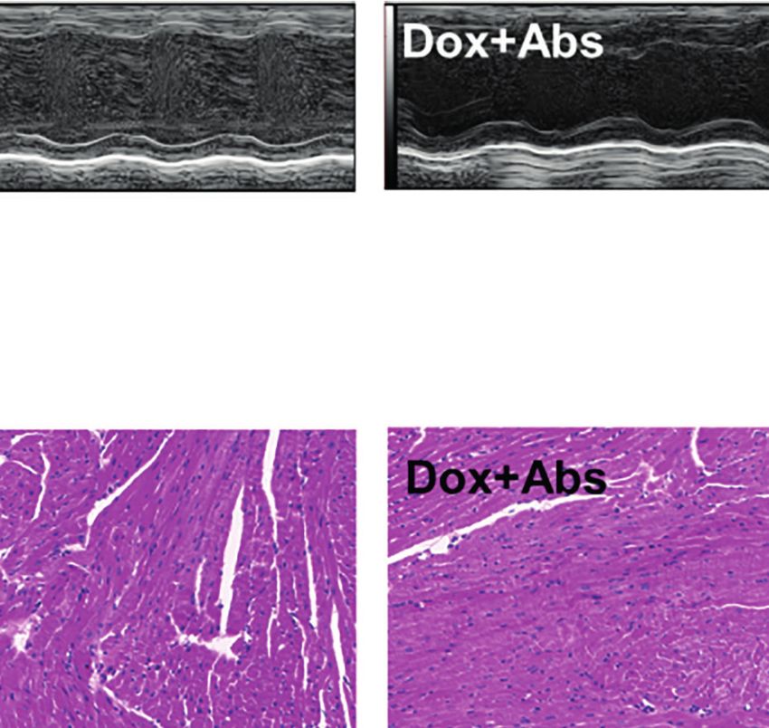
Huang et al.                                                                                                                Gut Microbiota in Cardiotoxicity

                                            Conclusions: The study here shows that composition imbalance and functional
                                            changes of the gut microbiota can be one of the etiological mechanisms underlying
                                            DIC. The gut microbiota may serve as new targets for the treatment of cardiotoxicity and
                                            cardiovascular diseases.
                                            Keywords: doxorubicin, cardiotoxicity, gut microbiota, 16S rRNA gene sequencing, metagenomic sequencing

INTRODUCTION                                                                          Here, to investigate the key microbes related to cardiotoxicity
                                                                                  and microbial functions from fecal samples, we constructed an
Doxorubicin (Dox) is a chemotherapeutic anthracycline agent with                  explicit mouse model of DIC. Using a combination of 16S rRNA
broad-spectrum and high efficacy used for the treatment of a                       gene sequencing and metagenomic sequencing analysis, we
variety of malignancies (Wu et al., 2021). However, its clinical                  strived to compare the composition and function of the gut
application is limited due to the dose-dependent cardiotoxicity,                  microbiome between the normal control (Con) mice and Dox
which may lead to acute pericarditis, irreversible cardiomyopathy,                mice. We also conducted an antibiotic intervention in mice to
and congestive heart failure (HF) (Wenningmann et al., 2019). The                 further understand the role of the gut microbiome in DIC.
pathogenesis of Dox may be associated with topoisomerase IIb
(Top2b) inhibition, oxidative stress, inflammation, and apoptosis
(Liu et al., 2020a; Tadokoro et al., 2020). Though a variety of
strategies including limitation of cumulative Dox doses, use of                   MATERIALS AND METHODS
antioxidant drugs, and common HF drugs have been proposed to
prevent or attenuate Dox-induced cardiotoxicity (DIC), none of                    Drugs and Reagents
these strategies have obtained satisfying efficacy (Vejpongsa and                  Dox for injection was obtained from Shenzhen Main Luck
Yeh, 2014; Varricchi et al., 2018; Yarmohammadi et al., 2021).                    Pharmaceuticals Co., Ltd. (Shenzhen, China). Four antibiotics
Given that the mechanism of DIC is a complex disturbance system,                  (Abs) including vancomycin, metronidazole, ampicillin, and
an alternative hypothesis is required to explain DIC, and a novel                 neomycin were purchased from Zhengde Pharmaceutical Co.,
therapeutic strategy needs to be established.                                     Ltd. (Taiwan, China), Huazhong Pharmaceutical Co., Ltd.
    From the outcome of recent studies, the intestinal microbiota                 (Xiangyang, China), Hunan Kangerjia Biomedical Technology
has been found to play an essential role in the health of the host                Co., Ltd. (Zhangjiajie, China), and BBI Life Sciences Co., Ltd.
organism. Multiple diseases, such as diabetes, obesity, cancer,                   (Shanghai, China), respectively.
and nervous system disease, are related to the changes of
intestinal microorganisms (Cheng et al., 2020; Fan and                            Animals and Treatments
Pedersen, 2020; Megur et al., 2020; Verhaar et al., 2020).                        Female C57BL/6J mice aged 6–8 weeks were obtained
Particularly, the gut microbiota and their metabolites have                       from Laboratory Animal Center, Xiangya School of Medicine,
been implicated in the progression of cardiovascular diseases                     Central South University (Changsha, China). All the procedures
(CVDs) including hypertension, dyslipidemia, atherosclerosis,                     in this research were operated in accordance with the National
thrombosis, HF, and ischemic stroke (Kasahara and Rey, 2019).                     Institutes of Health Guide (NIH publications no. 8023) for the
In addition, it has become evident that the gut microbiota affects                Care and Use of Laboratory Animals. The experimental protocol
the response to cancer therapy and susceptibility to toxic side                   was approved by the Medicine Animal Welfare Committee of
effects (Roy and Trinchieri, 2017). Besides, drugs can alter the                  Xiangya School of Medicine (SYXK-2015/0017).
microbiome and create secondary effects independent of the                           To establish the cardiotoxicity model, the mice were
drug molecule itself (Hitchings and Kelly, 2019). Recently,                       randomly divided into two groups with 10 animals in each
antitumor chemotherapy drugs, such as cisplatin and Dox,                          group, including the Con group and Dox group. The Dox
have been reported to cause alteration of the gut microbiota,                     group (15 mg/kg) was administrated with 3 mg/kg every other
which is involved in the pathogenesis of cardiotoxicity (Zhao                     day by intraperitoneal injection. For antibiotic treatment, the
et al., 2018; Wu et al., 2019). However, previous studies were                    mice were randomly divided into three groups including the Con
based on a 16S rRNA gene sequencing method to focus on                            group, Dox group, and Dox+Abs group. The Dox+Abs group
changes in microbial composition, which has the limitations                       was treated with drinking water containing 500 mg/L of each
such as the bias of PCR amplification (Silverman et al., 2021) and                 antibiotic for 28 days. Dox was injected intraperitoneally after 14
difficulty to identify most microbes at the strain and species levels              days of Abs. On the 28th day, the mice were subjected to
(Peng et al., 2018). Compared to the amplicon, the shotgun                        echocardiography for evaluating cardiac function. Besides, the
metagenome can provide functional gene profiles directly and                       fecal samples from the Con group and Dox group were collected
reach a much higher resolution of taxonomic annotation (Liu                       into sterile cryopreservation tubes, frozen quickly in liquid
et al., 2021). Therefore, further studies conducting metagenome                   nitrogen, and then refrigerated at −80°C for DNA extraction
sequencing are required to analyze the functional activity of the                 and amplification of the 16S rRNA gene. Subsequently, all
gut microbiome linked with DIC.                                                   animals were anesthetized with 1% pentobarbital sodium

Frontiers in Cellular and Infection Microbiology | www.frontiersin.org        2                                February 2022 | Volume 12 | Article 808837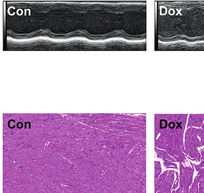
Huang et al.                                                                                                           Gut Microbiota in Cardiotoxicity

(50 mg/kg, i.p.). The venous blood was drawn from the orbit of                   OTUs were assigned to the closest taxonomic neighbors and
mice. The heart tissues were acquired after perfusing and rinsing            relative bacterial species by the RDP Classifier algorithm (http://
with cold saline and then preserved in 4% paraformaldehyde for               rdp.cme.msu.edu/) using a confidence threshold of 70%. The
histopathological analysis.                                                  species alignment database of 16S bacteria is the Silva database
                                                                             (https://www.arb-silva.de/). The relative abundance of each
M-Mode Echocardiograms                                                       taxonomic level was calculated using the QIIME tool. The
M-mode echocardiograms were performed by a VisualSonics                      indices of alpha diversity including Chao, Shannon, and Ace
Vevo 2100 (VisualSonics, Toronto, ON, Canada). The mice were                 were analyzed by MOTHUR (version 1.30.2, https://www.
anesthetized with 1.5% isoflurane, and an appropriate amount of               mothur.org/). Principal coordinate analysis (PCoA) projections
coupling agent was applied to the left anterior chest area after             were used to describe the beta diversity.
hair removal. Then the M-ultrasound changes of 10~20 cardiac
cycles were recorded. Finally, the parameters of cardiac function            Metagenome Sequence Analysis
including left ventricular (LV) ejection fraction (EF%) and LV               We also selected a subset of specimens (6 from the Con group
fractional shortening (FS%) were calculated.                                 and 6 from the Dox group) for metagenome sequencing
                                                                             following 16S rRNA gene sequence analysis. In brief, the
Histopathological Analysis                                                   extracted genomic DNA was detected by 1% agarose gel
After being fixed in 4% paraformaldehyde at room temperature,                 electrophoresis and fragmented to the appropriate length
the heart tissues were embedded in paraffin wax and cut into 3-               (approximately 400 bp) by using Covaris M220 (Gene
mm thin sections. After being dehydrated in a series of graded               Company Limited, Shanghai, China). The paired-end library
alcohols, the sections were stained with H&E and TdT-mediated                was constructed by the NEXTFLEX™ Rapid DNA-Seq Kit (Bioo
dUTP Nick-End Labeling (TUNEL) kit assay according to the                    Scientific, Austin, TX, USA) according to the manufacturer’s
manufacturer’s instructions.                                                 protocols. Then, the blunt-ends of fragments were ligated
                                                                             to adapters. Hiseq X sequencing systems (Illumina Inc.,
Biochemical Analysis                                                         San Diego, CA, USA) were used for metagenomic sequencing.
After Abs and Dox treatment, the collected blood samples                     The raw data were trimmed and decontaminated to obtain
were centrifuged at 3,000 rpm at 4°C for 15 min to obtain                    optimized reads, which were assembled using MEGAHIT
serum. The serum biochemical parameters including creatine                   (http://www.l3-bioinfo.com/products/megahit.htm). The
kinase (CK), CK-MB (CK-MB), lactic dehydrogenase (LDH),                      assembled contigs not less than 100 bp were used for further
and cardiac troponin T (cTnT) were analyzed by using kits                    gene prediction and annotation.
with an automatic biochemical analyzer according to the                          Subsequently, the open reading frames (ORFs) of the
manufacturers’ instructions.                                                 assembled sequences were predicted by using the MetaGene
                                                                             platform (http://metagene.cb.k.u-tokyo.ac.jp/). The redundant
16S rRNA Gene Sequence Analysis                                              genes were filtered out to construct non-redundant gene sets.
Fecal bacterial DNA was extracted using the E.Z.N.A.® Stool                  Genes with sequence identity greater than 95% were clustered
DNA Kit (Omega Biotech, Norcross, GA, USA) according to the                  together using CD-HIT (http://www.bioinformatics.org/cd-hit/),
manufacturer’s protocol. The extracted genomic DNA was                       and the longest sequences from each cluster were the
detected by 1% agarose gel electrophoresis. To assess bacterial              representative gene. The genetic functions were annotated and
diversity, the V3–V4 hypervariable regions of the bacterial 16S              classified by the functional databases. Cluster of orthologous
rRNA gene were amplified with a set of primers (338F 5′ACTCC                  groups of proteins (COG) annotation was conducted by using
TACGGGAGGCAGCAG-3′, 806R 5′GGACTACHVGGGT                                     BLASTP (version 2.3.0) against the eggNOG database (version
WTCTAAT-3′). Then, the PCR products were detected using                      4.5.1, http://eggnogdb.embl.de/#/app/home) with an e-value
2% agarose gel electrophoresis, recovered using AxyPrepDNA                   cutoff of 1e−5. Kyoto Encyclopedia of Genes and Genomes
Gel Recovery Kit (Axygen Biosciences, Union City, CA, USA),                  (KEGG) pathway annotation was conducted by using BLASTP
and quantified using QuantiFluor™-ST (Promega, Madison, WI,                   (version 2.3.0) against the KEGG database (http://www.genome.
USA) according to the quantitative results of electrophoresis.               jp/kegg/) with an e-value cutoff of 1e−5.
Subsequently, purified amplicons were mixed in appropriate                        The discrimination in COG and KEGG categories between
proportions and paired-end sequenced on an Illumina MiSeq                    the Con and Dox groups was identified by using linear
platform (Illumina, San Diego, CA, USA) according to the                     discriminant analysis (LDA) effect size (LEfSe; http://
standard protocols by Majorbio Bio-Pharm Technology Co.                      huttenhower.sph.harvard.edu/galaxy/root?tool_id=lefse_
Ltd. (Shanghai, China).                                                      upload). Only LDA values >2.0 at a p-value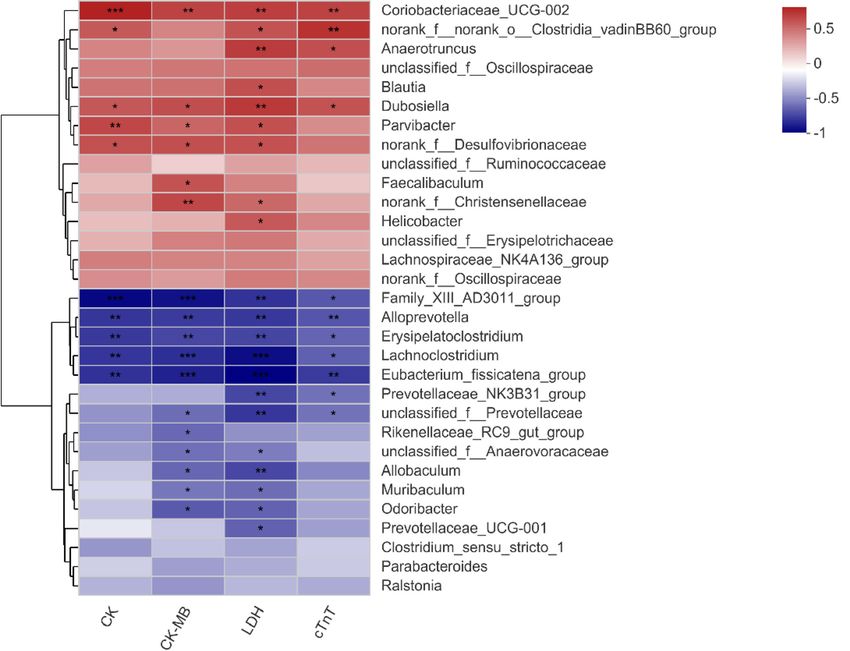
Huang et al.                                                                                                                                Gut Microbiota in Cardiotoxicity

to perform statistical analysis. A p-value less than 0.05 was                            inflammatory cell infiltration, myocardial fragmentation, and
considered statistically significant.                                                     disorder of cardiac fiber arrangement. TUNEL assay was used
                                                                                         to assess the cardiomyocyte apoptosis, which is an important
                                                                                         event in the process of DIC. The results revealed that mice with
RESULTS                                                                                  Dox treatment showed obvious cardiomyocyte apoptosis as
                                                                                         compared with the Con group (Figures 1D, E). Myocardial
Myocardial Injury and Cardiomyocyte                                                      injury was also determined by the serum levels of CK, CK-MB,
Apoptosis Induced by Doxorubicin                                                         LDH, and cTnT, and all of these myocardial enzymes were
First, we constructed the heart injury model induced by Dox in                           elevated in Dox-treated mice (Figure 1F). The above results
mice. After treatment with Dox, the M-mode echocardiograms                               suggested that Dox caused severe myocardial lesions.
showed LV dilation (Figure 1A). The EF% and FS% of the Dox
group were decreased as compared with the Con group,                                     Similar Gut Microbial Diversity Between
indicating that Dox reduced LV contractility in mice                                     Control and Doxorubicin Mice
(Figure 1B). To further determine the histological changes of                            To characterize the effect of Dox on gut microbial communities,
the heart induced by Dox, H&E staining and TUNEL staining                                we initially used 16S rRNA gene sequencing. After size filtering,
were conducted. As shown in Figure 1C, we observed regular cell                          quality control, and chimera removal, a total of 968,758 high-
distribution and normal morphology in the myocardium of the                              quality reads, ranging from 39,221 to 61,778 per sample, with an
Con group. However, the Dox-treated group showed                                         average length of 421.05 bp (421.05 ± 1.35 bp), were obtained

                             A                                                                           B

                             C

                             D
                                                                                                       E

                             F

 FIGURE 1 | Dox-induced cardiac dysfunction and histological injury. (A) M-mode echocardiograms showing left ventricular dilation induced by Dox. (B) The
 parameters of left ventricular ejection fraction (EF%) and left ventricular fractional shortening (FS%). (C) H&E staining reflected the histological changes, magnification
 ×200. (D) Representative image of TUNEL staining, magnification ×400; white arrows indicate the apoptotic cells. (E) Statistical result of TUNEL staining. (F) The
 serum level of creatine kinase (CK), creatine kinase-MB (CK-MB), lactic dehydrogenase (LDH), and cardiac troponin T (cTnT). Con, control; Dox, doxorubicin. Data
 are mean ± SEM. n = 6–10. *p < 0.05, **p < 0.01 vs. Con.

Frontiers in Cellular and Infection Microbiology | www.frontiersin.org               4                                        February 2022 | Volume 12 | Article 808837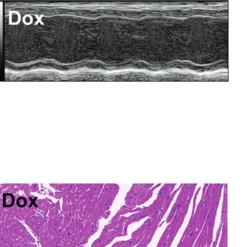
Huang et al.                                                                                                                    Gut Microbiota in Cardiotoxicity

from fecal samples of mice. These reads were matched into 827                     structure. All p-values >0.05 (Wilcoxon rank-sum test) excluding
OTUs (defined based on 97% sequence similarity) including 10                       Chao (p = 0.0392) at the phylum level (Supplementary Table 1),
phyla, 272 species, and 165 genera of gut microbes. A Venn                        which showed almost no significant differences between the Con
diagram showed that the two groups shared 699 OTUs, whereas                       and Dox groups.
85 and 43 OTUs were unique to the Con and Dox mice,
respectively (Figure 2A).                                                         Significant Differences in the Microbial
   The relative abundance in the two groups on phylum, family,                    Composition Between the Two Groups
and genus levels is displayed in the bar plot (Figures 2B–D). The                 The beta diversity analysis was carried out to reveal the difference
results showed that the gut microbiome compositions of the Con                    in the microbial composition between the Con and Dox samples.
and Dox mice were different. The phyla Bacteroidota, Firmicutes,                  As shown in PCoA, the first two principal coordinates explained
and Verrucomicrobiota were predominant in the gut microbiota                      36.74% and 52.22% of the total variance for unweighted (analysis
of mice. Muribaculaceae and Lactobacillaceae were the most                        of similarities (ANOSIM) R = 0.3811, p = 0.001) and weighted
abundant families in fecal samples from both the Con and Dox                      (ANOSIM R = 0.2420, p = 0.004) UniFrac, respectively
mice, but group Dox showed an upward trend compared with                          (Figures 3A, B). The partial least squares discriminant analysis
group Con.                                                                        (PLS-DA) showed that the bacterial communities of the two
   The microbial alpha diversity indices, including microbial                     groups clustered separately (Figure 3C). Thus, Dox-treated mice
community richness (Chao and Ace) and diversity (Shannon),                        possessed an obvious difference in a distinct clustering of fecal
were used to illustrate the changes in the microbiota community                   microbial structure as compared to the Con mice.

                   A                                                                   B

                   C                                                               D

 FIGURE 2 | Comparison of the microbial composition between the two groups. (A) Venn diagram depicting OTU richness and the overlap representing the shared
 OTUs in microbial communities. (B–D) Relative abundance of microbial community for each group at phylum, family, and genus levels. Con, control; Dox,
 doxorubicin; OTU, operational taxonomic unit. n = 10.

Frontiers in Cellular and Infection Microbiology | www.frontiersin.org        5                                    February 2022 | Volume 12 | Article 808837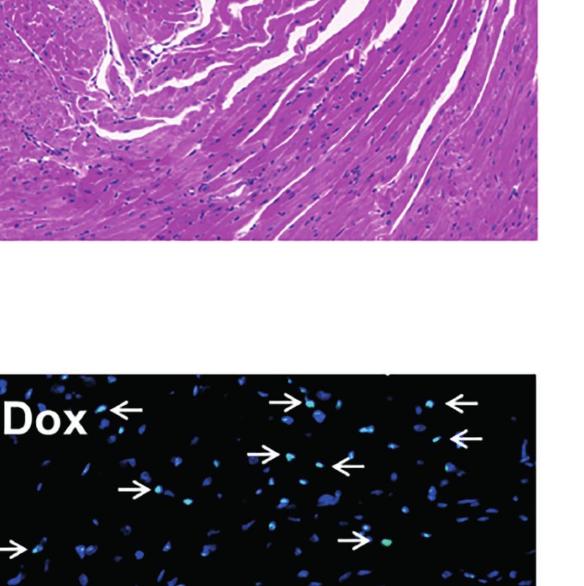
Huang et al.                                                                                                                                Gut Microbiota in Cardiotoxicity

          A                                                  B                                                    C

          D                                                                           E

 FIGURE 3 | Difference analysis of gut microbial composition between the two groups. (A, B) Principal coordinate analysis of unweighted (R = 0.3811, p = 0.001000)
 and weighted UniFrac (R = 0.2420, p = 0.004000) distances. PC1 and PC2 represent the top two principal coordinates that capture the maximum diversity. (C) Partial
 least squares discriminant analysis (PLS-DA). COMP 1 and COMP 2 represent the suspected influencing factors for the deviation of the microbial composition.
 (D) Cladogram analyzed by LEfSe (LDA > 2.0, p < 0.05) showing the phylogenetic distribution of the bacterial lineages. Circles indicate phylogenetic levels from phylum to
 genus. Nodes with different colors indicate microbial taxa that are enriched in the corresponding groups and have significant differences between groups; yellow nodes
 indicate microbial taxa that have no significant differences between groups. The diameter of each node is proportional to the abundance of the group. (E) Histogram of
 LEfSe analysis (LDA > 2.0, p < 0.05) showing the LDA scores for differentially abundant genera. Con, control; Dox, doxorubicin; LDA, linear discriminant analysis; LEfSe,
 linear discriminant analysis effect size. n = 10.

   To further identify the significant difference in specific                               g_Lachnospiraceae_NK4A136_group were significantly
bacterial taxa between the Con and Dox groups, the LEfSe                                  increased as compared with the Con group at the genus
analysis based on discriminative features cladogram and                                   level (Figure 3E).
histogram was performed, and the effect size cutoff of the LDA
score was set to 2.0. This analysis identified two phyla including
p_Actinobacteriota and p_Campilobacterota, and 31 genera,                                 The Microbial Correlation Networks
which were responsible for this discrimination (Figure 3D). In                            Between the Two Groups Were Different
the Dox group, the relative abundances of g_Allobaculum,                                  To investigate the microbial correlation network, we calculated
g_Muribaculum, and g_Lachnoclostridium were significantly                                  Spearman’s correlations among the 50 most abundant bacterial
decreased, whereas g_Faecalibaculum, g_Dubosiella, and                                    genera from each group. As shown in Figure 4, the Dox group

Frontiers in Cellular and Infection Microbiology | www.frontiersin.org               6                                        February 2022 | Volume 12 | Article 808837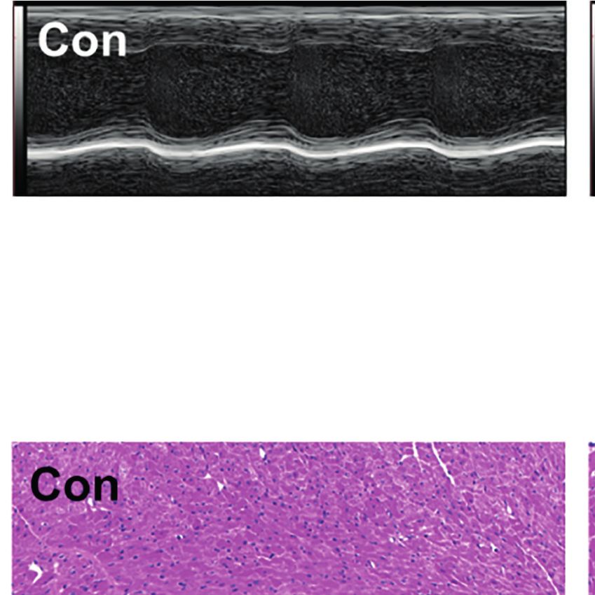
Huang et al. Gut Microbiota in Cardiotoxicity
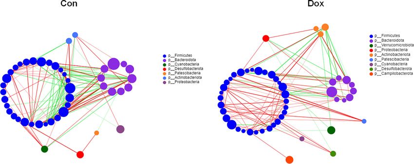
FIGURE 4 | Correlation network analysis of the 50 most abundant genera for each group. The size of the node indicates the genera abundance, and different colors
indicate different phyla. The lines indicate significant positive (red) and negative (green) pairwise correlations, and the thickness indicates the strength of the
correlation between two genera. Spearman’s value ≥0.05, p < 0.05. Con, control; Dox, doxorubicin. n = 10.
featured more phyla (7 vs. 9) and displayed a stronger positive Alternations of the Microbial
correlation among genera. The microbial community of the Dox Functional Profiles Were Revealed
group featured a more complicated network. In addition, the by Metagenomic Analysis
network constructed from the Dox group displayed fewer edges In this study, we used metagenomic sequencing analysis of the
(334 vs. 125) (Supplementary Table 2) and lower transitivity gut microbiomes to investigate the differences in the microbial
(0.5650 vs. 0.4127), suggesting that the correlation among the functional composition between the Dox-treated and Con mice.
microbiota in the Dox group was distinctly decreased compared Genomic DNA from the fecal specimens was extracted to obtain
to that of Con group. Moreover, we computed degree (DC), a total of 506,191,910 clean reads resulting in 2,646,123 contigs.
closeness (CC), and betweenness (BC) centrality to evaluate the A total of 4,092,442 ORFs predicted from the contigs were used
taxa importance at the genus level within the network. According for functional annotation in the COG and KEGG databases.
to the total scores of these coefficients (Supplementary Table 3), To identify protein function annotation, COG analysis was
the top three nodes from each group were selected as putative performed using LEfSe analysis between the Con and Dox mice.
keystone genera within this network (g_Prevotellaceae_ Based on the threshold LDA values >2.0 and p < 0.05, we
NK3B31_group, g_Alistipes, and g_Allobaculum for the Con identified 4 functional COG categories that showed high
group and g_norank_f_Oscillospiraceae, g_Bacteroides, and enrichment in the Dox group, which were related to the
g_Faecalibaculum for the Dox group). Taken together, the translation, ribosomal structure, and biogenesis [J]; cell cycle
above analyses suggested that the correlation structure of the control, cell division, and chromosome partitioning [D];
microbial community in the Dox group was distinctly different intracellular trafficking, secretion, and vesicular transport [U];
from that of the Con group. and extracellular structures [W] (Figure 6A). These functions
could be classified into two categories: information storage and
Correlations Between Significantly processing (Function J) and cellular processes and signaling
Different Species and Clinical Factors in (Functions D, U, and W). The remaining COG categories have
Two Groups of Mice no biologically significant differences. Overall, the cluster of
Spearman’s correlation heatmap showed that the relationship cellular processes and signaling was the predominant COG
between significantly different species (Figure 3E) and serum category associated with the Dox mice.
biochemical parameters (CK, CK-MB, LDH, and cTnT) was To further explore the functions of differentially expressed
different. Bacterial genera enriched in the Dox group were genes, the KEGG pathways were analyzed also by LEfSe analysis
positively correlated with these clinical factors, while those (LDA > 2.0, p < 0.05). At KEGG level 1 (Figure 6B), the LEfSe
enriched in the normal mice showed a negative correlation. bar showed that metabolism was the dominant signaling
Among them, Coriobacteriaceae_UCG-002 and Dubosiella pathway in the Con group, and genetic information processing
had significant positive correlations with all the clinical and environmental information processing were significantly
parameters, and Family_XIII_AD3011_group, Alloprevotella, enriched in the Dox group. At KEGG level 2 (Figure 6C), 10
Erysipelatoclostridium, Lachnoclostridium, and Eubacterium_ differential KEGG pathways (including membrane transport,
fissicatena_group showed extremely significant negative replication and repair, signal transduction, and others) were
correlations with all these biochemical parameters (Figure 5). identified in the gut microbiome of Dox-treated mice, while 8
Therefore, it is speculated that these different species may be KEGG pathways (including global and overview maps, amino
involved in the process of cardiotoxicity. acid metabolism, glycan biosynthesis, and metabolism and
Frontiers in Cellular and Infection Microbiology | www.frontiersin.org 7 February 2022 | Volume 12 | Article 808837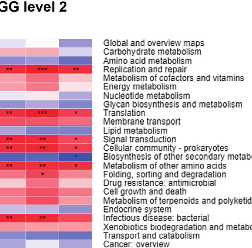
Huang et al.                                                                                                                               Gut Microbiota in Cardiotoxicity

 FIGURE 5 | Correlation heatmap of significantly different genera and clinical factors. The x- and y-axes are clinical factors and genera, respectively. R in different
 colors is shown; the right side of the legend is the color range of different R values. CK, creatine kinase; CK-MB, creatine kinase-MB; LDH, lactic dehydrogenase;
 cTnT, cardiac troponin T. n = 10. *p < 0.05, **p < 0.01, ***p < 0.001.

others) were significantly increased in the Con mice. At KEGG                            (Figure 8A), carbohydrate transport and metabolism [G]
level 3 (Figure 6D), we found a total of 38 statistically different                     showed a negative significant correlation with LDH level.
functional KEGG pathways between the two groups. Half of                                Inorganic ion transport and metabolism [P] revealed a
these functions were highly enriched in the Dox group, including                        significant negative correlation with the CK level. Translation,
two-component system, quorum sensing, and ribosome. In                                  ribosomal structure, and biogenesis [J] were significantly
contrast, the KEGG functions of the Con mice were enriched                              positively correlated with CK-MB, LDH, and cTnT levels.
in the biosynthesis of amino acids, other glycan degradation,                           Intracellular trafficking, secretion, and vesicular transport [U]
sphingolipid metabolism, and more. Thus, the Con mice and                               demonstrated a significant positive correlation with CK-MB and
Dox mice represented completely different multiple functional                           LDH levels. Cell cycle control, cell division, and chromosome
pathways in the gut microbiome.                                                         partitioning [D] were significantly positively correlated with all
    Furthermore, to visualize the association between the gut                           the biochemical factors. Extracellular structures [W] were
microbiome and functional properties, we determined the top                             significantly positively correlated with the CK level. Notably,
ten genera that mainly contributed to differences at KEGG level 1                       the COG categories including J, U, D, and W were enriched in
pathways between the Con and Dox mice (Figure 7). The main                              the Dox group (Figure 6A). At KEGG level 1 (Figure 8B), the
functions involved in these species were metabolism, genetic                            metabolism pathway tended to be negatively, but not
information processing, environmental information processing,                           significantly, correlated with these biochemical factors. Genetic
and cellular processes. G_Duncaniella was the main contributor                          information processing showed a significant positive correlation
of these functions and contributed significantly more to the Dox                         with CK-MB, LDH, and cTnT levels. Environmental information
samples than the Con samples. A reduced contribution by taxa                            processing and cellular processes were significantly positively
belonging to G_Prevotella and G_Bacteroides was also observed.                          correlated with CK-MB and LDH levels. At KEGG level 2
                                                                                        (Figure 8C), several pathways enriched in the Con group such
Correlations Between Microbial                                                          as amino acid metabolism, glycan biosynthesis, and metabolism
Functions and Clinical Factors                                                          and lipid metabolism were negatively correlated with the clinical
in the Two Groups of Mice                                                               factors, while several pathways enriched in the Dox group such
We next combined the bacterial functions and serum                                      as replication and repair, translation, and signal transduction
biochemical parameters into annotated heatmaps that provided                            were positively correlated with the clinical factors. Collectively,
several insights into the correlation between the microbial                             gut microbiota dysfunction may be at least partially related to
functional profiles and DIC (Figure 8). At the COG level                                 the DIC.

Frontiers in Cellular and Infection Microbiology | www.frontiersin.org              8                                        February 2022 | Volume 12 | Article 808837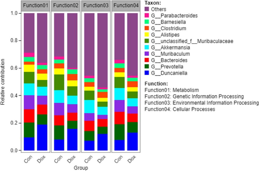
Huang et al.                                                                                                                               Gut Microbiota in Cardiotoxicity

                                    A

                                    B

                                    C

                                    D

 FIGURE 6 | The functional pathway comparisons in metagenome between Con and Dox groups were analyzed by LEfSe analysis (LDA > 2.0, p < 0.05). (A) Histogram
 of the LDA scores for the differences of COG functional categories. (B–D) Histograms of the LDA scores for the differences of KEGG functional pathways at three levels.
 Con, control; Dox, doxorubicin; LDA, linear discriminant analysis; LEfSe, linear discriminant analysis effect size; COG, Cluster of orthologous groups of proteins; KEGG,
 Kyoto Encyclopedia of Genes and Genomes. n = 6.

Depletion of Gut Microbiota Attenuated                                                  DISCUSSION
Doxorubicin-Induced Cardiotoxicity
Next, we depleted the gut microbiota with a cocktail of Abs in                          The gut microbiota has been shown to have a greater impact on
Dox-treated mice to determine whether the progression of DIC                            multiple diseases including CVDs (Kasahara and Rey, 2019) and
is related to gut microbiota dysbiosis. Results displayed that                          the therapeutic effects of drugs (Tarasiuk and Fichna, 2019). This
Abs treatment attenuated the LV dilation (Figure 9A) and the                            study firstly integrated the 16S rRNA gene and metagenomic
decrease of EF% and FS% caused by Dox injection (Figure 9B).                            sequencing to explore the association between DIC and the gut
H&E staining results showed that inflammatory infiltrations                               microbiota. Sequencing information not only can identify
and disorder of cardiac fiber arrangement in Dox-treated mice,                           bacteria at different taxonomic levels but also can obtain
while Abs administration could ameliorate this myocardial                               functional information on the microbiome. In the current
damage (Figure 9C). We also found that the Dox-induced                                  study, the mice with Dox treatment had markedly different
cardiomyocyte apoptosis was ameliorated by treatment with Abs                           structural compositions and functional networks on the gut
(Figures 9D, E). Moreover, Abs inhibited the effects of Dox on the                      microbiota as compared with the normal mice. Moreover, the
serum levels of myocardial enzymes including CK, CK-MB, LDH,                            cardiomyocyte apoptosis and myocardial damage caused by Dox
and cTnT (Figure 9F). These data suggested that depletion of the                        could be suppressed by depleting the gut microbiota. Therefore,
gut microbiota using a cocktail of Abs could alleviate Dox-induced                      it is reasonable for us to propose that the gut microbiota and
myocardial injury and cardiomyocyte apoptosis.                                          their functions, at least in part, contribute to DIC development.

Frontiers in Cellular and Infection Microbiology | www.frontiersin.org              9                                        February 2022 | Volume 12 | Article 808837
Huang et al.                                                                                                                                  Gut Microbiota in Cardiotoxicity

 FIGURE 7 | Histogram of the main species composition and their functional contribution at KEGG level 1 in Con and Dox groups. The top 10 genera and the top 4
 functions are displayed. Con, control; Dox, doxorubicin; KEGG, Kyoto Encyclopedia of Genes and Genomes. n = 6.

           A                                                                              C

          B

 FIGURE 8 | Correlation heatmaps of functional pathways and clinical factors. (A) Heatmap at COG level. (B, C) Heatmaps at KEGG level. The x- and y-axes are
 clinical factors and functional terms. R in different colors is shown; the right side of the legend is the color range of different R values. CK, creatine kinase; CK-MB,
 creatine kinase-MB; LDH, lactic dehydrogenase; cTnT, cardiac troponin T; COG, Cluster of orthologous groups of proteins; KEGG, Kyoto Encyclopedia of Genes
 and Genomes. n = 6. *p < 0.05, **p < 0.01, ***p < 0.001.

   According to the results of microbial alpha diversity analysis,                        the relative abundance of Firmicutes-to-Bacteroidetes ratio,
the Chao index showed a statistical difference with a downward                            contrary to the trend of this research, was decreased in Dox-
trend in Dox-treated mice. Another research on a rat model                                treated rats (Wu et al., 2019). Lactobacillus, one of the Firmicutes
indicated that Dox significantly decreased the species diversity of                        bacteria, is frequently either positively or negatively related to
fecal bacteria, which was consistent with our results. However,                           human disease and chronic conditions (Heeney et al., 2018;

Frontiers in Cellular and Infection Microbiology | www.frontiersin.org               10                                        February 2022 | Volume 12 | Article 808837
Huang et al. Gut Microbiota in Cardiotoxicity
A
B
C
D E
F
FIGURE 9 | The effect of Abs on the Dox-induced cardiac dysfunction and histological injury. (A) M-mode echocardiograms showing Abs treatment attenuated the
Dox-induced left ventricular dilation. (B) The parameters of left ventricular ejection fraction (EF%) and left ventricular fractional shortening (FS%). (C) H&E staining
reflected the histological changes, magnification ×200. (D) Representative image of TUNEL staining, magnification ×400; white arrows indicate the apoptotic cells.
(E) Statistical result of TUNEL staining. (F) The serum level of creatine kinase (CK), creatine kinase-MB (CK-MB), lactic dehydrogenase (LDH), and cardiac troponin T
(cTnT). Con, control; Dox, doxorubicin; Abs, antibiotics. Data are mean ± SEM. n = 6–10. *p < 0.05, **p < 0.01 vs. Con; #p < 0.05, ##p < 0.01 vs. Dox.
Slattery et al., 2019). Cisplatin was found to decrease the relative study demonstrated that the combination of multi-walled carbon
abundance of Lactobacillus in the fecal bacterial community, and nanotubes with Dox increased the abundance of family
supplementation with Lactobacillus could prevent cisplatin- Coriobacteriaceae within the phylum Actinobacteria in mice,
induced cardiotoxicity (Zhao et al., 2018). In contrast to our polarized colonic macrophages to an M1-like pro-inflammatory
study using Dox-treated mice, we observed a slight increase in phenotype, and thus upregulated proinflammatory factors TNF-
the proportion of this bacterium, but there was no significant a and IL-1b in DIC (Liu et al., 2020b). Here, we found that genus
difference compared to the Con mice. Given the conflicting Coriobacteriaceae_UCG-002 not only significantly increased in
reports, intestinal Lactobacillus level and its role in DIC need the Dox group but also positively correlated with the serum levels
further investigation. of myocardial enzymes. Further experiment at this genus level
The correlation network analysis also revealed the disorder of seemed to be necessary. Genus Dubosiella is a member of short-
gut microbiota structure in Dox-treatment mice, which chain fatty acid (SCFA) producers (Mao et al., 2019; Bojovic
performed fewer relationships but more complex networks. It et al., 2020). A previous study showed that chlorogenic acid
should be noted that the harmful bacterium Bacteroides was increased the abundance of Dubosiella and improved metabolic
chosen as one of the putative keystone genera in the Dox endotoxemia (Ye et al., 2021). In contrast, the protective effects of
network. It is an obligate anaerobic, gram-negative rod-shaped yellow wine polyphenolic compounds were associated with a
bacterium that is usually symbiotic and a common opportunistic lower abundance of Dubosiella in Dox-treated rats (Lin et al.,
pathogen in clinical infections (Rocha and Smith, 2013). A 16S 2021). Here, we demonstrated that Dubosiella increased in the
rRNA sequencing study showed that, compared with the no- Dox-treated mice and positively correlated with all the
treatment Wistar rats, Dox treatment caused intestinal flora myocardial enzyme levels. Further experiments are being
disorder, increasing the harmful flora Bacteroides fragilis (Zhao carried out to investigate the role of these microbes (e.g.,
et al., 2021). Coriobacteriaceae_UCG-002, Dubosiella, Family_XIII_
In line with our bacterial difference analysis, Liu et al. AD3011_group, and Alloprevotella) in DIC.
reported that phylum Actinobacteriota becomes abundant in At the functional level with metagenomic sequencing, we
the Dox mice (Liu et al., 2020b), suggesting that Actinobacteriota used COG and KEGG analyses to annotate the functional
may be a minus factor in the cardiotoxicity process. A previous discrimination of the gut microbiota between the two groups.
Frontiers in Cellular and Infection Microbiology | www.frontiersin.org 11 February 2022 | Volume 12 | Article 808837Huang et al. Gut Microbiota in Cardiotoxicity For COG functional annotation, cellular processes and signaling changes or diseases. However, further research needs to be were the predominant categories associated with the Dox mice, conducted for exploring the link between metabolites of the which included cell cycle control, cell division, intracellular gut microbiome and DIC. trafficking, secretion, vesicular transport, and extracellular It has been reported that the heart is a priority target for Dox structures. The KEGG pathway analysis further acknowledged toxicity. However, this anticancer drug also damages other organs the gut microbiota functions might contribute to DIC like the brain, kidney, and liver (Carvalho et al., 2009). For pathogenesis through cellular processes, such as membrane example, Dox administration can induce the decline of cognitive transport, replication and repair, and signal transduction. function (Jansen et al., 2008) and liver injury (Greupink et al., Correlation heatmaps also revealed those cellular processes 2006). An experiment in a high-sugar and high-fat diet model were significantly positively correlated with the clinical factors. showed that the abundance of Coriobacteriaceae involved in Extracellular structures of Gram-negative bacteria contain an cholesterol metabolism was increased, and the altered gut endotoxin called lipopolysaccharide (LPS) (Cheng et al., 2018). microbiota and their metabolites resulted in systemic impacts Cancer chemotherapy, such as Dox, can induce intestinal on both hepatic metabolism and cognitive function (Jena et al., mucositis and damage (Kaczmarek et al., 2012). Thus, LPS can 2020). In our study, Coriobacteriaceae level was also found to be enter the bloodstream through the impaired intestinal barrier elevated in the Dox mice and positively correlated with clinical and lead to the expression of a wide array of inflammatory factors. We speculate that those increased bacteria in the Dox downstream products (such as tumor necrosis factor (TNF), IL- group were not only associated with cardiac toxicity but may also 1, and IL-6) via the toll-like receptor 4 (TLR4) pattern affect other comorbidities induced by Dox. recognition receptor (Lu et al., 2008; Tang et al., 2019). This study proved the correlations between the composition/ Dysregulation of the cellular structure and function of the function of the gut microbiota and DIC in the mouse model, but microbiota may lead to increased LPS transport, which is further rigorous experimental models depleted or colonized with involved in the process of cardiotoxicity. Edematous patients a specific microbiota should be performed to identify the key with chronic HF were also found to have higher blood levels of bacteria. Even though several biological functions and pathways endotoxin and cytokines (Niebauer et al., 1999). Therefore, it can appear involved in the DIC process have been explained, we still be inferred that the alleviation of DIC after depletion of the gut need to further clarify these results of the biometric analysis by microbiota may be attributed to the lower endotoxin levels. molecular signaling experiments. Furthermore, metabolomics Moreover, our functional enrichment analysis revealed and metatranscriptomics are ultimately required to explore the obvious variation of metabolism processes such as amino acid changes in the levels of metabolites of the gut microbiome and metabolism, glycan biosynthesis and metabolism, lipid understand the metabolism mechanism of DIC. metabolism, and other secondary metabolites between the two groups of mice. The correlation heatmaps at the KEGG level also demonstrated the relationship between altered metabolic functions of the gut microbiota and DIC. Accumulating CONCLUSION evidence has suggested that gut microbial metabolites, including bile acids, SCFAs, trimethylamine N-oxide (TMAO), Taken together, our results demonstrated that Dox modified the and amino acid metabolites are mechanistically linked to the composition and function of the gut microbiome in mice. We pathogenesis of CVD (Mamic et al., 2021). Bile acids can activate provide important information that supports that the gut the bile acid receptor (known as FXR) and G-protein-coupled microbiota promotes DIC partially through influencing cell receptors (Tang et al., 2019). FXR modulates metabolism and processes and biochemical metabolism. The gut microbiota inflammation and is involved in myocardial apoptosis and might be a vital participant in a potential therapeutic strategy fibrosis (Calkin and Tontonoz, 2012; Pu et al., 2013). Several to attenuate the cardiotoxicity of chemotherapeutic drugs. SCFAs exert anti-inflammatory effects through regulatory T-cell activation to mitigate cardiac hypertrophy and fibrosis (Bartolomaeus et al., 2019). Butyric acid is beneficial to the DIC models, and its derivative phenylalanine-butyramide DATA AVAILABILITY STATEMENT could reduce Dox cardiotoxicity in human cellular models, The datasets presented in this study can be found in online thereby attenuating Dox-induced reactive oxygen species repositories. The names of the repository/repositories and accession production (Russo et al., 2019). TMAO is a gut microbiota- number(s) can be found in the article/Supplementary Material. dependent metabolite of specific dietary nutrients, which is mainly produced from the bacterial phyla Firmicutes and Actinobacteria (Romano et al., 2015). DOX-induced cardiac fibrosis could be aggravated by TMAO through activation of ETHICS STATEMENT the NLRP3 inflammasome (Li et al., 2019). Here, we found that the proportions of Firmicutes and Actinobacteria had an upward The animal study was reviewed and approved by the Medicine trend in the Dox mice compared with the Con mice. These Animal Welfare Committee of Xiangya School of Medicine findings facilitate our understanding of Dox-related cardiac (SYXK-2015/0017). Frontiers in Cellular and Infection Microbiology | www.frontiersin.org 12 February 2022 | Volume 12 | Article 808837
Huang et al.                                                                                                                                      Gut Microbiota in Cardiotoxicity

AUTHOR CONTRIBUTIONS                                                                          of Central South University (No. 2021zzts1057), Hunan
                                                                                              Provincial Natural Scientific Foundation (Nos. 2019JJ50849
BZ and WL conceived and designed the experiments. JH, SW, CJ,                                 and 2020JJ4823), Scientific Research Project of Hunan
and ZX performed the experiments. JH, JL, and WP analyzed the                                 Provincial Health and Family Planning Commission (No.
data. JH, BZ, and WL wrote the paper. All authors contributed to                              202113050843), and Bethune Quest-Pharmaceutical Research
the article and approved the submitted version.                                               Capacity Building Project (No. B-19-H-20200622).

FUNDING                                                                                       SUPPLEMENTARY MATERIAL
This study was supported by grants from the National Natural                                  The Supplementary Material for this article can be found online
Scientific Foundation of China (Nos. 82173911 and 81973406),                                   at: https://www.frontiersin.org/articles/10.3389/fcimb.2022.
Fundamental Research Funds for the Central Universities                                       808837/full#supplementary-material

                                                                                              Li, X., Geng, J., Zhao, J., Ni, Q., Zhao, C., Zheng, Y., et al. (2019). Trimethylamine
REFERENCES                                                                                        N-Oxide Exacerbates Cardiac Fibrosis via Activating the NLRP3
Bartolomaeus, H., Balogh, A., Yakoub, M., Homann, S., Marko, L., Hoges, S., et al.                Inflammasome. Front. Physiol. 10. doi: 10.3389/fphys.2019.00866
   (2019). Short-Chain Fatty Acid Propionate Protects From Hypertensive                       Lin, H., Meng, L., Sun, Z., Sun, S., Huang, X., Lin, N., et al. (2021). Yellow Wine
   Cardiovascular Damage. Circulation 139 (11), 1407–1421. doi: 10.1161/                          Polyphenolic Compound Protects Against Doxorubicin-Induced
   CIRCULATIONAHA.118.036652                                                                      Cardiotoxicity by Modulating the Composition and Metabolic Function of
Bojovic, K., Ignjatovic Eth, I., Sokovic Bajic, S., Vojnovic Milutinovic, D., Tomic,              the Gut Microbiota. Circ. Heart Fail 14 (10), e008220. doi: 10.1161/
   M., Golic, N., et al. (2020). Gut Microbiota Dysbiosis Associated With Altered                 CIRCHEARTFAILURE.120.008220
   Production of Short Chain Fatty Acids in Children With Neurodevelopmental                  Liu, X., Liu, Y., Chen, X., Wang, C., Chen, X., Liu, W., et al. (2020b). Multi-Walled
   Disorders. Front. Cell Infect. Microbiol. 10. doi: 10.3389/fcimb.2020.00223                    Carbon Nanotubes Exacerbate Doxorubicin-Induced Cardiotoxicity by
Calkin, A. C., and Tontonoz, P. (2012). Transcriptional Integration of Metabolism                 Altering Gut Microbiota and Pulmonary and Colonic Macrophage
   by the Nuclear Sterol-Activated Receptors LXR and FXR. Nat. Rev. Mol. Cell                     Phenotype in Mice. Toxicology 435, 152410. doi: 10.1016/j.tox.2020.152410
   Biol. 13 (4), 213–224. doi: 10.1038/nrm3312                                                Liu, C., Ma, X., Zhuang, J., Liu, L., and Sun, C. (2020a). Cardiotoxicity of
Carvalho, C., Santos, R. X., Cardoso, S., Correia, S., Oliveira, P. J., Santos, M. S.,            Doxorubicin-Based Cancer Treatment: What Is the Protective Cognition
   et al. (2009). Doxorubicin: The Good, the Bad and the Ugly Effect. Curr. Med.                  That Phytochemicals Provide Us? Pharmacol. Res. 160, 105062. doi: 10.1016/
   Chem. 16 (25), 3267–3285. doi: 10.2174/092986709788803312                                      j.phrs.2020.105062
Cheng, N., Liang, Y., Du, X., and Ye, R. D. (2018). Serum Amyloid A Promotes                  Liu, Y. X., Qin, Y., Chen, T., Lu, M., Qian, X., Guo, X., et al. (2021). A Practical
   LPS Clearance and Suppresses LPS-Induced Inflammation and Tissue Injury.                        Guide to Amplicon and Metagenomic Analysis of Microbiome Data. Protein
   EMBO Rep. 19 (10), 1–14. doi: 10.15252/embr.201745517                                          Cell 12 (5), 315–330. doi: 10.1007/s13238-020-00724-8
Cheng, Y., Ling, Z., and Li, L. (2020). The Intestinal Microbiota and Colorectal              Lu, Y. C., Yeh, W. C., and Ohashi, P. S. (2008). LPS/TLR4 Signal Transduction
   Cancer. Front. Immunol. 11. doi: 10.3389/fimmu.2020.615056                                      Pathway. Cytokine 42 (2), 145–151. doi: 10.1016/j.cyto.2008.01.006
Fan, Y., and Pedersen, O. (2020). Gut Microbiota in Human Metabolic Health and                Mamic, P., Chaikijurajai, T., and Tang, W. H. W. (2021). Gut Microbiome - A
   Disease. Nat. Rev. Microbiol. 19 (1), 55–71. doi: 10.1038/s41579-020-0433-9                    Potential Mediator of Pathogenesis in Heart Failure and Its Comorbidities:
Greupink, R., Bakker, H. I., Bouma, W., Reker-Smit, C., Meijer, D. K., Beljaars, L.,              State-Of-the-Art Review. J. Mol. Cell Cardiol. 152, 105–117. doi: 10.1016/
   et al. (2006). The Antiproliferative Drug Doxorubicin Inhibits Liver Fibrosis                  j.yjmcc.2020.12.001
   in Bile Duct-Ligated Rats and can be Selectively Delivered to Hepatic Stellate             Mao, G., Li, S., Orfila, C., Shen, X., Zhou, S., Linhardt, R. J., et al. (2019).
   Cells In Vivo. J. Pharmacol. Exp. Ther. 317 (2), 514–521. doi: 10.1124/jpet.105.               Depolymerized RG-I-Enriched Pectin From Citrus Segment Membranes
   099499                                                                                         Modulates Gut Microbiota, Increases SCFA Production, and Promotes the
Heeney, D. D., Gareau, M. G., and Marco, M. L. (2018). Intestinal Lactobacillus in                Growth of Bifidobacterium Spp., Lactobacillus Spp. And Faecalibaculum Spp.
   Health and Disease, a Driver or Just Along for the Ride? Curr. Opin. Biotechnol.               Food Funct. 10 (12), 7828–7843. doi: 10.1039/c9fo01534e
   49, 140–147. doi: 10.1016/j.copbio.2017.08.004                                             Megur, A., Baltriukiene, D., Bukelskiene, V., and Burokas, A. (2020). The
Hitchings, R., and Kelly, L. (2019). Predicting and Understanding the Human                       Microbiota-Gut-Brain Axis and Alzheimer’s Disease: Neuroinflammation Is
   Microbiome’s Impact on Pharmacology. Trends Pharmacol. Sci. 40 (7), 495–                       to Blame? Nutrients 13 (1), 1–24. doi: 10.3390/nu13010037
   505. doi: 10.1016/j.tips.2019.04.014                                                       Niebauer, J., Volk, H. D., Kemp, M., Dominguez, M., Schumann, R. R., Rauchhaus,
Jansen, C. E., Dodd, M. J., Miaskowski, C. A., Dowling, G. A., and Kramer, J.                     M., et al. (1999). Endotoxin and Immune Activation in Chronic Heart Failure:
   (2008). Preliminary Results of a Longitudinal Study of Changes in Cognitive                    A Prospective Cohort Study. Lancet 353 (9167), 1838–1842. doi: 10.1016/
   Function in Breast Cancer Patients Undergoing Chemotherapy With                                S0140-6736(98)09286-1
   Doxorubicin and Cyclophosphamide. Psychooncology 17 (12), 1189–1195.                       Peng, W., Yi, P., Yang, J., Xu, P., Wang, Y., Zhang, Z., et al. (2018). Association of
   doi: 10.1002/pon.1342                                                                          Gut Microbiota Composition and Function With a Senescence-Accelerated
Jena, P. K., Sheng, L., Nguyen, M., Di Lucente, J., Hu, Y., Li, Y., et al. (2020).                Mouse Model of Alzheimer’s Disease Using 16S rRNA Gene and Metagenomic
   Dysregulated Bile Acid Receptor-Mediated Signaling and IL-17A Induction are                    Sequencing Analysis. Aging (Albany NY) 10 (12), 4054–4065. doi: 10.18632/
   Implicated in Diet-Associated Hepatic Health and Cognitive Function.                           aging.101693
   biomark. Res. 8 (1), 59. doi: 10.1186/s40364-020-00239-8                                   Pu, J., Yuan, A., Shan, P., Gao, E., Wang, X., Wang, Y., et al. (2013).
Kaczmarek, A., Brinkman, B. M., Heyndrickx, L., Vandenabeele, P., and Krysko,                     Cardiomyocyte-Expressed Farnesoid-X-Receptor is a Novel Apoptosis
   D. V. (2012). Severity of Doxorubicin-Induced Small Intestinal Mucositis is                    Mediator and Contributes to Myocardial Ischaemia/Reperfusion Injury. Eur.
   Regulated by the TLR-2 and TLR-9 Pathways. J. Pathol. 226 (4), 598–608.                        Heart J. 34 (24), 1834–1845. doi: 10.1093/eurheartj/ehs011
   doi: 10.1002/path.3009                                                                     Rocha, E. R., and Smith, C. J. (2013). Ferritin-Like Family Proteins in the
Kasahara, K., and Rey, F. E. (2019). The Emerging Role of Gut Microbial                           Anaerobe Bacteroides Fragilis: When an Oxygen Storm is Coming, Take
   Metabolism on Cardiovascular Disease. Curr. Opin. Microbiol. 50, 64–70.                        Your Iron to the Shelter. Biometals 26 (4), 577–591. doi: 10.1007/s10534-
   doi: 10.1016/j.mib.2019.09.007                                                                 013-9650-2

Frontiers in Cellular and Infection Microbiology | www.frontiersin.org                   13                                        February 2022 | Volume 12 | Article 808837
Huang et al. Gut Microbiota in Cardiotoxicity
Romano, K. A., Vivas, E. I., Amador-Noguez, D., and Rey, F. E. (2015). Intestinal Doxorubicin-Induced Cardiotoxicity in Rats. Food Funct. 10 (9), 5587–5604.
Microbiota Composition Modulates Choline Bioavailability From Diet and doi: 10.1039/c9fo01034c
Accumulation of the Proatherogenic Metabolite Trimethylamine-N-Oxide. Wu, X., Zhang, N., Kan, J., Tang, S., Sun, R., Wang, Z., et al. (2021). Polyphenols
mBio 6 (2), e02481. doi: 10.1128/mBio.02481-14 From Arctium Lappa L Ameliorate Doxorubicin-Induced Heart Failure and
Roy, S., and Trinchieri, G. (2017). Microbiota: A Key Orchestrator of Cancer Improve Gut Microbiota Composition in Mice. J. Food Biochem. 00 (e13731),
Therapy. Nat. Rev. Cancer 17 (5), 271–285. doi: 10.1038/nrc.2017.13 1–17. doi: 10.1111/jfbc.13731
Russo, M., Guida, F., Paparo, L., Trinchese, G., Aitoro, R., Avagliano, C., et al. Yarmohammadi, F., Rezaee, R., and Karimi, G. (2021). Natural Compounds Against
(2019). The Novel Butyrate Derivative Phenylalanine-Butyramide Protects Doxorubicin-Induced Cardiotoxicity: A Review on the Involvement of Nrf2/
From Doxorubicin-Induced Cardiotoxicity. Eur. J. Heart Fail 21 (4), 519– ARE Signaling Pathway. Phytother. Res. 35 (3), 1163–1175. doi: 10.1002/
528. doi: 10.1002/ejhf.1439 ptr.6882
Silverman, J. D., Bloom, R. J., Jiang, S., Durand, H. K., Dallow, E., Mukherjee, S., Ye, X., Liu, Y., Hu, J., Gao, Y., Ma, Y., and Wen, D. (2021). Chlorogenic Acid-
et al. (2021). Measuring and Mitigating PCR Bias in Microbiota Datasets. PloS Induced Gut Microbiota Improves Metabolic Endotoxemia. Front. Endocrinol.
Comput. Biol. 17 (7), e1009113. doi: 10.1371/journal.pcbi.1009113 (Lausanne) 12. doi: 10.3389/fendo.2021.762691
Slattery, C., Cotter, P. D., and O’Toole, P. W. (2019). Analysis of Health Benefits Zhao, X., Feng, X., Ye, N., Wei, P., Zhang, Z., and Lu, W. (2021). Protective Effects
Conferred by Lactobacillus Species From Kefir. Nutrients 11 (6), 1–24. and Mechanism of Coenzyme Q10 and Vitamin C on Doxorubicin-Induced
doi: 10.3390/nu11061252 Gastric Mucosal Injury and Effects of Intestinal Flora. Korean J. Physiol.
Tadokoro, T., Ikeda, M., Ide, T., Deguchi, H., Ikeda, S., Okabe, K., et al. (2020). Pharmacol. 25 (4), 261–272. doi: 10.4196/kjpp.2021.25.4.261
Mitochondria-Dependent Ferroptosis Plays a Pivotal Role in Doxorubicin Zhao, L., Xing, C., Sun, W., Hou, G., Yang, G., and Yuan, L. (2018). Lactobacillus
Cardiotoxicity. JCI Insight 5 (9), 1–20. doi: 10.1172/jci.insight.132747 Supplementation Prevents Cisplatin-Induced Cardiotoxicity Possibly by
Tang, W. H. W., Li, D. Y., and Hazen, S. L. (2019). Dietary Metabolism, the Gut Inflammation Inhibition. Cancer Chemother. Pharmacol. 82 (6), 999–1008.
Microbiome, and Heart Failure. Nat. Rev. Cardiol. 16 (3), 137–154. doi: 10.1038/ doi: 10.1007/s00280-018-3691-8
s41569-018-0108-7
Tarasiuk, A., and Fichna, J. (2019). Gut Microbiota: What Is Its Place in
Pharmacology? Expert Rev. Clin. Pharmacol. 12 (10), 921–930. doi: 10.1080/ Conflict of Interest: The authors declare that the research was conducted in the
17512433.2019.1670058 absence of any commercial or financial relationships that could be construed as a
Varricchi, G., Ameri, P., Cadeddu, C., Ghigo, A., Madonna, R., Marone, G., et al. potential conflict of interest.
(2018). Antineoplastic Drug-Induced Cardiotoxicity: A Redox Perspective.
Front. Physiol. 9. doi: 10.3389/fphys.2018.00167 Publisher’s Note: All claims expressed in this article are solely those of the authors
Vejpongsa, P., and Yeh, E. T. (2014). Prevention of Anthracycline-Induced and do not necessarily represent those of their affiliated organizations, or those of
Cardiotoxicity: Challenges and Opportunities. J. Am. Coll. Cardiol. 64 (9), the publisher, the editors and the reviewers. Any product that may be evaluated in
938–945. doi: 10.1016/j.jacc.2014.06.1167 this article, or claim that may be made by its manufacturer, is not guaranteed or
Verhaar, B. J. H., Prodan, A., Nieuwdorp, M., and Muller, M. (2020). Gut endorsed by the publisher.
Microbiota in Hypertension and Atherosclerosis: A Review. Nutrients 12
(10), 1–22. doi: 10.3390/nu12102982 Copyright © 2022 Huang, Wei, Jiang, Xiao, Liu, Peng, Zhang and Li. This is an open-
Wenningmann, N., Knapp, M., Ande, A., Vaidya, T. R., and Ait-Oudhia, S. (2019). access article distributed under the terms of the Creative Commons Attribution
Insights Into Doxorubicin-Induced Cardiotoxicity: Molecular Mechanisms, License (CC BY). The use, distribution or reproduction in other forums is permitted,
Preventive Strategies, and Early Monitoring. Mol. Pharmacol. 96 (2), 219–232. provided the original author(s) and the copyright owner(s) are credited and that the
doi: 10.1124/mol.119.115725 original publication in this journal is cited, in accordance with accepted academic
Wu, R., Mei, X., Wang, J., Sun, W., Xue, T., Lin, C., et al. (2019). Zn(ii)-Curcumin practice. No use, distribution or reproduction is permitted which does not comply with
Supplementation Alleviates Gut Dysbiosis and Zinc Dyshomeostasis During these terms.
Frontiers in Cellular and Infection Microbiology | www.frontiersin.org 14 February 2022 | Volume 12 | Article 808837You can also read