Investigating the Antioxidant and Antimicrobial Activity of Artocarpus heterophyllus Lam. (Jackfruit) Latex
←
→
Page content transcription
If your browser does not render page correctly, please read the page content below
Article
 Volume 12, Issue 3, 2022, 3019 - 3033
 https://doi.org/10.33263/BRIAC123.30193033

Investigating the Antioxidant and Antimicrobial Activity
of Artocarpus heterophyllus Lam. (Jackfruit) Latex
Antony V. Samrot 1,* , Tan Chuan Sean 1
1 School of Bioscience, Faculty of Medicine, Bioscience and Nursing, MAHSA University, Jenjarom, Selangor 42610,
 Malaysia
* Correspondence: antonysamrot@gmail.com (A.V.S.);
 Scopus Author ID 36100751800
 Received: 27.05.2021; Revised: 10.07.2021; Accepted: 15.07.2021; Published: 8.08.2021

Abstract: Plant latex is well-established as a defensive exudate against natural herbivory. However, its
bioactive properties and potential use as a clinical adjunct are not fully explored in the current literature.
In this study, latex was procured from Artocarpus heterophyllus Lam. (jackfruit) and subsequently
evaluated for its antioxidant and antimicrobial activity. Aqueous extract (AE) and trifluoroethanol
extract (TFE) of jackfruit latex were prepared using distilled water and trifluoroethanol. Both extracts
were then characterized via thin-layer chromatography (TLC), ultraviolet-visible (UV-Vis)
spectroscopy, and gas chromatography-mass spectrometry (GC-MS) analysis. Antimicrobial activity
and antioxidant activity were determined for the extracts. Antibacterial activity was exhibited by TFE
against P. aeruginosa, S. aureus, and Bacillus sp. The antioxidant activity of AE was significantly
higher than TFE.

Keywords: Artocarpus heterophyllus Lam.; latex; antioxidant activity; antimicrobial activity.
© 2021 by the authors. This article is an open-access article distributed under the terms and conditions of the Creative
Commons Attribution (CC BY) license (https://creativecommons.org/licenses/by/4.0/).

1. Introduction

 The jackfruit is a widely known member of the breadfruit, fig, and mulberry family [1].
Originating as a regional native of Malaysia [2] and the Western Ghats of Southern India [3],
the jackfruit is also actively cultivated in other tropical areas solely for its large and nutrient-
rich fruit. Although jackfruit is a latex-producing plant, there is not much commercial interest
in its latex. Exuding from the jack tree's leaves, bark, stem, and fruits, it appears as a milky-
white fluid that is extremely sticky upon touch [4]. It coagulates on exposure to air and has a
fruity aroma resembling that of bananas and pineapples [1]. In terms of its chemical
composition, jackfruit latex is rich in lipid-derived waxy substances, proteins, secondary plant
metabolites (i.e., tannin, alkaloids, and flavonoids), and other minerals [5]. Chemical
examination by Balakrishna and Seshadri [6] found that the latex also consists of alpha-
artostenones (a ketone unique to Artocarpus) occurring in the form of enol-wax-esters. The
high amount of resin content and gum-like properties are also notable characteristics of
jackfruit latex [7].
 Rare cases of anaphylactic reactions towards the latex have been documented in the
existing literature, often inadvertently due to consumption of unwashed fruit or cross-reactivity
of food allergens with the latex [8]. A case report by Wongrakpanich et al. [9] described how
a 34-year-old female suffered from dyspnoea, facial angioedema, cough, urticaria, and chest
discomfort just 15 minutes after consuming dried jackfruit. This incites the false notion

https://biointerfaceresearch.com/ 3019
https://doi.org/10.33263/BRIAC123.30193033

amongst many that jackfruit latex is only harmful to humans and does not provide any
beneficial boons for health. However, as reviewed by Swami et al. [10], jackfruit latex is
capable of alleviating various ophthalmic ailments, pharyngitis, snakebites, and even glandular
swellings. Besides that, deterioration of wound abscesses can also be mitigated by utilizing a
mix of jackfruit latex and vinegar [11]. These aforementioned statements strongly support the
fact that latex-bearing plants, such as that of jackfruit, are still endowed with magico-medical
significance amongst indigenous tribes, as they believe that plant latex is capable of
ameliorating various ailments if prepared correctly [12].
 There is a very minimal description regarding the possible exploitations of jackfruit
latex for medicinal uses in the existing literature. In addition, the exact chemical composition
and bioactive identity of jackfruit latex are unbeknownst to many, inadvertently blurring the
lines between currently established knowledge and possible applications for human welfare.
Henceforth, the present study aims to investigate the bioactivity of jackfruit latex in an effort
to establish its use in the medical and scientific community.

2. Materials and Methods

 2.1. Latex samples.

 With the use of a small blade to make incisions, 7.6 g of jackfruit latex was successfully
acquired from the tree bark, stems, leaves, and fruits of Artocarpus heterophyllus Lam. located
at Jalan Sri Sarawak 4, Klang, Selangor, Malaysia (latitude: 3°0' 50.4" N and longitude: 101°27'
18"E). The collected latex was stored in a glass container and subsequently kept in a hot air
oven at 65 oC for 12 h. The dried latex was then scraped out from the container and weighed
as the dry weight of the latex. After weighing, the scraped latex was immediately kept in a
separate glass container (sterile) and refrigerated for storage.

 2.2. Test microorganisms.

 Four bacterial species comprising 2 Gram-negatives and 2 Gram-positives were
selected to investigate the antimicrobial activity of jackfruit latex: Escherichia coli,
Pseudomonas aeruginosa, Staphylococcus aureus, and Bacillus sp.

 2.3. Characterization of jackfruit latex.

 1 g of latex was dissolved in 10 mL of dimethyl sulfoxide (DMSO) (AR grade,
 TM
Fisher ) to prepare 10 % latex solution. 1 mL of the prepared 10 % latex solution was then
pipetted into a standard quartz glass cuvette before subjecting it for UV-Vis spectroscopy
(GENESYS 10S UV-Vis). An absorption spectrum between the wavelengths of 200 nm to 800
nm was then generated for the prepared latex solution. The DMSO extract, a bipolar solvent
(in order to find possible polar and nonpolar compounds), was used for analyzing it in GC-MS
(QP2010 Plus) where the following criteria have been kept - column oven temperature:50.0
°C, injection temperature:280.00 °C, injection mode: split, pressure: 49.5 kpa, total flow:13.4
ml/min, column flow: 0.95 ml/min, linear velocity: 35.3 cm/sec purge flow: 3.0 ml/min split
ratio:10.0.

https://biointerfaceresearch.com/ 3020
https://doi.org/10.33263/BRIAC123.30193033

 2.4. Preparation of jackfruit latex extracts.

 The solvent extraction method was utilized to prepare two different jackfruit latex
extracts [13]. For the preparation of 10 % w/v AE / TFE, 10 g of dried latex was mixed with
100 mL of distilled water/trifluoroethanol (AR grade, R&M Chemicals). The contents were
then centrifuged at 3000 rpm for 10 minutes. After centrifugation, the supernatant was poured
into a 50 mL beaker while the sediments were discarded. Subsequently, the beaker was sealed
with perforated aluminum foil and kept in a hot air oven (24 hours, 60 oC) to dry. The dried
AE and TFE were then scraped out from their respective beakers and weighed separately to
calculate the latex yield efficiency, according to Eqn. (1).

 Dry weight of extract {g}
Latex Yield Efficiency = x 100% (1)
 Dry weight of latex (g)

 2.5. Thin layer chromatography of latex extracts.

 Both AE and TFE were subjected to TLC [14]. 2 pieces of TLC plates measuring 2 cm
x 8 cm were cut out using sterile scissors. A pencil was then used to label the origin line,
approximately 1.5 cm apart from the bottom. A sterile wooden applicator stick was then used
to gently 'scrape' the spotting area at the origin line. Using another applicator stick, the prepared
AE was then spotted in the spotting area. Care was taken not to spot too much or too little of
the extract. A small amount of methanol (AR grade, HmBG Chemicals) was poured into a 500
mL beaker, approximately 1 cm above the chamber bottom. A strip of filter paper (soaked with
methanol) was then placed against the chamber wall. Using sterile forceps, the TLC plate was
then placed into the TLC chamber to allow the mobile phase (methanol) to rise to the stationary
phase (TLC plate). The chamber was then closed with a petri dish cover, acting as the lid.
Subsequently, the TLC plate was left alone in the chamber for the development of spots. Just
before the solvent reaches the uppermost end of the TLC plate, the TLC plate was removed
and the solvent front (final distance traveled by the solvent) was labeled with a pencil. The
plate was left to air dry before subjecting for staining. A pinch of resublimed iodine chips (AR
grade, DEXO Chem) was placed into a new 500 mL beaker for staining. The dried TLC plate
was then placed into the beaker and sealed with a petri dish cover. After leaving for staining,
any visualized spots were circled with a pencil, and the distance traveled by each spot (mm)
was measured. By measuring the distance traveled by the solvent (mm), the retention factors
(Rf) for each spot were calculated by using Eqn. (2). The procedure was repeated for TFE using
similar steps, and their Rf values were also calculated.

 ( )
 ( ) = ( ) (2)

 2.6. UV-Vis spectroscopy of latex extracts.

 1 mL of AE was pipetted into a standard quartz glass cuvette before subjecting it for
UV-Vis spectroscopy (Genesys 10S UV-Vis). An absorption spectrum between the
wavelengths of 200 nm to 800 nm was then generated for the prepared extract. For AE, 1 mL
of distilled water was used as the blank. The AE was diluted in a 1:1 ratio with distilled water
until maximum absorbance at a wavelength (λmax) of less than 1.000 was attained.

https://biointerfaceresearch.com/ 3021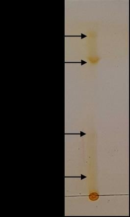
https://doi.org/10.33263/BRIAC123.30193033

 2.7. DPPH assay of latex extracts.

 The DPPH assay of the prepared extracts was done according to the procedure outlined
by Akowuah et al. [15]. 20 mL of 0.1 mM DPPH (extra pure 95%, SRL Chemicals) was
prepared with methanol as the solvent. Extract concentrations of 100 – 500 μg/mL for both AE
and TFE were prepared via simple dilution, starting from a stock concentration of 500 μg/mL.
The stock solutions were prepared by mixing 0.05 grams of a dried extract with 100 mL of its
corresponding solvent (distilled water or trifluoroethanol). Each prepared extract concentration
was then diluted again with methanol in a 1:5 ratio: 200 µl of an extract with 800 µl methanol.
2 mL of the prepared DPPH-methanolic solution was then pipetted to each of the diluted extract
concentrations. The mixtures were then mixed with a stirring glass rod and subsequently kept
under a shade for an hour. A negative control is simulated by pipetting 2 mL of DPPH-
methanolic solution to 1 mL of methanol. Using a UV-Vis spectrophotometer (GENESYS 10S
UV-Vis), three absorbance readings were measured at 517 nm for each extract concentration.
The mean absorbance readings for each extract concentration were then obtained and
incorporated onto Eqn. (3) to calculate the DPPH radical scavenging activity (%). A graph was
then plotted (for AE and TFE) to illustrate the relationship between the concentration of
extracts (μg/mL) and their respective DPPH scavenging activities (%).

 ( −
 (%) :
 100 (3)

 2.8. Phosphomolybdenum assay of latex extracts.

 Phosphomolybdenum assay was done according to the revised procedures outlined by
Saha et al. [16], which was first proposed by Prieto et al. [17]. To prepare the reagent solution,
0.6M of concentrated sulphuric acid (95-97% EMSURE®, Merck Chemicals) was mixed with
4 mM of ammonium molybdate (pure 98%, SRL Chemicals). 28 mM of sodium dihydrogen
phosphate (EMSURE®, Merck Chemicals) was then added into the mixture and stirred with a
stirring glass rod. 3 mL of the prepared solution was then pipetted into separate test tubes.
Subsequently, 0.3 mL of each of the different extract concentrations prepared earlier (100 - 500
μg/mL) were separately pipetted into the test tubes. For the negative control (blank), 0.3 mL of
methanol was added instead of the latex extract. All the test tubes were then incubated in the
hot air oven at 95 oC for 90 minutes. After cooling to room temperature, each of the test
mixtures' absorbance was measured at 695 nm. A graph was then plotted (for AE and TFE) to
illustrate the relationship between the concentration of extracts (μg/mL) and their respective
absorbance readings at 695 nm (a.u.).

 2.9. Agar well diffusion assay of latex extracts.

 Before carrying out the agar well diffusion assay, extract concentrations ranging from
500 – 2000 μg/mL for both AE and TFE were prepared by simple dilution, starting from a stock
concentration of 2000 μg/mL. The stock solutions were prepared by mixing 0.2 grams of the
dried extract with 100 mL of its corresponding solvent (distilled water or trifluoroethanol).
Agar well diffusion assay was done according to the procedures outlined by Purkayastha and
Dahiya [18]. Using sterile cotton swabs, inoculums of 4 selected bacteria (E. coli, P.
aeruginosa, S. aureus, and Bacillus sp.) were spread evenly on 2 sets of 4 nutrient agar
(HiMedia) plates (total of 8 plates). The plates for AE were labeled as A, B, C, and D while
the plates for TFE were labeled as E, F, G and H. Wells measuring 8 mm in diameter were
https://biointerfaceresearch.com/ 3022
https://doi.org/10.33263/BRIAC123.30193033

then aseptically punched into the agar with a gel puncher (6 equidistant wells per agar plate).
Subsequently, 10 μL of the different extract concentrations were pipetted into the wells in an
anti-clockwise manner. For the positive control, 5 μg of ciprofloxacin disc was used for all the
agar plates. 10 μL of distilled water was used as the negative control for AE, whilst 10 μL of
trifluoroethanol was used for TFE. The latex extracts were left to diffuse in the agar at room
temperature for 2 hours. All the agar plates were then incubated (upright position) at 37 oC for
24 hours. Post-incubation, the inhibitory zone diameters to the nearest millimeter (mm) were
measured and tabulated.

3. Results and Discussion

 3.1. Characterization of jackfruit latex.

 UV-Vis spectrum of dried latex mixed in DMSO is shown in Figure 1. As apparent
from the single sharp peak observable in the spectrum, the maximum absorbance (λmax) was
achieved at 290 nm. This denotes that the latex compounds' compounds maximally absorb
wavelengths of light within the ultraviolet B (UVB) band (280-315 nm).
 GC-MS analysis of latex demonstrated the presence of 1,2-benzoldicarbonsaeure, di-
(he) at RT - 20.704 as a major peak (Figure 2). Similar findings were reported in the ethanolic
extracts of Caralluma indica and Brassica oleracea var. capitata f. rubra [19, 20].

 3.2. Extraction of compounds from jackfruit latex.

 The latex yield efficiency for AE was calculated to be 15.26 %, whilst the latex yield
efficiency for TFE was 45.53 %. The yield for TFE was much higher than AE, denoting that
more nonpolar compounds were extracted from the dried latex (Table 1). Yield from Euphorbia
antiquorum and Calatropis gigantea was obtained as 91.05% and 59.47% respectively in
earlier studies [21].

 Table 1. Dry weights (g) and latex yield efficiencies (%) of AE and TFE.
 Extract Latex yield efficiency (%)
 AE 15.26
 TFE 45.53

 1.6
 1.4
 1.2
 Absorbance

 1
 0.8
 0.6
 0.4
 0.2
 0
 332

 376

 662

 706
 200
 222
 244
 266
 288
 310

 354

 398
 420
 442
 464
 486
 508
 530
 552
 574
 596
 618
 640

 684

 728
 750
 772
 794

 Wavelength
 Figure 1. UV-Vis spectrum of jackfruit latex.

https://biointerfaceresearch.com/ 3023
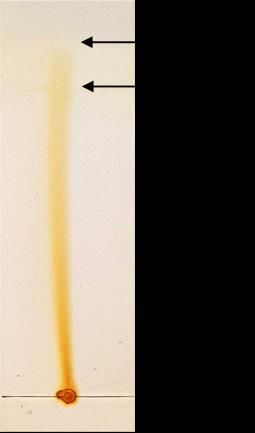
https://doi.org/10.33263/BRIAC123.30193033

 Figure 2. GC-MS analysis of jackfruit latex.

 3.3. Characterization of jackfruit latex extracts.

 As compared to the spectrum obtained for the dried latex, the UV-Vis spectrum for AE
(Figure 3) shows a significant shift to the left. As evident from the decrease in λmax (200 nm
vs. 290 nm), the compounds in AE absorb lower wavelengths of light at a higher frequency.
Besides that, the blunt absorption peak between 250 and 350 nm contrasts with the sharp peak
previously observed in Figure 1. With reference from (Figure 4), the decrease in λmax (269
nm vs. 290 nm) also signifies that the compounds in TFE absorb lower wavelengths of light at
a higher frequency. A sharp peak (albeit of lower absorbance) was also observed at 290 nm,
suggesting that some of the compounds in the dried latex lie in the aromatic range of carbonyl
groups (C=O).

 1
 0.9
 0.8
 0.7
 0.6
 Absorbance

 0.5
 0.4
 0.3
 0.2
 0.1
 0
 273

 537

 729
 200
 225
 249

 297
 321
 345
 369
 393
 417
 441
 465
 489
 513

 561
 585
 609
 633
 657
 681
 705

 753
 777

 Wavelength
 Figure 3. UV-Vis spectrum of AE with distilled water as the blank.

 After performing TLC for AE, there were 4 different spots visibly separated from one
another (Figure 5). With reference to Table 2, the distance traveled by spots 1, 2, 3, and 4 were
measured to be 7 mm, 21 mm, 50 mm, and 60 mm, respectively. As the solvent (methanol)
traveled a total distance of 70 mm, the retention factors of spots 1 to 4 were calculated to be
0.10, 0.30, 0.71, and 0.86, respectively.

https://biointerfaceresearch.com/ 3024
https://doi.org/10.33263/BRIAC123.30193033

 0.7

 0.6

 0.5

 0.4
 Absorbance

 0.3

 0.2

 0.1

 0
 223

 361

 499

 637
 200

 246
 269
 292
 315
 338

 384
 407
 430
 453
 476

 522
 545
 568
 591
 614

 660
 683
 706
 729
 752
 775
 798
 Wavelength
 Figure 4. UV-Vis spectrum of TFE with trifluoroethanol as the blank.

 Figure 5. TLC of AE using methanol as a solvent; stained with iodine.

 Table 2. Distance traveled (mm) by solvent and separated spots with their calculated Rf (AE).
 Identity Distance travelled (mm) Rf

 Solvent 70 -

 Spot 1 7 0.10

 Spot 2 21 0.30

 Spot 3 50 0.71

 Spot 4 60 0.86
 After subjecting TFE to TLC, it can be inferred that there were only 2 different 'spots'
visibly separated from one another (Figure 6). With reference to Table 3, the distance traveled
by spots 1 and 2 was measured to be 53 mm and 62 mm, respectively. As the solvent (methanol)
traveled a total distance of 70 mm, the retention factors of spots 1 and 2 were calculated to be
0.76 and 0.89, respectively. The similarity in Rf values between spot 3 and 4 of AE; and spot
1 and 2 of TFE may be due to the result of isomers having structural similarity but with different

https://biointerfaceresearch.com/ 3025
https://doi.org/10.33263/BRIAC123.30193033

functional groups, resulting in differences in polarity. This allows them to be separated during
solvent extraction with distilled water as well as trifluoroethanol.

 Figure 6. TLC of TFE using methanol as a solvent; stained with iodine.

 Table 3. Distance traveled (mm) by solvent and separated spots with their calculated Rf (TFE).
 Identity Distance travelled (mm) Rf

 Solvent 70 -

 Spot 1 53 0.76

 Spot 2 62 0.89

 3.4. Antioxidant activity of jackfruit latex extracts.

 Figure 7 shows the relationship between different jackfruit latex extract concentrations
(μg/mL) and DPPH scavenging activity (%).

 70
 DPPH Scavenging activity (%)

 60

 50

 40 Ascorbic
 acid
 30 AE

 20 TFE

 10

 0
 100 200 300 400 500
 Concentration (µg)

 Figure 7. DPPH scavenging activity (%) of jackfruit extracts.

https://biointerfaceresearch.com/ 3026
https://doi.org/10.33263/BRIAC123.30193033

 At 100 μg/mL, the DPPH scavenging activity of ascorbic acid, AE, and TFE were found
to be 37.53 ± 1.04, 18.89 ± 1.20 and 1.76 ± 0.19 respectively, whilst the DPPH scavenging
activity of ascorbic acid, AE, and TFE at 500 μg/mL were 64.72±0.80, 55.17±1.10 and
8.47±1.16 respectively. More DPPH radicals were evidently scavenged by 500 μg/mL of AE
and TFE (linear increments per 100 μg/mL of extracts). This signifies that the DPPH
scavenging activity increases as the concentration of the extracts increases. Although the
antioxidant activity of TFE showed an increasing trend, it is much lower than AE at all
concentrations.
 Figure 8 shows the results of the phosphomolybdenum assay, depicting the relationship
between different jackfruit latex extract concentrations (μg/mL) and absorbance at 695 nm. At
100 μg/mL, ascorbic acid, AE, and TFE had an absorbance reading of 0.343±0.002,
0.156±0.002, and 0.024±0.003 respectively, whilst the absorbance reading for ascorbic acid,
AE, and TFE at 500 μg/mL were 1.095±0.008, 0.735±0.003, and 0.118±0.003, respectively.
The linear increase in absorbance readings from 100 to 500 μg/mL reinforces the findings from
the DPPH assay. Similarly, TFE demonstrated the lowest absorbance readings at all
concentrations.

 1.2

 1

 Ascorbic
 Absorbance at 695 nm

 0.8 acid

 AE
 0.6
 TFE
 0.4

 0.2

 0
 100 200 300 400 500
 Concentration (μg/mL)

 Figure 8. Phosphomolybdenum assay of jackfruit extracts.

 Unraveling the bioactive capacity of jackfruit latex has been the forefront objective of
many researchers and scholars alike. As evident from the antioxidant findings in the present
study, linear relationships were apparent as the concentration of the jackfruit latex extract
increases. From the DPPH and phosphomolybdenum essay, it can be inferred that the
antioxidant activity of AE is on par with that of ascorbic acid, bearing values of proximity.
This coincides with the recent findings by Sundarrajan and Pottail [5], as they found that the
% inhibition of DPPH by jackfruit latex is comparable to that of ascorbic acid, with the lowest
inhibition difference being approximately 20%. As inferred by the authors, the aforementioned
trend is plausibly attributed to the polyphenol, alkaloid, flavonoid, and tannin content in the
latex, which was well-established to have significant levels of radical scavenging efficiency
[22- 24]. As surmised by Asfar et al. [25], these phyto-antioxidants proceed in a linear bio-
efficient mechanism; free radicals are actively reduced via electron donation, effectively
https://biointerfaceresearch.com/ 3027
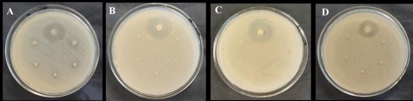
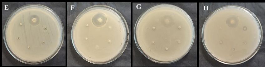
https://doi.org/10.33263/BRIAC123.30193033

hindering their capacity to inflict oxidative stress (and ultimately cell damage). Besides that,
the abundance of α-artostenone in jackfruit latex should also be acknowledged, plausibly
attributing to its antioxidant activity [6]. As this compound exists freely as enol-wax-esters,
they are thus composed of fatty acids and fatty alcohols, which are known to confer antioxidant
activity. An in vitro study by Shahinuzzaman et al. [26] affirms the aforementioned statement
as they found that the latex of Ficus carica (fig) demonstrated high levels of antioxidant
activity. Additionally, findings from Zhang et al. [27] elucidated the capacity of ascorbate,
thiols, and tocotrienol to confer antioxidant activity in the latex of Hevea brasiliensis. These
compounds may also be present in the latex of jackfruit, which must be unraveled in future
studies.
 The antioxidant capacity of jackfruit latex can be reinforced by relating to studies done
on other latex-producing plants. A study by Aljane et al. [28] found varying levels of
antioxidant activity among various fig ecotypes before concluding that figs are appreciable
sources of natural antioxidants. As the latex of figs was also determined to confer antioxidant
activity [26], the possibility that jackfruit latex possesses antioxidant activity should not be
overlooked. Devi et al. [29] reviewed the antioxidant capacity of many different parts of
jackfruit is well-acknowledged. As such, present findings of antioxidant activity in jackfruit
latex were to be expected. To further reinforce, the antioxidant activity of Aloe schelpei latex
was evidently determined by Teka and Kassahun [30], in which the antioxidant capacity of
Aloe variants was already well-acknowledged in the existing literature [31, 32]. Similar
findings were also apparent for other latex-producing plants, including studies on Euphorbia
dendroides [33] and Carica papaya [34].

 3.5. Antimicrobial activity of jackfruit latex extracts.

 Figure 9 shows the agar plates after performing agar well diffusion assay on AE (0 –
2000 μg/mL). With reference to Table 4, the positive control (5 μg of ciprofloxacin) was the
only well with a visible zone of inhibition (ranging from 24-29 mm). Increasing the
concentration of AE does not influence its antimicrobial capacity, as evident from the absence
of inhibitory zones (0 mm) in all the wells diffused with AE. Plates A-D swabbed with S.
aureus, Bacillus sp., E. coli, and P. aeruginosa, respectively were resistant to the extract
concentrations.

 Figure 9. Agar well diffusion results for AE; (A) S. aureus; (B) Bacillus sp.; (C) E. coli; (D) P. aeruginosa.

 On the other hand, Figure 10 shows the agar plates after performing agar well diffusion
assay on TFE (0 – 2000 μg/mL). With reference to Table 5, increasing the concentration of
TFE does influence its antimicrobial capacity, as evident from the inhibitory zones visible
surrounding the wells. The inhibitory zones were observed at the highest concentration of TFE.
For instance, plate H had inhibitory zones measuring 6 mm for 2000 μg/mL of TFE. E. coli
(plate G) was found to be resistant to TFE.
https://biointerfaceresearch.com/ 3028
https://doi.org/10.33263/BRIAC123.30193033

 Table 4. Inhibitory zone diameters of S. aureus, Bacillus sp., E. coli, and P. aeruginosa plates inoculated with
 ciprofloxacin (5 μg) and AE of increasing concentration (0 – 2000 μg/mL).
 Antibiotic AE concentration Inhibitory zone diameter to the nearest millimeter (mm)
 (μg/mL)
 (A) (B) (C) (D)
 S. aureus Bacillus sp. E. coli P. aeruginosa

 Ciprofloxacin - 29 25 28 24
 (5 μg)

 0 0 0 0 0

 500 0 0 0 0

 1000 0 0 0 0

 1500 0 0 0 0

 2000 0 0 0 0

 Figure 10. Agar well diffusion results for TFE; (E) S. aureus; (F) Bacillus sp.; (G) E. coli; (H) P. aeruginosa.

Table 5. Inhibitory zone diameters of S. aureus, Bacillus sp., E. coli, and P. aeruginosa plates inoculated with
ciprofloxacin (5 μg) and TFE of increasing concentration (0 – 2000 μg/mL).
 Antibiotic TFE Inhibitory zone diameter to the nearest millimeter (mm)
 concentration
 (μg/mL) (E) (F) (G) (H)
 S. aureus Bacillus sp. E. coli P. aeruginosa

 Ciprofloxacin - 27 30 28 25
 (5 μg)

 0 0 0 0 0

 500 0 0 0 0

 1000 0 0 0 0

 1500 0 0 0 3

 2000 3 2 0 6

 The antimicrobial capacity of jackfruit latex was made apparent from the agar well
diffusion assay in the present study. This is evident from the linear increase in inhibitory zones
in wells pipetted with increasing concentrations of the trifluoroethanol extract. This coincides
with the findings obtained by Sundarrajan and Pottail [5], as the incorporation of jackfruit latex
onto bimetallic silver-gold nanoparticles significantly improved the contraption's antimicrobial
activity. Various postulations regarding the antimicrobial capacity of jackfruit latex have been
theorized in the existing literature, including the role of proteinaceous enzymes. Siritapetawee
et al. [35] successfully isolated a 48-kDa antimicrobial serine protease from jackfruit latex,
which was then found to be capable of inhibiting the growth of P. aeruginosa and Candida
albicans. These findings were reinforced by atomic force microscopy (AFM) images of cell

https://biointerfaceresearch.com/ 3029
https://doi.org/10.33263/BRIAC123.30193033

suspensions treated with the purified protease, which significantly altered the normal
morphology of P. aeruginosa.
 The resin content of jackfruit latex may also be attributed to its antimicrobial activity
[7]. A study by Shuaib et al. [36] found that resin-rich methanolic extracts (RRMEs) of
Commiphora myrrha and Pinus roxburghii could inhibit the growth of Staphylococcus aureus
and Bacillus subtilis, with minimal sensitivity against Gram-negative bacteria. This coincides
with the findings from the present study, as E. coli was found to be resistant to the
trifluoroethanol extract of jackfruit latex. However, this does not apply to P. aeruginosa as it
was found to be the most sensitive amongst the 4 bacterial strains tested. To reinforce, a review
by Bahmani et al. [37] documented that the phytochemicals of medicinal plants (e.g., carvacrol,
limonene, borneol, linalool) were capable of inhibiting pseudomonal growth due to their
hydrophobic nature. These chemicals can separate lipids from bacterial cell walls, causing
membrane instability, thereby leading to ion expelling an electron imbalance. However, E. coli
can resist the aforementioned biomechanism. As evident from a study by Keweloh et al. [38],
E. coli responds to such stressors by modifying the fatty acid composition in their lipid
membranes, allowing them to resist oxidative damage [39]. This reinforces the absence of
inhibitory zones for E. coli as it was for this study.
 The antimicrobial activity of other jackfruit parts (e.g., seeds and leaves) are well-
acknowledged in the existing literature. To reinforce, Eve et al. [40] determined the
antibacterial capacity of jackfruit seeds on various diarrhea-inducing bacteria. The leaves of
jackfruit were also found to exhibit antifungal activity, a feat made possible by Vázquez-
González and colleagues [41]. A study in Malaysia found that essential oils extracted from
jackfruit seeds exhibited antibacterial activity, along with satisfactory radical scavenging
capacity (via DPPH assay) [42]. Since so many other parts of jackfruit exhibit antimicrobial
activity, the antimicrobial capacity of jackfruit latex extracts can be generalized to a small
extent. Latex from other plant species, including Jatropha sp. [43] and Euphorbia heterophylla
[44], showed appreciable antimicrobial activity, a relationship that is also apparent in the
present study done on jackfruit latex.

4. Conclusions

 As a concluding statement, it is evident that jackfruit latex possesses bioactive
properties. Antioxidant activity was found in both AE and TFE, while antimicrobial activity
was found only in TFE. These findings could propel future studies to pinpoint the exact
biological molecules involved in the aforesaid bioactivities. This can also pave the way for a
paradigm shift regarding the bio-significance of jackfruit latex for human applications,
ultimately improving its commercial, medical, and scientific interest.

Funding

This research received no external funding.

Acknowledgments

The authors wish to express their sincere gratitude to the School of Biosciences, MAHSA
University, for providing the necessary lab facilities, chemicals, reagents, disposables, and
equipment to conduct this study.

https://biointerfaceresearch.com/ 3030
https://doi.org/10.33263/BRIAC123.30193033

Conflicts of Interest

The authors declare no conflict of interest.

References

1. Morton, J. Jackfruit. Fruits of warm climates 1987, 58-64.
2. Kassim, N.; Hambali, K.; Amir, A. Nutritional Composition of Fruits Selected by Long-Tailed Macaques
 (Macaca fascicularis) in Kuala Selangor, Malaysia. Trop. Life Sci. Res. 2017, 28, 91–101,
 https://doi.org/10.21315/tlsr2017.28.1.6.
3. Jagadeesh, S.L.; Reddy, B.S.; Swamy, G.S.K.; Gorbal, K.; Hegde, L.; Raghavan, G.S.V. Chemical
 composition of jackfruit (Artocarpus heterophyllus Lam.) selections of Western Ghats of India. Food Chem.
 2007, 102, 361–365, https://doi.org/10.1016/j.foodchem.2006.05.027.
4. Garner, R.J.; Chaudhri, S.A. The propagation of tropical fruit trees. United Nations Food and Agricultural
 Organization: Rome, Italy, 1985.
5. Sundarrajan, K.S.; Pottail, L. Green synthesis of bimetallic Ag@Au nanoparticles with aqueous fruit latex
 extract of Artocarpus heterophyllus and their synergistic medicinal efficacies. Appl. Nanosci. 2021, 11, 971–
 981, https://doi.org/10.1007/s13204-020-01657-8.
6. Balakrishna, K.J.; Seshadri, T.R. Chemical examination of Jack tree latex and Jack fruit gum. Proc. Indian
 Acad. Sci. Section A 1947, 26, 46, https://doi.org/10.1007/BF03170948.
7. Nepacina, M.; Linis, V.; Janairo, J. Physical Characterization of Latex from Artocarpus heterophyllus Lam.
 (Jackfruit) and Four Related Artocarpus spp. Key Eng. Mater. 2020, 833, 107-117,
 https://doi.org/10.4028/www.scientific.net/KEM.833.107.
8. Kabir, S.; Fatteh, S. Jackfruit Induced Anaphylaxis Associated Birchpollen-Related Food Allergies. Ann.
 Allergy Asthma Immunol. 2018, 121, S120, https://doi.org/10.1016/j.anai.2018.09.398.
9. Wongrakpanich, S.; Klaewsongkram, J.; Chantaphakul, H.; Ruxrungtham, K. Jackfruit anaphylaxis in a latex
 allergic patient. Asian Pac. J. Allergy Immunol. 2015, 33, 65-68, https://doi.org/10.12932/AP0416.33.1.2015.
10. Swami, S.B.; Thakor, N.J.; Haldankar, P.M.; Kalse, S.B. Jackfruit and Its Many Functional Components as
 Related to Human Health: A Review. Compr. Rev. Food Sci. Food Saf. 2012, 11, 565-576,
 https://doi.org/10.1111/j.1541-4337.2012.00210.x.
11. Haq, N. Fruits for the Future 10. Jackfruit (Artocarpus heterophyllus). Southampton Centre for Underutilised
 Crops: Southampton, England, 2006; p. 192.
12. Abarca, L.F.S..; Klinkhamer, P.G.L.; Choi, Y.H. Plant Latex, from Ecological Interests to Bioactive Chemical
 Resources. Planta Med. 2019, 85, 856-868, https://doi.org/10.1055/a-0923-8215.
13. Ramirez‐Cadavid, D.A.; Cornish, K.; Hathwaik, U.; Kozak, R.; McMahan, M.; Michel, F.C. Development of
 novel processes for the aqueous extraction of natural rubber from Taraxacum kok‐saghyz (TK). J. Chem.
 Technol. Biotechnol., 2019, 94, 2452-2464, https://doi.org/10.1002/jctb.6027.
14. Samrot, A.; Selvarani, J.; Durga, A.; Lavanya, P.; Lavanya, J.; Paulraj, J.; Petchi, I. A Handbook on
 Phytochemical extraction, screening and its in-vitro bioactivity assays. SARAS Publisher: Nagercoil, India,
 2020.
15. Akowuah, G.A.; Ismail, Z.; Norhayati, I.; Sadikun, A. The effects of different extraction solvents of varying
 polarities of polyphenols of Orthosiphon stamineus and evaluation of the free radical-scavenging activity.
 Food Chem. 2005, 93, 311-317, https://doi.org/10.1016/j.foodchem.2004.09.028.
16. Saha, M.R.; Hasan, S.M.R.; Akter, R.; Hossain, M.M.; Alam, M.S.; Alam, M.A.; Mazumder, M.E.H. In vitro
 free radical scavenging activity of methanol extract of the leaves of Mimusops elengi Linn. Bangladesh J.
 Vet. Med. 2008, 6, 197-202, https://doi.org/10.3329/bjvm.v6i2.2336.
17. Prieto, P.; Pineda, M.; Aguilar, M. Spectrophotometric quantitation of antioxidant capacity through the
 formation of a Phosphomolybdenum Complex: Specific application to the determination of vitamin E. Anal.
 Biochem. 1999, 269, 337-341, https://doi.org/10.1006/abio.1999.4019.
18. Purkayastha, S.; Dahiya, P. Phytochemical screening and antimicrobial activity of some medicinal plants
 against multi-drug resistant bacteria from clinical isolates. Indian J. Pharm. Sci. 2012, 74, 443-450,
 https://doi.org/10.4103/0250-474x.108420.
19. Gnanashree, G.; Sirajudeen, P.M. Determination of bioactive compounds in ethanolic extract of Caralluma
 indica using GCMS technique. J. Pharmacogn. Phytochem. 2018; 7, 1675-1677.

https://biointerfaceresearch.com/ 3031
https://doi.org/10.33263/BRIAC123.30193033

20. Rajamani, L. Phytochemical and Gc-Ms analysis of Brassica oleracea var. capitata f. rubra. World J. Pharm.
 Res. 2018, 7, 1392 -1400.
21. Samrot, A.V.; Sahiti, K.; Bhavya, K.S.; Suvedhaa B. Synthesis of Plant Latex Based Hybrid Nanocarriers
 Using Surfactants for Curcumin Delivery. J Clust Sci. 2019. 30, 281–296 https://doi.org/10.1007/s10876-
 018-1472-5
22. Kasote, D. M.; Katyare, S. S.; Hegde, M. V.; Bae, H. Significance of antioxidant potential of plants and its
 relevance to therapeutic applications. Int. J. Biol. Sci. 2015, 11, 982–991,
 https://doi.org/10.7150%2Fijbs.12096.
23. Hussain, T.; Tan, B.; Yin, Y.; Blachier, F.; Tossou, M. C.; Rahu, N. Oxidative Stress and Inflammation: What
 Polyphenols Can Do for Us?. Oxid. Med. Cell Longev. 2016, 2016, 7432797,
 https://doi.org/10.1155/2016/7432797.
24. Maisetta, G.; Batoni, G.; Caboni, P.; Esin, S.; Rinaldi, A.C.; Zucca, P. Tannin profile, antioxidant properties,
 and antimicrobial activity of extracts from two Mediterranean species of parasitic plant Cytinus. BMC
 Complement Altern. Med. 2019, 19, 82, https://doi.org/10.1186/s12906-019-2487-7.
25. Asfar, A.A.; Zaheer, Z.; Aazam, E.S. Eco-friendly green synthesis of Ag@Fe bimetallic nanoparticles:
 antioxidant, antimicrobial and photocatalytic degradation of bromothymol blue. J. Photo. Chem. Photobiol.
 B. Biol. 2018, 185, 143–152, https://doi.org/10.1016/j.jphotobiol.2018.05.028.
26. Shahinuzzaman, M.; Yaakob, Z.; Anuar, F. H.; Akhtar, P.; Kadir, N.; Hasan, A.; Sobayel, K.; Nour, M.;
 Sindi, H.; Amin, N.; Sopian, K.; Akhtaruzzaman, M. In vitro antioxidant activity of Ficus carica L. latex from
 18 different cultivars. Sci. Rep. 2020, 10, 10852, https://doi.org/10.1038/s41598-020-67765-1.
27. Zhang, Y.; Leclercq, J.; Montoro, P. Reactive oxygen species in Hevea brasiliensis latex and relevance to
 Tapping Panel Dryness. Tree Physiol. 2017, 37, 261-269, https://doi.org/10.1093/treephys/tpw106.
28. Aljane, F; Neily, M.H.; Msaddak, A. Phytochemical Characteristics And Antioxidant Activity Of Several Fig
 (Ficus Carica L.) Ecotypes. Ital. J. Food Sci. 2020, 32, 755-768, https://doi.org/10.14674/IJFS.1884.
29. Devi, P.S.; Kumar, N.S.; Sabu, K.K. Phytochemical profiling and antioxidant activities of different parts
 of Artocarpus heterophyllus Lam. (Moraceae): A review on current status of knowledge. Futur. J. Pharm.
 Sci. 2021, 7, 30, https://doi.org/10.1186/s43094-021-00178-7.
30. Teka, T.; Kassahun, H. Characterization and Evaluation of Antioxidant Activity of Aloe schelpei Reynolds.
 Drug Des. Devel. Ther. 2020, 14, 1003-1008, https://doi.org/10.2147/DDDT.S241412.
31. Kaparakou, E.H., Kanakis, C.D., Gerogianni, M., Maniati, M., Vekrellis, K., Skotti, E.; Tarantilis, P.A.
 Quantitative determination of aloin, antioxidant activity, and toxicity of Aloe vera leaf gel products from
 Greece. J. Sci. Food Agric. 2021, 101, 414-423, https://doi.org/10.1002/jsfa.10650.
32. Klaikeaw, N.; Wongphoom, J.; Werawatganon, D.; Chayanupatkul, M.; Siriviriyakul, P. Anti-inflammatory
 and antioxidant effects of aloe vera in rats with non-alcoholic steatohepatitis. World J. Hepatol. 2020, 12,
 363-377, https://doi.org/10.4254/wjh.v12.i7.363.
33. Smeriglio, A.; Ragusa, S.; Monforte, M.T.; D'angelo, V.; Circosta, C. Phytochemical analysis and evaluation
 of antioxidant and anti-acetylcholinesterase activities of Euphorbia dendroides L. (Euphorbiaceae) latex.
 Plant Biosyst. 2019, 153, 498-505, https://doi.org/10.1080/11263504.2018.1498405.
34. Sharma, A.; Bachheti, A.; Sharma, P.; Bachheti, R.K.; Husen, A. Phytochemistry, pharmacological activities,
 nanoparticle fabrication, commercial products and waste utilization of Carica papaya L.: A comprehensive
 review. Curr. Res. Biotechnol. 2020, 2, 145-160, https://doi.org/10.1016/j.crbiot.2020.11.001.
35. Siritapetawee, J.; Thammasirirak, S.; Samosornsuk, W. Antimicrobial activity of a 48-kDa protease (AMP48)
 from Artocarpus heterophyllus latex. Eur. Rev. Med. Pharmacol. Sci. 2012, 16, 132–137.
36. Shuaib, M.; Ali, A.; Ali, M.; Panda, B. P.; Ahmad, M. I. Antibacterial activity of resin rich plant extracts. J.
 Pharm. Bioallied Sci. 2013, 5, 265-269, https://doi.org/10.4103/0975-7406.120073.
37. Bahmani, M.; Rafieian-Kopaei, M.; Hassanzadazar, H.; Taherikalani, M. Pseudomoniasis phytotherapy: a
 review on most important Iranian medicinal plants effective on Pseudomonas aeruginosa. Iran. J. Microbiol.
 2016, 8, 347–350.
38. Keweloh, H.; Diefenbach, R.; Rehm, H.J. Increase of phenol tolerance of Escherichia coli by alterations of
 the fatty acid composition of the membrane lipids. Arch. Microbiol. 1991, 157, 49–53,
 https://doi.org/10.1007/BF00245334.
39. Smirnova, G.; Samoylova, Z.; Muzyka, N.; Oktyabrsky, O. Influence of polyphenols on Escherichia coli
 resistance to oxidative stress. Free Radic. Biol. Med. 2009, 46, 759-768,
 https://doi.org/10.1016/j.freeradbiomed.2008.11.017.

https://biointerfaceresearch.com/ 3032
https://doi.org/10.33263/BRIAC123.30193033

40. Eve, A.; Aliero, A.A.; Nalubiri, D.; Adeyemo, R.O.; Akinola, S.A.; Pius, T.; Nabaasa, S.; Nabukeera, S.;
 Alkali, B.; Ntulume, I. In Vitro Antibacterial Activity of Crude Extracts of Artocarpus heterophyllus Seeds
 against Selected Diarrhoea-Causing Superbug Bacteria. Sci. World J. 2020, 2020, 9813970,
 https://doi.org/10.1155/2020/9813970.
41. Vázquez-González, Y.; Ragazzo-Sánchez, J.A.; Calderón-Santoyo, M. Characterization and antifungal
 activity of jackfruit (Artocarpus heterophyllus Lam.) leaf extract obtained using conventional and emerging
 technologies. Food Chem. 2020, 330, 127211, https://doi.org/10.1016/j.foodchem.2020.127211.
42. Ramli, A.; Badrulzaman, S.; Hamid, H.; Bhuyar, P. Antibacterial and antioxidative activity of the essential
 oil and seed extracts of Artocarpus heterophyllus for effective shelf‐life enhancement of stored meat. J. Food
 Process Preserv. 2020, 45, e14993, https://doi.org/10.1111/jfpp.14993.
43. Prastiyanto, M.E.; Tama, P.D.; Ananda, N.; Wilson, W.; Mukaromah, A.H. Antibacterial Potential
 of Jatropha sp. Latex against Multidrug-Resistant Bacteria. Int. J. Microbiol. 2020, 2020, 8509650,
 https://doi.org/10.1155/2020/8509650.
44. Pruthvi, M.L.; Mahesh, M.K.; Mary, R.S. Efficacy Of Euphorbia Heterophylla Latex Against Pathogenic
 Bacteria And Fungi. Asian J. Pharm. Clin. Res. 2020, 13, 141-145,
 https://doi.org/10.22159/ajpcr.2020.v13i6.37341.

https://biointerfaceresearch.com/ 3033
You can also read