Invasive snails, parasite spillback, and potential parasite spillover drive parasitic diseases of Hippopotamus amphibius in artificial lakes of ...
←
→
Page content transcription
If your browser does not render page correctly, please read the page content below
Schols et al. BMC Biology    (2021) 19:160
https://doi.org/10.1186/s12915-021-01093-2

 RESEARCH ARTICLE                                                                                                                                  Open Access

Invasive snails, parasite spillback, and
potential parasite spillover drive parasitic
diseases of Hippopotamus amphibius in
artificial lakes of Zimbabwe
Ruben Schols1,2*† , Hans Carolus3†, Cyril Hammoud1,4, Kudzai C. Muzarabani5, Maxwell Barson5,6,7 and Tine Huyse1

  Abstract
  Background: Humans impose a significant pressure on large herbivore populations, such as hippopotami, through
  hunting, poaching, and habitat destruction. Anthropogenic pressures can also occur indirectly, such as artificial lake
  creation and the subsequent introduction of invasive species that alter the ecosystem. These events can lead to
  drastic changes in parasite diversity and transmission, but generally receive little scientific attention.
  Results: In order to document and identify trematode parasites of the common hippopotamus (Hippopotamus
  amphibius) in artificial water systems of Zimbabwe, we applied an integrative taxonomic approach, combining
  molecular diagnostics and morphometrics on archived and new samples. In doing so, we provide DNA reference
  sequences of the hippopotamus liver fluke Fasciola nyanzae, enabling us to construct the first complete Fasciola
  phylogeny. We describe parasite spillback of F. nyanzae by the invasive freshwater snail Pseudosuccinea columella, as
  a consequence of a cascade of biological invasions in Lake Kariba, one of the biggest artificial lakes in the world.
  Additionally, we report an unknown stomach fluke of the hippopotamus transmitted by the non-endemic snail
  Radix aff. plicatula, an Asian snail species that has not been found in Africa before, and the stomach fluke
  Carmyerius cruciformis transmitted by the native snail Bulinus truncatus. Finally, Biomphalaria pfeifferi and two Bulinus
  species were found as new snail hosts for the poorly documented hippopotamus blood fluke Schistosoma
  edwardiense.
  Conclusions: Our findings indicate that artificial lakes are breeding grounds for endemic and non-endemic snails
  that transmit trematode parasites of the common hippopotamus. This has important implications, as existing
  research links trematode parasite infections combined with other stressors to declining wild herbivore populations.
  Therefore, we argue that monitoring the anthropogenic impact on parasite transmission should become an integral
  part of wildlife conservation efforts.
  Keywords: Trematodiasis1, Xenomonitoring2, One Health3, Barcoding4, Artificial lake5, Integrative taxonomy6,
  Taxonomic impediment7, Parasitology8, Conservation9, Biological invasions10

* Correspondence: ruben.schols@africamuseum.be
†
 Ruben Schols and Hans Carolus contributed equally to this work.
1
 Department of Biology, Royal Museum for Central Africa, Tervuren, Belgium
2
 Laboratory of Aquatic Biology, KU Leuven Kulak, Kortrijk, Belgium
Full list of author information is available at the end of the article

                                       © The Author(s). 2021 Open Access This article is licensed under a Creative Commons Attribution 4.0 International License,
                                       which permits use, sharing, adaptation, distribution and reproduction in any medium or format, as long as you give
                                       appropriate credit to the original author(s) and the source, provide a link to the Creative Commons licence, and indicate if
                                       changes were made. The images or other third party material in this article are included in the article's Creative Commons
                                       licence, unless indicated otherwise in a credit line to the material. If material is not included in the article's Creative Commons
                                       licence and your intended use is not permitted by statutory regulation or exceeds the permitted use, you will need to obtain
                                       permission directly from the copyright holder. To view a copy of this licence, visit http://creativecommons.org/licenses/by/4.0/.
                                       The Creative Commons Public Domain Dedication waiver (http://creativecommons.org/publicdomain/zero/1.0/) applies to the
                                       data made available in this article, unless otherwise stated in a credit line to the data.
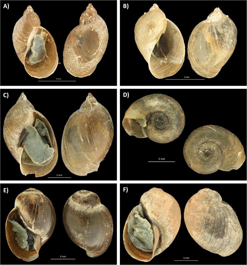
Schols et al. BMC Biology   (2021) 19:160                                                                       Page 2 of 21

Background                                                      North American snail Pseudosuccinea columella, which
Large artificial lakes can be a major driver of the socio-      in turn supports an extremely high prevalence (65%) of
economic development of a region by improving water             an unknown fasciolid trematode species [5]. The impres-
accessibility, stimulating agricultural irrigation and gen-     sive success of P. columella snails in colonizing Lake Ka-
erating renewable energy [1, 2]. Globally, 3700 medium          riba, as well as the high trematode infection prevalence,
to large size artificial lakes (those with a capacity larger    suggests a significant health burden for the affected de-
than 1 MW) have been built or are under construction            finitive host(s). The high parasite transmission by this
[2, 3]. Nevertheless, dam construction also has tremendous      invasive snail can be designated as either a spillback or a
repercussions on the surrounding environment as it can          spillover phenomenon [22]. To discriminate between
lead to the dislodgement and extinction of local aquatic        both scenarios, the identity of the parasite and final host
communities, altered migration of aquatic biota, changes in     are key. Carolus and colleagues [5] showed that the
aquatic biochemistry and physiology, disturbances of flood      trematode transmitted by P. columella is phylogenetic-
dynamics, introduction of exotic and invasive species, and      ally closely related to Fasciola gigantica and Fasciola
the creation of breeding grounds for (non-)endemic vectors      hepatica. However, the high genetic distances between
and intermediate hosts of (non-)endemic parasite species        this unknown species and other Fasciola species (based
[4–11]. Additionally, the subsequent intensification of agri-   on mitochondrial cytochrome c oxidase subunit I (COI)
culture, animal husbandry and aquaculture near artificial       sequences) suggested it to be (1) a new species, (2) a
lakes can lead to increased parasite transmission to            species not represented in current molecular databases,
humans, wildlife and domesticated animals [6, 12–15].           or (3) a hybrid species, as fasciolids are well-known for
   Snails play a key role as the intermediate host in the       their hybridization potential [23–25]. Therefore, the aim
life cycle of trematode parasites and are a prime example       of this study was to revisit Lake Kariba in order to col-
of intermediate hosts that thrive in artificial lakes, as       lect more information on this parasite and its intermedi-
shown by multiple studies [5, 7, 10, 11, 16]. Conse-            ate and definitive hosts. This subsequent sampling
quently, the health burden on the definitive host of these      campaign involved the collection of snails and adult liver
parasites increases, as illustrated by the construction of      and stomach flukes from a culled hippopotamus, a prime
the Diama dam in Northern Senegal, which caused dras-           suspect as definitive host [5]. An integrative taxonomic
tic changes in the aquatic chemistry (e.g., reduced salin-      approach [26], combining nuclear and mitochondrial
ity and increased pH) that favored the colonization of          markers, was applied to reveal the identity of the un-
planorbid snails and the subsequent establishment of            known Fasciola sp. and other trematodes. In addition,
one of the world’s biggest foci of human intestinal schis-      we investigated whether similar phenomena of biological
tosomiasis [17]. Another study suggests that dams block         invasions coupled with parasite transmission are occur-
migratory river prawns that predate on planorbid snails         ring in other artificial water systems in Zimbabwe.
and thereby increase the risk of schistosomiasis for mil-
lions of people worldwide [16]. The use of agrochemicals        Results
such as fertilizers and pesticides on irrigated land can        Trematode identification from the dissected
further stimulate snail—and thus trematode—prevalence           hippopotamus
by reducing snail predator populations and increasing           Morphological analysis
algal growth, the main food source of freshwater snails         Two morphologically distinct liver flukes (hereafter re-
[12, 15]. Besides favoring the colonization and prolifera-      ferred to as “Hippo liver fluke(s)”) were collected from
tion of endemic snail species, artificial lakes can also fa-    the liver bile ducts of the culled hippopotamus. In
cilitate the establishment of non-endemic snails that           addition, several hundred bright red stomach flukes
have the potential to drastically alter the dynamics of         (hereafter referred to as “Hippo stomach fluke type 1”)
trematode transmission [5, 18, 19]. Invasive non-               and two small yellowish stomach flukes (hereafter re-
endemic snails can affect parasite transmission in three        ferred to as “Hippo stomach fluke type 2”) were col-
distinct ways: (1) by introducing non-endemic parasites,        lected from the stomach wall of the hippopotamus. A
defined as “spillover”, (2) by successfully transmitting en-    high-resolution photograph of each adult fluke morpho-
demic parasites with an overall increased transmission          type is shown in Fig. 1. Scanning electron microscope
as a result, defined as “spillback”, or (3) by transmitting     (SEM) imaging was only possible for Hippo stomach
endemic parasites but with a lower transmission poten-          fluke type 1 due to the low sample size of the other mor-
tial, resulting in a “dilution” effect [20–22].                 photypes (Additional file 1: Figure S1). Specimens of
   Recently, we found strong indications that in Lake Ka-       Hippo stomach fluke type 1 had a cylindrical shape that
riba, the world’s biggest artificial lake by volume, the        tapered towards the anterior end and showed a marked
proliferation of the invasive water hyacinth (Eichhornia        color change from bright red in fresh specimens to
crassipes) has facilitated the establishment of the invasive    brown upon fixation. The Hippo stomach fluke type 2
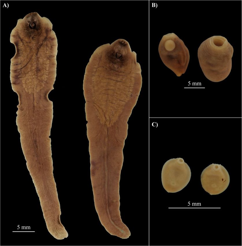
Schols et al. BMC Biology     (2021) 19:160                                                                                             Page 3 of 21

 Fig. 1. The collected flukes from the male hippopotamus at Lake Kariba. A Hippo liver flukes 1 and 2 (resp. left and right), B Hippo stomach fluke
 type 1, and C Hippo stomach fluke type 2. Scale bars represent 5 mm. Pictures were cropped and pasted to an artificial black background for
 contour visibility

had a discoid body shape and one specimen appeared to                       of F. hepatica, F. gigantica, and F. nyanzae [27, 30, 31].
have two small caudal appendages (Fig. 1c, right speci-                     Notably, the observed shedding time between 9 and 11
men). A comparative morphometric analysis (Table 1) of                      pm (see the “Trematode infection prevalence in snails”
the fixed Hippo liver flukes described above and fixed                      section), coincides with the increased foraging activity of
museum specimens of F. hepatica, F. gigantica, and F.                       hippos at night [32].
nyanzae (all shown in Additional file 1: Figure S2), com-
bined with the descriptions in literature [27–29], indi-                    Molecular analysis
cates that the hitherto unknown Fasciola sp. present in                     The obtained COI sequences for Hippo stomach fluke
Lake Kariba and Mwenje reservoir is morphologically                         type 1 and 2 (871 base pairs [bp] and 675 bp, “GenBank:
most similar to F. nyanzae. The body width at 2 mm                          MT909560 and MT909561” resp.) did not closely match
from the posterior end, the testis location, the testis to                  any of the available sequences on the GenBank or BOLD
body length ratio, and the vitellaria morphology differen-                  databases. The closest BLAST hits were Gastrothylax
tiate F. nyanzae from F. hepatica and F. gigantica. This,                   crumenifer (89% identity score) and Explanatum expla-
together with the fact that the definitive host is the com-                 natum (87% identity score), respectively. Our phylogen-
mon hippopotamus, supports the morphological identifi-                      etic analyses show that Hippo stomach fluke type 1 has
cation of the Hippo liver fluke(s) as F. nyanzae.                           close affinity with the Carmyerius genus (Fig. 2). Add-
Additionally, measurements (Table 1) of the metacercar-                     itionally, the sequence proved 100% identical (COI, 376
iae (Additional file 1: Figure S3) that were released from                  bp) to an amphistome infection isolated from Bulinus
two P. columella snails collected in Lake Kariba, fall                      truncatus in 2017 in Lake Kariba (“GenBank: MT013349
within the range reported in literature for metacercariae                   and MT013355” resp. for the snail and the amphistome

Schols et al. BMC Biology       (2021) 19:160                                                                                              Page 4 of 21

Table. 1 Morphometric analysis of Fasciola species. Three reference measurements from literature are provided for Fasciola nyanzae
collected from Hippopotamus amphibius [27–29]. All measurements are provided in mm, except measurements of metacercariae
which are displayed in μm. Metacercarial measurements for Hippo liver fluke 1 and 2 were taken from two shedding Pseudosuccinea
columella snails from site 3 in Kariba (Additional file 1: Figure S3). Metacercarial cyst measurements for Fasciola gigantica and Fasciola
hepatica were taken from Alicata [30] and Vareille-Morel et al. [31], respectively. Variables that differentiate between the three
Fasciola species are in bold italics. Measurements were taken with ImageJ software on 70% EtOH fixed samples with exception of
the reports from literature. Measurement methodology is shown in Additional file 1: Figure S2. Abbreviations: body length to body
width ratio (BL/BW), body length (BL), body width at widest point (BW), posterior width at 2 mm from the posterior end (BW2),
cephalic cone length until shoulders (CL), ventral sucker anteroposterior diameter (VS), testis location in body length (Testis loc.),
testis to body length ratio, vitellaria location in body length, outer diameter metacercaria (OD cyst), inner diameter metacercaria
(without fibrous layer, ID cyst)
Species        Source               Host        BL/BW     BL    BW      BW2 CL      VS       Testis loc.    Testis/ Vitellaria OD cysts       ID cysts
                                                                                                            BL                 (μm)           (μm)
F. nyanzae     Leiper (1910)        H.        ~ 7/1       69    ~9      3     3     1.25     /              /        25         /             /
                                    amphibius
F. nyanzae     Jackson (1921)       ?           3.6/1-    35-   4.5-    /     /     /        Anterior third /        /          /             /
                                                7.8/1     59    13.5
F. nyanzae     Dinnik and Dinnik H.        ~ 7/1          49-   ~9      /     /     1.46-    Anterior third 25%-     /          242-272       212-228
               (1961)            amphibius                91                        1.97                    40%
Hippo liver    This study           H.        4.9/1       52.4 10.6     3.1   /     1.47     Anterior third 24%      34.1       261           221
fluke 1                             amphibius
Hippo liver    This study           H.        3.5/1       46.9 13.3     3.2   2.2   1.72     Anterior third 29%      27.1       259           228
fluke 2                             amphibius
F. nyanzae     This study           H.        4.2/1       41.2 9.8      3     2.04 1.35      Anterior third 25%      25.4       /             /
RMCA                                amphibius
F. gigantica   This study           Bovine      3.6/1     35.3 9.9      5.1   3.1   0.83     Anterior two   41%      10.8       238-268       180-206
RMCA                                                                                         third
F. hepatica    This study           Bos taurus 3.1/1      26.8 8.6      4.6   1.67 1.47      Anterior two   42%      10.16      205-256       /
RMCA                                                                                         third

  Fig. 2. Maximum likelihood analysis of the available sequences of the Superfamily Paramphistomoidea using the GTR + G (= 0.80) + I (= 0.47)
  model on 758 bp of the COI marker. Homalogaster paloniae (“GenBank: KT266674”), from the family Gastrodiscidae, was used as an outgroup.
  Nodal support is indicated as bootstrap percentages (1000 bootstraps) and posterior probabilities, respectively before and after the “/” separator.
  Specimens from this study are indicated in blue. GenBank accession numbers are provided after the “|” separator
Schols et al. BMC Biology   (2021) 19:160                                                                        Page 5 of 21

infection; preprint, [33]). As no deeper taxonomic reso-        file 1: Figure S2) in our phylogenetic analysis. None of
lution could be obtained from the molecular data, we            the tested datasets showed conclusive evidence of substi-
further refer to this species as “Carmyerius sp.” Based on      tution saturation (see supplementary file: “Raw_data” for
COI sequencing, we can conclude that the Hippo stom-            the Steel’s and Xia’s method outputs per dataset).
ach fluke type 2 belongs to the superfamily Paramphisto-
moidea, but it could not be identified to a lower               Snail identification
taxonomic level in our phylogenetic analysis (Fig. 2).          Six different snail species were sampled in this study:
The COI sequence was, however, 100% identical (420              Pseudosuccinea columella and Radix aff. plicatula at
bp) to an amphistome infection in Radix sp. (“GenBank:          Lake Kariba and Radix natalensis, Biomphalaria pfeifferi,
MT013350”; preprint, [33]), here identified as Radix aff.       Bulinus globosus, and Bulinus sp. at Mwenje reservoir
plicatula (see the “Snail identification” section), from        (Fig. 4). Out of 16 sampled sites in Lake Kariba, only site
Lake Kariba [[5]; preprint, [33]]. All pairwise distances       3 and site 16 harbored snails (see Table 2 for the num-
with COI reference sequences on GenBank exceed 12%              ber of snails per site and Fig. 4 for information on sam-
and thereby surpass the 10% divergence of congeneric            pling locations). High-resolution photographs of one
trematodes [34]. Therefore, we further refer to this spe-       fixed specimen per species are depicted in Fig. 5.
cies as “unidentified amphistome species”. DNA sequen-             COI sequences of P. columella were 100% identical
cing of Hippo liver fluke 1 and 2 resulted in COI               (463 and 433 bp, “GenBank: MT888842”) to the invasive
fragments of 815 and 862 bp (“GenBank: MT909542 and             P. columella haplotype reported across Africa, Australia,
MT909543” resp.) and rDNA fragments of 3083 and                 and Northern America (i.e., Egypt [“BOLD:
2987 bp (“GenBank: MT909821 and MT909820”), re-                 GBMIN110283-17”], Australia [“BOLD: GBMLG0711-
spectively. The rDNA region, which was identical be-            06”], and USA [“BOLD: GBMPL484-13”]). We confirm
tween the two flukes, covered almost the entire 18S             the presence of this invasive haplotype in Lake Kariba,
rDNA (1829 bp, see the “Methods” section and Add-               as published before [5] (see “GenBank: MK333465”).
itional file 1: Figure S4), internal transcribed spacer 1       Carolus and colleagues [5] reported an unidentified
(ITS1, 454 bp), 5.8S rDNA (160 bp), internal transcribed        Radix sp., presumably originating from Asia, but the au-
spacer 2 (ITS2, 360 bp), and partial 28S rDNA (281 and          thors were not able to identify the species, as closely re-
185 bp resp. for Hippo liver fluke 1 and 2) regions. The        lated reference sequences were missing at that time. The
COI sequences differed 2.2% over 814 nucleotides. The           COI sequences from the Radix species sampled in this
COI and rDNA sequences showed the highest similarity            study (“GenBank: MT888847”) show only one mutation
(twice 99.3% for COI [“GenBank: MK330628-                       in the 350 bp overlapping COI sequence compared to
MK330630”] and 99.9% for rDNA [“GenBank:                        the Radix sp. sequenced by Carolus et al. [5] (see “Gen-
MK330623-MK330625”]) to the Fasciola sp. infections             Bank: MK333466”). In our phylogenetic analyses, the
in P. columella and Radix sp. (here identified as R. aff.       Radix sp. shows well supported clustering with a re-
plicatula, see the “Snail identification” section) from         cently generated reference sequence of Radix plicatula
Lake Kariba, reported by Carolus et al. [5]. The COI            from China [37] (Additional file 1: Figure S5 and Table
pairwise distances of Hippo liver fluke 1 and 2 with F.         S3). However, many radicine snail species are absent
gigantica (10.6% and 14.4%, resp.) and F. hepatica              from online genetic databases, and shell morphology dif-
(16.4% and 18%, resp.) exceed the 5% threshold for spe-         fers from the description in Vinarski et al. [38]. There-
cies differentiation based on the COI barcoding marker          fore, and in combination with the pairwise distance of
[34, 35]. The rDNA pairwise distances (rDNA sequences           3.6% between the Radix sp. from Kariba and the refer-
were identical between Hippo liver fluke 1 and 2) to F.         ence sequence of R. plicatula on a 463 bp COI align-
gigantica and F. hepatica were less pronounced com-             ment, we identify our current specimen and the
pared to COI distances (0.4% and 0.2%, resp.) (Add-             previously reported Radix sp. from Lake Kariba as the
itional file 1: Table S1 and S2). Phylogenetic analysis         non-endemic R. affinis plicatula (Additional file 1: Table
based on COI sequences place the Hippo liver fluke as a         S3). The qualifier affinis is utilized in accordance with
sister taxon to F. gigantica within the Fasciola genus          the open nomenclature guidelines of Sigovini and col-
(Fig. 3a and Additional file 1: Table S1). The placement        leagues [39], as our specimens show clear molecular af-
in the Fasciola genus is confirmed by the rDNA-based            finity to R. plicatula, but are not identical to it.
phylogeny (Fig. 3c and Additional file 1: Table S2). No            The identity of R. natalensis, B. pfeifferi, and B. globo-
DNA sequences could be obtained from the museum                 sus collected in Mwenje (446 bp, 446 bp, and 463 bp
samples, probably due to DNA degradation or PCR in-             resp.; “GenBank: MT888844-MT888846”) was confirmed
hibition as a result of long-term preservation, first in for-   based on phylogenetic clustering and pairwise distances
maldehyde and later in 70% ethanol. This prevented the          below 5%, compared to COI reference sequences in the
inclusion of the museum samples (shown in Additional            BOLD and NCBI databases (Additional file 1: Figure S5,
Schols et al. BMC Biology    (2021) 19:160                                                                                           Page 6 of 21

 Fig. 3. A Maximum likelihood analysis of the available sequences of the subfamily Fasciolinae using the GTR + G (= 0.25) model on 814 bp of the
 COI marker. Fasciolopsis buski (“GenBank: NC_030528”), from the subfamily Fasciolopsinae, was used as an outgroup. B The Fasciola nyanzae COI
 haplotype network of 11 sequences based on 412 positions, visualized using the TCS model in PopArt®. One hatch mark represents one
 mutation. The legend indicates the used color-code and haplotype abundance through circle size. C Maximum likelihood analysis of the available
 sequences of the subfamily Fasciolinae using the GTR + G (= 0.05) model on 2771 bp of the rDNA region. Fasciolopsis buski (“GenBank:
 MN970005”), from the subfamily Fasciolopsinae, was used as an outgroup. Nodal support is indicated as bootstrap percentages (1000 bootstraps)
 and posterior probabilities, respectively before and after the “/” separator. Specimens from this study are indicated in blue. GenBank accession
 numbers are provided after the “|” separator

S6 and Table S3, S4 and S5). A second Bulinus species                      Trematode infection prevalence in snails
from Mwenje (446 bp, “GenBank: MT888843”) could                            Cercarial shedding (i.e., release of trematode larvae) was
not be identified to species level based on COI barcoding                  exclusively observed between 9 pm and 11 pm in six out
but appears to be part of the Bulinus truncatus/tropicus                   of 103 examined P. columella snails (5.8%) collected in
species complex as indicated by the firm clustering                        Kariba. No other snail species was found to release cer-
within this group in our phylogenetic analysis (Add-                       cariae during shedding experiments. When screening for
itional file 1: Figure S6a and Table S5). Further taxo-                    infections through diagnostic PCRs, more infected snails
nomic identification was not possible as the species                       were detected. Table 2 shows the number of snails
group contains 14 species that are morphologically                         tested, the number of trematode infections detected and
nearly indistinguishable, many of which still lack reliable                the number of samples where a Schistosoma sp. or Fas-
reference genetic material [36, 40].                                       ciola sp. infection was detected in the multiplex PCR
Schols et al. BMC Biology                (2021) 19:160                                                                                                 Page 7 of 21

    Fig. 4. A Map indicating the sampling locations in the Zambezi basin including “Kariba Town” at Lake Kariba and “Mwenje” at Mwenje reservoir in Zimbabwe.
    B Sampling sites at Lake Kariba; figure adapted from Carolus et al. [5]. C Sampling site at Mwenje, a temporary puddle adjacent to the main reservoir located at
    S 17°14′ 47.9″ E 31°01′ 07.7″; figure adapted from Schols et al. [41]. D Sampling site 13 at Lake Kariba. E Sampling site at Mwenje reservoir

Table. 2 Snail abundance and trematode infections per site. All snail species are listed per site along with the number of specimens
collected (Sampled), the number of snails that shed cercariae in the shedding experiment (Shedding), the number of snails for
which DNA was extracted for diagnostic multiplex PCRs (Tested), the number of samples that tested positive for a trematode
infection (Trematoda inf.), and how many of those trematode infections had a Fasciola nyanzae (F. nyanzae inf.), Schistosoma
edwardiense (S. edwardiense inf.), or Schistosoma haematobium infection (S. haematobium inf.). For Kariba sites, the number between
brackets refers to exact sample location adopted from Carolus et al. [5]. High-resolution pictures of each snail morphotype are
shown in Fig. 5
Site            Species                         Sampled   Shedding      Tested     Trematoda inf.      F. nyanzae inf.    S. edwardiense inf.    S. haematobium inf.
Kariba (3)      Pseudosuccinea columella        60        6             24         21                  21                 0                      0
Kariba (3)      Radix aff. plicatula            12        0             12         3                   1                  0                      0
Kariba (16)     Pseudosuccinea columella        43        0             24         24                  24                 0                      0
Mwenje          Radix natalensis                17        0             17         13                  11                 0                      0
                                                                                                                           a
Mwenje          Biomphalaria pfeifferi          6         0             6          6                   0                  3                      0
Mwenje          Bulinus sp.                     1         0             1          1                   0                  1                      0
Mwenje          Bulinus globosus                9         0             9          9                   0                  6                      1
One more B. pfeifferi showed infection signals indicating a Schistosoma sp. infection but no sequences could be obtained, inhibiting species identification
a
Schols et al. BMC Biology (2021) 19:160 Page 8 of 21
Fig. 5. Pictures of snail morphotypes collected in Mwenje and Kariba. A Pseudosuccinea columella, B Radix sp. molecularly identified as Radix aff.
plicatula, C Radix natalensis, D Biomphalaria pfeifferi, E Bulinus globosus, and F Bulinus sp. Scale bars represent 5 mm. Pictures were cropped and
pasted to an artificial black background for contour visibility
assay. In summary, 77 out of 93 samples (82.8%) had hippopotamus (based on 408 bp). Eleven specimens of
(pre)patent trematode infections. Of all detected infec- Radix natalensis from Mwenje with a positive Fasciola
tions, a total of 57 out of the 61 (93.4%) infected lym- sp. PCR signal were used for COI and ITS1-5.8S-ITS2 se-
naeid snails (P. columella and R. aff. plicatula from quencing. Five COI sequences (~ 400 bp) were ~ 99%
Kariba and R. natalensis from Mwenje) were infected identical to a Diplostomidae sp. collected from the adja-
with a Fasciola sp., while 12 out of the 16 (75%) infected cent reservoir by Schols et al. [41] (“GenBank:
planorbid snails (B. pfeifferi, B. globosus, and Bulinus sp. MT994279”), two did not provide sequencing results of
from Mwenje) were infected with a Schistosoma species. sufficient quality and the remaining four amplicons (424
bp–428 bp, “GenBank: MT909546-MT909548 and
Trematode identification from snail infections MT909550”) showed a pairwise distance below 2% to
Tissue of one P. columella specimen and one R. aff. pli- the two adult F. nyanzae specimens from Kariba (results
catula specimen from Lake Kariba with a positive Fas- not shown, 408 bp). The ITS1-5.8S-ITS2 amplification
ciola sp. PCR signal was used for trematode COI and sequencing was successful for 10 out of the 11 Fas-
barcoding (425 bp and 482 bp resp., “GenBank: ciola sp. positive samples (464 bp, “GenBank:
MT909545 and MT909549” resp.). Both COI sequences MT893586-MT893595”). All 10 sequences were 100%
showed a pairwise distance of less than 0.5% compared identical to each other, to the two adult F. nyanzae and
to each other and less than 0.8% compared to the two to the S151 haplotype from Carolus et al. [5] (“GenBank:
adult F. nyanzae specimens from the culled MK330624”).Schols et al. BMC Biology (2021) 19:160 Page 9 of 21
Using the two-step diagnostic PCR assay of Schols Discussion
et al. [42] (see the “Methods” section, “detection of In this study, we report four trematode species that in-
infected snails”), we identified S. haematobium in a B. fect the common hippopotamus and show that they are
globosus snail. This was confirmed by sequencing part transmitted by six snail species that thrive in Lake Ka-
of the COI marker (335 bp, “GenBank: MT886703”) riba and Mwenje reservoir. We are able to complete the
and rDNA region (985 bp, “GenBank: MT884914”) in life cycle for three of these species by identifying them
this specimen. The remaining Schistosoma sp. infec- in both the final and the intermediate host. Below, we
tions in B. pfeifferi (4), B. globosus (6) and Bulinus sp. discuss the biological phenomena behind parasite trans-
(1) gave a Schistosoma-specific amplicon in the first mission in artificial lakes and the possible implications
multiplex PCR but did not yield a species-specific for threatened hippopotamus populations.
amplicon in the second multiplex PCR, indicating it
was not Schistosoma haematobium, Schistosoma man- 1. Completing the Fasciola phylogeny
soni, Schistosoma mattheei, Schistosoma bovis, Schisto- The adult liver flukes sampled in this study match the
soma curassoni, or Schistosoma guineensis [42]. We, morphological descriptions of Fasciola nyanzae made by
therefore, sequenced the COI marker and the Schisto- Leiper in 1910 [27]. Since then, only two scientific re-
soma-specific 5.8S-ITS2 amplicon generated in the ports have elaborated on the morphology and life cycle
first multiplex PCR. The COI marker (335 bp, “Gen- of F. nyanzae [27, 28]. Along with morphological mea-
Bank: MT886702”) was successfully amplified for only surements of the cercariae that were genetically identical
one B. pfeifferi infection and showed a pairwise dis- to the adult liver flukes, these morphological descrip-
tance of 0.2% to the preliminary identified S. edwar- tions enable us to identify F. nyanzae and provide the
diense isolated from Biomphalaria sudanica in first genetic record of this species. As a result, we can
Uganda (“GenBank: AY197347”). The 5.8S-ITS2 (~ provide the last missing link in the molecular phylogeny
340 bp, “GenBank: MT884915-MT884924”) marker of the Fasciola genus and show that F. nyanzae is most
was successfully sequenced for ten out of the eleven closely related to Fasciola gigantica based on partial COI
remaining samples and all sequences were 100% iden- mtDNA (Fig. 3a and Additional file 1: Table S1). This
tical to each other and to the S. edwardiense refer- close relationship is also supported by the observation
ence sequence (“GenBank: AY197344”). Based on that both species can infect the intermediate snail host
these results, we identify all ten remaining Schisto- R. natalensis and the definitive mammalian host H.
soma sp. infections in B. pfeifferi (3), B. globosus (6), amphibius, while no records exist of Fasciola hepatica in
and Bulinus sp. (1) as S. edwardiense. hippopotami [27, 43]. This phylogenetic relationship is,
however, less clear based on the more conserved nuclear
markers, where the distances between F. nyanzae and
Haplotype network of Fasciola nyanzae both F. hepatica and F. gigantica are of the same order
The genetic diversity of F. nyanzae in Lake Kariba and below 1% (Fig. 3c and Additional file 1: Table S2).
and Mwenje reservoir was assessed using all available This apparent mito-nuclear discordance can either be
COI sequences of F. nyanzae. To build the haplotype explained by drastically different evolutionary rates be-
network, we included both adult liver flukes collected tween nuclear and mitochondrial DNA [44] or by intro-
from the hippopotamus, F. nyanzae sequences isolated gressive hybridization [24, 45]. A higher genomic
from four infected P. columella from Lake Kariba (of coverage is needed to conclude on this, as concerted
which one from this study and three from Carolus evolution in the rDNA region can blur biological phe-
et al. [5]: “GenBank: MK330623-MK330625”), one se- nomena, such as a bias towards one of the two parental
quence from an infected R. aff. plicatula from Lake species after hybridization [46]. Our phylogenetic ana-
Kariba and F. nyanzae sequences from four infected lyses also enable us to contribute to the taxonomic dis-
R. natalensis from Mwenje reservoir. The resulting cussion on whether or not Fasciola jacksoni should be
alignment (412 bp) consisted of 11 sequences, of reclassified as Fascioloides jacksoni, as suggested by Lotfy
which nine were unique, and the associated haplotype et al. [47]. Mas-Coma et al. [43] opposed this reclassifi-
diversity (Hd) was 0.96. The highest pairwise distance cation, because the reference sequences used by Lotfy
between two haplotypes was 1.7%. The haplotype net- et al. [47] originated from regions where F. gigantica x F.
work shows no spatial genetic structure (Fig. 3b). hepatica hybrids are known to occur, F. nyanzae and
All GenBank identifiers generated by this study, and Tenuifasciola tragelaphi were missing from the phylo-
those of Carolus et al. [5] and Muzarabani et al. (pre- genetic analysis, and the mitochondrial marker (nad1)
print, [33]) involved in the analyses, are listed together used in the study of Lotfy et al. [47] is too variable, lead-
with the sample type, marker, taxonomic identification ing to substitution saturation. The inclusion of well-
and region of origin in Additional file 1: Table S6. selected reference sequences from regions where noSchols et al. BMC Biology (2021) 19:160 Page 10 of 21
hybridization has been reported, the addition of F. nyan- Africa [54–56]. Amphistomes are most commonly stud-
zae, and the use of a less variable mtDNA marker [48] ied in livestock, especially ruminants, but many species
in this study, results in a dataset without substitution infect wild animals. Here, we provide sequences, the
saturation and resolves most of the aforementioned is- intermediate host identity, and high–resolution photo-
sues. Our maximum likelihood and Bayesian phylogenies graphs of two amphistome species of the common
of mitochondrial and nuclear markers strongly support hippopotamus (i.e., Carmyerius cruciformis and an un-
the position of F. jacksoni within the genus Fascioloides identified amphistome species). We link these stomach
(Fig. 3a and c and Additional file 1: Table S1 and S2, fluke species to infections in the endemic snail B. trun-
respectively). catus reported by Muzarabani et al. (preprint, [33]) and
the non-endemic snail R. aff. plicatula, respectively.
2. Parasite spillback of the hippo liver fluke Fasciola Based on its morphology (Fig. 1b and Additional file 1:
nyanzae in Lake Kariba Figure S1), its final and intermediate hosts and close
Fasciola nyanzae infections were detected in three lym- phylogenetic affinity to Carmyerius exoporus (Fig. 2), we
naeid snail species: the endemic Radix natalensis, the tentatively identify the amphistome Hippo stomach fluke
Asian Radix aff. plicatula, and the North American type 1, genotyped as “Carmyerius sp.” in this study, as
Pseudosuccinea columella. The latter two species are in- Carmyerius cruciformis. First of all, the reported size,
vasive non-endemic snails that could affect parasite shape, and the distinct color change upon fixation in our
transmission in three distinct ways: parasite spillover, specimens match prior descriptions [29, 57]. Secondly, it
parasite spillback or a dilution effect (see the “Back- is the only Carmyerius species described from the hippo-
ground” section for explanations). Co-invading parasites, potamus so far [58]. Thirdly, members of the Bulinus
like other invading fauna and flora, typically go through genus are known intermediate hosts of this parasite [59],
a genetic bottleneck, resulting in low genetic diversity in which corresponds with the prior infections detected in
the invasive population [49]. In contrast, our F. nyanzae B. truncatus (“GenBank: MT013350, MT013355”; pre-
samples show a high genetic diversity with 9 out of the print, [33]), that match our genotype. Fourthly, the oral
11 sequenced COI haplotypes being unique. Moreover, opening and tegumental papillae (Additional file 1: Fig-
their definitive host (H. amphibius) and at least one of ure S1) correspond to prior reports of C. cruciformis [57,
their intermediate hosts (R. natalensis) are endemic to 60]. And finally, no reference sequence exists of C. cruci-
Zimbabwe [50, 51], supporting the endemicity of F. formis, probably explaining the failed attempt to identify
nyanzae to this region. In addition, another requirement this species through molecular barcoding. To our know-
to comply with a “parasite spillback” hypothesis was ledge, this is the first molecular record and southern-
met, as a higher F. nyanzae infection prevalence was de- most report (other reports are from Kenya, Uganda,
tected in the invasive snail P. columella compared to na- Benin, and Chad [58]) of C. cruciformis.
tive snails. We can therefore confirm that the cascade of The amphistome Hippo stomach fluke type 2 (Fig. 1c)
biological invasions, in which the invasion of P. colu- could not be identified based on the COI phylogeny that
mella was facilitated by the introduction and subsequent includes all families of the superfamily Paramphistomoi-
colonization of water hyacinth from South America as dea present in online molecular databases (Fig. 2). The
described in Carolus et al. [5], has led to “parasite spill- absence of a morphologically similar hippopotamus
back” of the endemic parasite F. nyanzae in Lake Kariba. trematode described in literature and the lack of refer-
Water hyacinth [52] and lymnaeid snails such as P. colu- ence sequences for many amphistome families (e.g., fam-
mella [53] generally thrive in nutrient rich, still, or slow- ily Brumptinae) make the identification of these
moving water, but not in fast moving riverine systems, specimens currently impossible. To further elucidate its
like the Zambezi river, which occupied the Kariba gorge phylogenetic position and identify it to species level,
before the construction of Kariba dam. Therefore, we future research should study the morphological char-
hypothesize that the parasite spillback phenomenon we acteristics such as the tegumental surface and the
witness here, is a result of this man-made genital pore through scanning electron microscopy
impoundment. [60] and transversal sections [57], respectively. Fur-
thermore, an integrative taxonomic approach could be
3. Two hippopotamus stomach flukes, one case of applied to generate reference sequences for the miss-
parasite spillover? ing amphistome families [26, 41]. For the Hippo
Although amphistomiasis, or stomach fluke disease, is stomach fluke type 2, sampled from the Asian snail
one of the most prevalent and pathogenic animal trema- R. aff. plicatula, no prior genetic and morphological
todiases, there is a significant knowledge gap concerning records exist, which raises the question whether this
the life cycle, host compatibility, prevalence, and geo- parasite is endemic to Africa. The answer will deter-
graphic range of many amphistome species, especially in mine whether this is a case of a potential “parasiteSchols et al. BMC Biology (2021) 19:160 Page 11 of 21
spillback” (like for F. nyanzae), or a “parasite spill- fluke, Fascioloides magna, is thought to be the single
over,” as a result of co-introduction of a non-endemic major cause of mortality in a declining population of
parasite with a non-endemic snail host. moose in the USA [68]. Schistosomes or blood flukes
are also thought to be an important yet overlooked cause
4. New intermediate hosts for the hippopotamus blood of animal mortality and productivity losses in the live-
fluke Schistosoma edwardiense? stock industry, with a major underestimated economic
Three endemic planorbid snail species, namely B. pfeif- impact [69]. Certain schistosome species have also been
feri, B. globosus, and an unidentified Bulinus sp. from suggested to be of concern to conservation of endan-
the Bulinus truncatus/tropicus complex, were found to gered animals such as rhinos and elephants in Asia [70]
be infected with the hippopotamus blood fluke S. edwar- and chimpanzees in Africa [71]. In addition to liver and
diense. Although schistosome blood flukes are among blood flukes, immature stomach flukes, or amphistomes,
the most studied trematode species due to their public inflict damage to the intestine, leading to anorexia and
health importance, wildlife schistosomes are severely diarrhea, while severe infections can be lethal [72, 73].
understudied. S. edwardiense has so far only been re- Amphistome infections are regarded as one of the most
ported in Uganda [61] and South Africa [62]. The spe- prevalent parasitic disease in livestock [55]. Due to their
cies is known to be transmitted through Biomphalaria high morbidity and mortality in young animals, some
snails [61], and so far, Bulinus snails have not been re- authors hypothesize that the economic losses due to
ported as an intermediate host. We cannot conclude amphistomiases are greater than many other parasitic
whether the molecular detection of S. edwardiense in the diseases in livestock [74].
two Bulinus species reflects true, compatible host- Few records exist on the pathology and impact of
parasite combinations or whether they are a result of trematode infections in hippopotami, but a similar im-
aborted infections, since we did not observe cercarial pact as described above can be expected. The most elab-
shedding in these samples (neither in the infected Biom- orate study on hippopotamus trematodiases dates back
phalaria snails). However, we found a positive PCR sig- to 1967 and reports one hundred culled hippopotami of
nal in seven bulinid specimens, and the closest known the Kruger National Park in South Africa [75]. It de-
relative of S. edwardiense, Schistosoma hippopotami, is scribes unknown fasciolids that colonized the bile duct
also transmitted through bulinid snails [61]. These facts in large numbers, causing lesions and fibrosis of the duct
strengthen our hypothesis that apart from B. pfeifferi, wall and damage extending into the liver parenchyma.
bulinid snails are compatible hosts for S. edwardiense The same study reports a high load of schistosomes in
too. Our results strengthen the statement of Morgan the heart and most major blood vessels, especially in the
et al. [61] that S. edwardiense and S. hippopotami belong venae cavae, pulmonary and hepatic veins, associated
to a different group, ancestral to the S. mansoni and S. with inflammation, macroscopic lesions, and endocardi-
haematobium species groups. The ability to use mem- tis [75]. Since then, no further pathological reports on
bers of both the Biomphalaria and Bulinus genera ap- trematode infections in the common hippopotamus have
pears as a “primitive” trait, which is lost in the derived been published to the best of our knowledge.
species groups that specialized in only one of the two Here, we provide a parasitological survey of a single
snail genera, respectively. culled hippopotamus from Lake Kariba, in which only
parts of the liver and stomach lining were studied. This
5. The potential health burden of hippopotamus revealed three different trematode species (i.e., F. nyan-
trematodiases zae, C. cruciformis and an unidentified amphistome)
Trematode parasites can have detrimental effects on the with a very high infection intensity of the stomach fluke
health, lifespan, and reproduction of wild and domestic C. cruciformis. In addition, the high trematode infection
animals. For instance, liver flukes cause hepatic fibrosis, prevalence in snail intermediate hosts, reported here and
cirrhosis, internal hemorrhage, and calcification of the in Carolus et al. [5], indicates that the exposure risk of
bile duct [63]. As a result, fascioliasis in livestock is asso- hippopotami to these parasites is high.
ciated with an increased mortality and abortion rate, re-
duced growth and a lower productivity [64, 65], which 6. Do artificial lakes and biological invasions pose a
accounts for global economic losses exceeding 3.2 billion burden on Hippopotamus amphibius?
US dollars [64]. Fasciola jacksoni and Protofasciola Considering our findings and the aforementioned bur-
rubusta can form a major health burden and source of den of trematodes on wildlife populations, we argue that
(calf) mortality in Asian [66] and African [67] elephants, artificial lakes can act as a breeding ground for (non-)en-
respectively. Additionally, F. jacksoni has been suggested demic intermediate snail hosts that transmit hippopot-
to be a major contributor to the drastic decline of en- amus infecting parasites, and thereby indirectly affect
dangered elephant populations in Asia [66]. Another hippopotamus (and possibly other wildlife) populations.Schols et al. BMC Biology (2021) 19:160 Page 12 of 21 Nevertheless, the actual impact of dam construction on infections with the liver fluke P. robusta have a syner- hippopotami cannot be directly verified. No data is avail- getic effect with drought-related starvation in mass- able on hippopotamus populations and their trematode mortality of African elephants in Kenya. Parasitic dis- fauna prior to dam construction or the arrival of invasive eases can thus have major consequences for threatened snail species. However, key historical reports do show a populations, making it a critical, although understudied link between the Kariba dam construction and altered issue for the conservation of species [92]. snail and trematode dynamics in the area. A health risk assessment prior to the Kariba dam construction con- Conclusions cluded that the chance for a schistosomiasis outbreak The data presented here suggests that the construction was minimal given the rocky substrate of Kariba gorge, of artificial lakes along with the introduction and estab- which is unsuitable for freshwater snails [76]. At that lishment of endemic and non-endemic species might time, only urinary schistosomiasis was present in the have increased or at least altered the burden of snail- surrounding communities at low prevalence, while B. borne diseases on hippopotami. Still, to quantify what pfeifferi and thus intestinal schistosomiasis was absent. the exact impact might be, we lack historic and current However, soon after dam construction, urinary schisto- information on the prevalence and infection intensities somiasis increased and for the first time, intestinal schis- of trematode species in the hippopotamus populations tosomiasis was reported [77]. Furthermore, Bulinus, in this region. Such information is hard to obtain and re- Biomphalaria, and lymnaeid species, all potentially suit- quires collaborations between researchers in the fields of able intermediate snail hosts for the hippopotamus para- ecology, veterinary science, and conservation biology as sites we describe here, colonized the lake [77]. Later suggested by previous studies [92, 93]. Nevertheless, studies confirmed the presence of planorbid snails and enriching the genetic database of wildlife trematodes to increased levels of schistosomiasis in nearby towns [78– design non-invasive sampling methods can generate 81]. Ultimately, the invasive snails P. columella and valuable resources in conservation studies [92]. For ex- Radix sp. (here identified as R. aff. plicatula), which we ample, by providing the genetic reference of parasites find to be compatible intermediate hosts for hippopot- like F. nyanzae and C. cruciformis, its prevalence can be amus parasites, were first reported in the lake in 2017 monitored by designing species-specific diagnostic PCRs [5]. In conclusion, these historical records and our find- to screen stool samples from hippos and other wildlife ings strongly suggest that the creation of Lake Kariba species. has drastically altered parasite transmission dynamics by favoring the colonization and proliferation of (non-)en- Methods demic snail species that transmit trematode parasites, in- Adult fluke collection and morphometric characterization cluding those infecting hippopotami. A subadult male Hippopotamus amphibius was culled by Large herbivores such as hippos play pivotal roles in rangers as part of the wildlife governance quota set for their ecosystems and are therefore crucial targets for population control, problem animal management, com- conservation efforts, which rely on a profound under- munity benefits, or other aspects of sustainable standing of the factors that put the target population at utilization by Zimbabwe Parks and Wildlife Manage- risk [82–85]. Among the major anthropogenic distur- ment Authority. The hippopotamus was culled near Ka- bances, such as habitat loss, hunting, and poaching [82, riba Town (Fig. 4), and the liver, bile ducts, and stomach 86], the impact of parasitic diseases remains hard to as- were dissected and inspected for adult trematode para- sess due to knowledge gaps in the diversity and epidemi- sites. The entire liver and a 15 by 15 cm part of the ology of most wildlife parasites [87]. Therefore, the stomach were screened for liver and stomach flukes, re- impact of trematode parasites on wildlife populations spectively. Fresh adult flukes were stored in 70% ethanol might be severely underestimated [88]. The common for later DNA analysis and morphometrics. The fixed hippopotamus is listed as vulnerable to extinction [50], flukes collected in this study, along with museum (Royal yet remains populous in southern and eastern Africa Museum for Central Africa, RMCA) specimens of Fas- with an estimated 5000 specimens in Zimbabwe [50, 85]. ciola nyanzae, F. gigantica, and F. hepatica (“RMCA tis- However, models show that these populations are two to sue vouchers: 31048, 29430, and 22595, resp.”), that five times more likely to cross the risk threshold of dras- were collected in the Democratic Republic of Congo in tic decline in the next 60 years, if mild human-induced 1956, 1952, and 1938, respectively, were photographed perturbations continue, alongside the present natural using a Canon EOS 600D camera equipped with a disturbance [85]. The combination of disease with other Macro Photo Lens according to Brecko et al. [94]. Pic- environmental or anthropogenic stressors can impose tures were taken at different focal depths while the spec- significant conservation threats [88–90]. This is illus- imens remained submerged in 70% ethanol and stacked trated by Obanda et al. [91], which suggest that using the Zerene Stacker™ software. Stacked pictures of
Schols et al. BMC Biology   (2021) 19:160                                                                    Page 13 of 21

the adult flukes were used for measurements in the Ima-       tube in the event of shedding. Snails that released cer-
geJ software (version 1.8.0). Morphometric measure-           cariae were individually stored together with the
ments were taken of body width at the widest point            emerged cercariae, while non-shedding snails were
(BW), body length (BL), cephalic cone length until            pooled per species per site. One representative of each
shoulders (CL), width at 2 mm from the posterior end          snail morphotype per site was, prior to DNA extraction,
(BW2), and the anteroposterior diameter of the ventral        fixed on black clay and photographed from its ventral
sucker (VS) (see Additional file 1: Figure S2 for a de-       and dorsal side according to Brecko et al. [94] using the
tailed visualization of how the measurements were             focus stacking system described above. The obtained
done). Specimens for scanning electron microscopy were        pictures were processed in Microsoft PowerPoint® by re-
transferred in acetone for 24 h, then airdried, mounted       moving the image background and combining the front
on aluminum stubs, coated with gold, and studied using        and rear perspective of the shell in one picture while en-
a JEOL JSM-6480LV scanning electron microscope.               suring a constant scale size. The (meta)cercariae were
                                                              placed in 80% ethanol on a glass slide with a glass cover.
Snail sampling and shedding experiments                       For taking and compiling photos, we used a digital cam-
The study area constituted of Mwenje reservoir, and the       era QImaging MicroPublisher 5.0 RTV mounted on a
northeastern shoreline of Lake Kariba, which are two          Leitz Dialux 22 stereomicroscope and piloted with the
artificial reservoirs of the Zambezi river basin in           Syncroscopy’s Image Reconstruction Software Auto-
Zimbabwe. Mwenje dam was erected in 1970, covers              Montage Pro (version 5.03.0061). The resulting images
roughly 5 km2 when full and has a catchment area of ap-       of (meta)cercariae were used for measurements in the
proximately 557 km2 [95]. The Kariba dam was com-             ImageJ software (version 1.8.0).
pleted in 1959 and Lake Kariba covers about 5,580 km2
when full and has an approximated catchment area of           DNA extraction
815,000 km2 [96]. Sampling sites are displayed in Fig. 4.     DNA of ethanol preserved adult trematodes was ex-
Locations sampled at Lake Kariba were adopted from            tracted using the DNeasy Blood and Tissue kit (Qiagen™)
prior studies [5, 79]. The Mwenje sampling site was lo-       according to the manufacturer’s protocol. For each DNA
cated at S 17°14′ 47.9″ E 31°01′ 07.7″ in close vicinity      extraction, we used a small piece of tissue isolated from
to the reservoir. Snails were collected in October 2018       the posterior side of the specimen with a sterile scissor.
from water, aquatic vegetation, and sediment by two           The DNeasy Blood and Tissue kit (Qiagen™) was not
persons for 30 min per site, using both a scooping net        successful for the museum samples of adult trematodes.
and manual collection. Collected snails were stored in        The specimens are currently stored in 70% ethanol yet
jars with lake water from the site of origin and kept at a    were initially fixed in formaldehyde at the time of collec-
constant temperature during transport. Upon arrival,          tion. Formaldehyde, a storage medium often used in the
snails were sorted per morphotype, counted, and prelim-       past by museums, is known to inhibit proteinase K-based
inary identified based on the morphological identifica-       DNA extractions [101]. Therefore, Chelex® (Biorad™) and
tion keys of Brown [97] and Mandahl-Barth [98]. Next,         phenol chloroform isoamyl alcohol (PCI) based DNA ex-
shedding experiments were conducted: snails were sepa-        traction methods were assessed for the same museum
rated in 12-well cell culture plates and incubated in fil-    samples, which do not rely on proteinase K for lysis. For
tered and aged lake water in complete darkness                the Chelex-based extraction, tissue was first incubated
overnight, followed by bright light exposure for 5 h from     for 24 h in ATL buffer (Qiagen™) to flush excess formal-
7 am to 12 pm. This treatment induces the natural emer-       dehyde as suggested by Shedlock et al. [101]. Next, sam-
gence (i.e., shedding) of larval stages, the so-called cer-   ples were homogenized with a sterile scalpel on a glass
cariae, of several trematode species [55, 99]. Well           plate. Tissue homogenate was incubated for 1 h at 56 °C
contents and surfaces were inspected with a stereo-           and 30 min at 95 °C in 200 μL of 5% Chelex® (Biorad™)
microscope for the presence of cercariae and/or meta-         solution in a shaking Thermocycler™. Next, samples were
cercariae, respectively, as cercariae from liver flukes are   centrifuged for 7 min at 13,000×g, and supernatant DNA
known to rapidly encyst and form metacercariae on bi-         extract was collected and stored at − 20 °C. Regarding
otic or abiotic surfaces [100]. This was done at the start    the PCI-based DNA extraction, tissue samples were
of the shedding experiment, 10 pm, 7 am, and hourly be-       dried with sterile absorbent paper, 300 μL Tris-EDTA
tween 7 am and 12 pm during the bright light exposure         (TE) buffer and 300 μL PCI solution (phenol pH 6.7-
experiment to detect nocturnal and diurnal shedding, re-      chloroform-isoamylalcohol [25:24:1]) was added, and tis-
spectively. All snails were sacrificed by heat shock in ~     sue was lysed by micro-bead shearing in a FastPrep-
70 °C water to prevent contraction and simplify later         24TM Classic lysis system (20 s, 6 m/s) (MP biomedical™).
DNA extraction, followed by fixation in 80% ethanol,          The DNA containing fraction was separated from lipids
and then we added the released cercariae to the same          and cellular debris by 10 min of centrifugation at 14,000
Schols et al. BMC Biology (2021) 19:160 Page 14 of 21
rpm. Next, 15 μL sodium acetate (3 M, pH 5.2) and a Schistosoma spp. infection were subsequently analyzed
450 μL ethanol (100%) were added and the mixture was using a second multiplex PCR according to the two-step
placed in a freezer at − 20 °C for 20 min followed by cen- approach described by Schols et al. [42]. This PCR dif-
trifugation for 10 min at 4 °C (14,000 rpm) for DNA pre- ferentiates between several schistosomes of veterinary
cipitation. Pellets were washed by adding ice cold and medical importance (i.e., S. haematobium, S. man-
ethanol (70%) followed by centrifugation (10 min at 4 °C, soni, S. mattheei and S. bovis/S. curassoni/S. guineensis)
14,000 rpm). The resulting DNA was dissolved in TE by generating species-specific COI amplicons of different
buffer and stored at − 20 °C. Successful DNA isolation lengths. By selectively screening all snail DNA extracts
was verified by gel electrophoresis (0.8% agarose gel, using either of the two methods (i.e., Lymnaeidae with
SYBR™ Safe). the method of Carolus et al. [5] and Planorbidae with
The DNA of (meta)cercariae was extracted using a the method of Schols et al. [42]), we identified Schisto-
proteinase K-based lysis buffer as described in Ziętara soma spp. and Fasciola spp. infections that were used
et al. [102]. In short, 10 μL of the prepared proteinase K for further analysis.
lysis buffer was added to a single cercaria in 10 μL of
MilliQ water (Merck, Darmstadt, Germany). A first and PCR and DNA sequencing
second incubation step followed at 65 °C for 25 min and A fragment of the mitochondrial COI and a nuclear
95 °C at 10 min, respectively. After the protocol, DNA rDNA region covering the 18S rDNA marker, the in-
extracts were stored undiluted at − 20 °C. ternal transcribed spacer 1 (ITS1), the 5.8S rDNA
For DNA extractions from ethanol preserved snail marker, the internal transcribed spacer 2 (ITS2), and/or
samples, soft tissue was isolated from the shell by using the partial 28S rDNA marker were amplified to assess
a sterile needle to make a hole in the apex to push the the taxonomic position of trematode species sampled in
soft tissue out of the shell. Next, the isolated tissue was this study. Additional file 1: Figure S4 indicates the an-
dried using absorbent paper and homogenized using a nealing sites of the used primers for amplification of the
sterile scalpel. DNA was extracted using the E.Z.N.A.® rDNA region of adult liver flukes. The primers and
Mollusc DNA Kit (OMEGA Bio-tek, Norcross, GA, resulting amplicon lengths are listed in Table 3, together
USA) according to the manufacturer’s protocol and with those for snail and trematode identification. We
eluted through two elution steps of 75 μL, totaling to barcoded (COI sequencing) two random specimens per
150 μL of DNA extract. Additionally, the more cost- and snail species and all Schistosoma and Fasciola spp. in-
time efficient Chelex® (Biorad™) method [5, 103] was fected snails. For the parasites, we selected all Schisto-
used on 24 non-shedding P. columella specimens per soma and Fasciola spp. infections, all adult fasciolids and
site in Lake Kariba, as described previously. All DNA ex- one adult amphistome specimen per morphotype. A new
tracts were diluted 1:10 with MilliQ water (Merck, Fasciola-specific reverse primer COI_FAS_R (‘5-
Darmstadt, Germany) to prevent inhibition of PCR GACAAACAAACACAAGCAGG-3’), targeting a shorter
reactions. COI fragment of 148 nucleotides when combined with
the COI1_DIG_F primer (see Table 3), was designed to
Molecular diagnosis of infected snails amplify DNA of the Fasciola museum samples, as no
The DNA extracts of a maximum of 24 snails per spe- successful PCR amplification was obtained using the lon-
cies per site were subjected to the diagnostic multiplex ger amplicon. It has become apparent that shorter
PCR methods described in Schols et al. [42] and Carolus amplicons (< 400 bp) outperform longer amplicons due
et al. [5]. These approaches enable rapid and cost- to severe DNA fragmentation and/or degradation in old
efficient diagnosis of trematode infections in snail DNA samples [107–109], such as the museum specimens in
extracts, with simultaneous detection of Schistosoma this study. Simplex PCR reactions and programs for se-
spp. [42] or Fasciola spp. [5]. Both methods are based quencing were performed, according to Carolus et al.
on the amplification of multiple amplicons of different [5], in a 15 μL volume with 1.5 μL of DNA extract using
lengths in a single PCR reaction: an internal control that the Qiagen™ Taq DNA polymerase kit containing 1.5
amplifies 18S snail rDNA to confirm the success of the mM PCR buffer (Qiagen™), 0.6 mM dNTP mix (Qia-
DNA extraction and PCR reaction, a general trematode gen™), 1.5 mM MgCl2, 0.45 units of Taq Polymerase
primer pair designed to amplify the 18S rDNA of all (Qiagen™), 0.8 μM forward primer, and 0.8 μM reverse
trematode genera that have a reference sequence avail- primer. The PCR reactions were performed as follows:
able in GenBank and Schistosoma- [42] or Fasciola- [5] initial denaturation at 94 °C for 15 min, 39 cycles of
specific internal transcribed spacer 2 (ITS2) or mito- 94 °C for 30 s, annealing temperature depending on pri-
chondrial cytochrome c oxidase subunit I (COI) primers, mer pair as listed in Table 3 for 45 s and 72 °C for 45 s
respectively. All used primers and PCR protocols are de- and a final elongation step at 72 °C for 10 min in a Tpro-
scribed elsewhere [5, 42]. Samples that were positive for fessional Thermocycler (Biometra™). PCR products wereYou can also read