Integrative Biology REVIEW ARTICLE
←
→
Page content transcription
If your browser does not render page correctly, please read the page content below
Integrative Biology
This article is licensed under a Creative Commons Attribution-NonCommercial 3.0 Unported Licence.
View Article Online
REVIEW ARTICLE View Journal | View Issue
Modeling of the mechano-chemical behaviour
of the nuclear pore complex: current research
Open Access Article. Published on 21 September 2016. Downloaded on 6/11/2022 10:35:09 AM.
Cite this: Integr. Biol., 2016,
8, 1011 and perspectives
Alberto Garcia,* Jose F. Rodriguez Matas and Manuela T. Raimondi
Recent evidence suggests that mechanical deformation of the cell nucleus regulates the nuclear import
of the transcriptional activators of genes involved in primary physiological cell responses such as stem
cell differentiation. In addition, this nuclear mechanosensing response is de-regulated in pathological
states, such as cancer and neurodegeneration. One hypothesis that could greatly advance the field is
that the deformation of the nuclear envelope activates nuclear pore complexes through a direct mechanical
link. The understanding of this possible mechanism for nuclear pore complex stretch-activation entails
studying the mechanical connection of this complex to the nuclear envelope at the nanoscale. The
nanomechanics of the nuclear pore complex is thus emerging as a novel research field, bridging
nanoscience with nanotechnology. This review examines the frontier of research methodologies that are
potentially useful for building a computational model of this interaction. This includes, for example,
Received 3rd August 2016, electron tomography to assess the geometrical features of the nuclear pore complex and nanoindentation
Accepted 19th September 2016 to estimate its mechanical properties and that of the nuclear envelope. In order to summarize the
DOI: 10.1039/c6ib00153j state-of-the-art and perspectives in the field of NPC nanomechanics, this review covers highly
interdisciplinary experimental and theoretical research methodologies pertaining to the fields of physics,
www.rsc.org/ibiology chemistry, biology, materials and mechanics.
Insight, innovation, integration
We explore the literature supporting the hypothesis of a relation between transport through the nuclear pore complex (NPC) and mechanical forces that it
experiences from the surroundings, providing insights into a possible mechanism of NPC stretch-activation. The frontier technology for nuclear pore complex
characterization together with computational simulations would be a powerful tool to interpret research into a possible mechanism of nuclear pore stretch
activation. We demonstrate integration of technology and biology regarding: (1) characterization techniques for the nanostructure of the NPC and the assembly
of the nuclear envelope/lamina/NPC; (2) techniques to obtain the pore architecture and boundary conditions for numerical analysis; and (3) modelling
techniques of the relationship between nucleocytoplasmic transport and the mechanical forces transmitted on the NPC.
Cell responses are based on biochemical signals, which enable models to describe cell behavior.6–9 Similarly to the physiological
structural internal changes such as cytoskeletal remodeling, case, the appearance of cellular pathological processes and
contraction and stretching. The cell’s ability to feel external diseases10–12 can also be linked to external stimuli when they
stimuli and transform them into internal chemical reactions is have a negative impact on cell functions.13
known as mechanosensing and mechanotransduction, respec- In eukaryotic cells, nuclear pore complexes (NPCs) are the
tively.1–4 A recently published review5 provides a highly detailed gates through which molecular exchange and genetic transport
history of scientific publications regarding the mechanical between the cytoplasm and the nucleus take place (Fig. 1). The
interaction of cells with their microenvironment under physio- NPC literally pierces the nuclear envelope (NE) of the nucleus
logical conditions, mostly from an experimental/microstructural allowing the exchange of molecules between the nucleus and the
point of view. However, the work also reports on efforts linking the cytoplasm. The exchange of molecules and genetic information
observed microstructural aspects of the cell with computational through the nuclear envelope, and how this affects cell differ-
entiation, adaptability, and also disease, depend on how this
Department of Chemistry, Materials and Chemical Engineering ‘‘Giulio Natta’’,
trafficking takes place through the NPC. Both chemical and
Politecnico di Milano, Piazza L. da Vinci, 32, 20133 Milan, Italy. mechanical factors are involved in cell motility and remodeling,
E-mail: alberto.garcia@polimi.it; Tel: +39 02 2399 4729 and such responses are led by the response of the NPC in
This journal is © The Royal Society of Chemistry 2016 Integr. Biol., 2016, 8, 1011--1021 | 1011
View Article Online
Review Article Integrative Biology
regulating the transport of signaling molecules between the This review aims to highlight these advances and their
nucleus and the cytoplasm. The question as to how the NPC potential application in order to unveil the nanomechanics of
guides this molecular exchange is still unanswered. However, the NPC.
over the last few years researchers have made major break- The review is organized as follows. In Section 1, ‘‘Geometry
This article is licensed under a Creative Commons Attribution-NonCommercial 3.0 Unported Licence.
throughs in the fields of nanoscale imaging and mechanical and structure of the nuclear pore complex’’, we focus on the
characterization in terms of understanding the main geometrical current knowledge of the NPC architecture and geometry, and the
and structural features of the NPC. On the other hand, the most advanced imaging techniques used for accurately describ-
mechanical behavior of the NPC and the mechanisms by which ing these features. Section 2, entitled ‘‘Chemo-mechanics of the
this behavior can affect nucleocytoplasmic transport remain nuclear pore complex’’, describes the multiscale and multiphy-
Open Access Article. Published on 21 September 2016. Downloaded on 6/11/2022 10:35:09 AM.
poorly understood. The new frontier techniques available today, in sics nature of the relationship between cell deformation and
both experimental and computational fields, are potentially useful nucleocytoplasmic transport. Section 3, ‘‘Mechanical properties
for building a computational model of the mechanical behavior of the nuclear pore complex: modeling at multiple scales, from
of the NPC in response to nuclear envelope stretching.14–16 coarse-grained to atomistic scales’’, describes the main efforts
concentrated on characterizing the material properties of the
NE–NPC assembly, the recent contributions to understanding the
mechanical behavior of the NPC, and its role in the regulation of
Alberto Garcı́a obtained his the molecular exchange through the NPC. At the end of Section 3
degree in Industrial Engineering there is a summary of the main advances in the computer
(Mechanics) from the ‘‘Univer- simulation of NPC mechanics. Section 4, ‘‘Conclusions and
sidad de Oviedo’’ (Spain) and perspectives’’, provides our concluding remarks and perspectives
his PhD (European Mention) in for studying the dependence of the NPC on mechanical stimuli,
Computational Mechanics (bio- with an emphasis on the role that computational modeling will
engineering) from the ‘‘Universidad play in this regard.
de Zaragoza’’ (Spain). After working
as a mechanical designer and as
an analyst (freelance), he started 1 Geometry and structure of the
as a postdoctoral researcher at nuclear pore complex
‘‘Dublin City University’’ (Ireland)
Alberto Garcı́a in biomechanics and medical This section summarizes the main advances regarding the mecha-
imaging (MRI and DTI). In 2015 nical dependency of the nucleoplasmic exchange of the NPC.
he became a senior researcher at the Laboratory of Biological Particular emphasis is placed on the mechanical factors affecting
Structure Mechanics (LaBS) of the ‘‘Politecnico di Milano’’, the transport of solutes, and on the visualization and computa-
focusing his research on the nuclear pore complex. Recently he tional techniques used to study this process. The term pore
moved to the ‘‘Universitat Politècnica de Catalunya’’ as a lecturer complex was introduced in ref. 17 to define the nucleocyto-
and member of the Lacàn Group. plasmic structure channel in animal cells visualized using an
José F. Rodrı́guez Matas received Manuela T. Raimondi is an asso-
his BE degree in mechanical engi- ciate professor of bioengineering
neering from the Universidad at Politecnico di Milano, where
Simón Bolı́var, Caracas, Venezuela, she teaches the course ‘‘technol-
in 1993, and his PhD in mechanical ogies for regenerative medicine’’
engineering from the University of to graduate students of the MS
Notre Dame, IN, in 1999. He is program in biomedical engineer-
currently an associate professor ing. She is the founder and head
of bioengineering in the Depart- of the Mechanobiology Lab and
ment of Chemistry, Materials and of the Interdepartmental Live
Chemical Engineering ‘‘Giulio Cell Imaging lab. She integrates
Natta’’ at Politecnico di Milano. multiphysics/multiscale computa-
José F. Rodrı́guez Matas His research interests concern Manuela T. Raimondi tional modeling with advanced
computational mechanics applied cell culture techniques, including
to the biomechanics of soft tissues and couple biophysical problems. synthetic stem cell niches and micromechanical bioreactors, for
He is particularly involved in nonlinear finite element applications, investigating basic stem cell mechanobiology. She is currently
and the development of efficient computational tools for cardiac the Principal Investigator of an ERC-funded project entitled
electrophysiology and couple problems. ‘‘Mechanobiology of nuclear import of transcription factors
modelled within a bioengineered stem cell niche’’.
1012 | Integr. Biol., 2016, 8, 1011--1021 This journal is © The Royal Society of Chemistry 2016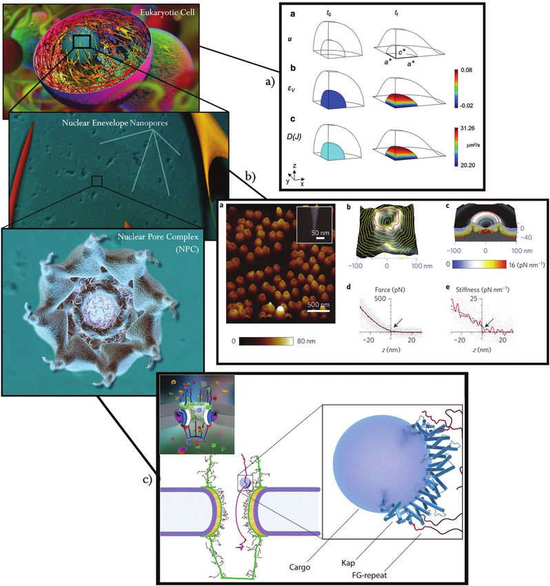
View Article Online
Integrative Biology Review Article
This article is licensed under a Creative Commons Attribution-NonCommercial 3.0 Unported Licence.
Open Access Article. Published on 21 September 2016. Downloaded on 6/11/2022 10:35:09 AM.
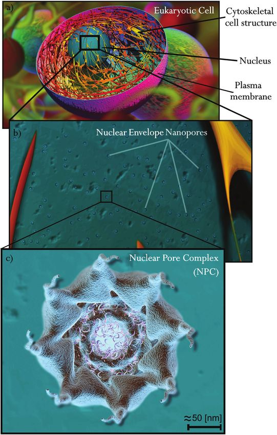
Fig. 1 3D rendered illustration of a eukaryotic cell and its NPC distribution in the NE. (a) Cytoskeletal structure. (b) Distributed nanopores in the nuclear
envelope, zoom-in of a eukaryotic cell, and a cytoplasmic side view. (c) Zoom-in of one of the 8-fold rotational symmetric nanopores; the nuclear pore
complex, represented in an axial direction (cytoplasmic side).
This journal is © The Royal Society of Chemistry 2016 Integr. Biol., 2016, 8, 1011--1021 | 1013
View Article Online
Review Article Integrative Biology
electron microscope (EM) and with an NPC diameter average electron microscopy (FE-SEM), a variety of electron microscopy,
of 100 nm and an inner-to-outer membrane separation between was used in ref. 32 to study the overall structure of NPCs, and
20 and 30 nm. The molecular structure of the NPC and how this they were able to identify its main parts.
influences nucleocytoplasmic transport is a very active field of Transmission electron microscopy (TEM) along with cryo-
This article is licensed under a Creative Commons Attribution-NonCommercial 3.0 Unported Licence.
research. Nucleoporins that build the structure of the pores also genic samples (cryo-electron microscopy and tomography,
participate in NPC activity in different ways depending above all Cryo-ET) has become a very useful tool to obtain ultra high-
on their location within the NPC structure. In this regard, resolution images of the general structure of NPCs in their
important progress has been recently made in understanding almost native environment. Using this technique,33 the authors
the composite structure and functionality of the NPCs.18–20 These showed electron micrographs of Xenopus NPCs with which it
Open Access Article. Published on 21 September 2016. Downloaded on 6/11/2022 10:35:09 AM.
studies demonstrate similarities in the higher-order structure of was possible to create a 3D reconstruction of the NPCs. Their
the inner and outer rings of the NPC,20 but also show that the results were very similar to the 3D reconstructions obtained
structure of the molecular machines within the NPC can undergo using Cryo-ET enhanced with advanced image processing
compositional arrangements as a function of cell types.18 A more techniques.34 In this regard,34 the authors describe the NPC
recent study has performed a reconstruction of the composite structure of Dictyostelium discoideum during cargo (labeled with
structure for the entire NPC symmetric core using combined gold nanoparticles) translocation along the channel. Cryo-ET
bottom-up and top-down approaches.21–23 The results of this has also been key in identifying different nucleoporins and
study show a high degree of symmetry in the NPC structure, structural arrangements of the NPCs in different states and
which provides the basis to explain how a limited vocabulary of cells.25,27,35,36 The advent of super-resolution fluorescence
proteins can generate such a large macromolecular structure. The microscopy techniques such as direct stochastic optical recon-
thickness of a single NPC has been demonstrated to be less than struction microscopy (dSTORM) has enabled the position of
180 nm along its maximum length (perpendicular to the NE24–26). proteins of nuclear complexes in an isolated nucleus to be
Therefore, visualization techniques at the nanoscale resolution mapped. A combined SEM and dSTORM study was used to
are required in order to study how the structures of the nucleo- generate a super-resolution protein map of the NPCs in a
porins and the NPC change under the effect of external chemo- Xenopus laevis oocyte NE.37 In addition, the work in ref. 38
mechanical factors. shows an analysis of the structure of the nuclear pore complex
Advances in imaging techniques have now made it possible scaffold by the use of stochastic super-resolution microscopy in
to visualize the NPC structure at the nanoscale, in addition to combination with single-particle averaging. In addition to the
providing important insights into nucleocytoplasmic transport. image-based structural analysis techniques used for the study
Recent studies have reported similar dimensions of the NPC and visualization of pore complexes, single molecule fluores-
structure, with a maximum external diameter of the nucleoporin– cence microscopy can be used to perform in vivo studies of the
NE connector of 125 nm for the Dictyostelium discoideum nuclei, nucleocytoplasmic transport of cargos and smaller molecules
and 114 nm for the hNPC (human nuclear pore complex), by passive diffusion through a single pore.39–41
respectively.24,27 These results are consistent with other studies.25 The modern visualization techniques and imaging protocols
New imaging techniques and image acquisition protocols are detailed above provide insights into the molecular structure
constantly being developed in order to improve the accuracy of the NPCs. As previously mentioned, the NPC (Fig. 2) is a
of the image, thus leading to better characterization of the multiprotein structure embedded in the NE that connects the
NPC structure, and nucleocytoplasmic cargo transport. In the outer nuclear membrane with the inner nuclear membrane.
reviews in ref. 28 and 29, atomic force microscopy/scanning It is mainly made up of three coaxial 8-fold rotational sym-
force microscopy (AFM/SFM) are highlighted as useful techni- metric rings assembled together,24,25,33,42 see Fig. 1c for an
ques to analyze changes in the NPC shape in relation to axial view of the cytoplasmic side, and Fig. 2a and b for 3D
different functional states. These reviews mention the work in illustrations of the NPC from the cytoplasmic and nucleo-
ref. 30 that demonstrates the potential of using SFM to analyze plasmic sides, respectively. This 8-fold rotational symmetry
structural changes in native NPCs and the central channel, can present, in some cases, 9 or 10-fold variations which lead
according to different calcium-rated environments. Similarly,31 to changes in the dimensions of the NPC central channel
we can use AFM and energy-filtered transmission electron and in NPC structural dimensions.43 Such modifications could
microscopy (EFTEM)25 on the cytoplasmic face of the NPC in be caused by local NE inhomogeneity as suggested in ref. 37.
an Xenopus laevis oocyte to analyze the occlusion of the free and The three main rings of the NPC (see Fig. 2) are differentiated
passive diffusion of small and intermediate-sized molecules by their spatial location: (i) the spoke ring, (ii) the cytoplasmic
across the central channel under different calcium depletion ring, and (iii) the nuclear ring. The spoke ring serves as the
conditions. One of the first studies on the characterization of junction/scaffold between the outer nuclear membrane and
the mechanical behavior of NPCs was performed in ref. 14. In the inner nuclear membrane. Initial insights into the connec-
this work, the authors use AFM to both develop NPC density tion between the NE and the NPC are provided in ref. 33 and 35.
maps and to measure the stiffness of the NPC. Furthermore, in A more recent work developed at the molecular resolution
ref. 91 the authors show the first experimental work that reveals showed a more detailed nucleoporin connection between the
FG Nup conformational dynamics inside the native NPC by NPC and the NE.27,44 The cytoplasmic ring, which is located on
high-speed atomic force microscopy. Field emission scanning the cytoplasm side of the NPC, is connected directly to the
1014 | Integr. Biol., 2016, 8, 1011--1021 This journal is © The Royal Society of Chemistry 2016
View Article Online

                                                                                                                 Integrative Biology                                                                                                                        Review Article
             This article is licensed under a Creative Commons Attribution-NonCommercial 3.0 Unported Licence.
Open Access Article. Published on 21 September 2016. Downloaded on 6/11/2022 10:35:09 AM.

                                                                                                                 Fig. 2 The nuclear pore complex. (a) Cytoplasmic and (b) nucleoplasmic sides. (c) Phenylalanine–glycine (FG) nucleoporin filament illustration of the
                                                                                                                 central channel. (d) A section cut of one symmetric fold and illustration of the nucleoporins (Nups) that form the NPC. (e) An axial/vertical section cut of
                                                                                                                 the NPC. The cytoplasmic ring (CR) in blue is directly connected to the cytoplasmic filaments and the central ring/spoke ring (SR) is shown in purple,
                                                                                                                 which connects to the nucleoplasmic ring (NR) shown in green with the nuclear basket filaments. The translocation paths are represented in light blue
                                                                                                                 (the central channel) and red (two of the secondary channels for passive diffusion).

                                                                                                                 spoke ring and also weakly connected to the outer nuclear                      the NPC for molecules and cargos with the help of the cyto-
                                                                                                                 membrane. The cytoplasmic ring works as the initial filter of                  plasmic filaments that serve as cargo receptors. The nuclear

                                                                                                                 This journal is © The Royal Society of Chemistry 2016                                                             Integr. Biol., 2016, 8, 1011--1021 | 1015
View Article Online
Review Article Integrative Biology
ring is submerged in the nucleoplasm and is directly connected migration may lead to a rupture of the nuclear envelope that
with the spoke ring as in the case of the cytoplasmic ring. A provokes nucleocytoplasmic leakage and subsequent DNA damage
specific structure of filaments of 10 nm diameter, known as the leading to cell death.58,59
nuclear basket, grows from the nuclear ring and ends in a This perviousness of the NE is possible thanks to the
This article is licensed under a Creative Commons Attribution-NonCommercial 3.0 Unported Licence.
smaller ring called the distal ring.32 Around 30 different groups presence of nanopores, called nuclear pore complexes (NPCs),
of nucleoporin repetitions can be found in the NPC.24,45 For distributed along the NE, which are responsible for selecting
detailed descriptions of the most important breakthroughs in which molecules and cargos are allowed to pass through the
the last few years, see reviews in ref. 44 and 46–50 and the NE60 (Fig. 1b and c). These NPCs (Fig. 1c and 2)28,41,46 are made
papers in ref. 21, 27, 45, 51 and 52. These studies provide a up of a large number of proteins called nucleoporins45 (Fig. 2d).
Open Access Article. Published on 21 September 2016. Downloaded on 6/11/2022 10:35:09 AM.
thorough overview of the molecular arrangements of the most These nucleoporins form a structure that acts as: (i) a mecha-
important Nups according to their main tasks, both structural nical scaffold between the inner and outer nuclear membranes
and translocational. In particular, the review in ref. 47 gives a of the NE by joining both of them34,61 and (ii) a selective
very detailed description of the NPC structure from the macro filter for the import–export transport of molecules between
to the atomic level. the nucleus and the cytoplasm.40 This transport is dependent
Differences in the structure and diameter of the transport on the size of the molecules to be exchanged through the NPC.
channels are also reported in ref. 26. In this study, the authors The transport of small molecules normally occurs via passive
show an ultra high-resolution 3D reconstruction of the X. laevis diffusion, whereas larger molecules are actively transported
oocytes NPC with strong differences in the nucleoporin arrange- cargos.39,62
ment and structure due to transcription inhibition induction. The molecular exchange between the nucleus and cyto-
In their work, X. laevis oocytes were treated with Actinomycin D plasm, and the role of the NPC in nucleocytoplasmic transport,
leading above all to changes in the central channel ring (the has gained considerable attention in the last decade. Along
internal part of the spoke ring) and the nuclear ring. These the NPC, there are two different types of channels where the
changes caused modifications in both the central channel molecular exchange takes place, see Fig. 2e: (i) the main central
diameter and the secondary channels, which the authors channel controlled by phenylalanine–glycine repeats nucleo-
demonstrated to be around the central channel ring, and of a porins (Fig. 2c) that ‘‘hook’’ both small molecules and cargos,34
smaller diameter. There are thus still a number of unanswered and serves as the main permeability barrier, and (ii) a group
questions regarding the NPC. For instance, the role of the of secondary channels around the central channel. The role
central plug/transporter24 was highly unclear until 3D recon- of the permeability barrier played by the nucleoporins has
structed Cryo-Electron Tomography (Cryo-ET) images demon- been demonstrated in a number of recently published
strated that the central plug/transporter is related to cargo works.28,40,50,60,63 In this regard, the reviews in ref. 64 and 65
translocation instead of a proper structural part belonging to and the work in ref. 66 present a detailed description of the
the NPC spoke ring.34 cargo transport (import–export of large molecules) through the
NPC in association with phenylalanine–glycine–nucleoporins
(FG–nucleoporins). The authors of ref. 40 proposed a brush-like
2 Chemo-mechanics of the nuclear mechanism as a cargo selective barrier of the central channel in
pore complex which the density of phenyl–glycine–repeat nucleoporins acts
as the barrier by reducing the effective space within the central
The NE separates the cytoplasmic and nucleoplasmic environ- pore. The strong influence of the FG-repeats, and therefore the
ments and acts as the selective permeable membrane in the permeability barrier of the central channel, is also studied
exchange of nanoconstituents and genetic information.53 Thus, in ref. 67 and 68. These studies also propose a model of trans-
the NE is of vital importance since such exchange selectivity location in which the FG-repeats are incorporated as a selecti-
regulates cell chemo-mechanical responses, adaptability, and vity filter that restricts passage through the central channel. In
transformations according to its particular cell function as ref. 51 the authors propose a Brownian affinity gating mecha-
part of a complex multicellular organism. Deformation of the nism for this nucleocytoplasmic transport. The authors suggest
plasma membrane of the eukaryotic cell, caused by cell con- that the diffusive movement of FG–nucleoporins may contri-
traction when interacting with the extracellular matrix, induces bute to the selective binding of macromolecules. The calcium
large deformations in the cell cytoskeleton (Fig. 1a), and the in the cytosol has also been found to play an important role.
nuclear lamina to which the cytoskeleton is connected. This Stoffler et al.30 demonstrated how the NPC of Xenopus oocytes
then deforms the bilayered nuclear envelope, connected to the opens or closes its distal ring on the nucleoplasmic side with
nuclear lamina from the nuclear side.54,55 The layer of the the absence or presence of calcium in the cytosol. Similar
nucleus in contact with the cytoplasm is known as the outer calcium responses of NPCs were also found in the work out-
nuclear membrane, whereas the layer on the nuclear side is lined in ref. 31. The group of secondary channels with a smaller
known as the inner nuclear membrane (Fig. 2a). This deforma- diameter around the central channel is believed to ensure the
tion mechanism suggests that the NE is subjected to large nucleocytoplasmic exchange of small molecules and ions
deformations due to external factors.56,57 Two recent studies via passive diffusion.26,35 A more detailed description is given
have shown that the large nuclear deformation induced during in Section 1.
1016 | Integr. Biol., 2016, 8, 1011--1021 This journal is © The Royal Society of Chemistry 2016View Article Online
Integrative Biology Review Article
Consequently, it seems that cell adaptability, structural The estimated values were in the order of a few kilopascals to
changes and thus normal and abnormal cell functionality are 10 KPa (as the authors mention), and lower values for the
dependent on both internal and external chemical reactions as elastic modulus in the experiments carried out in the areas over
well as mechanical factors. Therefore, the cell response is highly the nucleus were found, and higher values in the peripherical
This article is licensed under a Creative Commons Attribution-NonCommercial 3.0 Unported Licence.
dependent on the reaction of the NE and the NPC to these areas of the cells. Aspiration experiments of the NE–NPC–lamina
chemomechanical stimuli and how that affects the nucleocyto- macrostructure73,74 suggested the existence of a mechanical
plasmic exchange of solutes. coherence between the lamina network and the NPC nucleoporins
at the nuclear ring level, and the solid-shear force resistance of the
whole NE complex assembly.
Open Access Article. Published on 21 September 2016. Downloaded on 6/11/2022 10:35:09 AM.
3 Mechanical properties of the nuclear It has been suggested that the mechanical response of
the NPC affects the diffusion of cargos and smaller molecules
pore complex: modeling at multiple through the NPC,71 and in particular that NPC stiffness and
scales, from coarse-grained to deformation can affect this process.15,75 However, this hypo-
atomistic scales thesis is still under debate among the scientific community. The
review in ref. 47 presents the state of the art regarding in silico
NPCs are multi-protein assemblies (linked together with the NE studies of phenyl–glycine nucleoporins serving as the selective
and nuclear fibrous lamina) which work as a selective barrier barrier of the NPC. In a series of papers, Moussavi-Baygi et al.71,76
between the nucleus and the cytoplasm. Thus, as a structural developed a numerical model to examine the mechanism of
assembly, it seems reasonable to hypothesize that the deforma- nucleocytoplasmic exchange. They proposed a coarse-grained
tion of the NE due to mechanosensing could affect the structure model where the chemo-mechanical interactions at the atomic-
of the NPC and therefore induce translocation of molecules.69 scale are homogenized to the macromolecular scale to mimic
It is known that the NE is deformed due to cell response to the cargo transport interaction with phenyl–glycine nucleo-
external stimuli, i.e. forces corresponding to the instant when porins. The models were validated against experimental data.
mechanotransduction takes place.70 Over the last two decades, Fig. 3c shows one of the images resulting from their work,71
there have been several breakthroughs in understanding the which depicts the average path (in purple) of over 150 inde-
influence of chemical factors on the nucleocytoplasmic trans- pendent simulations of a 15 nm-sized cargo complex during the
port of molecules regulated by NPCs.21,26,33,34,42,44 However, the translocation process. The work in ref. 16 also shows the
influence of mechanical factors on this molecular exchange importance of nucleoporin-like charge regions with the use of
through the nuclear membrane is still a frontier field.14,15,71 a coarse-grained model. The molecular dynamics numerical
Mechanical testing has been demonstrated to be very useful method has been recently shown to be a potential tool to
for quantitative estimations of the nanomechanical properties analyze and simulate these interactions at the atomistic scale
of the pore and translocation information within the central as shown in ref. 77 and 78.
channel as revealed in ref. 14. In this study, nanoscale stiffness The energetic efficiency and stability of nucleocytoplasmic
topography analysis of the cytoplasmic side of the NPCs was transport and NPC motion have also been analysed using static,
performed using AFM nanoindentation, thus providing an estimate modal, and transient analysis simulations, carried out on the
of the density of the NPC in the NE. For example, Fig. 3b, taken 8-fold rotational symmetry of the NPC.15 The results suggest
from the author’s publication and corresponding to their Fig. 1, that this 8-fold distribution symmetry maximizes the bending
shows an image of the AFM results, from the cytoplasmic side, of stiffness of each fold, which is desirable for the normal function-
an isolated nuclear envelope, the average force data measured and ing of the pore as the authors mention. Modal analysis simula-
the stiffness calculated in the order of [pN nm 1]. tions are useful when searching for more mechanically favorable
The study of the NPC mechanical/structural response also modes of the deformation of complex protein structures, which
requires knowledge of its interaction with the NE/lamina have direct implications on selective cargo translocation.75,79 An
assembly. In contrast to the many studies performed on the agent-based model was also proposed in ref. 80 and 81 to analyze
structure of NPCs and the biochemical signaling associated nucleocytoplasmic molecular diffusion through the NPC. The
with NPC translocation, only a few studies have looked into the results suggest that that the affinity gradients in the nucleoporins
morphological changes of NPCs induced by the deformation directly affect the transport rate.
of the NE/lamina assembly. The NE is considered to have a At a nuclear scale, the authors of ref. 70 attempt to numerically
viscous fluid-like behavior. However, the NPCs are physically predict a strain-dependent passive diffusion of solutes between
attached to the NE and the lamina network, which is directly the nucleus and the cytoplasm (Fig. 3a). In their analysis, the
connected to the cytoskeleton. Therefore, the stretching of the whole nucleus is deformed and assumed as a permeable material
lamina by the cytoskeleton induces a deformation of the NE. In in order to calculate the diffusive flux as a function of nuclear
other words, the lamina gives solid–elastic material properties deformation. The results reinforce the hypothesis that under
to the whole bilayer envelope assembly, as suggested in ref. 72, higher levels of strain of the nucleus, there is an increase in the
where the authors developed an AFM system to carry out force flux of solutes through the NE, likely contributing to the nuclear
measurements through indentation on HeLa cells, in order import of mechanobiological transcription factors involved in
to quantify the elastic modulus of the NE–lamina assembly. stem cell differentiation.
This journal is © The Royal Society of Chemistry 2016 Integr. Biol., 2016, 8, 1011--1021 | 1017View Article Online
Review Article Integrative Biology
This article is licensed under a Creative Commons Attribution-NonCommercial 3.0 Unported Licence.
Open Access Article. Published on 21 September 2016. Downloaded on 6/11/2022 10:35:09 AM.
Fig. 3 Published examples of experimental and numerical simulations of the mechanics of the NPC and the nucleocytoplasmic transport at different scales.
(a) Finite element simulation of the passive diffusive flux from the cytoplasm to the nucleus as a function of nuclear deformation. The image was taken from ref. 70,
licensed under CC BY 4.0 https://creativecommons.org/licences/by/4.0/ – corresponding to Fig. 6 in the scientific publication. (b) Tapping-mode AFM image of an
isolated NE. The figure was taken from ref. 14, corresponding to Fig. 1 in the scientific publication. Adapted and reprinted by permission from Macmillan Publishers
Ltd: [Nat. Nanotechnol.] (ref. 14), copyright (2015). (c) Numerical simulation of a cargo trajectory through an NPC. Numerical prediction of a 15 nm cargo trajectory in
purple (left image) by its interaction with the FG-repeats (red), right image. The image was taken and slightly modified from ref. 71, licensed under CC BY 4.0 https://
creativecommons.org/licences/by/4.0/ – corresponding to Fig. 1 and 7 in the scientific publication, both figures have been joined together into one single figure.
4 Conclusions and perspectives described how the NPC is a highly complex macromole-
cular structure, which plays a vital role in regulating the
In this review we have briefly summarized the main aspects nucleocytoplasmic exchange of constituents at the nanoscale
which, from a mechanistic perspective, play a significant and in gene expression. We have also described how many
role in the behavior and functioning of the NPC. We have different studies have focused on analyzing NPC behavior,
1018 | Integr. Biol., 2016, 8, 1011--1021 This journal is © The Royal Society of Chemistry 2016View Article Online
Integrative Biology Review Article
with strong insights and breakthroughs from a biochemical In addition, the use of very innovative numerical methods,
point of view.47,50 such as particle-based methods,83 makes it possible to simulate
Finally, we have described how the mechanical modeling the interaction of fluids/solids with different properties. This is
and simulation of NPC behaviour, which depends on nuclear possible by the discretization of the model as small moving
This article is licensed under a Creative Commons Attribution-NonCommercial 3.0 Unported Licence.
stretching, is a highly challenging field which remains essen- particles and calculating their interaction based on the physical
tially unexplored. From a mechanobiological point of view, this rules described by the user. An extensive review on the applica-
is due to the highly complex nanostructure of the NPCs and the tion of this technique in cells for tissue mechanics problems
microstructural assembly of the NE–lamina–NPC. Another reason has recently been published.84 A multiscale finite element
is that frontier techniques and protocols are needed to obtain approach driven by the surface tension of fluids is presented
Open Access Article. Published on 21 September 2016. Downloaded on 6/11/2022 10:35:09 AM.
experimental measurements and the quasi-realistic boundary in ref. 85. Here the authors show a Eulerian–Lagrangian finite
conditions at the nanoscale that can be fed into a numerical element method based on a surface tension approach. This
analysis. makes it possible to simulate the highly nonlinear deformation
Today, the advent of novel ultra-high resolution imaging and merging of meshes as the authors show with an example
techniques, in addition to the continuous growth of computa- of a water drop with different viscosities. Furthermore, the
tional power that permits the implementation of sophisticated immersed finite element method that allows modeling of solid–
visualization algorithms and complex numerical simulations of fluid interactions is an attractive choice for the simulation of
molecular/macromolecular structures, open the door to a whole cell mechanics.86 Thin shell dynamic re-meshing models and
new world of possibilities and questions. For example, how the textile-like mechanical behavior have also been presented for
mechanical strains that are transmitted to the NPC regulate pore modeling highly nonlinear deformable structures.87–89 The
activation, and thus the translocation of molecules? Several authors present simulations of thin shells with different material
studies have started to look into these mechanisms,15,79 however properties such as textiles, plastics or metal under several stress
there is still a long way to go before gaining a full understanding conditions such as fractures or crumpling. Also, physics-based
of this complex correlation. The mechanics of the NPC and its numerical computational algorithms that allow the recreation of
specific response to the deformation of the NE, due to the cell’s the high deformations of solids, and non-linear material proper-
mechanosensitive loop, may be key to understanding a plethora ties, are worth considering when highly complex interactions
of vital processes such as cell remodeling, proliferation, mecha- require realistic simulations.90 In the context of a NPC stretch-
notransduction and cell adaptation. In addition, modeling the activation model, these methods could be used to better predict
mechanical behavior of the NE and its failure mechanisms will the interactions and responses of highly nonlinear materials and
help to explain the process of nuclear envelope rupture and multiphase models, as in the case of a viscous-fluid material
repair during cell migration in highly confined spaces and behavior of the nuclear envelope in interaction with stiffer and
depletion of nuclear lamins, a key mechanism behind cancer complex structures such as the nuclear pores in addition to a
metastasis.58,59 fibred anisotropic material like the lamina.
Numerical modeling and computational simulations of the Despite the fact that these frontier modeling techniques are
NPC structure will play a crucial role in understanding the normally applied in very different fields of mechanobiology, all
behavior of cell mechanosensing and mechanotransduction in of them are based on well-established physical, mechanical and
the future. However, current numerical techniques require a thermodynamic laws. Thus they have great potential for the
significantly large number of assumptions and simplifications prediction of NPC mechanics, responses, fluxes and the nucleo-
in order to achieve physically significant results. In this regard, cytoplasmic exchange of nanoconstituents.
viscous fluid–solid interaction methods, highly nonlinear material
behavior of the different components, and complex boundary
conditions need to be formulated and applied. Furthermore, Conflict of interest
numerical models need to satisfy a number of requirements in
The authors declare that they have no conflict of interest.
terms of geometry, mechanical properties and boundary condi-
tions, according to experimental observations in order to validate
them, and to demonstrate the importance and viability of the Acknowledgements
in silico modeling of the NPCs. For example, in ref. 70 and 82,
the authors measured different NE deformations in hundreds We would like to thank Dr Michele M. Nava’s scientific advice
of differentially-spread cells. To control the nuclear shape, the and expertise in cell mechanotransduction. Politecnico di Milano
authors of ref. 82 used a flat substrate with variable stiffness while supported Jose Felix Rodriguez Matas under the ‘‘Enrolment of
the authors of ref. 70 used a 3D environment (i.e. a laser-printed International Faculty’’ program (grant no. RMJ5RIST01). This
‘‘nichoid’’ microstructure), which allows the cells to interact with project received funding from the European Research Council
each other, as well as with the engineered extracellular matrix. (ERC) under the European Union’s Horizon 2020 research and
Such biomimetic environments provide a close to in vivo mimicking innovation programme (grant agreement no. 646990 – NICHOID).
of the nuclear deformations. These experimental studies lead the This publication only reflects the author’s view and the Agency is
way towards a first approximation of the appropriate boundary not responsible for any use that may be made of the information
conditions that should be used in simulations of the NPC–NE. it contains.
This journal is © The Royal Society of Chemistry 2016 Integr. Biol., 2016, 8, 1011--1021 | 1019View Article Online
Review Article Integrative Biology
References 24 M. Beck, F. Förster, M. Ecke, J. M. Plitzko, F. Melchior,
G. Gerisch, W. Baumeister and O. Medalia, Science, 2004,
1 D.-H. Kim, A. B. Chambliss and D. Wirtz, Soft Matter, 2013, 306, 1387–1390.
9, 5516–5523. 25 D. Stoffler, B. Feja, B. Fahrenkrog, J. Walz, D. Typke and
This article is licensed under a Creative Commons Attribution-NonCommercial 3.0 Unported Licence.
2 D.-H. Kim, S. B. Khatau, Y. Feng, S. Walcott, S. X. Sun, U. Aebi, J. Mol. Biol., 2003, 328, 119–130.
G. D. Longmore and D. Wirtz, Sci. Rep., 2012, 2, 555. 26 M. Eibauer, M. Pellanda, Y. Turgay, A. Dubrovsky, A. Wild
3 K. N. Dahl, A. J. Ribeiro and J. Lammerding, Circ. Res., 2008, and O. Medalia, Nat. Commun., 2015, 6, 7532.
102, 1307–1318. 27 K. H. Bui, A. von Appen, A. L. DiGuilio, A. Ori, L. Sparks,
4 S. Dupont, L. Morsut, M. Aragona, E. Enzo, S. Giulitti, M.-T. Mackmull, T. Bock, W. Hagen, A. Andrés-Pons and
M. Cordenonsi, F. Zanconato, J. Le Digabel, M. Forcato
Open Access Article. Published on 21 September 2016. Downloaded on 6/11/2022 10:35:09 AM.
J. S. Glavy, et al., Cell, 2013, 155, 1233–1243.
and S. Bicciato, et al., Nature, 2011, 474, 179–183. 28 R. Y. Lim, U. Aebi and D. Stoffler, Chromosoma, 2006, 115,
5 M. M. Nava, M. T. Raimondi and R. Pietrabissa, Biomech. 15–26.
Model. Mechanobiol., 2014, 13, 929–943. 29 M. Stolz, D. Stoffler, U. Aebi and C. Goldsbury, J. Struct.
6 P. Cañadas, S. Wendling-Mansuy and D. Isabey, J. Biomech. Biol., 2000, 131, 171–180.
Eng., 2006, 128, 487–495. 30 D. Stoffler, K. N. Goldie, B. Feja and U. Aebi, J. Mol. Biol.,
7 D. Kardas, U. Nackenhorst and D. Balzani, Biomech. Model. 1999, 287, 741–752.
Mechanobiol., 2013, 12, 167–183. 31 H. Wang and D. E. Clapham, Biophys. J., 1999, 77, 241–247.
8 V. S. Deshpande, R. M. McMeeking and A. G. Evans, Proc. 32 M. W. Goldberg and T. D. Allen, J. Mol. Biol., 1996, 257,
Natl. Acad. Sci. U. S. A., 2006, 103, 14015–14020. 848–865.
9 J. L. Tan, J. Tien, D. M. Pirone, D. S. Gray, K. Bhadriraju and 33 C. W. Akey and M. Radermacher, J. Cell Biol., 1993, 122, 1–19.
C. S. Chen, Proc. Natl. Acad. Sci. U. S. A., 2003, 100, 1484–1489. 34 M. Beck, V. Lučić, F. Förster, W. Baumeister and O. Medalia,
10 U. Günther, D. Schuppan, M. Bauer, H. Matthes, A. Stallmach, Nature, 2007, 449, 611–615.
A. Schmitt-Gräff, E.-O. Riecken and H. Herbst, Am. J. Pathol., 35 T. Maimon, N. Elad, I. Dahan and O. Medalia, Structure,
1999, 155, 493–503. 2012, 20, 998–1006.
11 M. A. Gimbrone, J. N. Topper, T. Nagel, K. R. Anderson and 36 A. Rigort, F. J. Bäuerlein, E. Villa, M. Eibauer, T. Laugks,
G. Garcia-Cardeña, Ann. N. Y. Acad. Sci., 2000, 902, 230–240. W. Baumeister and J. M. Plitzko, Proc. Natl. Acad. Sci. U. S. A.,
12 M. J. Paszek, N. Zahir, K. R. Johnson, J. N. Lakins, G. I. 2012, 109, 4449–4454.
Rozenberg, A. Gefen, C. A. Reinhart-King, S. S. Margulies, 37 A. Löschberger, C. Franke, G. Krohne, S. van de Linde and
M. Dembo and D. Boettiger, et al., Cancer Cell, 2005, 8, M. Sauer, J. Cell Sci., 2014, 127, 4351–4355.
241–254. 38 A. Szymborska, A. de Marco, N. Daigle, V. C. Cordes,
13 D. Ingber, Ann. Med., 2003, 35, 564–577. J. A. Briggs and J. Ellenberg, Science, 2013, 341, 655–658.
14 A. Bestembayeva, A. Kramer, A. A. Labokha, D. Osmanović, 39 U. Kubitscheck, D. Grünwald, A. Hoekstra, D. Rohleder,
I. Liashkovich, E. V. Orlova, I. J. Ford, G. Charras, A. Fassati T. Kues, J. P. Siebrasse and R. Peters, J. Cell Biol., 2005, 168,
and B. W. Hoogenboom, Nat. Nanotechnol., 2015, 10, 233–243.
60–64. 40 L.-C. Tu, G. Fu, A. Zilman and S. M. Musser, EMBO J., 2013,
15 C. Wolf and M. R. Mofrad, Biophys. J., 2008, 95, 2073–2085. 32, 3220–3230.
16 M. Peyro, M. Soheilypour, A. Ghavami and M. R. Mofrad, 41 R. L. Adams and S. R. Wente, Cell, 2013, 152, 1218–1221.
PLoS One, 2015, 10, e0143745. 42 C. W. Akey, J. Mol. Biol., 1995, 248, 273–293.
17 M. L. Watson, J. Biophys. Biochem. Cytol., 1959, 6, 147–156. 43 J. E. Hinshaw and R. A. Milligan, J. Struct. Biol., 2003, 141,
18 A. Ori, N. Banterle, M. Iskar, A. Andrés-Pons, C. Escher, 259–268.
H. K. Bui, L. Sparks, V. Solis-Mezarino, O. Rinner and 44 T. U. Schwartz, Curr. Opin. Struct. Biol., 2005, 15, 221–226.
P. Bork, et al., Mol. Syst. Biol., 2013, 9, 648. 45 L. Mi, A. Goryaynov, A. Lindquist, M. Rexach and W. Yang,
19 A. von Appen, J. Kosinski, L. Sparks, A. Ori, A. L. DiGuilio, Sci. Rep., 2015, 5, 9372.
B. Vollmer, M.-T. Mackmull, N. Banterle, L. Parca and 46 M. Suntharalingam and S. R. Wente, Dev. Cell, 2003, 4,
P. Kastritis, et al., Nature, 2015, 140–143. 775–789.
20 J. Kosinski, S. Mosalaganti, A. von Appen, R. Teimer, A. L. 47 R. Y. Lim, K. S. Ullman and B. Fahrenkrog, Int. Rev. Cell Mol.
DiGuilio, W. Wan, K. H. Bui, W. J. Hagen, J. A. Briggs and Biol., 2008, 267, 299–342.
J. S. Glavy, et al., Science, 2016, 352, 363–365. 48 J. Fernandez-Martinez and M. P. Rout, Curr. Opin. Cell Biol.,
21 T. Stuwe, A. R. Correia, D. H. Lin, M. Paduch, V. T. Lu, 2012, 24, 92–99.
A. A. Kossiakoff and A. Hoelz, Science, 2015, 347, 1148–1152. 49 S. Bilokapic and T. U. Schwartz, Curr. Opin. Cell Biol., 2012,
22 T. Stuwe, C. J. Bley, K. Thierbach, S. Petrovic, S. Schilbach, 24, 86–91.
D. J. Mayo, T. Perriches, E. J. Rundlet, Y. E. Jeon and L. N. 50 M. Beck and J. S. Glavy, Nucleus, 2014, 5, 119–123.
Collins, et al., Science, 2015, 350, 56–64. 51 M. P. Rout, J. D. Aitchison, A. Suprapto, K. Hjertaas, Y. Zhao
23 D. H. Lin, T. Stuwe, S. Schilbach, E. J. Rundlet, T. Perriches, and B. T. Chait, J. Cell Biol., 2000, 148, 635–652.
G. Mobbs, Y. Fan, K. Thierbach, F. M. Huber and L. N. Collins, 52 N. C. Leksa, S. G. Brohawn and T. U. Schwartz, Structure,
et al., Science, 2016, 352, aaf1015. 2009, 17, 1082–1091.
1020 | Integr. Biol., 2016, 8, 1011--1021 This journal is © The Royal Society of Chemistry 2016View Article Online
Integrative Biology Review Article
53 M. W. Hetzer, Cold Spring Harbor Perspect. Biol., 2010, 72 M. Yokokawa, K. Takeyasu and S. Yoshimura, J. Microsc.,
2, a000539. 2008, 232, 82–90.
54 M. L. Lombardi, D. E. Jaalouk, C. M. Shanahan, B. Burke, 73 A. C. Rowat, L. Foster, M. Nielsen, M. Weiss and J. H. Ipsen,
K. J. Roux and J. Lammerding, J. Biol. Chem., 2011, 286, J. R. Soc., Interface, 2005, 2, 63–69.
This article is licensed under a Creative Commons Attribution-NonCommercial 3.0 Unported Licence.
26743–26753. 74 A. Rowat, J. Lammerding and J. H. Ipsen, Biophys. J., 2006,
55 S. Osmanagic-Myers, T. Dechat and R. Foisner, Genes Dev., 91, 4649–4664.
2015, 29, 225–237. 75 T. R. Lezon, A. Sali, I. Bahar and M. Levitt, PLoS Comput.
56 N. Wang, J. D. Tytell and D. E. Ingber, Nat. Rev. Mol. Cell Biol., 2009, 5, e1000496.
Biol., 2009, 10, 75–82. 76 R. Moussavi-Baygi, Y. Jamali, R. Karimi and M. Mofrad,
Open Access Article. Published on 21 September 2016. Downloaded on 6/11/2022 10:35:09 AM.
57 P. Isermann and J. Lammerding, Curr. Biol., 2013, 23, Biophys. J., 2011, 100, 1410–1419.
R1113–R1121. 77 C. L. Zhao, S. H. Mahboobi, R. Moussavi-Baygi and M. R.
58 M. Raab, M. Gentili, H. de Belly, H. Thiam, P. Vargas, Mofrad, PLoS One, 2014, 9, e93709.
A. Jimenez, F. Lautenschlaeger, R. Voituriez, A. Lennon- 78 S. H. Mahboobi, A. A. Javanpour and M. R. Mofrad, PLoS
Duménil and N. Manel, et al., Science, 2016, 352, 359–362. One, 2015, 10, e0112969.
59 C. M. Denais, R. M. Gilbert, P. Isermann, A. L. McGregor, 79 D.-N. Kim, C.-T. Nguyen and M. Bathe, J. Struct. Biol., 2011,
M. te Lindert, B. Weigelin, P. M. Davidson, P. Friedl, K. Wolf 173, 261–270.
and J. Lammerding, Science, 2016, 352, 353–358. 80 M. Azimi and M. R. Mofrad, PLoS One, 2013, 8, e81741.
60 E. J. Tran, M. C. King and A. H. Corbett, Biochim. Biophys. 81 M. Azimi, E. Bulat, K. Weis and M. R. Mofrad, Mol. Biol. Cell,
Acta, Mol. Cell Res., 2014, 1843, 2784–2795. 2014, 25, 3643–3653.
61 D. Devos, S. Dokudovskaya, F. Alber, R. Williams, B. T. Chait, 82 D. E. Discher, P. Janmey and Y.-l. Wang, Science, 2005, 310,
A. Sali and M. P. Rout, PLoS Biol., 2004, 2, e380. 1139–1143.
62 B. B. Hülsmann, A. A. Labokha and D. Görlich, Cell, 2012, 83 S. Premžoe, T. Tasdizen, J. Bigler, A. Lefohn and R. T. Whitaker,
150, 738–751. Computer Graphics Forum, 2003, pp. 401–410.
63 A. A. Labokha, S. Gradmann, S. Frey, B. B. Hülsmann, H. Urlaub, 84 P. Van Liedekerke, M. Palm, N. Jagiella and D. Drasdo,
M. Baldus and D. Görlich, EMBO J., 2013, 32, 204–218. Computational Particle Mechanics, 2015, 2, 401–444.
64 T. Jamali, Y. Jamali, M. Mehrbod and M. Mofrad, Int. Rev. 85 N. Thürey, C. Wojtan, M. Gross and G. Turk, ACM Transac-
Cell Mol. Biol., 2011, 287, 233–286. tions on Graphics (TOG), 2010, p. 48.
65 H. B. Schmidt and D. Görlich, Trends Biochem. Sci., 2016, 41, 86 T. Rüberg and J. M. G. Aznar, Advanced Modeling and
46–61. Simulation in Engineering Sciences, 2016, 3, 1.
66 M. Peyro, M. Soheilypour, B. Lee and M. Mofrad, Sci. Rep., 87 R. Narain, A. Samii and J. F. O’Brien, ACM Transactions on
2015, 5, 1–14. Graphics (TOG), 2012, vol. 31, p. 152.
67 R. Peters, Traffic, 2005, 6, 421–427. 88 R. Narain, T. Pfaff and J. F. O’Brien, ACM Transactions on
68 R. Peters, BioEssays, 2009, 31, 466–477. Graphics (TOG), 2013, vol. 32, p. 51.
69 S. Gupta, N. Marcel, A. Sarin and G. Shivashankar, PLoS One, 89 T. Pfaff, R. Narain, J. M. de Joya and J. F. O’Brien, ACM
2012, 7, e53031. Transactions on Graphics (TOG), 2014, vol. 33, p. 110.
70 M. M. Nava, R. Fedele and M. T. Raimondi, Biomech. Model. 90 G. Irving, C. Schroeder and R. Fedkiw, ACM Transactions on
Mechanobiol., 2015, 1–11. Graphics (TOG), 2007, p. 13.
71 R. Moussavi-Baygi, Y. Jamali, R. Karimi and M. R. Mofrad, 91 Y. Sakiyama, A. Mazur, L. E. Kapinos and R. Y. H. Lim,
PLoS Comput. Biol., 2011, 7, e1002049. Nat. Nanotechnol., 2016, 11, 719–723.
This journal is © The Royal Society of Chemistry 2016 Integr. Biol., 2016, 8, 1011--1021 | 1021You can also read