Notch1/Hes1 PTEN/AKT/IL 17A feedback loop regulates Th17 cell differentiation in mouse psoriasis like skin inflammation
←
→
Page content transcription
If your browser does not render page correctly, please read the page content below
Molecular Medicine REPORTS 26: 223, 2022

      Notch1/Hes1‑PTEN/AKT/IL‑17A feedback loop regulates Th17
       cell differentiation in mouse psoriasis‑like skin inflammation
        YA‑WEN LIN1*, XIN‑XIN LI1,2*, FANG‑HUI FU1, BIN LIU3, XIAOYUN XING1, RUIQUN QI4 and LEI MA1

                  1
                   Department of Dermatology, Binzhou Medical University Hospital, Binzhou, Shandong 256603;
                     2
                      Department of Dermatology, Liaocheng Veterans Hospital, Liaocheng, Shandong 252000;
                   3
                    Institute for Metabolic and Neuropsychiatric Disorders, Binzhou Medical University Hospital,
                     Binzhou, Shandong 256603; 4Department of Dermatology, The First Affiliated Hospital of
                                  China Medical University, Shenyang, Liaoning 110001, P.R. China

                                       Received December 23, 2021; Accepted May 3, 2022

                                                   DOI: 10.3892/mmr.2022.12739

Abstract. IL‑17A, the effector cytokine of T helper                  skin inflammation, splenomegaly and lymphadenopathy.
(Th) 17 cells, plays a crucial role in the pathogenesis of           LY294002 treatment significantly alleviated the severity of
psoriasis. The Notch1 and PI3K/AKT signaling pathways                psoriasis‑like skin inflammation in the intervention mice,
are implicated in Th17 cell differentiation and IL‑17A               attenuated the degree of epidermal hyperplasia and dermal
production. The present study aimed to evaluate the regu‑            inflammatory cell infiltration, and mitigated splenomegaly
latory effect of the Notch1/hairy and enhancer of split 1            and lymphadenopathy. In addition, LY294002 treatment
(Hes1)‑PTEN/AKT/IL‑17A feedback loop on Th17 cell                    reversed the increased Th17 cell percentage, as well as
differentiation via the PI3K/AKT inhibitor LY294002 in a             the increased expression of Notch1, NICD1, Hes1, AKT,
mouse model of psoriasis. Mice were randomly divided into            p‑AKT, mTORC1, p‑mTORC1, S6K1, S6K2 and IL‑17A,
3 groups: a control group, a model group [5% imiquimod               and the decreased expression of PTEN. In vitro study
(IMQ)‑induced group] and an intervention group (5%                   from 5% IMQ‑induced mouse splenic mononuclear cells
IMQ‑induced plus LY294002‑treated group). Skin structural            presented that high dose of LY294002 exerted more obvi‑
characteristics were recorded and evaluated by hematoxylin           ously regulatory effect on Notch1/Hes1‑PTEN/AKT/IL‑17A
and eosin staining. The weights of the spleens and inguinal          feedback loop. The current findings suggested that the
lymph nodes were measured. Th17 cell percentage, as well as          Notch1/Hes1‑PTEN/AKT/IL‑17A feedback loop regulates
the mRNA and protein expression levels of Notch1, Notch1             Th17 cell differentiation within the disease environment of
intracellular domain (NICD1), Hes1, PTEN, AKT, phosphor‑             psoriasis. Blocking the Notch1/Hes1‑PTEN/AKT/IL‑17A
ylated (p)‑AKT, mTOR complex 1 (mTORC1), p‑mTORC1,                   feedback loop may thus be a potential therapeutic method for
S6 kinase (S6K)1, S6K2 and IL‑17A were detected in skin              management of psoriatic inflammation.
samples of the three experimental groups. Additionally,
splenic mononuclear cells from model mice were treated by            Introduction
10 and 50 µM LY294002 to further evaluate its regulatory
effect on Notch1/Hes1‑PTEN/AKT/IL‑17A feedback loop.                 Psoriasis is an immune‑mediated chronic inflammatory
Increased Th17 cell percentage, increased expression of              skin disease, in which intralesional T lymphocytes and their
Notch1, NICD1, Hes1, AKT, p‑AKT, mTORC1, p‑mTORC1,                   proinflammatory signals trigger keratinocytes to rapidly
S6K1, S6K2 and IL‑17A, and decreased PTEN levels were                proliferate and initiate the inflammatory process (1). Previous
observed in model mice alongside marked psoriasis‑like               studies reported that the IL‑23/T helper (Th)17 axis played a
                                                                     crucial role in the pathogenesis of psoriasis (2,3). IL‑17A is
                                                                     the most important effector cytokine of Th17 cells, which is
                                                                     overexpressed in psoriasis, resulting in apparent epidermal
                                                                     hyperplasia and inflammatory reaction. Targeted anti‑IL‑17
Correspondence to: Dr Lei Ma, Department of Dermatology,             therapies have been demonstrated to be of great efficacy in
Binzhou Medical University Hospital, 661 Second Huanghe Road,        moderate and severe plaque psoriasis (4). Furthermore, in a
Binzhou, Shandong 256603, P.R. China
                                                                     mouse model of psoriasis, the crucial roles of IL‑17 have also
E‑mail: doctor_malei@hotmail.com
                                                                     been demonstrated (5).
*
    Contributed equally                                                  Notch signaling is an evolutionarily conserved intracellular
                                                                     signal transduction system, which is involved in the regulation
Key words: psoriasis, Notch1 signaling, AKT signaling, Th17 cells,   of processes of differentiation, proliferation, migration and
LY294002                                                             apoptosis of epidermal cells, and participates in the pathogen‑
                                                                     esis of certain human skin diseases, including psoriasis (6). In
                                                                     mammals, four Notch receptors (Notch1, 2, 3 and 4) and five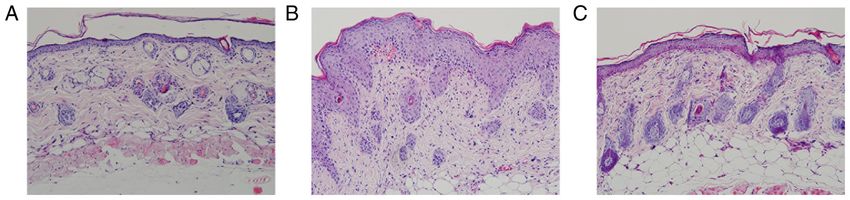
2          LIN et al: NOTCH1/HES1-AKT/IL-17A LOOP REGULATES Th17 CELLS IN MOUSE PSORIASIS-LIKE INFLAMMATION

ligands (jagged 1 and 2, as well as δ‑like 1, 3 and 4) have been    PI3K/AKT signaling was blocked in the present study using
confirmed. Although the process of activation and transduc‑         LY294002 to explore the possible regulatory effect of the
tion of Notch signaling is complex, in essence, it involves the     Notch1/Hes1‑PTEN/AKT/IL‑17A feedback loop on Th17 cell
cleavage of cytoplasmic Notch by γ‑secretase and the release        differentiation and IL‑17A production in mouse psoriasis‑like
of the Notch intracellular domain (NICD) that possesses a           skin inflammation.
nuclear localization signal (NLS) that can translocate signals to
the nuclei and further regulate downstream target genes, such       Materials and methods
as hairy and enhancer of split 1 (Hes1) (7). Notch signaling has
been reported to be crucial for Th17 cell differentiation, which    Mice and treatment. A total of 24 male BALB/c mice (age,
is activated in Th17 cell polarized conditions in humans and        6‑8 weeks old; weight range, 16‑20 g) were purchased from
mice (8‑12). A previous study suggested that blocking Notch         Jinan Pengyue Experimental Animal Breeding Co., Ltd.,
signaling with a γ‑secretase inhibitor could significantly alle‑    China and bred in a specific pathogen‑free environment in
viate mouse psoriasis‑like skin inflammation, which resulted in     the animal center of Binzhou Medical University Hospital
a dose‑dependent decrease in the percentage of Th17 cells and       (Binzhou, China). They were group‑housed at up to five mice
its transcription factor RAR‑related orphan receptor (RORγt),       per cage in a ventilated, temperature‑controlled 23±1˚C,
as well as a decrease in Notch1, Hes1 and IL‑17A mRNA               55% relative humidity condition with a 12‑h light/dark cycle.
expression and IL‑17A secretion in splenic CD4+ T cells in          Autoclaved water and food were available ad libitum to
a 5% imiquimod (IMQ)‑induced mouse psoriasis‑like skin              mice. The experimental mice were randomly divided into
model, indicating that Notch1/Hes1 signaling can regulate           3 groups, namely the control group (n=8), the model group
Th17 cell differentiation and function in the disease‑specific      (5% IMQ‑induced group; n=8) and the intervention group
inflammation of psoriasis (13). NF‑ΚB activator1(Act1) is           (5% IMQ‑induced plus LY294002‑treated group; n=8). Daily
an important adaptor molecule in IL‑17 signaling transduc‑          topical application of 5% IMQ cream (62.5 mg; 3M Health
tion (14), which can interact with Notch1 signaling and form        Care Limited) on the shaved back skin of the model and inter‑
an Act1‑NICD1 complex to induce Notch1 activation and               vention mice were employed to induce psoriasis‑like skin
facilitate IL‑17 crosstalk with Notch1 signaling (15).              inflammation in these groups, as previously described (5),
     PI3K/AKT (also called protein kinase B) and mTOR               while an equivalent quantity of Vaseline (Qingdao Hainuo
signaling is important in the regulation of innate and adaptive     Biological Engineering Co., Ltd.) was applied to the control
immune responses (16), which has been reported to be upregu‑        mice. LY294002 (cat. no. HY‑10108; MedChemExpress)
lated in lesions of patients with psoriasis and in IMQ‑induced      dissolved in DMSO (cat. no. D2650; Sigma‑Aldrich; Merck
mouse psoriasiform inflammation (17,18). mTOR exists in two         KGaA) was intraperitoneally injected (10 mg/kg/day) in the
distinct protein complexes, namely mTORC1 and mTORC2.               intervention mice since the beginning of IMQ application.
PI3K/AKT/mTORC1 signaling has been reported to positively           Control and model mice received an equivalent volume of
regulate Th17 cell differentiation through several means,           DMSO intraperitoneal injection. The experimental mice
including the regulation of transcription factor hypoxia‑induc‑     were anesthetized by 4% isoflurane for the induction and
ible factor 1α expression, signal transducers and activators of     2% for the maintenance to complete daily treatment. After
transcription 3 tyrosine phosphorylation, downregulation of         6 consecutive days, mice were euthanized by inhaling
the Th17 cell differentiation negative regulator growth factor      10% isoflurane for 5 min. Animal death was confirmed by
independent 1 (Gfi1) and RORγt nuclear translocation (19,20),       observing respiratory and cardiac arrest and the absence of
among which, Gfi1 expression and RORγt nuclear transloca‑           active paw reflex for >5 min. Next, the skin tissues, inguinal
tion are dependent on the signaling molecules 40S ribosomal         lymph nodes and spleens were acquired to conduct subse‑
S6 kinase (S6K)1 and S6K2, which are located downstream of          quent experiments. During the experimental process, if
mTORC1. Gfi1 expression is suppressed by the transcription          their weights reduced 20%, the skins occurred infection and
factor early growth response protein 2, which is positively         suppuration or they appeared obvious fidgety, mice would be
regulated by S6K1 (19). RORγ t does not possess a NLS but           subjected to euthanasia. All animal procedures were approved
is localized in the nuclei of Th17 cells. S6K2, the nuclear         (approval no. 20190104‑15) by the Laboratory Animal
counterpart of S6K1, possesses a RORγ t‑associated NLS,             Ethics Committee of Binzhou Medical University Hospital
and can bind and transport RORγt to nuclei. Thus, mTORC1            (Binzhou, China), followed the 3R‑principle (replacement,
can accelerate RORγ t translocation into nuclei during Th17         reduction, refinement) and carried out in accordance with the
differentiation (19). PTEN is the critical negative regulator       UK Animals (Scientific Procedures) Act, 1986, and associ‑
of PI3K/AKT/mTOR signaling, which was reported to be                ated guidelines, and the EU Directive 2010/63/EU for animal
a target of activated Notch1 in T‑cell acute lymphoblastic          experiments.
leukemia, via Hes1‑mediated suppression of the PTEN
promoter (21,22). In addition, the regulatory function of the       Skin structural characteristics scoring and histopathological
Notch1/Hes1/PTEN axis has also been identified in other             examination. Changes in skin structural characteristics were
human diseases, including asthma, diabetic nephropathy and          recorded daily, and the severity of psoriasis‑like inflammation
hepatocellular carcinoma (23‑27). Based on these previous           was estimated by the target lesion score based on the clinical
studies and our previous research, it could be hypothesized         psoriasis area and severity index (5). Erythema, thickening
that a feedback loop between Notch1 and IL‑17A exists               and scaling were scored on a scale from 0 to 4. The cumulative
via the Notch1/Hes1‑PTEN/AKT/mTORC1 signaling axis                  score of the three indicators from 0 to 12 served as a measure
within the pathological environment of psoriasis. Thus,             of the severity of psoriasis‑like inflammation. Skin samples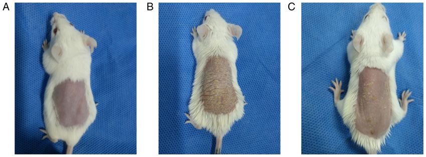
Molecular Medicine REPORTS 26: 223, 2022 3
were fixed in 10% neutral formalin for 24 h at 4˚C, embedded 30 min at 4˚C in the dark. Detection and analysis were conducted
with paraffin, sectioned, and stained with hematoxylin and with a FACSCanto flow cytometer (BD Biosciences). The charac‑
eosin using standard procedures (hematoxylin staining terized phenotype of Th17 cells was IL‑17A+ CD4+ and the results
for 8 min and eosin staining for 1 min at room tempera‑ are expressed as a percentage of total CD4+ T cells.
ture) (28). Histopathological characteristics were evaluated
by well‑trained pathologists in a double‑blinded manner. Reverse transcription‑quantitative PCR (RT‑qPCR) analysis.
Image‑Pro Plus 6.0 imaging system (Media Cybernetics, Inc.) According to the manufacturer's instructions, total RNA was
was employed to measure the epidermis thickness from the extracted from each skin sample and spleen using TRIzol®
stratum corneum to the basement membrane. (cat. no. B511311; Sangon Biotech Co., Ltd.). The purity and
integrity of each RNA sample were confirmed, respectively,
Preparation of single cell suspension from skin tissues and by measuring an absorbance 260/A280 ratio of 1.8‑2.0, as
spleen. Skin samples were cut into 0.5 cm x 0.5 cm pieces and detected by spectrophotometry (NV3000; Vastech Inc.),
incubated in the presence of 0.5% trypsin (cat. no. Y0002311; and by observing intact 28S and 18S ribosomal RNA bands
Sigma‑Aldrich; Merck KGaA) at 37˚C for 2 h. Upon separa‑ with a 2:1 ratio in 1.5% agarose gel electrophoresis with
tion of the epidermis, the dermis was digested with DNase I AG‑CelRed Nucleic acid gel stain (cat. no. AG11918; Hunan
(cat. no. D8071; Beijing Solarbio Science & Technology Co., Accurate Biotechnology Co., Ltd.). cDNA was synthesized
Ltd.) and collagenase IV (cat. no. C5138; Sigma‑Aldrich; utilizing the Evo M‑MLV Mix Kit with gDNA Clean for
Merck KGaA) in DMEM (cat. no. E600008‑0500; Sangon qPCR (cat. no. AG11728; Hunan Accurate Biotechnology Co.,
Biotech Co., Ltd.) for 37˚C, 1 h. Cells were harvested and Ltd.) according to the manufacturer's protocols. RT‑qPCR
resuspended (1x10⁶ cells/ml) for use in subsequent flow cytom‑ was performed with gene‑specific primers using SYBR®
etry analysis. Green Premix Pro Taq HS qPCR kit (cat. no. AG11701;
Spleen samples were triturated and filtered through cell Hunan Accurate Biotechnology Co., Ltd.) on a CFX96
strainers (70 and 40 µm; cat. no. F613462 and F613461; Touch™ Real‑Time PCR Detection System (Bio‑Rad
Sangon Biotech Co., Ltd.). Cell suspension was collected Laboratories, Inc.). All primers were designed by Hunan
and treated according to the protocol of mouse splenic Accurate Biotechnology Co., Ltd. The National Center for
mononuclear cell isolation kit (cat. no. LDS1090PK; Biotechnology Information accession numbers are as follows:
Tianjin Haoyang Biological Products Technology Co., Ltd.). 14433 (GAPDH), 18128 (Notch1), 15205 (Hes1), 19211
Splenic mononuclear cells were acquired and suspended in (PTEN), 11651 (AKT), 74370 (mTORC1), 72508 (S6K1),
RPMI‑1640 medium (cat. no. E600028; Sangon Biotech Co., 58988 (S6K2) and 16171 (IL‑17A). The primer sequences
Ltd.) with 10% fetal bovine serum (cat. no. E510008‑0500; were as follows: Notch1 sense, 5'‑TGCCAGTATGATGTG
Sangon Biotech Co., Ltd.), 1% penicillin‑streptomycin liquid, GATGAG‑3' and antisense, 5'‑GGTCCCTGTGTAACCTTC
1% nonessential amino acid and 0.01% β ‑mercaptoethanol TGT‑3'; Hes1 sense, 5'‑AGCCCACCTCTCTCT TCTGA‑3',
(cat. nos. P1400, N1250 and M8210, respectively; Beijing and antisense, 5'‑AGGCGCAATCCAATATGAAC‑3'; PTEN
Solarbio Science & Technology Co., Ltd.). Then cells were sense, 5'‑AAGG GACGGACTG GTGTAATGATT TG‑3' and
counted and adjusted to 106 cells/ml for subsequent treatment antisense, 5'‑CGCCTCTGACTGG GAATTGTGAC‑3'; AKT
and experiments. sense, 5'‑GACTGACACCAGG TATTTCGATGA‑3' and
antisense, 5'‑CTCCGCTCACTGTCCACACA‑3'; mTORC1
Splenic mononuclear cells treatment by LY294002. Splenic sense, 5'‑CCATCGGTGCAAACCTACAG‑3' and antisense,
mononuclear cells isolated from 5% IMQ‑induced model mice 5'‑TCCATTCACTGTAGGCCTG G‑3'; S6K1 sense, 5'‑TTC
were divided into 0, 10 and 50 µM LY294002‑treated group. TGTCGG GAGTAGCACTG‑3' and antisense, 5'‑CCCCTT
In LY294002‑treated groups, LY294002 was dissolved in TACCAAGTACCCGA‑3'; S6K2 sense, 5'‑TCACTGCAG
DMSO. At the same time, splenic mononuclear cells isolated AGAACCGGAAGA‑3' and antisense, 5'‑GGGGTTCCGCTT
from control mice were used as control group and treated with CAGAAACTT‑3'; IL‑17A sense, 5'‑TTTAACTCCCTTG GC
DMSO only. GCAAAA‑3' and antisense, 5'‑CTTTCCCTCCGCATTGAC
AC‑3'; and GAPDH sense, 5'‑AAATGGTGAAGGTCGGTG
Flow cytometric analysis of the proportion of Th17 cells TGAAC‑3' and antisense, 5'‑CAACAATCTCCACTT TGC
(IL‑17A+ CD4+ T cells/CD4+ T cells). Cells were firstly CACTG‑3'. The thermocycling conditions were as follows:
stimulated with phorbol 12‑myristate 13‑acetate, ionomycin, 95˚C for 5 min, followed by 40 cycles of denaturation for
brefeldin A and monensin (Cell Stimulation Cocktail Plus 10 sec at 95˚C and annealing/elongation for 30 sec at 60˚C.
Protein Transport Inhibitors; cat. no. 4975; eBioscience; Thermo GAPDH was used as the internal control, and the expression
Fisher Scientific, Inc.) for 4 h at 37˚C under 5% CO2. Next, the levels of target genes were calculated based on the following
cells were collected (630 x g for 10 min at room temperature), formula 2‑ΔΔCq (ΔΔCq=ΔCq‑treated‑ΔCq‑control) (29).
washed two times with ice‑cold phosphate‑buffered saline
(PBS) (cat. no. E607008‑0500; Sangon Biotech Co., Ltd.) and Western blot detection and analysis. Total protein from skin
surface‑stained with an Allophycocyanin (APC) anti‑mouse CD4 samples and treated splenic mononuclear cells were extracted
Antibody (0.8 µg/ml, cat. no. 100516; BioLegend, Inc.) for 30 min using RIPA lysis buffer (cat. no. R0020; Beijing Solarbio
at 4˚C in the dark. Next, the cells were fixed and permeabilized Science & Technology Co., Ltd.). Equal quantities of dena‑
using Perm/Fix (cat. no. 88‑8824‑00; eBioscience) solution and tured protein (50 µg) were separated by 10% sodium dodecyl
intracellularly stained with a P‑phycoerythrin (PE) anti‑mouse sulphate‑polyacrylamide gel electrophoresis and transferred
IL‑17A Antibody (2.5 µg/ml, cat. no. 506904; BioLegend, Inc.) for to polyvinylidene fluoride (cat. no. ipvh00010; Sigma‑Aldrich;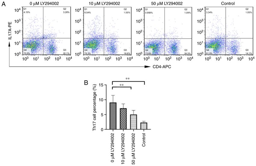
4            LIN et al: NOTCH1/HES1-AKT/IL-17A LOOP REGULATES Th17 CELLS IN MOUSE PSORIASIS-LIKE INFLAMMATION

Figure 1. Structural characteristics of the skin of the experimental mice. (A) Control mice did not present any signs of erythema, scaling or thickening.
(B) Model mice displayed significant signs of psoriasis‑like inflammation. (C) Intervention mice showed similar signs of psoriasis‑like inflammation as model
mice; however, the degree of severity was notably alleviated compared with model mice.

Merck KGaA) membranes. After blocking in 5% skim milk in                        Results
0.1% TBS‑Tween 20 (TBST; cat. no. T1082; Beijing Solarbio
Science & Technology Co., Ltd.) for 2 h at room temperature, the                LY294002 alleviates the severity of psoriasiform skin lesions.
membranes were incubated overnight at 4˚C with primary anti‑                    Control mice presented no signs of erythema, thickening or
bodies (all from Affinity Biosciences) against Notch1 (1:1,000;                 scaling during the 6 days. By contrast, the model mice presented
cat. no. AF5307), NICD1 (1:1,000; cat. no. AF5307), Hes1                        signs of psoriasis‑like inflammation on their shaved back skin
(1:1,000; cat. no. AF7575), PTEN (1:1,000; cat. no. AF6351),                    from the second day onwards, which exacerbated progressively
AKT (1:1,000; cat. no. AF6261), phosphorylated (p)‑AKT                          and was most severe on the 6th day. LY294002 inhibition
(Thr308) (1:1,000; cat. no. AF3262), p‑AKT (Ser473) (1:1,000;                   alleviated the degree of erythema, thickening and scaling in
cat. no. AF0016), mTORC1 (1:1,000; cat. no. AF6308), p‑mTOR                     the intervention mice (Fig. 1). The scores of lesions of control
(Ser2448) (1:1,000, cat. no. AF3308), S6K1 (p70S6K) (1:1,000;                   mice, model mice and intervention mice were listed in Table I
cat. no. AF6226), S6K2 (p70S6kβ) (1:1,000; cat. no. AF3486) and                 and were significantly different among the three experimental
IL‑17A (1:1,000; cat. no. DF6127). The next day, each membrane                  groups (F=1751.182, P
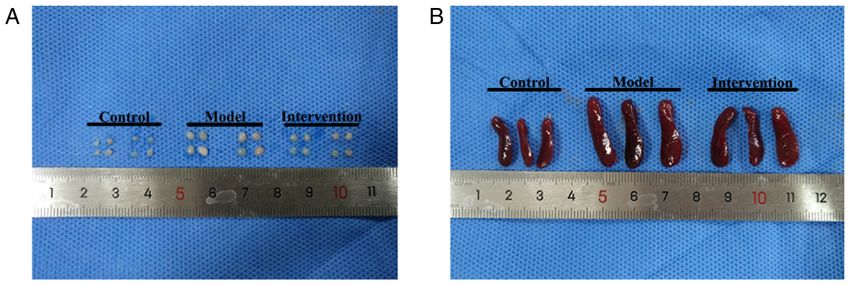
Molecular Medicine REPORTS 26: 223, 2022                                                                  5

Table I. Psoriasis area and severity index scores of experimental mice.

Group	Day 1	Day 2	Day 3	Day 4	Day 5	Day 6

Control mice                0.000±0.000          0.000±0.000            0.000±0.000           0.000±0.000            0.000±0.000            0.000±0.000
Model mice                  0.000±0.000          2.875±0.354            5.500±0.535            7.00±0.535            8.625±0.518           10.125±0.835
Intervention mice           0.000±0.000          2.000±0.535a           3.625±0.518a          4.375±0.518a           6.500±0.535a           6.000±0.535a

Data are presented as the means ± SD. Repeated measures ANOVA followed by Bonferroni's adjustments are used for statistical analysis.
a
 P
6           LIN et al: NOTCH1/HES1-AKT/IL-17A LOOP REGULATES Th17 CELLS IN MOUSE PSORIASIS-LIKE INFLAMMATION

Table II. Inguinal lymph node weight, spleen mass and spleen index of experimental mice (each n=8).

Group	Inguinal lymph node weight (g)                                                     Spleen mass (g)                       Spleen index (mg/g)

Control                                     4.863±0.544                                    0.253±0.024                             0.118±0.010
Model                                      15.760±0.716a                                   0.408±0.024a                            0.214±0.013a
Intervention                                9.943±0.701b                                   0.348±0.009b                            0.175±0.006b

Data are presented as the means ± SD. One‑way ANOVA followed by Student‑Newman‑Keuls test were used for statistical analysis. aP
Molecular Medicine REPORTS 26: 223, 2022                                                           7

Table III. mRNA expression of Notch1, Hes1, PTEN, AKT, mTORC1, S6K1, S6K2 and IL‑17A in skin samples of experimental
mice (each n=8).

Group	Notch1                         Hes1            PTEN	AKT                       mTORC1             S6K1             S6K2	IL‑17A

Control      0.966±0.442 0.617±0.501 1.369±0.411 1.024±0.238 1.021±0.224 0.789±0.189 0.621±0.305 0.302±0.156
Model        2.351±0.958a 3.704±1.823a 0.504±0.215a 3.864±0.891a 1.455±0.389a 1.323±0.313a 1.888±0.615a 1.097±0.543a
Intervention 1.178±0.590b 1.341±1.085b 1.052±0.359b 2.352±0.706b 1.030±0.274c 1.016±0.185c 1.015±0.189b 0.545±0.252b
F‑value          9.146       13.154       13.361       35.941        5.339       11.473       19.893       10.402
P‑value
8          LIN et al: NOTCH1/HES1-AKT/IL-17A LOOP REGULATES Th17 CELLS IN MOUSE PSORIASIS-LIKE INFLAMMATION

Table IV. Protein levels of Notch1, NICD1, Hes1, PTEN, p‑AKT (Thr308), p‑AKT (Ser473), p‑mTOR (Ser2448), S6K1
(p70s6kα), S6K2 (p70s6kβ) and IL‑17A in skin samples of experimental mice (each n=8).

Group	Notch1	NICD1                                                             Hes1                  PTEN	p‑AKT(Thr308)

Control               0.688±0.365                0.996±0.205              0.735 (0.130)          1.011±0.077             0.908±0.273
Model                 1.211±0.339b               1.229±0.095b             1.188 (0.680)b         0.770±0.053a            1.928±0.536a
Intervention          0.824±0.220d               0.798±0.141c             0.819 (0.210)d         0.984±0.079c            1.111±0.540d
F‑value/χ2                4.476                     11.800                    9.696                 21.027                   8.037
P‑value
Molecular Medicine REPORTS 26: 223, 2022                                                              9

Figure 6. Protein levels of (A) Notch1, (B) NICD1, (C) hairy and enhancer of split 1, (D) PTEN, (E‑1) p‑AKT (Thr308), (E‑2) AKT, (E‑3) p‑AKT (Ser473),
(F‑1) p‑mTOR (Ser2448), (F‑2) mTORC1, (G) S6K1 (p70S6Kα), (H) S6K2 (p70S6Kβ) and (I) IL‑17A in skin samples of experimental mice. *P
10          LIN et al: NOTCH1/HES1-AKT/IL-17A LOOP REGULATES Th17 CELLS IN MOUSE PSORIASIS-LIKE INFLAMMATION

Table VI. Protein levels of Hes1, PTEN, p‑AKT(Thr308), p‑AKT(Ser473), p‑mTOR (Ser2448) and IL‑17A in splenic mono‑
nuclear cells of experimental mice (each n=6).

Group                        Hes1              PTEN	          p‑AKT (Thr308) p‑AKT (Ser473) p‑mTOR (Ser2448)	IL‑17A

0 µM LY294002   1.179±0.060 0.530±0.149                          1.264±0.072           1.304±0.065             1.256±0.095           1.173±0.134
10 µM LY294002 0.939±0.070a 0.788±0.097a                        0.992±0.077a          1.039±0.088a            1.026±0.061a          0.951±0.074a
50 µM LY294002 0.756±0.073a,b 0.948±0.100a                      0.845±0.062a,c        0.866±0.108a,c          0.857±0.109a,c        0.810±0.085a
Control        0.569±0.111a,b 1.151±0.119a,b                    0.608±0.073a,b        0.450±0.084a,b          0.550±0.107a,b        0.522±0.107a,b
F‑value            62.235        29.712                             88.796               100.548                  58.847                42.321
P‑value
Molecular Medicine REPORTS 26: 223, 2022                                                        11

Figure 8. mRNA expression of (A) hairy and enhancer of split 1, (B) PTEN, (C) AKT, (D) mTOR complex 1 and (E) IL‑17A in mouse splenic mononuclear
cells of control group, 0 µM LY294002 group, 10 µM LY294002 group and 50 µM LY294002 group. *P
12           LIN et al: NOTCH1/HES1-AKT/IL-17A LOOP REGULATES Th17 CELLS IN MOUSE PSORIASIS-LIKE INFLAMMATION

Figure 9. Protein levels of (A) hairy and enhancer of split 1, (B) PTEN, (C‑1) p‑AKT (Thr308), (C‑2) AKT, (C‑3) p‑AKT (Ser473), (D‑1) mTORC1, (D‑2) p‑mTOR
(Ser2448), (E) IL‑17A in mouse splenic mononuclear cells of control group, 0 µM LY294002 group, 10 µM LY294002 group and 50 µM LY294002 group.
**
  P
Molecular Medicine REPORTS 26: 223, 2022                                                       13

notably, the even more obvious intervention effects of 50 µM             5. van der Fits L, Mourits S, Voerman JS, Kant M, Boon L,
                                                                            Laman JD, Cornelissen F, Mus AM, Florencia E, Prens EP and
LY294002 were confirmed. Related in vitro/in vivo studies                   Lubberts E: Imiquimod‑induced psoriasis‑like skin inflamma‑
shall be performed in the future to more deeply evaluate the                tion in mice is mediated via the IL‑23/IL‑17 axis. J Immunol 182:
potential therapeutic effect of LY294002 in psoriatic inflam‑               5836‑5845, 2009.
                                                                         6. Gratton R, Tricarico PM, Moltrasio C, Lima Estevão de
mation.                                                                     Oliveira AS, Brandão L, Marzano AV, Zupin L and Crovella S:
                                                                            Pleiotropic role of Notch signaling in human skin diseases. Int J
Acknowledgements                                                            Mol Sci 21: 4214, 2020.
                                                                         7. Gratton R, Tricarico PM, d'Adamo AP, Bianco AM, Moura R,
                                                                            Agrelli A, Brandão L, Zupin L and Crovella S: Notch signaling
Not applicable.                                                             regulation in autoinflammatory diseases. Int J Mol Sci 21: 8847,
                                                                            2020.
                                                                         8. Fiúza UM and Arias AM: Cell and molecular biology of Notch.
Funding                                                                     J Endocrinol 194: 459‑474, 2007.
                                                                         9. Auderset F, Coutaz M and Tacchini‑Cottier F: The role of Notch
The present study was supported by National Natural Science                 in the differentiation of CD4+ T helper cells. Curr Top Microbiol
                                                                            Immunol 360: 115‑134, 2012.
Foundation of China (grant no. 81803145).                               10. Eagar TN, Tang Q, Wolfe M, He Y, Pear WS and Bluestone JA:
                                                                            Notch 1 signaling regulates peripheral T cell activation.
Availability of data and materials                                          Immunity 20: 407‑415, 2004.
                                                                        11. Keerthivasan S, Suleiman R, Lawlor R, Roderick J, Bates T,
                                                                            Minter L, Anguita J, Juncadella I, Nickoloff BJ, Le Poole IC, et al:
All data generated or analyzed during this study are included               Notch signaling regulates mouse and human Th17 differentia‑
in this manuscript or are available from the corresponding                  tion. J Immunol 187: 692‑701, 2011.
                                                                        12. Radtke F, Fasnacht N and MacDonald HR: Notch signaling in the
author on reasonable request.                                               immune system. Immunity 32: 14‑27, 2010.
                                                                        13. Ma L, Xue H, Qi R, Wang Y and Yuan L: Effect of γ‑secretase
Authors' contributions                                                      inhibitor on Th17 cell differentiation and function of mouse
                                                                            psoriasis‑like skin inflammation. J Transl Med 16: 59, 2018.
                                                                        14. Qian Y, Liu C, Hartupee J, Altuntas CZ, Gulen MF, Jane‑Wit D,
YWL and XXL carried out the experiments, performed the                      Xiao J, Lu Y, Giltiay N, Liu J, et al: The adaptor Act1 is required
statistical analysis and wrote the first draft of the manuscript.           for interleukin 17‑dependent signaling associated with autoim‑
                                                                            mune and inflammatory disease. Nat Immunol 8: 247‑256, 2007.
FHF participated in the design of the study and carried out the         15. Wang C, Zhang CJ, Martin BN, Bulek K, Kang Z, Zhao J,
experiments, particularly for the development of the experi‑                Bian G, Carman JA, Gao J, Dongre A, et al: IL‑17 induced
mental model. BL and RQQ participated in the design of the                  NOTCH1 activation in oligodendrocyte progenitor cells
                                                                            enhances proliferation and inflammatory gene expression. Nat
study and analyzed the data. XYX helped to carry out the                    Commun 8: 15508, 2017.
experiments. LM was responsible for the design of the study,            16. Weichhart T and Säemann MD: The PI3K/Akt/mTOR pathway
reviewed and edited this article. All authors read and approved             in innate immune cells: Emerging therapeutic applications. Ann
                                                                            Rheum Dis 67 (Suppl 3): iii70‑iii74, 2008.
the final manuscript and agree to be accountable for all aspects        17. Mercurio L, Albanesi C and Madonna S: Recent updates on
of the work in ensuring that questions related to the accuracy or           the involvement of PI3K/AKT/mTOR molecular cascade in the
integrity of the work are appropriately investigated and resolved.          pathogenesis of hyperproliferative skin disorders. Front Med
                                                                            (Lausanne) 8: 665647, 2021.
LM and YWL confirm the authenticity of all the raw data.                18. Chamcheu JC, Chaves‑Rodriquez MI, Adhami VM, Siddiqui IA,
                                                                            Wood GS, Longley BJ and Mukhtar H: Upregulation of
Ethics approval and consent to participate                                  PI3K/AKT/mTOR, FABP5 and PPARβ/δ in human psoriasis and
                                                                            imiquimod‑induced murine psoriasiform dermatitis model. Acta
                                                                            Derm Venereol 96: 854‑856, 2016.
The present study was approved (approval no. 20190104‑15) by            19. Kurebayashi Y, Nagai S, Ikejiri A, Ohtani M, Ichiyama K,
the Laboratory Animal Ethics Committee of Binzhou Medical                   Baba Y, Yamada T, Egami S, Hoshii T, Hirao A, et al:
                                                                            PI3K‑Akt‑mTORC1‑S6K1/2 axis controls Th17 differentiation
University Hospital (Binzhou, China).                                       by regulating Gfi1 expression and nuclear translocation of RORγ.
                                                                            Cell Rep 1: 360‑373, 2012.
Patient consent for publication                                         20. Nagai S, Kurebayashi Y and Koyasu S: Role of PI3K/Akt and
                                                                            mTOR complexes in Th17 cell differentiation. Ann N Y Acad
                                                                            Sci 1280: 30‑34, 2013.
Not applicable.                                                         21. Palomero T, Sulis ML, Cortina M, Real PJ, Barnes K, Ciofani M,
                                                                            Caparros E, Buteau J, Brown K, Perkins SL, et al: Mutational
                                                                            loss of PTEN induces resistance to NOTCH1 inhibition in T‑cell
Competing interests                                                         leukemia. Nat Med 13: 1203‑1210, 2007.
                                                                        22. Hales EC, Taub JW and Matherly LH: New insights into Notch1
The authors declare that they have no competing interests.                  regulation of the PI3K‑AKT‑mTOR1 signaling axis: Targeted
                                                                            therapy of γ‑secretase inhibitor resistant T‑cell acute lympho‑
                                                                            blastic leukemia. Cell Signal 26: 149‑161, 2014.
References                                                              23. Li X, Zou F, Lu Y, Fan X, Wu Y, Feng X, Sun X and Liu Y:
                                                                            Notch1 contributes to TNF‑α‑induced proliferation and migra‑
 1. Yamanaka K, Yamamoto O and Honda T: Pathophysiology of                  tion of airway smooth muscle cells through regulation of the
    psoriasis: A review. J Dermatol 48: 722‑731, 2021.                      Hes1/PTEN axis. Int Immunopharmacol 88: 106911, 2020.
 2. Furue K, Ito T, Tsuji G, Kadono T and Furue M: Psoriasis and the    24. Liu X, Zhang Y, Shi M, Wang Y, Zhang F, Yan R, Liu L, Xiao Y
    TNF/IL23/IL17 axis. G Ital Dermatol Venereol 154: 418‑424, 2019.        and Guo B: Notch1 regulates PTEN expression to exacerbate
 3. Molinelli E, Campanati A, Brisigotti V and Offidani A: Biologic         renal tubulointerstitial fibrosis in diabetic nephropathy by inhib‑
    therapy in psoriasis (part II): Efficacy and safety of new treat‑       iting autophagy via interactions with Hes1. Biochem Biophys Res
    ment targeting IL23/IL‑17 pathways. Curr Pharm Biotechnol 18:           Commun 497: 1110‑1116, 2018.
    964‑978, 2017.                                                      25. Tian T, Fu X, Lu J, Ruan Z, Nan K, Yao Y and Yang Y:
 4. Ly K, Smith MP, Thibodeaux Q, Reddy V, Liao W and Bhutani T:            MicroRNA‑760 inhibits doxorubicin resistance in hepatocellular
    Anti IL‑17 in psoriasis. Expert Rev Clin Immunol 15: 1185‑1194,         carcinoma through regulating Notch1/Hes1‑PTEN/Akt signaling
    2019.                                                                   pathway. J Biochem Mol Toxicol 32: e22167, 2018.
14 LIN et al: NOTCH1/HES1-AKT/IL-17A LOOP REGULATES Th17 CELLS IN MOUSE PSORIASIS-LIKE INFLAMMATION
26. Zhang X, Hu Y, Gao H, Lan X and Xue Y: Downregulation of 40. Chamcheu JC, Adhami VM, Esnault S, Sechi M, Siddiqui IA,
Notch1 inhibits the invasion and metastasis of human gastric Satyshur KA, Syed DN, Dodwad SJ, Chaves‑Rodriquez MI,
cancer cells SGC7901 and MKN74 in vitro through PTEN Longley BJ, et al: Dual inhibition of PI3K/Akt and mTOR
activation and dephosphorylation of AKT and FAK. Mol Med by the diet a r y a ntioxida nt, delph in idin, a meliorates
Rep 16: 2318‑2324, 2017. psoriatic features in vitro and in an imiquimod‑induced
27. Sokolowski KM, Balamurugan M, Kunnimalaiyaan S, Wilson J, psoriasis‑like disease in mice. Antioxid Redox Signal 26:
Gamblin TC and Kunnimalaiyaan M: Role of Akt inhibition on 49‑69, 2017.
Notch1 expression in hepatocellular carcinoma: Potential role for 41. Coutaz M, Hurrell BP, Auderset F, Wang H, Siegert S, Eberl G,
dual targeted therapy. Am J Surg 211: 755‑760, 2016. Ho PC, Radtke F and Tacchini‑Cottier F: Notch regulates Th17
28. Lan XO, Wang HX, Qi RQ, Xu YY, Yu YJ, Yang Y, Guo H, differentiation and controls trafficking of IL‑17 and metabolic
Gao XH and Geng L: Shikonin inhibits CEBPD downregula‑ regulators within Th17 cells in a context‑dependent manner. Sci
tion in IL‑17‑treated HaCaT cells and in an imiquimod‑induced Rep 6: 39117, 2016.
psoriasis model. Mol Med Rep 22: 2263‑2272, 2020. 42. Nagase H and Nakayama K: γ‑Secretase‑regulated signaling
29. Livak KJ and Schmittgen TD: Analysis of relative gene expres‑ typified by Notch signaling in the immune system. Curr Stem
sion data using real‑time quantitative PCR and the 2(‑Delta Delta Cell Res Ther 8: 341‑356, 2013.
C(T)) method. Methods 25: 402‑408, 2001. 43. Palomero T, Dominguez M and Ferrando AA: The role of the
30. Greb JE, Goldminz AM, Elder JT, Lebwohl MG, Gladman DD, PTEN/AKT pathway in NOTCH1‑induced leukemia. Cell
Wu JJ, Mehta NN, Finlay AY and Gottlieb AB: Psoriasis. Nat Cycle 7: 965‑970, 2008.
Rev Dis Primers 2: 16082, 2016. 44. Liu S, Ma X, Ai Q, Huang Q, Shi T, Zhu M, Wang B and
31. Di Cesare A, Di Meglio P and Nestle FO: The IL‑23/Th17 axis Zhang X: NOTCH1 functions as an oncogene by regulating the
in the immunopathogenesis of psoriasis. J Invest Dermatol 129: PTEN/PI3K/AKT pathway in clear cell renal cell carcinoma.
1339‑1350, 2009. Urol Oncol 31: 938‑948, 2013.
32. Li B, Huang L, Lv P, Li X, Liu G, Chen Y, Wang Z, Qian X, 45. Ma L, Xue HB, Guan XH, Qi RQ, An RZ, Shu CM, Zhang YJ,
Shen Y, Li Y and Fang W: The role of Th17 cells in psoriasis. Wei YH and Zhang JH: Possible role of Th17 cells and IL‑17
Immunol Res 68: 296‑309, 2020. in the pathogenesis of atopic dermatitis in northern China.
33. Hawkes JE, Chan TC and Krueger JG: Psoriasis pathogenesis and J Dermatol Sci 68: 66‑68, 2012.
the development of novel targeted immune therapies. J Allergy 46. Ma L, Xue HB, Wang F, Shu CM and Zhang JH: MicroRNA‑155
Clin Immunol 140: 645‑653, 2017. may be involved in the pathogenesis of atopic dermatitis by
34. Ma L, Xue HB, Guan XH, Shu CM, Wang F, Zhang JH and modulating the differentiation and function of T helper type 17
An RZ: The imbalance of Th17 cells and CD4(+) CD25(high) (Th17) cells. Clin Exp Immunol 181:142‑149, 2015.
Foxp3(+) Treg cells in patients with atopic dermatitis. J Eur Acad 47. Peng X, Zhou J, Li B, Zhang T, Zuo Y and Gu X: Notch1
Dermatol Venereol 28:1079‑1086, 2014. and PI3K/Akt signaling blockers DAPT and LY294002 coor‑
35. Ma L, Xue H, Gao T, Gao M and Zhang Y: Notch1 signaling dinately inhibit metastasis of gastric cancer through mutual
regulates the Th17/Treg immune imbalance in patients with enhancement. Cancer Chemother Pharmacol 85: 309‑320,
psoriasis vulgaris. Mediators Inflamm 2018: 3069521, 2018. 2020.
36. Wang Y, Li X, Xing X, Xue H, Qi R, Ji H and Ma L: Notch‑Hes1 48. Zhao J, Di T, Wang Y, Liu X, Liang D, Zhang G and Li P:
signaling regulates IL‑17A+ γδ +T cell expression and IL‑17A Multi‑glycoside of tripterygium wilfordii Hook. f. ameliorates
secretion of mouse psoriasis‑like skin inflammation. Mediators imiquimod‑induced skin lesions through a STAT3‑dependent
Inflamm 2020: 8297134, 2020. mechanism involving the inhibition of Th17‑mediated inflamma‑
37. Korman NJ: Management of psoriasis as a systemic disease: tory responses. Int J Mol Med 38: 747‑757, 2016.
What is the evidence? Br J Dermatol 182: 840‑848, 2020.
38. Kim CH, Yoo JK, Jeon SH, Lim CY, Lee JH, Koo DB and This work is licensed under a Creative Commons
Park MY: Anti‑psoriatic effect of myeloid‑derived suppressor Attribution-NonCommercial-NoDerivatives 4.0
cells on imiquimod‑induced skin inflammation in mice. Scand J International (CC BY-NC-ND 4.0) License.
Immunol 89: e12742, 2019.
39. Frenzel DF, Borkner L, Scheurmann J, Singh K, Scharffetter-
Kochanek K and Weiss JM: Osteopontin deficiency affects
imiquimod‑induced psoriasis‑like murine skin inflammation
and lymphocyte distribution in skin, draining lymph nodes and
spleen. Exp Dermatol 24: 305‑307, 2015.You can also read