Inositol 1,4,5 trisphosphate receptor type 3 is involved in resistance to apoptosis and maintenance of human hepatocellular carcinoma
←
→
Page content transcription
If your browser does not render page correctly, please read the page content below
ONCOLOGY LETTERS 23: 32, 2022

             Inositol 1,4,5‑trisphosphate receptor type 3 is involved
                   in resistance to apoptosis and maintenance
                       of human hepatocellular carcinoma
        MARCONE LOIOLA DOS SANTOS1,2, ANDRESSA FRANÇA2, ANTÔNIO CARLOS MELO LIMA FILHO2,
            RODRIGO M. FLORENTINO2, PAULO HENRIQUE DINIZ3, FERNANDA OLIVEIRA LEMOS2,
                   CARLOS ALBERTO XAVIER GONÇALVES4, VITOR LIMA COELHO4,
               CRISTIANO XAVIER LIMA5, GISELLE FOUREAUX1, MICHAEL H. NATHANSON6,
                       PAULA VIEIRA TEIXEIRA VIDIGAL7 and M. FÁTIMA LEITE2

    Departments of 1Morphology, 2Physiology and Biophysics and 3Internal Medicine, Federal University of Minas Gerais,
    Belo Horizonte, Minas Gerais 31270‑901; 4Coordination of Biotechnology, SENAI's Innovation Institute for Biosynthetics
     and Fibers, SENAI CETIQT, Rio de Janeiro 20961‑020; 5Department of Surgery, Federal University of Minas Gerais,
     Belo Horizonte, Minas Gerais 30 130‑100, Brazil; 6Section of Digestive Diseases, Department of Internal Medicine,
       Yale University School of Medicine, New Haven, CT 06520‑8056, USA; 7Department of Pathological Anatomy
                     and Forensic Medicine of Hospital das Clínicas, Federal University of Minas Gerais,
                                     Belo Horizonte, Minas Gerais 30 130‑100, Brazil

                                         Received March 2, 2021; Accepted September 24, 2021

                                                         DOI: 10.3892/ol.2021.13150

Abstract. The expression of the inositol 1,4,5‑trisphosphate               known whether ITPR3 expression in hepatocytes is involved
receptor type 3 (ITRP3) in hepatocytes is a common event in                in tumor maintenance. The aim of the present study was to
the pathogenesis of hepatocellular carcinoma (HCC), regard‑                determine whether there is an association between ITPR3
less of the type of underlying liver disease. However, it is not           expression and clinical and morphological parameters using
                                                                           HCC samples obtained from liver explants from patients
                                                                           (n=53) with different etiologies of underlying chronic liver
                                                                           disease (CLD). ITPR3 expression, mitosis and apoptosis were
                                                                           analyzed in human liver samples by immunohistochemistry.
Correspondence to: Professor Paula Vieira Teixeira Vidigal,                Clinical and event‑free survival data were combined to assess
Department of Pathological Anatomy and Forensic Medicine of                the relationship between ITPR3 and liver cancer growth in
Hospital das Clínicas, Federal University of Minas Gerais, 3rd             patients. RNA sequencing analysis was performed to identify
floor, Av. Alfredo Balena 190, Santa Efigênia, Belo Horizonte,
                                                                           apoptotic genes altered by ITPR3 expression in a liver tumor
Minas Gerais 30 130‑100, Brazil
                                                                           cell line. ITPR3 was highly expressed in HCC tumor cells
E‑mail: pvidigal@medicina.ufmg.br
                                                                           relative to adjacent CLD tissue and healthy livers. There was
Abbreviations: AFP, α fetoprotein; ALD, alcoholic liver disease;           an inverse correlation between ITPR3 expression and mitotic
CAAE, Certificado de Apresentação de Apreciação Ética (Certificate         and apoptotic indices in HCC, suggesting that ITPR3 contrib‑
of Presentation of Ethical Appreciation); CC, cryptogenic cirrhosis;       uted to the maintenance of HCC by promoting resistance to
CHILD, CHILD‑Pugh scores; CHOP, C/EBP homologous protein;                  apoptosis. This was confirmed by the upregulation of CTSB,
CLD, chronic liver disease; CTSB, cathepsin B; EFS, event‑free             CHOP and GADD45, genes involved in the apoptotic pathway
survival; H&E, hematoxylin and eosin; HCC, hepatocellular                  in HCC. The expression of ITPR3 in the liver may be a prom‑
carcinoma; HCV, hepatitis C virus; HepG2, immortalized liver cancer        ising prognostic marker of HCC
cell line; HPF, high‑power fields; INR, international normalized
ratio; ITPR, inositol 1,4,5‑trisphosphate receptor; ITPR1, inositol
                                                                           Introduction
1,4,5‑trisphosphate type 1 receptor; ITPR2, inositol 1,4,5‑trisphosphate
type 2 receptor; ITPR3, inositol 1,4,5‑trisphosphate type 3 receptor;
MELD, model for end‑stage liver disease; OLT, orthotopic liver             Hepatocellular carcinoma (HCC) is the commonest primary
transplantation; PH, partial hepactomy; RNA‑seq, RNA sequencing;           liver malignancy and its incidence is increasing worldwide (1).
TBIL, total bilirubin                                                      According to the GLOBOCAN database, in 2020, including
                                                                           intrahepatic bile ducts, there were 905,677 new cases of
Key words: apoptosis, calcium signaling, chronic diseases, liver           liver cancer around the world, accompanied by 830,180 new
cancer, mitosis                                                            deaths (2). Liver cirrhosis is a major risk factor for HCC (3).
                                                                           Although liver transplantation is the primary long term cura‑
                                                                           tive treatment, HCC still has a recurrence rate of 10‑15% in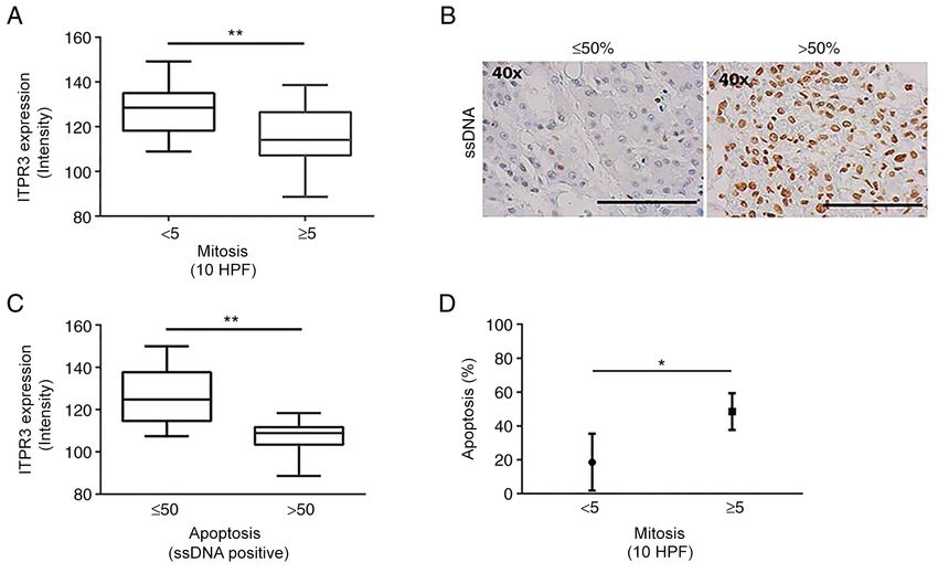
2                              SANTOS et al: ITPR3 IN HEPATOCELLULAR CARCINOMA MAINTENANCE

transplant patients (4). Since biopsy is not mandatory for HCC        in the thermal oven), followed by four immersions in xylene
diagnosis (5), the availability of tumor specimens for analysis       (the first for 20 min and the others with 1 min duration) and
is limited, which in turn has limited our understanding of the        three immersions in absolute alcohol (the first for 5 min and
molecular mechanisms involved in carcinogenesis.                      the others with 1 min duration). Afterwards, the slides were
     The calcium ion (Ca2+) is an intracellular second messenger      immersed in running water for 3 min. The Novolink Polymer
involved in energy metabolism, cell cycle control, gene expres‑       Detection System (cat. no. RE7200‑CE; Leica Microsystems,
sion, cell proliferation, cell migration, necrosis and apoptosis in   Inc.) was used in the subsequent steps as described previ‑
nearly every cell type, including hepatocytes (6). The inositol       ously (21). Primary antibodies against ITPR3 (anti‑ITPR3;
1,4,5‑trisphosphate receptor (ITPR) is the only intracellular         Sigma‑Aldrich; Merck KGaA; cat. no. HPA003915) and
Ca 2+ release channel expressed in the liver, with all three          ssDNA labeling (anti‑Single Stranded DNA; cat. no. 18731;
isoforms described in humans and rodents (7,8). The expression        Immuno‑Biological Laboratories, Co., Ltd.) were incubated
of each ITPR varies among cell type, having distinct subcel‑          overnight at room temperature in a 1:100 dilution, followed
lular distribution and biophysical properties (9). For instance,      by incubation with detection polymer for 40 min at room
under physiological conditions type 1 (ITPR1) and 2 (ITPR2)           temperature. DAB was used for signal detection.
are the predominant isoforms in hepatocytes (10,11), while                Clinical pre‑OLT data and histopathological parameters
isoform 3 (ITPR3) is absent or minimally expressed (12).              of the explanted liver were retrospectively collected from the
However, ITPR3 becomes significantly expressed in hepato‑             medical records, including sex, age, etiology of the underlying
cytes in acute (13,14) and chronic liver diseases, as well as in      chronic liver disease (CLD), model for end‑stage liver disease
HCC and in liver cancer cell lines (15,16). ITPR3 expression is       (MELD) and CHILD‑Pugh scores (CHILD); prognostic
also increased in other malignancies, including cholangiocar‑         models that estimate the severity of underlying liver condi‑
cinoma (17), colon cancer (18), melanoma (19), mesothelioma           tion, pre‑OLT serum levels of α fetoprotein (AFP), number of
and prostate cancer (20).                                             nodules, size of the greatest nodules, microvascular invasion
     Epigenetic events have been associated with the expres‑          and histological grade. The medians of MELD, AFP, total
sion of ITPR3 in HCC, with hypomethylation of the promoter            bilirubin, international normalized ratio, creatinine, alanine
region of the ITPR3 gene upregulating its expression (16).            aminotransferase, aspartate aminotransferase and serum
In contrast, the promoter regions of ITPR1 and ITPR2 are              albumin were obtained from the sum of the values of each
already demethylated in normal hepatocytes (16), consistent           of these clinical data for each patient. Therefore, the medians
with the observation that ITPR1 and ITPR2 are constitutively          reflect the mean of the total sum of the 53 patients in this study,
expressed in this cell type (10).                                     between the minimum and maximum values.
     To understand the biological role of ITPR3 in HCC, the               Outcomes, such as death from any cause or tumor recur‑
present study compared ITPR3 expression in HCC and adja‑              rence since OLT and event‑free survival (EFS), defined as the
cent cirrhotic parenchyma in patients with different types of         time interval between the OLT date and the occurrence of the
underlying chronic liver disease. It also correlated ITPR3            event or ending of the follow‑up period (December 20, 2017),
expression with clinical and morphological parameters in              were analyzed.
order to investigate whether ITPR3 could serve as a prognostic
marker.                                                               Analysis of ITPR3 expression intensity and ssDNA labeling
                                                                      index. To evaluate ITPR3 expression, images of immunohis‑
Materials and methods                                                 tochemistry slides stained for ITPR3 from 10 different fields
                                                                      (magnification, x40) were captured in tumor and adjacent
Sample characterization. A total of 53 liver explants from            cirrhotic parenchyma through an optical microscope (Zeiss
patients who underwent orthotopic liver transplantation               GmbH). The intensity of ITPR3 staining was evaluated by
(OLT) in Hospital das Clínicas, Universidade Federal                  delimiting 10 cytoplasmic regions, excluding the nucleus,
de Minas Gerais (UFMG; Federal University of Minas                    using ImageJ 1.50i software (National Institutes of Health).
Gerais), Brazil, between January 2002 and December 2017,              The software performed this analysis through a histogram,
were retrospectively reviewed after the study was approval            which expressed the intensity of the pixels in each selected
by the local Ethics Committee, COEP‑UFMG, in the                      region on a scale between 0‑255. ITPR3 expression intensity
city of Belo Horizonte, state of Minas Gerais, Brazil                 was expressed as the mean of 10 analyzed cells in each of
(CAAE 71206617.8.0000.5149) and written informed                      the 10 imaged fields.
consent was obtained from the patients or their relatives.
Histologically normal tissues were obtained from liver resec‑         Mitotic index evaluation. A total of two experienced liver
tions of patients with metastatic colon cancer between 2010           pathologists identified areas of interest and counted the
and 2017 at Hospital das Clínicas (UFMG), after rigorous              number of mitotic cells in 10 high‑power fields (HPF) of
examination by an experienced pathologist.                            H&E stained slides (magnification, x20) in order to identify
    A representative slide of each case was selected and              areas containing mitotic figures, in metaphase or anaphase.
analyzed after staining with conventional hematoxylin and             Following Baak (22), the mitotic cells were counted only with
eosin (H&E) and immunohistochemistry. Formalin‑fixed                  a complete agreement between the two pathologists through a
(concentration 10%; duration 12 h at room temperature) and            multi‑head microscope. The delineation between low mitotic
paraffin‑embedded samples were deparaffinized and antigen             index in samples with >5 mitotic figures at 10 HPF and high
retrieval was performed in citrate buffer (10 mM) containing          mitotic index in samples with ≥5 mitotic figures at 10 HPF was
0.6% peroxide of hydrogen (heating temperature, 65˚C for 8 h          based on previous studies (23‑27).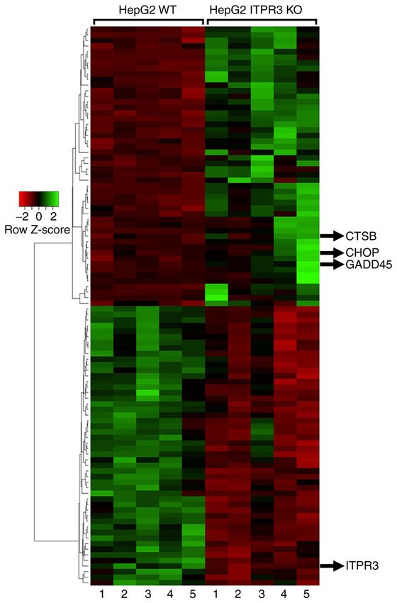
ONCOLOGY LETTERS 23: 32, 2022                                                     3

Pre‑processing of raw RNA sequencing (RNA‑seq) data.               normality test (Shapiro‑Wilk) was performed for each contin‑
Raw read sequences were pre‑processed by filtering out read        uous variable. For comparison between the etiological groups,
sequences with less than 36 bp, removing low quality or N bases    χ2 or the Fisher's exact test was applied in categorical variables
from the read ends, trimming Illumina adapters, scanning the       and Mann Whitney or Kruskal‑Wallis test in continuous data.
read with a 4‑base wide sliding window and cutting when            In multiple comparisons, Dunn's test was applied after the
the average quality per base dropped below 15 (LEADING:3           Kruskal‑Wallis test. Multivariate COX regression model with
TRAILING:3 SLIDINGWINDOW:4:15 MINLEN:36) by                        covariance structure was performed to determine the model for
Trimmomatic v0.39 (28).                                            EFS. Moreover, to evaluate the correlation between the clini‑
    The raw RNA‑seq data were obtained in our previous publi‑      copathological parameter and ITPR3 expression, Spearman's
cation, through experiments with HepG2 (16), considered a cell     coefficient was used. SPSS software, v20 (IBM Corp.), was
line of liver cancer. A commercially available CRISPR/Cas9         used for statistical analysis. Three technical replicates were
system was used to eliminate ITPR3 in HepG2 liver cancer           used per sample in the analyses. Final results were expressed
cells (Santa Cruz Biotechnology, Inc.). Cells were grown in        as the mean of the three values. P
4                              SANTOS et al: ITPR3 IN HEPATOCELLULAR CARCINOMA MAINTENANCE

Table I. Clinical and laboratory data of patients with hepato‑       Table III. Correlation between ITPR3 labeling intensity with
cellular carcinoma.                                                  anatomopathological and clinical data parameters.

Parameter                Minimum         Median       Maximum        Clinical                           Cirrhosis              Tumor
                                                                     parameters                         P‑value                P‑value
Age (years)                41.11         57.09           74.03
MELD                        6.0           15.0            32.0       Number nodules       0.120 0.296
AFP (ng/ml)                1.00          123.78         2883.00      Vascular invasion    0.637 0.746
TBIL (mg/dl)                0.50           2.65           8.70       Necrosis             0.114 0.461
INR                        0.990          1.045          2.690       Fibrosis             0.752 0.456
Creatinine (mg/dl)          0.14           1.02           3.00       Tumor inflammation   0.383 0.552
ALT (U/l)                   24.0           91.3          325.0       Intracellular        0.927 0.825
AST (U/l)                  25.00          85.54         267.00       characteristics
ALB (g/dl)                  2.00           3.20           4.80       Differentiation      0.677 0.668
                                                                     degree
MELD, model for end‑stage liver disease; AFP α fetoprotein; TBIL,    Histological pattern 0.601 0.941
total bilirubin; INR, international normalized ratio; ALT, alanine
                                                                     MELD                 0.395 0.393
aminotransferase; AST, aspartate transaminase; ALB, albumin.
                                                                     AFP (ng/ml)          0.703 0.781
                                                                     TBIL (mg/dl)         0.496 0.694
                                                                     INR                  0.403 0.344
Table II. Anatomopathological characteristics of liver from          Creatinine (mg/dl)   0.216 0.248
patients with hepatocellular carcinoma.                              ALT (U/l)            0.410 0.281
                                                                     AST (U/l)            0.497 0.018a
Clinical parameters                                     Value
                                                                     Mitosis (10 HPF)       ‑   0.0098b
Nodules numbera
                                                                     MELD, model for end‑stage liver disease; AFP, α fetoprotein; TBIL,
 ≤3                                                  26 (55.3%)      total bilirubin; INR, international normalized ratio; ALT, alanine
 >3                                                  21 (44.7%)      aminotransferase (aP
ONCOLOGY LETTERS 23: 32, 2022 5
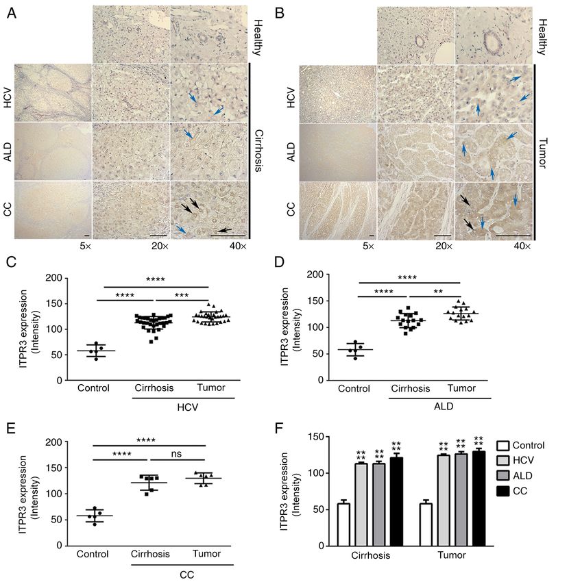
Figure 1. Representative images of ITPR3 labeling in HCC, underlying cirrhotic parenchyma and non‑tumor livers (controls). (A) ITPR3 labeling in cirrhosis
in patients diagnosed with HCV (n=31), ALD (n=16), CC (n=6) and healthy (control; n=2). Scale bar=200 µm (magnification, x5), 50 µm (magnification, x 20)
and 25 µm (magnification, x40). Black arrows indicate ITPR3 perinuclear localization in hepatocytes. Blue arrows indicate ITPR3 cytoplasmatic localization
in hepatocytes. (B) ITPR3 labeling in tumor (HCC) region of patients diagnosed with HCV (n=31), ALD (n=16), CC (n=6) and healthy (control; n=3). Black
arrows indicate ITPR3 perinuclear localization in hepatocytes. Blue arrows indicate ITPR3 cytoplasmatic localization in hepatocytes. (C) Representative
graph of the intensity of ITPR3 marking in HCC regions and cirrhotic parenchyma of patients with HCV compared with non‑tumor livers (control; n=31;
****
P6 SANTOS et al: ITPR3 IN HEPATOCELLULAR CARCINOMA MAINTENANCE Figure 2. Correlation between ITPR3 labeling intensity and mitotic and apoptosis percentages (ssDNA). (A) Low mitotic (
ONCOLOGY LETTERS 23: 32, 2022                                                     7

between CC and HCV etiologies (P=0.001). However, neither              high mitotic index (≥5/10 HPF is associated with intrahepatic
ITPR3 expression in the tumor nor in the cirrhotic region              metastasis, larger tumor size, higher AFP levels and advanced
was associated with EFS outcome. Together, the results                 tumor stage (23,27), as well as vascular invasion (26), poor
suggested that ITPR3 expression in transformed hepatocytes             histological differentiation and high recurrence rate (23). In
is implicated in evasion of apoptosis, which contributes to the        the samples in the present study, most patients (62.8%) had a
maintenance of HCC.                                                    low mitotic index (
8                               SANTOS et al: ITPR3 IN HEPATOCELLULAR CARCINOMA MAINTENANCE

    In the context of HCC, CHOP is pro‑apoptotic (39),                 to apoptosis. This resistance to apoptosis and alteration
performing a pro‑tumorigenic function, being an oncogene in            of tumor cell survival occurred, at least in part, through an
hepatic carcinogenesis (39). CHOP is ubiquitously expressed            intracellular signaling pathway in which ITPR3 demonstrated
at very low levels normally, but is strongly expressed in cells        a negative effect on the expression of CHOP and GADD45,
subjected to severe stress (64). However, activation of this           providing a reduction in apoptotic cell death. The expression
gene is not universally observed in human HCC, which leads             of ITPR3 in the liver may be a promising prognostic marker of
to the suggestion that there are other oncogenic pathways that         HCC and other types of liver cancer.
promote hepatocarcinogenesis independently of CHOP. Thus,
this gene may participate more in the progression of HCC               Acknowledgements
rather than in its induction (39). The data from the present
study suggested that the action of ITPR3 on CHOP expres‑               Not applicable.
sion was associated with the apoptotic signaling pathway by
promoting maintenance of liver cancer development.                     Funding
    Growth arrest and damage to DNA 45 (GADD45) is a
family composed of three homologous acidic proteins (α, β and          The present study was supported by grants from CNPq,
γ) (65), which respond to cellular stresses of environmental and       FAPEMIG and NIH (grant nos. P01‑DK57751, P30‑DK34989,
physiological origin (41). GADD45 proteins are also involved in        R01‑DK114041 and R01‑DK112797).
hepatic tumorigenesis, in both carcinogenesis and established
HCC (65). In the context of HCC, GADD45β has a pro‑apoptotic           Availability of data and materials
action and occurs at a low or absent expression level in neoplastic
cells, a specific characteristic of this type of tumor (41). This      The datasets analyzed during the current study are avail‑
is critically correlated with fundamental clinical‑pathological        able in the NCBI Sequence Read Archive under the
characteristics of tumor development (41). The synergistic             Accession code PRJNA758563 (https://www.ncbi.nlm.nih.
effect of aspirin and sorafenib treatment induces an increase in       gov/sra/?term=PRJNA758563). All data generated or analyzed
GADD45β in HCC, promoting apoptosis and control of tumor               during this study are included in this published article.
growth (66), an opposite effect of growth in the non‑neoplastic
liver (65), reflecting the opposing effects of this gene depending     Authors' contributions
on the biological context. GADD45β responses reflect its
dynamics in rapid adaptations (65), being a possible diagnostic        The entire project was conducted, written and analyzed by
and selection biomarker for the treatment of HCC (66). These           MLDS, including carrying out the experiments. AF contrib‑
data suggest that the appearance of ITPR3 in liver cancer may          uted in all aspects, mainly in the analysis and interpretation
promote tumor maintenance through the suppressive effect of            of data. RMF contributed with the assistance in carrying out
GADD45 expression, especially of the β isoform.                        the experiments, analysis and discussion of data and with the
    The present study demonstrated the upregulation of impor‑          preparation of graphics and images. ACMLF contributed with
tant genes involved in the apoptotic pathway in liver cancer,          the assistance in carrying out the experiments, analysis and
such as GADD45 and CHOP. However, high‑throughput                      discussion of data and with the preparation of graphics and
technology would add stronger evidence. Thus, the absence of           images. PHD contributed to the statistical analysis and in the
microarray experiments is a limitation of the present study.           organization of the survival data. CAXG and VLC elaborated
    In other types of neoplasms or liver diseases, ITPR3 can           all the bioinformatics data, analysis and RNA‑seq results.
assume several roles. In prostate cancer, the mechanisms that          PVTV performed the mitotic cell count on the HCC samples in
promote the degradation of ITPR3 and prevent apoptosis have            the tumor mitotic index experiment. In addition, PVTV helped
been elucidated (19). In ischemia‑reperfusion injury, ITPR3            to analyse and interpret the data in conjunction with the litera‑
serves a protective role in which expression of the protein serves     ture already published and was involved in the writing of the
to change the mechanism of hepatocyte death from necrosis to           manuscript and in the critical review of important intellectual
apoptosis (15). This demonstrates the versatility that ITPR3           content. PVTV also helped with the construction of the data
assumes within the varied contexts of different diseases and           discussion. MFL participated in carrying out the experiments
organs, but all associated with apoptotic events. It has been          and articulating the data obtained from the works previously
demonstrated that ITPR3 serves a role in proliferative stimulation     published by the group. FOL was involved in the writing of
and apoptosis block in liver cell lines and normal mouse liver (16).   the manuscript and critical review of important intellectual
The current study provided evidence in human HCC samples               content, and also contributed to the analysis of some of the
that ITPR3 serves a role in hepatocarcinogenesis by modulating         data presented. MHN contributed to the analysis and discus‑
apoptosis. Additional work will be needed to better understand         sion of the data in this study, in addition to approving the
the mechanism by which de novo expression of ITPR3 correlates          final version submitted for publication. GF participated in the
with mitosis and apoptosis in the pathogenesis of HCC, as well as      orientation and design of the study, particularly in the contri‑
whether this represents a potential target for therapy.                bution of immunohistochemical staining image analysis.
    In conclusion, ITPR3 was highly expressed in HCC tumor             CXL participated in the acquisition of clinical data from
cells relative to its expression level in the underlying CLD and       patients and in the interpretation of these data in conjunction
healthy livers. ITPR3 expression was inversely correlated with         with the results. MLDS and PVTV confirm the authenticity
apoptotic and mitotic indices in HCC, suggesting that ITPR3            of all the raw data. All authors reviewed and approved the
contributed to the maintenance of HCC, promoting resistance            final manuscript.
ONCOLOGY LETTERS 23: 32, 2022 9
Ethics approval and consent to participate 17. Ueasilamongkol P, Khamphaya T, Guerra MT, Rodrigues MA,
Gomes DA, Kong Y, Wei W, Jain D, Tramper t DC,
Ananthanarayanan M, et al: Type 3 inositol 1,4,5‑trisphosphate
The present study was approved by the Ethics Committee of receptor is increased and enhances malignant properties in chol‑
the Hospital das Clínicas, Federal University of Minas Gerais angiocarcinoma. Hepatology 71: 583‑599, 2020.
18. Shibao K, Fiedler MJ, Nagata J, Minagawa N, Hirata K, Nakayama Y,
(approval number CAAE 71206617.8.0000.5149) and written Iwakiri Y, Nathanson MH and Yamaguchi K: The type III inositol
informed consent was obtained from the patients or their rela‑ 1,4,5‑trisphosphate receptor is associated with aggressiveness of
tives, including for use of their tissue in the research. colorectal carcinoma. Cell Calcium 48: 315‑323, 2010.
19. Kuchay S, Giorgi C, Simoneschi D, Pagan J, Missiroli S,
Saraf A, Florens L, Washburn MP, Collazo‑Lorduy A,
Patient consent for publication Castillo‑Martin M, et al: PTEN counteracts FBXL2 to promote
IP3R3‑ and Ca 2+‑mediated apoptosis limiting tumour growth.
Not applicable. Nature 546: 554‑558, 2017.
20. Bononi A, Giorgi C, Patergnani S, Larson D, Verbruggen K,
Tanji M, Pellegrini L, Signorato V, Olivetto F, Pastorino S, et al:
Competing interests BAP1 regulates IP3R3‑mediated Ca 2+ flux to mitochondria
suppressing cell transformation. Nature 546: 549‑553, 2017.
21. Fonseca MC, França A, Florentino RM, Fonseca RC,
The authors declare that they have no competing interests. Lima Filho AC, Vidigal PT, Oliveira AG, Dubuquoy L,
Nathanson MH and Leite MF: Cholesterol‑enriched membrane
microdomains are needed for insulin signaling and proliferation
References in hepatic cells. Am J Physiol Gastrointest Liver Physiol 315:
G80‑G94, 2018.
1. Agni RM: Diagnostic histopathology of hepatocellular carcinoma: 22. Baak JP: Mitosis counting in tumors. Hum Pathol 21: 683‑685, 1990.
A case‑based review. Semin Diagn Pathol 34: 126‑137, 2017. 23. Ha SY, Choi M, Lee T and Park CK: The prognostic role of
2. Sung H, Ferlay J, Siegel RL, Laversanne M, Soerjomataram I, mitotic index in hepatocellular carcinoma patients after curative
Jemal A and Bray F: Global cancer statistics 2020: GLOBOCAN hepatectomy. Cancer Res Treat 48: 180‑189, 2016.
estimates of incidence and mortality worldwide for 36 cancers in 24. Haratake J, Takeda S, Kasai T, Nakano S and Tokui N: Predictable
185 countries. CA Cancer J Clin 71: 209‑249, 2021. factors for estimating prognosis of patients after resection of
3. Forner A, Reig M and Bruix J: Hepatocellular carcinoma. hepatocellular carcinoma. Cancer 72: 1178‑1183, 1993.
Lancet 391: 1301‑1314, 2018. 25. Nanashima A, Tanaka K, Yamaguchi H, Shibasaki S, Morino S,
4. Bodzin AS, Lunsford KE, Markovic D, Harlander‑Locke MP, Yoshinaga M, Sawai T, Nakagoe T and Ayabe H: Fibrosis and
Busuttil RW and Agopian VG: Predicting mortality in patients inflammatory activity in noncancerous tissue and mitotic index
developing recurrent hepatocellular carcinoma after liver of cancer tissue in patients with hepatocellular carcinoma:
transplantation: Impact of treatment modality and recurrence Relationship to clinicopathological factors and prognosis after
characteristics. Ann Surg 266: 118‑125, 2017. hepatic resection. Dig Dis Sci 48: 1517‑1522, 2003.
5. Villanueva A: Hepatocellular carcinoma. N Engl J Med 380: 26. Osório FM, Vidigal PV, Ferrari TC, Lima AS, Lauar GM and
1450‑1462, 2019. Couto CA: Histologic grade and mitotic index as predictors of
6. Amaya MJ and Nathanson MH: Calcium signaling in the liver. microvascular invasion in hepatocellular carcinoma. Exp Clin
Compr Physiol 3: 515‑539, 2013. Transplant 13: 421‑425, 2015.
7. Patel S, Joseph SK and Thomas AP: Molecular properties of inositol 27. Ouchi K, Sugawara T, Ono H, Fujiya T, Kamiyama Y, Kakugawa Y,
1,4,5‑trisphosphate receptors. Cell Calcium 25: 247‑264, 1999. Mikuni J, Yamanami H, Komatsu S and Horikoshi A: Mitotic
8. Wojcikiewicz RJ: Type I, II, and III inositol 1,4,5‑trisphosphate index is the best predictive factor for survival of patients with
receptors are unequally susceptible to down‑regulation and are resected hepatocellular carcinoma. Dig Surg 17: 42‑48, 2000.
expressed in markedly different proportions in different cell 28. Bolger AM, Lohse M and Usadel B: Trimmomatic: A flex‑
types. J Biol Chem 270: 11678‑11683, 1995. ible trimmer for illumina sequence data. Bioinformatics 30:
9. Yule DI, Ernst SA, Ohnishi H and Wojcikiewicz RJ: Evidence that 2114‑2120, 2014.
zymogen granules are not a physiologically relevant calcium pool. 29. Patro R, Duggal G, Love MI, Irizarry RA and Kingsford C:
Defining the distribution of inositol 1,4,5‑trisphosphate receptors Salmon provides fast and bias‑aware quantification of transcript
in pancreatic acinar cells. J Biol Chem 272: 9093‑9098, 1997. expression. Nat Methods 14: 417‑419, 2017.
10. Cruz LN, Guerra MT, Kruglov E, Mennone A, Garcia CR, Chen J and 30. Howe KL, Achuthan P, Allen J, Allen J, Alvarez‑Jarreta J,
Nathanson MH: Regulation of multidrug resistance‑associated protein Amode MR, Armean IM, Azov AG, Bennett R, Bhai J et al:
2 by calcium signaling in mouse liver. Hepatology 52: 327‑337, 2010. Ensembl 2021. Nucleic Acids Res 49D: D884‑D891, 2021.
11. Hirata K, Pusl T, O'Neill AF, Dranoff JA and Nathanson MH: The 31. Love MI, Huber W and Anders S: Moderated estimation of fold
type II inositol 1,4,5‑trisphosphate receptor can trigger Ca2+ waves change and dispersion for RNA‑seq data with DESeq2. Genome
in rat hepatocytes. Gastroenterology 122: 1088‑1100, 2002. Biol 15: 550, 2014.
12. Dufour JF, Lüthi M, Forestier M and Magnino F: Expression of 32. Kucukural A, Yukselen O, Ozata DM, Moore MJ and Garber M:
inositol 1,4,5‑trisphosphate receptor isoforms in rat cirrhosis. DEBrowser: Interactive differential expression analysis and visu‑
Hepatology 30: 1018‑1026, 1999. alization tool for count data. BMC Genomics 20: 6, 2019.
13. Lemos FO, França A, Lima Filho ACM, Florentino RM, 33. Kanehisa M and Goto S: KEGG: Kyoto encyclopedia of genes
Santos ML, Missiaggia DG, Rodrigues GOL, Dias FF, and genomes. Nucleic Acids Res 28: 27‑30, 2000.
Souza Passos IB, Teixeira MM, et al: Molecular mechanism for 34. UniProt Consortium: UniProt: The universal protein knowledge‑
protection against liver failure in human yellow fever infection. base in 2021. Nucleic Acids Res 49D: D480‑D489, 2021.
Hepatol Commun 4: 657‑669, 2020. 35. Kanehisa M and Sato Y: KEGG Mapper for inferring cellular
14. Lima Filho ACM, França A, Florentino RM, Dos Santos ML, functions from protein sequences. Protein Sci 29: 28‑35, 2020.
de Olivei ra L emos F, Missiaggia DG, Fonseca RC, 36. Babicki S, Arndt D, Marcu A, Liang Y, Grant JR, Maciejewski A
Gustavo Oliveira A, Ananthanarayanan M, Guerra MT, et al: and Wishart DS: Heatmapper: Web‑enabled heat mapping for all.
Inositol 1,4,5‑trisphosphate receptor type 3 plays a protective
role in hepatocytes during hepatic ischemia‑reperfusion injury. Nucleic Acids Res 44W: W147‑W153, 2016.
Cell Calcium 91: 102264, 2020. 37. Edmondson HA and Steiner PE: Primary carcinoma of the
15. Leite MF, Thrower EC, Echevarria W, Koulen P, Hirata K, liver: A study of 100 cases among 48,900 necropsies. Cancer 7:
Bennett AM, Ehrlich BE and Nathanson MH: Nuclear and cyto‑ 462‑503, 1954.
solic calcium are regulated independently. Proc Natl Acad Sci 38. Ruan J, Zheng H, Rong X, Rong X, Zhang J, Fang W, Zhao P and
USA 100: 2975‑2980, 2003. Luo R: Over‑expression of cathepsin B in hepatocellular carcinomas
16. Guerra MT, Florentino RM, França A, Lima Filho AC, Dos predicts poor prognosis of HCC patients. Mol Cancer 15: 17, 2016.
Santos ML, Fonseca RC, Lemos FO, Fonseca MC, Kruglov E, 39. Scaiewicz V, Nahmias A, Chung RT, Mueller T, Tirosh B and
Mennone A, et al: Expression of the type 3 InsP3 receptor is a Shibolet O: CCAAT/enhancer‑binding protein homologous
final common event in the development of hepatocellular carci‑ (CHOP) protein promotes carcinogenesis in the DEN‑induced
noma. Gut 68: 1676‑1687, 2019. Hepatocellular carcinoma model. PLoS One 8: e81065, 2013.10 SANTOS et al: ITPR3 IN HEPATOCELLULAR CARCINOMA MAINTENANCE
40. Liebermann DA and Hoffman B: Gadd45 in stress signaling. 55. Okada S, Shimada K, Yamamoto J, Takayama T, Kosuge T,
J Mol Signal 3: 15, 2008. Yamasaki S, Sakamoto M and Hirohashi S: Predictive factors
41. Qiu W, David D, Zhou B, Chu PG, Zhang B, Wu M, Xiao J, Han T, for postoperative recurrence of hepatocellular carcinoma.
Zhu Z, Wang T, et al: Down‑regulation of growth arrest DNA Gastroenterology 106: 1618‑1624, 1994.
damage‑inducible gene 45beta expression is associated with 56. Ruà S, Comino A, Fruttero A, Torchio P, Bouzari H, Taraglio S,
human hepatocellular carcinoma. Am J Pathol 162: 1961‑1974, Torchio B and Capussotti L: Flow cytometric DNA analysis of
2003. cirrhotic liver cells in patients with hepatocellular carcinoma can
42. Llovet JM, Peña CE, Lathia CD, Shan M, Meinhardt G and provide a new prognostic factor. Cancer 78: 1195‑1202, 1996.
Bruix J; SHARP Investigators Study Group: Plasma biomarkers 57. Soini Y, Virkajärvi N, Lehto VP and Pääkkö P: Hepatocellular
as predictors of outcome in patients with advanced hepatocel‑ carcinomas with a high proliferation index and a low degree of
lular carcinoma. Clin Cancer Res 18: 2290‑2300, 2012. apoptosis and necrosis are associated with a shortened survival.
43. Alves RC, Alves D, Guz B, Matos C, Viana M, Harriz M, Br J Cancer 73: 1025‑1030, 1996.
Terrabuio D, Kondo M, Gampel O and Polletti P: Advanced 58. Tannapfel A, Geissler F, Köckerling F, Katalinic A, Hauss J and
hepatocellular carcinoma. Review of targeted molecular drugs. Wittekind C: Apoptosis and proliferation in relation to histopath‑
Ann Hepatol 10: 21‑27, 2011. ological variables and prognosis in hepatocellular carcinoma.
44. Ho DW, Lo RC, Chan LK and Ng IO: Molecular pathogenesis of J Pathol 187: 439‑445, 1999.
hepatocellular carcinoma. Liver Cancer 5: 290‑302, 2016. 59. Alisi A, Mele R, Spaziani A, Tavolaro S, Palescandolo E and
45. Lu LC, Hsu CH, Hsu C and Cheng AL: Tumor heterogeneity in Balsano C: Thr 446 phosphorylation of PKR by HCV core
hepatocellular carcinoma: Facing the challenges. Liver Cancer 5: protein deregulates G2/M phase in HCC cells. J Cell Physiol 205:
128‑138, 2016. 25‑31, 2005.
46. Hoshida Y, Nijman SM, Kobayashi M, Chan JA, Brunet JP, 60. Clemens DL, Calisto LE, Sorrell MF and Tuma DJ: Ethanol
Chiang DY, Villanueva A, Newell P, Ikeda K, Hashimoto M, et al: metabolism results in a G2/M cell‑cycle arrest in recombinant
Integrative transcriptome analysis reveals common molecular Hep G2 cells. Hepatology 38: 385‑393, 2003.
subclasses of human hepatocellular carcinoma. Cancer Res 69: 61. Juríková M, Danihel L, Polák Š and Varga I: Ki67, PCNA, and
7385‑7392, 2009. MCM proteins: Markers of proliferation in the diagnosis of
47. Chiang DY, Villanueva A, Hoshida Y, Peix J, Newell P, breast cancer. Acta Histochem 118: 544‑552, 2016.
Minguez B, LeBlanc AC, Donovan DJ, Thung SN, Solé M, et al: 62. Michalopoulos GK: Liver regeneration after partial hepatectomy:
Focal gains of VEGFA and molecular classification of hepatocel‑ Critical analysis of mechanistic dilemmas. Am J Pathol 176:
lular carcinoma. Cancer Res 68: 6779‑6788, 2008. 2‑13, 2010.
48. Villanueva A, Hoshida Y, Battiston C, Tovar V, Sia D, Alsinet C, 63. Mangla A, Guerra MT and Nathanson MH: Type 3 inositol
Cornella H, Liberzon A, Kobayashi M, Kumada H, et al: 1,4,5‑trisphosphate receptor: A calcium channel for all seasons.
Combining clinical, pathology, and gene expression data to predict Cell Calcium 85: 102132, 2020.
recurrence of hepatocellular carcinoma. Gastroenterology 140: 64. Chikka MR, McCabe DD, Tyra HM and Rutkowski DT: C/EBP
1501‑1512.e2, 2011. homologous protein (CHOP) contributes to suppression of
49. Lee JS, Chu IS, Heo J, Calvisi DF, Sun Z, Roskams T, Durnez A, metabolic genes during endoplasmic reticulum stress in the liver.
Demetris AJ and Thorgeirsson SS: Classification and predic‑ J Biol Chem 288: 4405‑4415, 2013.
tion of survival in hepatocellular carcinoma by gene expression 65. Tian J and Locker J: Gadd45 in the liver: Signal transduction and
profiling. Hepatology 40: 667‑676, 2004. transcriptional mechanisms. In: Gadd45 Stress Sensor Genes.
50. Zucman‑Rossi J, Villanueva A, Nault JC and Llovet JM: Liebermann DA and Hoffman B (eds). Springer, New York, NY,
Genetic landscape and biomarkers of hepatocellular carcinoma. pp69‑80, 2013.
Gastroenterology 149: 1226‑1239.e4, 2015. 66. Xia H, Lee KW, Chen J, Kong SN, Sekar K, Deivasigamani A,
51. Masuzaki R and Omata M: Screening program in high‑risk popula‑ Seshachalam VP, Goh BKP, Ooi LL and Hui KM: Simultaneous
tions. In: Hepatocellular Carcinoma. 2nd edition. Mcmasters KM silencing of ACSL4 and induction of GADD45B in hepatocel‑
and Vauthey JN (eds). Springer, New York, NY, pp55‑68, 2011. lular carcinoma cells amplifies the synergistic therapeutic effect
52. Bloom HJ and Richardson WW: Histological grading and prog‑ of aspirin and sorafenib. Cell Death Discov 3: 17058, 2017.
nosis in breast cancer; a study of 1409 cases of which 359 have
been followed for 15 years. Br J Cancer 11: 359‑377, 1957. This work is licensed under a Creative Commons
53. Vang R, Shih IM and Kurman RJ: Ovarian low‑grade and Attribution-NonCommercial-NoDerivatives 4.0
high‑grade serous carcinoma: Pathogenesis, clinicopathologic International (CC BY-NC-ND 4.0) License.
and molecular biologic features, and diagnostic problems. Adv
Anat Patht 16: 267‑282, 2009.
54. Suehiro T, Matsumata T, Itasaka H, Yamamoto K, Kawahara N
and Sugimachi K: Clinicopathologic features and prognosis of
resected hepatocellular carcinomas of varied sizes with special
reference to proliferating cell nuclear antigen. Cancer 76:
399‑405, 1995.You can also read