Inhibitory Effects of Synthesize Silver Nanoparticles on Cucumber Mosaic Virus Infecting Tomato Plants
←
→
Page content transcription
If your browser does not render page correctly, please read the page content below
International Journal of Pharmaceutical and Phytopharmacological Research (eIJPPR) | April 2021 | Volume 11 | Issue 2 | Page 51-61 Esam Kamal Fahmy Elbeshehy, Inhibitory Effects of Synthesize Silver Nanoparticles on Cucumber Mosaic Virus Infecting Tomato Plants Inhibitory Effects of Synthesize Silver Nanoparticles on Cucumber Mosaic Virus Infecting Tomato Plants Esam Kamal Fahmy Elbeshehy1,2*, Areej Ali Mohamed Baeshen1, Wael Mohamed Hassan Mohamed2,3 1 Department of Biological Sciences, Faculty of Science, University of Jeddah, Jeddah, Saudi Arabia. 2 Botany Department, Faculty of Agriculture, Suez Canal University, Egypt. 3 Department of Biology, Quwayiyah College of Science and Humanities, Shaqra University, Saudi Arabia. ABSTRACT Nanoparticles have risen rapidly to be one of the superior research scopes. The most suitable in the manufacture of nanoparticles as an alternative to the traditional physical and chemical methods are biological methods, bacteria provide many advantages in this context. The silver nanoparticles show interesting with compared to other salts due to widest applications in medical, therapeutics, and cosmetics. Reports of silver nanoparticle activity as antimicrobials are numerous, but the search on activity against the plant viruses was unpretentious. Diverse leave cultivars of tomato plants (Lycopersicon esculentum L) infected by Cucumber Mosaic Virus (CMV) are showing yellowing leaves, mottling, leaf curl, and leaf shoestring as a result of deformation. To better understand, the antiviral properties of silver nanoparticles are the efficacy of CMV disease control strategies. In this article, we will look at the effective role of silver nanoparticles as an excellent antiviral to CMV isolated from naturally infected tomato plants. Viral identity well confirms by direct ELISA test with antiserum to CMV, TMV, TEV, PVY, TSWV, PMMoV, and ToMV, host range, electron microscopy, and (RT-PCR) studies. The present study will evaluate the effects of exogenously applied silver nanoparticles on the percentage of CMV infection, disease severity, virus concentration, main photosynthetic pigments Phenolic compounds, and total protein and protein profile. Key Words: Silver nanoparticles, Antiviral, Cucumoviruses, CMV, Physiological characteristic, Tomato. eIJPPR 2021; 11(2):61-61 HOW TO CITE THIS ARTICLE: Elbeshehy E K F, Baeshen A A M, Mohamed W M H. Inhibitory Effects of Synthesize Silver Nanoparticles on Cucumber Mosaic Virus Infecting Tomato Plants. Int j pharm phytopharm res. 2021;11(2):51-61. https://doi.org/10.51847/Dn3Y66NE2u INTRODUCTION The attention of scientists in various fields has been attracted about assessing nanoparticles to be one of the Solanum lycopersicon L. owned by many plants is called most important particles of modern science [10, 11]. There the Solanaceae. It is the most popular vegetable throughout are many ways to produce nanoparticles, such as chemical, the world and the most important commercially grown physical, and mixed methods, and these methods are quick vegetable in the Mecca regions. In the last years, the to produce, but are unsafe and have unwanted residues, and tomato plant has been productively used as a model plant are very expensive, and therefore scientists have tended to to examine the initiation of defense pathways after infected create an environmentally friendly and inexpensive way to viral bacterial, fungal, and biotic stress [1]. produce nanoparticles [12-14]. Many scientists in the Tomatoes are infected by several viruses, that is, TMV, recent period have made great efforts to take advantage of cucumber CMV [2], PVY [3], TEV [4], TSWV [5], and microorganisms as factories to create environmentally PMMoV [6]. The CMV virus follows the Bromoviridae friendly nanoparticles [15]. Scientists have used many family, and it follows the genus Cucumovirus. The virus eukaryotic microorganisms such as (yeasts, fungi, and has a wide range, [7] and can cause many symptoms of plants) and prokaryotes such as (actinomyces and bacteria) infection, for example, yellowing, mosaic, stunting, and in the synthesis of nanoparticles. Silver nanoparticles have deformation, thus causing serious economic losses [8, 9]. been used among many minerals as antiviral, antifungal, antibacterial, and current anticancer agents [16]. In this Corresponding author: Esam Kamal Fahmy Elbeshehy Address: Department of Biological Sciences, Faculty of Science, University of Jeddah, Jeddah, Saudi Arabia. E-mail: esamelbeshehy @ yahoo.com Received: 27 February 2021; Revised: 14 April 2021; Accepted: 19 April 2021 ISSN (Online) 2249-6084 (Print) 2250-1029 www.eijppr.com
International Journal of Pharmaceutical and Phytopharmacological Research (eIJPPR) | April 2021 | Volume 11 | Issue 2 | Page 51-61
Esam Kamal Fahmy Elbeshehy, Inhibitory Effects of Synthesize Silver Nanoparticles on Cucumber Mosaic Virus Infecting Tomato Plants

article, we hypothesize that the discovery of new silver reacted as were vein clearing, mosaic, blasters, and
reducing strains, the suppression activity against the plant leaf narrow [18].
viruses, and the use of charge-free or charge-credit
production route, may lead to the greater possibility for Mechanical Transmission of CMV
economical, eco-friendly silver nanoparticles production The virus was transmitted mechanically to healthy
method and inhibit the viral infection caused by CMV Squash Cucurbita pepo seedling, cv. (Vegetable
infecting tomato plants through virus isolation and morrow white bush) for virus propagation and
identification. In addition, studying the effect of different preserved under controlled conditions after
silver nanoparticles antiviral concentration will collect inoculation for three weeks. The presence of CMV
from different prokaryotic organisms on CMV and was appropriate with the ELISA test (Enzyme-linked
evaluate the effects of exogenous applied silver Immunosorbent Assay), and the samples that gave a
nanoparticles synthesized from different prokaryotic positive result for the ELISA test were used as a source
organisms on the infection percentage of CMV, the of the virus in the post. The infested sap was obtained
severity of the disease, concentration virus, the by grinding the leaves of zucchini with a buffered
concentrations of the photosynthetic pigments content, phosphate buffer at a pH7.
Phenolic compounds, and protein components.
 We can summarize the roles of silver nanoparticles Electron Microscope
 synthesized from different prokaryotic organisms in the Healthy and infected mesophyll tissue was pieced,
 effect on natural and artificial infected tomato plants by fixed, and stained for ultra-thin sectioning according
 CMV or protect healthy profitable tomato failed as listed to the standard procedures of [19]. The ultra-thin
 below: section was viewed with an electron microscope Unit
 A- Treatments of the plants with silver nanoparticles (JOEL-JEA100 CX).
 (NPs) can lead to the initiation of tolerance
 mediators, that characterized by confining of the DAS-ELISA
 multiplication of virus and repression of symptoms of We examined all samples by using DAS-ELISA with
 disease compared with healthy plants. a specific kit of CMV antibodies at 405 nm and was
 B- This study was undertaken to assess the effect of determined by using an ELISA plate reader (Model
 52
 silver nanoparticles (NPs) on CMV multiplication 550; Bio-Rad, Hercules, CA, USA) [20].
 and disease development.
 C- Increasing the productivity of tomato of the fruit crop Total RNA Extraction and RT-PCR
 We extracted total RNA from un-inoculated and
MATERIALS AND METHODS inoculated leaves of tomato by EZ-10 Spin Column
 Total TNA Minipreps Super Kit (BIO BASIC INC)
Isolation and Identification of CMV specific CMV1 oligonucleotide
Naturally infected tomatoes (Lycopersicon esculentum) (5´GCC/GTAA/GCT/GGA/TGG/AC/AA3´). CMV2
with the CMV that develop typical symptoms on upper and (5`TAT/GAT/AAG/AAG/CTT/GTT/TCG/CG 3`)
lower leaves were collected from Mecca regions. primers [21], with one-step RT-PCR (Reverse
 Transcription Polymerase Chain Reaction)
 Virus Isolation and Propagation amplification of CMV coat protein gene.
 Infected leaves of Lycopersicon esculentum showing
 a typical symptom suspected to be CMV infections Collection of Silver Nanoparticles Synthesized from
 were collected from Mecca regions. The collected Different Prokaryotic Organisms
 leaves samples were serologically checked for CMV Isolation and Characterization of Bacteria
 infection by DAS-ELISA test as explained by [17] The bacterial strains were isolated, cultured and the
 against the following viruses: CMV, ToMV, and morphological and physiological characterization of
 TMV. All infected samples were tested in triplicate. the isolates were performed according to the methods
 Samples with positive reactions were used as a source described by [22, 23].
 of CMV.
 CMV was isolated and biologically purified from Production of Biomass
 naturally infected tomato plants by on C. For product biosynthesis of bacterial strains in a
 amaranticolor Coste and Reyn plants produced nutrient broth medium, they were cultured and
 necrotic local legions surrounded with little halo edge. incubated on an orbital shaker at 27°C and agitated at
 It was propagated in healthy Squash Cucurbita pepo 220 rpm. After 24 hours, biomass was harvested and
 seedling, cv. (Vegetable morrow white bush) which

ISSN (Online) 2249-6084 (Print) 2250-1029 www.eijppr.com
International Journal of Pharmaceutical and Phytopharmacological Research (eIJPPR) | April 2021 | Volume 11 | Issue 2 | Page 51-61
Esam Kamal Fahmy Elbeshehy, Inhibitory Effects of Synthesize Silver Nanoparticles on Cucumber Mosaic Virus Infecting Tomato Plants

 centrifuged for 10 minutes at 5000 rpm. and collected from virus inoculation, group 3: spray by NPs after 24
 the supernatant material for nanoparticle synthesis. hours from virus infection.

 Silver Nanoparticles Synthesis Evaluation of Changes in Virus Infection Treated with
 The culture supernatant was added separately to CMV and NPs Treatment
 reaction vessels containing silver nitrate at The plant leaves were sprayed with 0.1 μg/μl AgNPs
 concentrations of 0.5 mM, 1 mM, and 2 mM. The including every part from the leaves. The infection
 reaction between this supernatant and silver ions was percentage and severity of disease were recorded after
 carried out for 24 hours in dark conditions. Same three weeks from inoculation, according to the
 time: The control was also triggered without silver following scale: 0 = no symptoms; 1 = crinkling and
 ions along with the experiment flasks [22, 24]. light mottling; 2 = crinkling and mild mosaic; 3=
 crinkling, severe mosaic, and size reduction; and 4 =
 Characterization of Silver Nanoparticles deformation. Disease Severity (DS) values were
 Primary detection of silver nanoparticles after 24 h of calculated using the following formula according to
 incubation of the supernatant and silver ions, was [26].
 done by changing the color of the filtrate. Then, these
 samples were subjected to optical measurements, DS (%)
 which by using a UV-Vis spectrophotometer, were ( ℎ × )
 performed. The Fourier Transform Infrared (FTIR) =
 ( × ℎ ℎ )
 analyzed the interaction between silver nanoparticles × 100 (1)
 and protein.
 Using an X-ray diffractometer (Philips PW 1710), the
 After three weeks from inoculation, the youngest
 X-ray Diffraction (XRD) checked the silver
 developed leaves from treated healthy and infected
 nanoparticles formation. Under sunlight, the
 tomato plants were collected for analysis of changes.
 supernatant treated with silver nitrate was evaporated
 to dryness. Using the Debye-Sherrer formula, the air-
 Effect of Silver Nanoparticles on Physiological
 dried biomass was analyzed [25].
 Parameter 53
 Inhibitory Effects of Different Silver Nanoparticles
 Determination of Photosynthetic Pigments
 Synthesized by some Bacteria on Cucumber Mosaic
 The photosynthetic (chlorophyll a, b and carotenoids)
 Virus Infecting Tomato plants
 pigments were determined according to [27, 28] in
 one-half gram of Tomato cv. Castle rock after 21 days
 Material of Plants and Treatments
 from inoculation by (CMV). Leaves were collected
 After surface sterilization, seeds of tomato susceptible
 and ground in a mortar with 10 ml of 90% acetone,
 cultivar, cv. Castle rock was germinated at 26 ºC in an
 approximately 2-3 gm purified sand, and 2 gm CaCo3
 incubator on moistened filter paper in sterile Petri
 were added to neutralize the acidity of the sap and to
 dishes for 2–3 days and after one day from
 prevent the transformation of chlorophyll into
 germination were transferred to sterilized soil-filled
 pheophytin. The acetone extract was filtered through
 pots kept in a growth chamber. CMV was prepared
 a center glass funnel of fine porosity (G4) and the
 from fresh severely infected leaves of Tomato Cv.
 remainders were washed with small volumes of
 Castle rock and used in these experiments. Plants of
 acetone 90% until being free from pigments. Each
 similar size were selected and divided into three
 filtrate was made up to 50 ml volume with 85%
 groups after 21 days of growth. Each group consists
 acetone and Pigment concentrations in [mg g−1 fresh
 of eight treatments from synthesized AgNPs. The
 weight (FW)] were calculated using the formula of
 names of the treatments were as follows, T1:
 Lichtenthaler:
 (Healthy, negative control) , T2: (Healthy & NPs1) ,
 T3: (Healthy & NPs2) , T4: (Healthy & NPs3), T5:
 = [(11.75 663 − 2.35 645) × 50]
 (Infected, positive control), T6: (Infected & NPs1),
 ∕ 500 = mg /g (F. wt. )
 T7: (Infected & NPs2), T8: (Infected & NPs3). Each
 (2)
 treatment contains 3 replicates (a replicate is one pot = [(18.61 645 − 3.96 663) × 50]
 containing three healthy seedlings). The names of the ∕ 500 = mg /g (F. wt. )
 groups were as follows, group 1: Inoculation by the (3)
 virus with NPs, group 2: spray by NPs before 72 hours + = chlorophyll a + chlorophyll b
 = mg /g (F. wt. ) (4)

ISSN (Online) 2249-6084 (Print) 2250-1029 www.eijppr.com
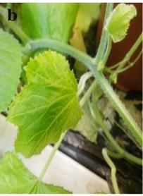
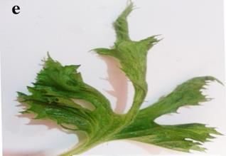
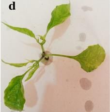
International Journal of Pharmaceutical and Phytopharmacological Research (eIJPPR) | April 2021 | Volume 11 | Issue 2 | Page 51-61
Esam Kamal Fahmy Elbeshehy, Inhibitory Effects of Synthesize Silver Nanoparticles on Cucumber Mosaic Virus Infecting Tomato Plants

 = [ 1000 470 − (2.27 × Chl a) electropherogram of 1.2% agarose gel from
 − (8.14 × Chl b) ∕ 227 × 50] infected tomato with CMV. Electropherogram of
 ∕ 500. (5) 1.2% agarose gel showing PCR amplification
 from infected tomato with CMV. Lane M, DNA
 Total Phenolic content mg/gm (D.Wt.) ladder marker, Lanes 1, 2, 3, and 4 represent PCR-
 A modified Folin-Ciocalteu method [29] was used as positive infected tomato seedling with CMV. Lane
 shown below. For the determination of total phenols 5 represents healthy tomato.
 in various leaf samples, 1 ml of ethanolic extracts of
 each leaf sample was added to 1 ml of 2N Folin-
 Samples that yielded a positive reaction in the DAS-ELISA
 Ciocalteu. Test tubes were heated to about 70 c◦ in a
 test to CMV were used as a source of virus infection.
 water bath, then left for slow cooling at room
 Thereafter, CMV was isolated and biologically purified
 temperature. Beak man DK-2 spectrophotometer at a
 from naturally infected tomato plants by a single local
 wavelength of 650 nm was used for measured optical
 lesion on Chenopodium amaranticolor plants that
 densities of these samples.
 produced necrotic local lesions surrounded with little halo
 edges. It was propagated in healthy squash seedlings cv.
 Protein Contents Analyses
 white vegetable marrow bush, which reacted with vein
 The tomato cultivar leaves were collected for
 clearing, mosaic, blasters, and leaf narrowing. This result
 determining soluble, insoluble and total protein
 was in agreement with those of [31].
 contents of infected and NPs treated leaves compared
 CMV symptoms showing a wide range of discoloration
 with control. These methods were according to [30].
 and fruit deformation on the pumpkin (Cucurbita maxima),
 pepper (Capsicum annuum L.), squash (Cucurbita pepo
Statistical Analyses
 L.), periwinkle (Catharanthus roseus), and Tomato
All data were subjected to analysis of variance (ANOVA)
 (Lycopersicon esculentum L.) appeared after 21 days from
and by two conventional methods of analysis, the means
 inoculation with CMV. Symptomatic mosaic appeared on
were compared. The SPSS software used in carrying out
 pumpkin; fern leaf, mosaic, and fruit deformation on
all statistical tests.
 squash; Yellowing mosaic and leaf deformation on
 periwinkle; severe mosaic, and leaf deformation on tomato 54
RESULTS AND DISCUSSION
 leaf; and mosaic, blasters, leaf deformation, and size
Isolation and Identification of CMV reduction on pepper leaves and watermelon leaves Figure
Tomato leaves showing typical symptoms of CMV, i.e., (2 a, b, c, d, e, f, and g). These results are in agreement
vein clearing, severe mosaic, fern leaf, size reduction, and with those of [32].
leaf deformation, were located and collected from the
Mecca region.

 a)

 Figure 1. Symptoms caused by natural infection
 with CMV on tomato leaves; and

ISSN (Online) 2249-6084 (Print) 2250-1029 www.eijppr.com
International Journal of Pharmaceutical and Phytopharmacological Research (eIJPPR) | April 2021 | Volume 11 | Issue 2 | Page 51-61
Esam Kamal Fahmy Elbeshehy, Inhibitory Effects of Synthesize Silver Nanoparticles on Cucumber Mosaic Virus Infecting Tomato Plants

 e)

 b)

 f)

 55

 g)
 c) Figure 2. CMV related symptoms show a wide range
 of fruit malformation and discoloration: (a)
 Shoestring effect, severe mosaic, leaf deformation on
 tomato leaf; (b) Symptomatic mosaic on cucumber
 leaves; (c) Mosaic and fruit malformation on squash;
 (d) Mosaic, blasters, leaf deformation and size
 reduction on pepper leaf; (e) Shoestring effect, severe
 mosaic, leaf deformation on squash leaf; (f) Mosaic,
 leaf deformation and stunting of plants; (g) Severe
 mosaic and leaf deformation on pumpkin

 Total RNA Extraction and RT-PCR
 CMV coat protein gene (CP-gene) was detected in the
 infected tomatoes using RT-PCR. PCR fragment of the
 expected size 512 bp was amplified and is shown in Figure
 1 [33, 34].
 d)
 Electron Microscope
 Ultra-structural studies of tomato leaves inoculated with
 CMV indicated that spherical shaped virus particles

ISSN (Online) 2249-6084 (Print) 2250-1029 www.eijppr.com
International Journal of Pharmaceutical and Phytopharmacological Research (eIJPPR) | April 2021 | Volume 11 | Issue 2 | Page 51-61
Esam Kamal Fahmy Elbeshehy, Inhibitory Effects of Synthesize Silver Nanoparticles on Cucumber Mosaic Virus Infecting Tomato Plants

associated with cytological changes were found nucleus of diseased plants was deformed, as well as viral
consistently in leaves with symptoms that were absent in particles and increasing in the chloroplast in a dark area of
symptomless leaves, at the same time, Figure 3 represents leaf tissue. These results are similar to the results obtained
the electron micrograph of an ultrathin section of dark by [35].
green area in infected tomato leaves with CMV, showed
the increasing number of chloroplasts with viral particles Bacterial Strains Identification
compared with light green area. The bacterial strains used to stimulate the production of
 silver nanoparticles with high efficiency by changing the
 color of the solution from yellow to dark brown and the
 stability of the brown color of the solution for not less than
 48 hours is evidence of the long-term stability of the
 created nanoparticles. The presence of characteristic
 Surface Plasmon Resonance (SPR) of silver nanoparticles
 at 450 nm measured by UV-Visible spectrophotometer was
 confirmed the AgNPs formation [24].
 For identification of B. persicus (KJ743245), and B.
 licheniformis (KJ743244), BLAST analysis, PCR
 amplification of the 16S rRNA gene and comparison with
 known sequences of the GenBank nucleotide database
 as Bacillus pumilus (KJ743246), were used [24].
 The DLS, which was found to be on average 80, 92, and
 Figure 3. Electron micrograph of an ultra-thin section
 77 nm, respectively, determines the size distribution of
 of light green area in infected tomato leaves with
 AgNPs synthesized by B. pumilus, B. persicus, and B.
 CMV showing few spherical viral particles which
 licheniformis. Hence, all silver nanoparticles synthesized
 were randomly distributed throughout the cytoplasm.
 by the three Bacillus strains were stable at room
 Although, the grana and lamellae degeneration and the
 temperature and they indicated a negative zeta potential
 myelin-like structures’ occurrence was prominent in
 [24]. 56
 the chloroplasts; and showing aggregated chloroplasts,
 The transmission electron microscope showed that the
 swollen chloroplast, amoeboid-shaped chloroplast,
 scale of the nanoparticles is in the range of the nanoparticle
 chloroplast with stromule, and chloroplast with
 scale, most of them being monodispersed with triangular,
 irregular out-membrane structures such as peripheral
 hexagonal, and spherical shapes [24].
 vesicle, cytoplasmic invagination, membrane
 proliferation, and broken envelope. On the other hand,
 Antiviral Activity of Silver Nanoparticles Against CMV
 showing deformation in the nucleus shape. Viral
 Healthy plants were not affected when exposed to the
 particles (VP). Cell organelles (i.e., Ch, chloroplasts;
 nanoparticles. Infected plants differ in their reactions when
 V, vesicles; CW, cell wall; N, nucleus) are also
 treated with nanoparticles depending on the treatment time
 displayed.
 (72 hours before infection, with virus infection, and 24
 hours after infection). Table 1 show that there is no effect
The viral particles occurred mainly in parenchyma cells
 on the concentration of the virus or on the disease severity
associated with several vesicles. These vesicles differed in
 of tomato plants infected with the virus when treated with
size and were bounded by a single membrane. In addition,
 nanoparticles 72 hours before viral inoculation.
the chloroplast from CMV infected tomato was invigilated
to include some of the ground cytoplasms of the cell. The

 Table 1. The effect of different silver nanoparticles antiviral on an estimation of the concentration of virus, infection
percentage and diseases severity of tomato, cv. Castle rock in the presence of Cucumber mosaic virus under greenhouse
 condition
 * Virus Infectivity Percentage of
 Virus Percentage
 Groups Treatments Disease Severity
 conc. R1 R2 R3 of Infection
 (DS %).
 Negative 0.034 0.00%
 0/3 0/3 0/3 0.00%
 Control (-) (0)
 Group 1
 1.153 100%
 Positive Control 3/3 3/3 3/3 100%
 (+) (4)

ISSN (Online) 2249-6084 (Print) 2250-1029 www.eijppr.com
International Journal of Pharmaceutical and Phytopharmacological Research (eIJPPR) | April 2021 | Volume 11 | Issue 2 | Page 51-61
Esam Kamal Fahmy Elbeshehy, Inhibitory Effects of Synthesize Silver Nanoparticles on Cucumber Mosaic Virus Infecting Tomato Plants

 8.33%
 Infected & NPs1 0.372 1/3 1/3 1/3 33.33%
 (1)
 44.44%
 Infected & NPs2 0.915 2/3 3/3 3/3 88.89%
 (2)
 5.56%
 Infected & NPs3 0.401 1/3 0/3 1/3 22.22%
 (1)
 Negative 0.035
 0/3 0/3 0/3 0.00% 0.00%
 Control (-)
 1.152 100%
 Positive Control 3/3 3/3 3/3 100%
 (+) (4)
 88.89%
 Group 2 Infected & NPs1 1.145 3/3 3/3 2/3 88.89%
 (4)
 100%
 Infected & NPs2 1.163 3/3 3/3 3/3 100%
 (4)
 33.33%
 Infected & NPs3 0.994 2/3 2/3 2/3 66.67%
 (2)
 Negative 0.035
 0/9 0/9 0/9 0.00% 0.00%
 Control (-)
 1.153 100%
 Positive Control 3/3 3/3 3/3 100%
 (+) (4)
 8.33%
 Group 3 Infected & NPs1 0.084 1/3 1/3 1/3 33.33%
 (1)
 22.22%
 Infected & NPs2 0.400 1/3 2/3 1/3 44.44%
 (2)
 5.56%
 Infected & NPs3 0.053 1/3 1/3 0/3 22.22%
 (1)

The values are the means (M) of three replicates. The viral infection on infected tomato leaves, which led to a
positive and negative control in the Elisa test for virus reduction in the concentration of the virus and symptoms
 57
concentration are 1.492 and 0.113, respectively. Positive of infection does not appear on the infected leaves by
control means that infected leaves have typical symptoms CMV.
and negative control means that infected leaves have no The concentration of CMV, infection percentage, and
symptoms. severity of disease were illustrated in Table 1. Low
On the other hand, the concentration of virus, the severity incidence of symptoms occurred with silver nanoparticles
of disease, and percentage of infection were remarkably NPs-1, NPs-2 and NPs-3 sprayed after 24 hr of infection
decreased when infected tomato seedling treated with and significant decreases in CMV concentration (0.084,
every one of NPs-1, NPs-2, and NPs-3 in the post-infection 0.400 and 0.053), percentage of infection (33.33%, 44.44%
treatment in which prevented all destructive symptoms and 22.22%) and disease severity (8.33%, 22.22% and
caused by the virus. 5.56%) respectively, compared with other treatments.
Systemic severe mosaic, vein clearing, fern leaf, size When the silver nanoparticles were sprayed at the same
reduction and leaf deformation yellow mosaic, crinkling, time of infection in Group 1, there was moderate
and leave malformation appeared on infected tomato reductions in all symptoms. This suggestion indicates that
leaves cv. Castle rock by CMV in comparison to healthy the silver nanoparticles were affected by the virus during
leaves. In opposition, silver nanoparticles submission the replication stage inside the cells of infected tomato
abridged the attendance of destructive symptoms caused plants. In contrast, weak to rare reduction occurred in virus
by virus progress, particularly when plants spread by silver concentration (1.145, 1.163 and 0.994), percentage of
nanoparticles after 24 hours from inoculation compared infection (88.89%,100% and 66.67%) and disease severity
with infected and healthy controls, while Mild CMV (88.89%, 100% and 33.33%), when silver NPs1, NPs2 and
symptoms were obtained when plants treated by silver NPs3 sprayed pre-viral infection. This may be due to the
nanoparticles with viral inoculums sap. silver nanoparticles inability to activate the plants’ induced
Silver nanoparticles treatment in Group 1 was recorded a systemic resistance against CMV infection.
middling velocity or decreased compared with control. Silver nanoparticles may affect the RNA copying during
Instead, we observed the negative effect of AgNPs treated viral multiplication and it is clear that silver nanoparticles
before 72 hours from inoculation on CMV infection. significantly influence the inhibition of viral nucleic acid
Ended that silver nanoparticles NPs-3 in Group 3 caused replication when silver nanoparticle particles become less
an almost complete reduction of the impact of the virus and than the size of the particles [36, 37].

ISSN (Online) 2249-6084 (Print) 2250-1029 www.eijppr.com
International Journal of Pharmaceutical and Phytopharmacological Research (eIJPPR) | April 2021 | Volume 11 | Issue 2 | Page 51-61
Esam Kamal Fahmy Elbeshehy, Inhibitory Effects of Synthesize Silver Nanoparticles on Cucumber Mosaic Virus Infecting Tomato Plants

Effect of Silver Nanoparticles on Physiological Parameter increased in all plants treated with silver nanoparticles,
Determination of Photosynthetic Pigments while in infected plants the concentration of the pigment
From the data exposed in Table 2, the concentrations of decreased. In addition, these results are in agreement with
photosynthetic pigments (chlorophyll a, b, total [24, 38].
chlorophyll a + b, carotenoids, and Chl a+b / Car)

 Table 2. Changes of photosynthetic pigments in control and cucumber mosaic virus-infected tomato leaves under the
 effect of different silver nanoparticles antiviral
 Car. (mg
 Chl an (mg g_1 Chl b (mg g_1 Chl a+b /
 Groups Treatments Ch a+b g_1 fresh
 fresh weight) fresh weight) Car.
 weight)
 Negative 1.046± 0.398± 1.444± 0.416± 3.471±
 Control 0.020502 0.0346747 0.0694718 0.018502 0.031528
 Positive Control 0.520± 0.194± 0.714± 0.254± 2.811±
 Infected & 1.048± 0.411± 1.459± 0.419± 3.482±
 Group 1
 NPs1 0.033151 0.033151 0.03923 0.017039 0.016083
 Infected & 0.852± 0.241± 1.093± 0.410± 2.667±
 NPs2 0.016653 0.015716 0.037723 0.016807 0.056471
 Infected & 1.052± 0.423± 1.475± 0.432± 3.414±
 NPs3 0.011015 0.032787 0.024705 0.043432 0.027638
 Negative 1.045± 0.397± 1.442± 0.415± 3.475±
 Control 0.020502 0.037859 0.014295 0.063553 0.015508
 0.517± 0.121± 0.638± 0.257± 2.483±
 Positive Control
 0.037541 0.020502 0.018583 0.049644 0.039815
 Group 2 Infected & 0.501± 0.111± 0.612± 0.200± 3.060±
 NPs1 0.016028 0.0458784 0.015044 0.01044 0.026351
 Infected & 0.495± 0.099± 0.594± 0,198± 3.000±
 NPs2 0.010017 0.064671 0.02611 0.011533 0.015948 58
 Infected & 0.810± 0.197± 1.007± 0.348± 2.894±
 NPs3 0.0336056 0.048872 0.0351493 0.035774 0.0997807
 Negative 1.046± 0.399± 1.445± 0.420± 3.441±
 Control 0.051316 0.025774 0.011547 0.011547 0.0430039
 0.521± 0.195± 0.716± 0.211± 3.393±
 Positive Control
 0.025774 0.020817 0.035119 0.015275 0.0241937
 Infected & 1.051± 0.378± 1.429± 0.507± 2.819±
 Group 3
 NPs1 0.021213 0.011547 0.060828 0.015275 0.0527695
 Infected & 1.043± 0.387± 1.430± 0.413± 3.463±
 NPs2 0.047258 0.036056 0.025865 0.01 0.0967720
 Infected & 1.098± 0.410± 1.510± 0.508± 2.972±
 NPs3 0.049329 0.032146 0.0623244 0.015678 0.0876603
 Mean of three replications and ± is the standard deviation

The total phenolics content (mg /g-1 dry weight) reached nanoparticles (Figure 4-A). These results agree with [24,
(0.362, 0.350 and 0.374) in infected tomato leaves cv. 39]. On the other hand, [40] mentioned that the under-
Castle rock when inoculated by CMV compared with stress condition, including viral infection, stimulation, and
healthy plants (0.532, 0.523, and 0.539). There was a increased activity of phenolic play role in defense
significant increase in phenolic content at all levels of mechanism.
treatment in tomato leave that were treated with silver

ISSN (Online) 2249-6084 (Print) 2250-1029 www.eijppr.com
International Journal of Pharmaceutical and Phytopharmacological Research (eIJPPR) | April 2021 | Volume 11 | Issue 2 | Page 51-61
Esam Kamal Fahmy Elbeshehy, Inhibitory Effects of Synthesize Silver Nanoparticles on Cucumber Mosaic Virus Infecting Tomato Plants

 Total phenols (mg g -1 dry weight)
 0.7

 0.6
 mg g -1 dry weight

 0.5
 Group1
 0.4
 Group2
 0.3 Group3

 0.2

 0.1

 0
 Negative Positive control Infected & Infected & Infected &
 control NPs1 NPs2 NPs3
 Tretments
 a)
 Soluble proteins (mg g_1 fresh weight)
 60

 50
 mg g -1 fresh weight

 40
 Group1
 30
 Group2
 20
 Group3
 10 59
 0
 Negative Positive Infected & Infected & Infected &
 control control NPs1 NPs2 NPs3
 Treatments
 b)
 Figure 4. a) Changes of Total Phenols (mg g -1 dry Weight) in Control and Cucumber Mosaic Virus-infected
 Tomato Leaves under Effect of Different Silver Nanoparticles Antiviral. b) Changes of Soluble Proteins (mg g_1
 Fresh Weight) in Control and Cucumber Mosaic Virus-Infected Tomato Leaves under Effect of Different Silver
 Nanoparticles Antiviral

All plants treated by silver nanoparticles showed a higher
accumulation of phenolic contents compared with infected CONCLUSION
leaves. [41-43] mentioned that a significant decrease in the
alkaloids and phenols contents was observed in the leaves In this study, tomato leaves showing typical symptoms of
of Datura stramonium inoculated with Potato virus x. CMV were located and collected from the Mecca region.
 CMV was isolated and biologically purified by a single
Protein Composition and Protein Patterns Analyses local lesion. CMV symptoms showed a wide range of
High values from contents of total soluble protein (mg g_1 discoloration and fruit deformation on many host plants
fresh weight) were recorded in infected tomato plants and detected the CP-gene of CMV in infected tomatoes by
treated with NPs-1, NPs-2, and NPs-3 after 24 hours from RT-PCR. Ultra-structural studies of tomato leaves
inoculation with CMV (37.64, 34.71, and 39.83), inoculated with CMV indicated that spherical-shaped virus
respectively. In comparison with healthy plants (30.41) particles associated with cytological changes were found
and other treatment groups shown in Figure 4-B. consistently in leaves with symptoms. The bacterial strains
Therefore, all treatments of silver nanoparticles showed used to stimulate the production of silver nanoparticles
lower accumulated total soluble protein content compared with high efficiency by changing the color of the solution
with infected leaves, [24, 38]. from yellow to dark brown. The presence of characteristic

ISSN (Online) 2249-6084 (Print) 2250-1029 www.eijppr.com
International Journal of Pharmaceutical and Phytopharmacological Research (eIJPPR) | April 2021 | Volume 11 | Issue 2 | Page 51-61
Esam Kamal Fahmy Elbeshehy, Inhibitory Effects of Synthesize Silver Nanoparticles on Cucumber Mosaic Virus Infecting Tomato Plants

Surface Plasmon Resonance (SPR) of silver nanoparticles [5] Yurtmen M, Guldur ME, Yilmaz MA. Tomato spotted
at 450 nm measured by UV-Visible spectrophotometer was wilt virus on peppers in Icel Province of Turkey.
confirmed the AgNPs formation. The spherical shapes Petria. 1999;9(3):243-344.
from AgNPs at the nanoscale were confirmed by using [6] Palloix A, Abak A, Gognalons P, Daubeze AM,
TEM (Transmission Electron Microscopy) images. The Guldur M, Memouchi G, et al. Virus diseases infecting
disease severity of tomato plants infected with the virus pepper crops in Turkiye. 9th Congr Mediterr
was not affected or virus concentration when treated with Phytopathol Union, Kusadasi- Aydin, Turkey.
nanoparticles 72 hours before viral inoculation. Instead, 1994:469-72.
viral concentration, disease severity, and percentage of [7] Çağlar BK, Fidan H, Elbeaino T. Detection and
infection were remarkably decreased when infected tomato Molecular Characterization of Pepper Mild Mottle
seedling treated with every one of NPs-1, NPs-2, and NPs- Virus from Turkey. J Phytopathol. 2013;161(6):434-
3 in the post-infection treatment in which prevented all 8.
destructive symptoms caused by the virus. [8] Garcia‐Luque I, Serra MT, Alonso E, Wicke B,
The concentrations of photosynthetic pigments increased Ferrero ML, Diaz‐Ruiz JR. Characterization of a
in all plants treated with silver nanoparticles, while in Spanish Strain of Pepper Mild Mottle Virus (PMMV‐
infected plants the concentration of the pigment decreased. S) and its Relationship to Other Tobamoviruses. J
There was a significant increase in phenolic content at all Phytopathol. 1990;129(1):1-8.
levels of treatment in tomato leave that were treated with [9] Kirita M, Akutsu K, Watanabe Y, Tsuda S. Nucleotide
silver nanoparticles. High values of total soluble protein sequence of the Japanese isolate of pepper mild mottle
contents were recorded in infected tomato plants treated tobamovirus. Ann Phytopathol Soc Jpn.
with AgNPs after 24 hours from inoculation with CMV. 1997;63(5):373-6.
 [10] Uddin I, Rachana N, Suraj N, Naveena N, Mounica P.
Acknowledgments: We would like to thank the Screening anti-cancer activity of colchicine loaded
Deanship of Scientific Research (DSR) at the University of chitosan nanoparticles. Pharmacophore.
Jeddah, Jeddah, Saudi Arabia. 2019;10(2):37-42.
 [11] Pogorelov AG, Stepanova TA, Panait AI, Pogorelova
Conflict of interest: None MA, Suvorov OA, Gulin AA. Nanoformulations: 60
 Clinoptilolite-Based Capsule with Lecithin Shell. Int J
Financial support: This project was funded by the Pharm Res Allied Sci. 2020;9(3):125-30.
Deanship of Scientific Research (DSR) at the University of [12] Babaei H, Sepahy AA, Amini K, Saadatmand S. The
Jeddah, Jeddah, Saudi Arabia., under grant number No. Effect of Titanium Dioxide Nanoparticles Synthesized
(UJ-02-101-DR). The authors, therefore, acknowledge and by Bacillus tequilensis on clb Gene Expression of
appreciate DSR for technical and financial support. Colorectal Cancer-causing Escherichia coli. Arch
 Pharm Pract. 2020;11(2):22-31.
Ethics statement: None [13] Mazumdar H, Ahmed GU. Synthesis of silver
 nanoparticles and its adverse effect on seed
REFERENCES
 germinations in Oryza sativa, Vigna radiata, and
[1] Green SK, Kim JS. Characteristics and control of Brassica campestris. Int J Adv Biotechnol Res.
 viruses infecting peppers: a literature review. Asian 2011;2(4):404-13.
 Vegetable Research and Development Center. Tech [14] Wang S, Zhang Y, Ma HL, Zhang Q, Xu W, Peng J,
 Bull. 1991;18:60. et al. Ionic-liquid-assisted facile synthesis of silver
[2] Kapoor S, Sharma A, Handa A. Correlation between nanoparticle-reduced graphene oxide hybrids by
 symptoms and ELISA for the detection of Cucumber gamma irradiation. Carbon. 2013;55:245-52.
 mosaic virus in bell Pepper. Int J Curr Microbiol Appl 10.1016/j.carbon.2012.12.033.
 Sci. 2018;7(6):400-6. [15] Song JY, Kim BS. Rapid biological synthesis of silver
[3] Erkan S. Potato virus Y on pepper, in Turkey. nanoparticles using plant leaf extracts. Bioprocess
 Phytopathol Mediterr. 1986;25(1-3):149-50. Biosyst Eng. 2009;32(1):79-84. DOI:
[4] Skelton A, Uzayisenga B, Fowkes A, Adams I, 10.1007/s00449-008-0224-6. Epub 2008 Apr 26.
 Buxton-Kirk A, Harju V, et al. First report of Pepper PMID: 18438688.
 veinal mottle virus, Pepper yellows virus and a novel [16] Galdiero S, Falanga A, Vitiello M, Cantisani M, Marra
 enamovirus in chilli pepper (Capsicum sp.) in V, Galdiero M. Silver nanoparticles as potential
 Rwanda. New Dis Rep. 2018;37(5):2044-0588. antiviral agents. Molecules. 2011;16(10):8894-918.
 [17] Yoshimoto R, Sasaki H, Takahashi T, Kanno H,
 Nanzyo M. Contribution of soil components to

ISSN (Online) 2249-6084 (Print) 2250-1029 www.eijppr.com
International Journal of Pharmaceutical and Phytopharmacological Research (eIJPPR) | April 2021 | Volume 11 | Issue 2 | Page 51-61
Esam Kamal Fahmy Elbeshehy, Inhibitory Effects of Synthesize Silver Nanoparticles on Cucumber Mosaic Virus Infecting Tomato Plants

 adsorption of Pepper Mild Mottle Virus by Japanese [31] Chauhan P, Singla K, Rajbhar M, Singh A, Das N,
 soils. Soil Biol Biochem. 2012;46:96-102. Kumar K. A systematic review of conventional and
[18] Al-Ani RA, Hassan SA. Effect of Henna, Thuja, and advanced approaches for the control of plant viruses.
 Tamarisk extracts on the multiplication of Tomato J Appl Biol Biotechnol. 2019;7(04):89-98.
 yellow leaf curl virus (TYLCV). Jerash J Res Stud. [32] Eiras M, Boari AJ, Colariccio A, Chaves AL, Briones
 2002;6(2):135-48. MR, Figueira AR, et al. Characterization of isolates of
[19] Alani RA, Alessawi UN, Almashaikhy SA. Isolation the Cucumovirus Cucumber mosaic virus present in
 of proteins from Datura stramonium has ability to Brazil. J Plant Pathol. 2004;86(1):61-9.
 inhibition the multiplication of potato virus Y (PVYn). [33] Khan I, Saeed K, Khan I. Nanoparticles: Properties,
 Jerash J Res Stud. 2002;7(1):9-21. applications and toxicities. Arab J Chem.
[20] Clark MF, Adams AN. Characteristics of the 2019;12(7):908-31.
 microplate method of enzyme-linked immunosorbent [34] Colariccio A, Chagas CM, Ferrari JT, Eiras M, Chaves
 assay for the detection of plant viruses. J Gen Virol. AL. Molecular Characterization and Phylogenetic
 1977;34(3):475-83. analysis of Cucumber mosaic virus in Zingiber
[21] Al-Ani RA, Diwan SN, Adhab MA. Efficiency of officinale in Brazil. InProc. XII International
 Thuja orientalis and Artimisia campestris extracts to Congress of Virology. The World of Microbes, Paris,
 control of Potato leaf roll virus (PLRV) in potato France 2002:443-4.
 plants. Agric Biol J North Am. 2010;1(4):579-83. [35] Singh L, Kruger HG, Maguire GE, Govender T,
[22] Prakash A, Sharma S, Ahmad N, Ghosh A, Sinha P. Parboosing R. The role of nanotechnology in the
 Synthesis of AgNps by Bacillus cereus bacteria and treatment of viral infections. Ther Adv Infect Dis.
 their antimicrobial potential. J Biomater 2017;4(4):105-31.
 Nanobiotechnol. 2011;2(02):155-62. [36] Galdiero S, Falanga A, Vitiello M, Cantisani M, Marra
[23] Kushwaha A, Singh VK, Bhartariya J, Singh P, V, Galdiero M. Silver nanoparticles as potential
 Yasmeen K. Isolation and identification of E. coli antiviral agents. Molecules. 2011;16(10):8894-918.
 bacteria for the synthesis of silver nanoparticles: 10.3390/molecules16108894.
 characterization of the particles and study of [37] Narasimha G, Khadri H, Alzohairy M. Antiviral
 antibacterial activity. Eur J Exp Biol. 2015;5(1):65-70. properties of silver nanoparticles synthesized by 61
[24] Elbeshehy EK, Elazzazy AM, Aggelis G. Silver Aspergillus spp. Pharm Lett. 2012;4(2):649-51.
 nanoparticles synthesis mediated by new isolates of [38] Hemida SK. Effect of bean yellow mosaic virus on
 Bacillus spp., nanoparticle characterization and their physiological parameters of Vicia faba and Phaseolus
 activity against Bean Yellow Mosaic Virus and human vulgaris. Int J Agric Biol. 2005;7(2):154-7.
 pathogens. Front Microbiol. 2015;6:453. DOI: [39] Balogun OS, Teraoka T. Time-course analysis of the
 10.3389/fmicb.2015.00453. accumulation of phenols in tomato seedlings infected
[25] Udapudi B, Praveenkumar N, Sabiha TS, Rupali S, with Potato Virus X and Tobacco mosaic virus.
 Samprita B. Synthesis and characterization of silver Biokemistri. 2004;16(2):112-20.
 nanoparticles. Int J Pharm Biol Sci. 2012;2(3):10-4. [40] Rai VP, Jaiswal N, Kumar S, Singh SP, Kumar R, Rai
[26] Yang X, Kang L, Tien PO. Resistance of tomato AB. Response of total phenols and peroxidase activity
 infected with cucumber mosaic virus satellite RNA to in Chilli exposed to pepper leaf curl virus disease. Veg
 potato spindle tuber viroid. Ann Appl Biol. Sci. 2010;37(1):78-80.
 1996;129(3):543-51. [41] Duarte LM, Salatino ML, Salatino A, Negri G,
[27] Holden M. Chlorophyll in chemistry and biochemistry Barradas MM. Effect of Potato virus X on total phenol
 of plant pigments. Academic Press Inc., New York. and alkaloid contents in Datura stramonium leaves.
 1965. Summa Phytopathol. 2008;34(1):65-7.
[28] Lichtenthaler HK. Chlorophylls and carotenoids: [42] Kruszka D, Sawikowska A, Selvakesavan RK,
 pigments of photosynthetic biomembranes. In: Douce, Krajewski P, Kachlicki P, Franklin G. Silver
 R. and Packer, L. (eds.), Methods Enzymol. Academic nanoparticles affect phenolic and phytoalexin
 Press Inc., New York. 1987;148:350-82. composition of Arabidopsis thaliana. Sci Total
[29] William H, Chichilo PAC, Reynolds H. Official Environ. 2020;716:135361.
 methods of analysis of the association of agricultural https://doi.org/10.1016/j.scitotenv.2019.135361.
 chemists. Tenth edition, Published by ASS. Off Agric [43] Elbeshehy EK, Almaghrabi OA, Mahmoud WM,
 Chem Washington D.C.P. 1965;158. Elazzazy AM. Effect of biosynthesized silver
[30] Lowry OH, Rosebrough NJ, Farr AL, Randall RJ. nanoparticles on physiological parameters of Vicia
 Protein measurement with the Folin phenol reagent. J faba infected by bean yellow mosaic virus. J Pure Appl
 Biol Chem. 1951;193(1):265-75. Microbiol. 2014;8(2):803-12.

ISSN (Online) 2249-6084 (Print) 2250-1029 www.eijppr.com
You can also read