Infraorbital Hyaluronic Acid Filler: Common Aesthetic Side Effects With Treatment and Prevention Options
←
→
Page content transcription
If your browser does not render page correctly, please read the page content below
Cosmetic Medicine

                                                                                                        Aesthetic Surgery Journal Open Forum
                                                                                                        2022, 1–13
Infraorbital Hyaluronic Acid Filler:                                                                    © 2022 The Aesthetic Society.
                                                                                                        This is an Open Access article distrib-
Common Aesthetic Side Effects With                                                                      uted under the terms of the Creative
                                                                                                        Commons Attribution-NonCommercial
Treatment and Prevention Options

                                                                                                                                                   Downloaded from https://academic.oup.com/asjopenforum/article/doi/10.1093/asjof/ojac001/6508878 by guest on 02 July 2022
                                                                                                        License (https://creativecommons.org/
                                                                                                        licenses/by-nc/4.0/), which permits
                                                                                                        non-commercial re-use, distribution, and
                                                                                                        reproduction in any medium, provided
                                                                                                        the original work is properly cited. For
Robyn Siperstein, MD                                                                                    commercial re-use, please contact jour-
                                                                                                        nals.permissions@oup.com
                                                                                                        https://doi.org/10.1093/asjof/ojac001
                                                                                                        www.asjopenforum.com

Abstract
Background: Infraorbital hollows can give a fatigued or aged appearance, which can be treated by volumizing the seg-
mented transition from the tear trough to the cheek with hyaluronic acid filler. Due to thin skin and the complex anatomy of
the infraorbital area, both short- and long-term side effects (SEs) from this treatment are very common. While some patients
are clear surgical candidates vs filler candidates, in real-world practice, many, if not most, patients are on a continuum where
either procedure is appropriate, and the treatment decision is individualized based on each person’s risk vs benefit profile.
Objectives: Common aesthetic SEs from hyaluronic acid filler treatment in the infraorbital area will be reviewed, including
their etiology, prevention, detection, and treatment.
Methods: The author’s experience from injecting the infraorbital areas of more than 800 patients in private clinical prac-
tice and observations from both short- and long-term follow-ups over 8 years is leveraged to provide detailed guidance.
Results: Recommendations on injection techniques, patient selection, and patient education are presented along with
algorithms for the prevention and management of bruising, short- and long-term swelling, bumps, and blue discoloration
(which is usually secondary to swelling from the filler rather than just the filler alone placed or migrating too superficially).
Conclusions: For nearly all patients, complete dissolution of filler with hyaluronidase is not required to address the issue,
and the guidelines provided here will assist clinicians in the management of SEs to increase patient satisfaction with their
treatment and aesthetic outcome.

Level of Evidence: 5

Editorial Decision date: December 27, 2021. online publish-ahead-of-print January 15, 2022.

The fatigued or sunken-in appearance and shadowing                volumization of the tear trough and areas of the upper malar
characteristic of infraorbital volume deficiency is a pre-        region. A nonsurgical approach to treating this area is gen-
senting concern for many patients in clinical practice. Due       erally aimed at improving skin quality and volumization
to the central role of the eyes in communication and per-         with hyaluronic acid (HA) filler, neuromodulators, and/or
ceptions of age and beauty and the predictable patterns of
aging in this area (including skin laxity, reduced volume, and    Dr Siperstein is a volunteer assistant professor of dermatology,
changes in the retaining ligaments and skeletal structures),      University of Miami, Coral Gables, FL, USA.
this area is often the target of facial rejuvenation.1 In indi-
                                                                  Corresponding Author:
viduals seeking treatment for the infraorbital area, the tran-    Dr Robyn Siperstein, 9897 Hagen Ranch Road, Boynton Beach, FL
sition between the lower eyelid and midface or cheeks is          33472, USA.
often segmented, and the resultant “hollowing” can require        E-mail: DoctorSip@SipDerm.com; Instagram: Dr_Robyn_Sip
2                                                                                       Aesthetic Surgery Journal Open Forum

energy-based devices. While the optimal intervention is sur-          In a study by Goldberg and Fiaschetti of 121 patients,
gery for some patients, nonsurgical intervention is needed        15% had edema following Restylane injection using a
to accommodate patient preference, fundamental objection          “layered feathering technique” in the suborbicularis plane
to surgery, and ineligibility for surgery due to medical condi-   with up to 0.5 cc each side placed in the orbital rim and zy-
tions, downtime, or financial considerations, even for those      gomatic hollow.8 In another study of 303 treatments using
patients with more significant deficits such as prolapsed         Restylane in the preperiosteal plane using serial puncture
fat pads. Thus, an understanding of the appropriate use of        with an average of 0.8 cc on each side, 6% had what the
fillers in the periorbital area, as well as knowledge of how to   author called “persistent fullness”; 7 of the 18 resolved
diagnose and manage common treatment side effects (SEs),          with hyaluronidase.9 In a study with 51 patients receiving

                                                                                                                                      Downloaded from https://academic.oup.com/asjopenforum/article/doi/10.1093/asjof/ojac001/6508878 by guest on 02 July 2022
is imperative for all cosmetic practitioners.                     Juvederm (Allergan, Irvine, CA) or Restylane (Galderma,
     Given the delicacy of the skin around the eye, the dy-       Lausanne, Switzerland) with a linear threading or serial
namic nature of the resident musculature, and minimal             puncture technique 2 to 8 mm below the infraorbital rim
subcutaneous fat, any topological irregularities resulting        extending along the length of the rim preperiosteally,
from injection are readily apparent. Furthermore, the com-        24% had prolonged edema lasting more than 1 month and
partmentalization of the area by ligaments (orbital retaining     25% of these patients received hyaluronidase. None of
ligament above and zygomatico-cutaneous ligament [ZCL]            the patients showed signs of inflammation, such as ery-
below) and the presence of large important vessels (an-           thema, pain, or heat.10 In a longer retrospective study of
gular and infraorbital artery) makes the area one of the          147 patients at least 5 years posttreatment, who received
most complicated areas to treat with a different SE profile       Restylane with a fanning technique in the suborbicularis
than the rest of the face.                                        plane, 11.5% had malar edema, 31.3% had bluish-gray
     In addition, the superficial lymphatic system in the         dyschromia, and 30.5% had contour irregularities,11 all of
infraorbital area is very delicate and often leads to malar       which could be related to edema. Unfortunately, most pa-
edema. There is less drainage through the superficial             tients and practitioners are not aware of the possibility of
lymphatics, possibly due to the presence of a malar septum        delayed-onset edema multiple years after treatment,12 so
as reported by Pessa and Garza,2 who showed a clear line          it is not uncommon for patients to receive extensive med-
of demarcation that limits the downward descent of subcu-         ical testing in search of a medical diagnosis when none
taneous dye, similar to a sharply demarcated black eye.3          exists. Here, guidance for the diagnosis and management
This malar septum is described as originating from the per-       of common short- and long-term nonischemic SEs is pre-
iosteum along the orbital rim and inserting into the skin         sented alongside practical tips for patient selection and
on the cheek 2.5 to 3 cm below the lateral canthus. It div-       education.
ides the suborbicularis oculi fat into superior and inferior
sections. The inferior section connects with the cheek fat,
and the superior section can contribute to a malar mound.         METHODS
This malar septum is thought to be a relatively imperme-
able barrier that allows tissue edema to accumulate above         The guidance presented here is based on the experience of
its cutaneous insertion.2 Therefore, it is vital to place most    the author in treating more than 800 patients (average age:
of the filler deep near the periosteum4 to prevent this SE.       62.6 years, age range: 26-89, female/male ratio: 95%/5%) in
     For all these reasons above, it is recommended to only       private clinical practice from April 2013 to November 2021.
use dissolvable HA fillers so hyaluronidase may be used           Most patients had long-term follow-up appointments ran-
if prolonged or serious adverse events occur. In addition,        ging from 1 to 7 years after their last treatment in addition to
knowledge of infraorbital anatomy5,6 and the skill set of an      their short-term follow-up. Due to the location of the author’s
experienced injector is recommended. However, even with           practice, most patients are above 50 years of age, with a
the above-listed criteria satisfied, SEs (swelling in partic-     significant number above 65 years, and have characteristics
ular) are relatively common, especially over time, and must       that would normally preclude nonsurgical management due
be discussed with patients before treatment so that they          to severity of the volumetric deficit, presence of prolapsed
can be managed appropriately. In fact, in a 7 year retro-         fat pads, sun damage, wrinkled skin, and skin laxity. For
spective review by the author,7 the rate of patient-reported      these patients, in particular, the risk of SEs, especially short-
short-term swelling after infraorbital hollow (IOH) treatment     and long-term swelling, is elevated. Thus, the guidance pre-
with a 27-gauge cannula was 51% and long-term or de-              sented here is shaped by clinical experience managing a
layed onset swelling, while less frequent, still occurred at      substantial number of patients with numerous risk factors
a rate of approximately 19% as rated by trained evaluators        for treatment SEs. However, for these same patients, the im-
counting even minimal swelling not noticeable to patients.7       provement to their appearance from the filler treatment is
Others using different techniques and patient popula-             often dramatic (Video 1), and the patient’s desire to retain
tions have also published similar incidences of long-term         the filler despite SEs is great. This constellation of factors
swelling in this area from 6% to 24%.                             has led to a developed understanding of SE management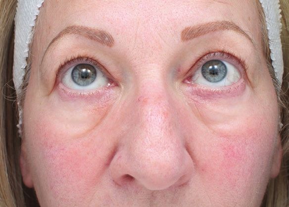
Siperstein3

Table 1. Criteria for Candidacy

Ideal candidate                                            Treat with caution                            Do not recommend treating

Good skin quality and elasticity                           Prolapsed fat pads                                Periocular erythema

Minimal volume loss (able to be corrected with 0.5             Skin laxity                         History of prolonged periocular swelling
  cc or less)

No superficial contour issues                                 Static rhytids                       Double contours (visibility of both orbicularis
                                                                                             retaining ligament and zygomatico-cutaneous ligament)

                                                                                                                                                     Downloaded from https://academic.oup.com/asjopenforum/article/doi/10.1093/asjof/ojac001/6508878 by guest on 02 July 2022
No visible veins                                        Need for more than 1.0 cc                     Prominent swollen malar mounds

                                                              Visible veins                                   Severe skin laxity

                                                                Thin skin

                                                           On blood thinners

                                                          Rosacea or eczema

                                                           History of allergies

                                                         Auto-immune disease

                                                           Mild malar edema

                                                        History of facial swelling

                                                                       first column of Table 1, in the author’s practice, few patients
                                                                       have these characteristics. Yet even patients with multiple
                                                                       “contraindications” (“Treat with caution,” Table 1) are often
                                                                       able to achieve dramatic improvement with fillers and are
                                                                       very satisfied as seen in Figure 1.
                                                                           Poor candidates for nonsurgical treatment have se-
                                                                       vere skin laxity that cannot be resolved with volume
                                                                       alone. These patients can be treated first with deep laser
                                                                       resurfacing with coagulation to tighten and smooth the
                                                                       skin to make them better filler candidates if surgery is
                                                                       not an option. Additionally, baseline chronic periocular
                                                                       edema, with or without double contours (tight ligaments
Video 1. Watch now at http://academic.oup.com/                         binding both above and below in infraorbital region) as
asjopenforum/article-lookup/doi/10.1093/asjof/ojac001                  shown in Figure 2, often responds poorly to both filler
                                                                       and surgery.
for the treatment of the infraorbital hollow (IOH). All patients
discussed here were treated in accordance with the prin-
ciples outlined in the Declaration of Helsinki, and each pa-           Patient Education and Informed Consent
tient consented to treatment and photography.
                                                                       Due to the increased risk of SEs associated with the treat-
                                                                       ment of IOH, a thorough discussion with the patient is ne-
RESULTS                                                                cessary to ensure that the patient is fully informed and in
                                                                       agreement with the procedure. In the author’s practice, a
Patient Selection                                                      specialized informed consent is reviewed with the patient.
                                                                       The following points are critical elements:
As with any treatment, patient selection is an important
part of preventing SEs. While some patients are clear and               1.     In clinical practice, the physician and patient can agree
absolute surgical candidates vs filler candidates, in real-                    to use FDA-approved injectable fillers “off label” in lo-
world practice, many, if not most, patients are on a con-                      cations not explicitly stated in the product label.
tinuum where either procedure is appropriate, and the                  2.      The areas around the eyes are more sensitive and
treatment decision is individualized through the analysis                      more prone to reactions.
of the patient’s risk vs benefit profile. Though the “ideal             3.     The SEs in this area are different than the rest of the
candidate” for infraorbital HA treatments is outlined in the                   face, with special attention paid to the potential for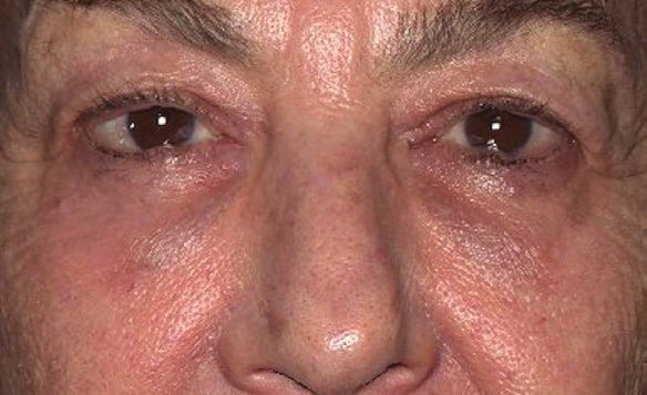
4 Aesthetic Surgery Journal Open Forum
A B
Downloaded from https://academic.oup.com/asjopenforum/article/doi/10.1093/asjof/ojac001/6508878 by guest on 02 July 2022
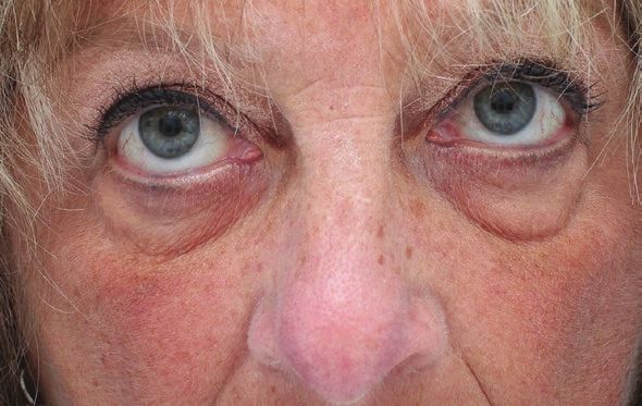
Figure 1. A 68-year-old female (A) before treatment and (B) 3 weeks later. The second photograph was taken 1 week after the
second infraorbital hyaluronic acid filler treatment to camouflage her prolapsed fat pads.
fillers, none of these SEs are irreversible, and they can
be managed nonsurgically.
6. Understanding the degree of improvement given the
severity of the baseline condition to ensure realistic
expectations (no change in the texture of the skin).
Treatment Planning and Prevention
Though a full review of resident anatomy is beyond
the scope of this manuscript, it should be emphasized
that the injector should have knowledge about the
nerves, vasculature, lymphatics, muscles, ligaments,
and fat compartments. Without this knowledge, the
risk of SEs increases dramatically, and the likelihood
Figure 2. A 62-year-old female with infraorbital edema of a good aesthetic result declines especially if an in-
before any cosmetic treatments with visible outlines of her jector is unable to discern the plane into which they are
both orbital retaining ligament and zygomatico-cutaneous injecting. Furthermore, while the SEs discussed here are
ligament (double contours). nonischemic and centered on those affecting aesthetic
outcomes, the proximity to the orbital rim and vessels
connected to the ophthalmic artery warrant extreme cau-
tion; therefore, the depth and location of filler placement
prolonged or delayed-onset swelling. The appearance
should be carefully selected.13-17
of bumps or lumps and/or bluish-gray discoloration
More broadly, the following considerations when
are most often due to distended vessels or swelling.
injecting this area should be considered:
The images included are shown in Figure 3. It is vital to
educate the patients to return to the office if they have 1. The volume of the injection is directly related to the risk
swelling in this area even years later before seeking of short-term swelling, and patients should be treated
alternative diagnoses to prevent unnecessary medical with the smallest possible volume, ideally ≤0.5 cc total.7
workups. 2. As much of the product as possible should be injected
4. Plan for SEs. While patients do not have to pay for deep, underneath the muscle, with only the smallest
SE management directly (it is built into the cost of the amount placed superficially if needed to smooth out
treatment), no refunds are given if the filler needs to superficial contours.
be dissolved as this is not due to the fault of the physi- 3. In the author’s opinion, the product should be in-
cian but instead a possible known reaction. jected not only just above the ZCL but also below
5. Though the risk of these events is higher in this area, in the malar fat pads and lateral on the zygoma so
the risk is taken together by the physician and patient. that there is support and balance with less product
In the author’s clinical experience treating with HA needed in the IOH.
Siperstein5

  A                                                                                      B

                                                                                                                                                        Downloaded from https://academic.oup.com/asjopenforum/article/doi/10.1093/asjof/ojac001/6508878 by guest on 02 July 2022
Figure 3. Possible side effects from infraorbital hyaluronic acid filler injections shown in consent form: (A) a 68-year-old female
with delayed-onset swelling and (B) a 78-year-old female with a linear vein appearing as a contour irregularity.

Table 2. Strategies for the Prevention and Treatment of                             6.   Inject as much as possible with a cannula to maximize
Short-Term Swelling Following Injection of HA Filler in the                              safety18 and reduce SEs such as bruising.19 A needle
Infraorbital Area
                                                                                         should be reserved for situations when the results
 Prevention of short-term swelling                                                       would differ such as treating a depression inferior to the
                                                                                         malar mound. The author uses a vertical needle injec-
  Use the correct product (HA filler with low propensity for swelling)
                                                                                         tion at the lateral cutaneous insertion of the ZCL starting
  Use lower volumes (ideally ≤0.5 cc per side per session)                               on the periosteum and continuing upward with a ret-
                                                                                         rograde injection throughout each layer (not possible
  Use antihistamines (for patients with a history of reactivity to injury, aller-
   gens, or history of hypersensitive foreign body response)                             with a cannula). Deep needle injections should only be
                                                                                         placed lateral to the lateral limbus for safety reasons.20
  Sleep upright with several pillows
                                                                                    7.   Inject retrograde with minimal force, while constantly
  Decrease salt intake and aerobic activity (lower blood pressure)                       moving the device with re-direction if resistance is en-
                                                                                         countered. This technique ensures that small amounts
  Mix in triamcinolone (1 mg/mL of filler)
                                                                                         of filler are injected in any one location at low pressures.
  Silicate cream overnight after treatment for compression                               One possible theory of HA filler-induced blindness is
                                                                                         that the force of the filler placed in the lumen of a vessel
 Treatment of short-term swelling
                                                                                         overcomes blood pressure and when there is sufficient
  Watchful waiting, firm massage (down and out with a lubricating cream or               volume, flows retrograde from vessels connected to the
   topical steroid) by injector, and reassurance
                                                                                         ophthalmic artery back to the bifurcation with the central
  Topical cortisone cream to massage at home several times per day                       retinal artery, though other theories are also described.
                                                                                    8.   If using a cannula, the entry point should not be placed
  Silicate cream during the day and overnight for compression
                                                                                         in the malar prominence (red outline in Figure 4A) be-
  Diuretics or antihistamines(only effective in select cases, if allergic compo-         cause trauma from the cannula going in and out of this
   nent is suspected)                                                                    location is very likely to cause acute malar mounds
  Local triamcinolone injection (no more than 0.1 cc of 2.5 mg/cc) subdermal             (Figure 4B) due to the ligament/septum below binding
   (avoid intradermal)                                                                   this area and the poor superficial lymphatic flow
                                                                                         above. The author starts with an injection port inferior
  Oral steroids (Medrol dose pack or 10 to 40 mg prednisone ×5 days
   depending on patient weight and severity of swelling).                                and then adds a lateral entry point if needed.
                                                                                    9.   Diligent before and after photography to assist in the
HA, hyaluronic acid.                                                                     diagnosis of SEs.

4.    Only soft HA fillers with a low hydrophilicity should be
                                                                                    Short-Term SEs
      used. In the author’s practice, treatment options are re-                     Postprocedure Edema
      stricted to NASHA (Restylane, Galderma Laboratories,                          Edema is the most common SE of IOH treatment. There
      L.P. Fort Worth, TX), CPM gel (Belotero Balance, Merz                         is significant inter-patient variability with regard to the risk
      North America, Inc., Raleigh. NC), VYC-15L (Juvéderm                          of swelling, and it is often impossible to predict whether a
      Volbella-XC, Allergan Inc., Irvine, CA), and RHA2 (TEOSYAL                    patient will be prone to swelling following infraorbital in-
      RHA 2, TEOXANE Laboratories, Geneva Switzerland).                             jection.10 The swelling response comes from a cascade of
5.    Permanent or non-dissolvable fillers are not recom-                           events that leads to the leakage of fluid from the vessels
      mended in this area due to the increased risk of SEs.                         in the area. The rate at which the area is filled with fluid is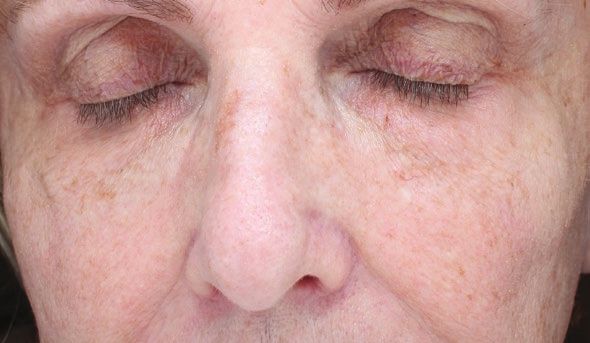
6                                                                                         Aesthetic Surgery Journal Open Forum

                A                                                  B

                                                                                                                                      Downloaded from https://academic.oup.com/asjopenforum/article/doi/10.1093/asjof/ojac001/6508878 by guest on 02 July 2022
Figure 4. (A) A 69-year-old female with the area outlined in red that is susceptible to swelling and, therefore, not
recommended as an entry point and the recommended area circled in green. (B) A 42-year-old female with swollen malar
mounds around the entry points 2 weeks after infraorbital hollow filler treatment, worse on her right (sleeping side).

               A                                                     B

Figure 5. A 63-year-old female with mild swelling presenting 2 months after her first infraorbital hyaluronic acid filler treatment
(A) before and (B) after application with sodium silicate for compression therapy.

dependent upon the rate of fluid entering (intensity of the             Compression, often with bandages, is a well-estab-
swelling response), the size of the area (confined by the           lished modality for both the prevention and treatment of
ZCL and malar septum below and the orbicularis retaining            mild swelling in other areas of the body. Similarly, for the
ligament above),21 and how fast the fluid can leave the area        face, the author recommends the use of sodium or magne-
(lymphatic system and septum permeability). Additional              sium silicate tightening creams for several days after treat-
environmental factors may include blockage by the filler            ment to compress the tissue to prevent or mitigate swelling
itself or reduced muscle contraction and movement in the            in the area (examples are Instant FirmX, Peter Thomas
area from treatment with neuromodulators.                           Roth Labs LLC, Saddle Brook, NJ or Rapid Reduction, True
    The methods for the prevention and treatment of im-             Earth Health Products, Farmingdale, NY). Most patients re-
mediate swelling following HA filler placement in the IOH           port worsening of swelling upon awakening, and thus the
are presented in Table 2. Mixing 0.1 mL (1 mg) of 50 mg/5           use of this cream before bed as overnight compression
mL of triamcinolone per 1 cc of HA filler dramatically re-          therapy is highly recommended to prevent swelling. While
duces short-term swelling (Video 2). In a retrospective             all brands with sodium silicate can cause a white residue
study from the author’s practice of more than 250 patients          if too much is used and not rubbed into the skin, the ap-
using this technique, there were no SEs reported among              pearance or ability to mix with makeup or sunscreen is not
all skin types, and the rate of short-term swelling was re-         a concern when using it overnight.
duced from 51% to 23%.7 In fact, multiple patients within               Treatment options for short-term swelling are outlined in
the author’s practice who experienced swelling with treat-          Table 2. In many instances, only watchful waiting and reas-
ment without triamcinolone have avoided this SE when                surance with massage may be needed. Compression with
triamcinolone is used for re-treatment.                             silicate compression/tightening cream may also speed up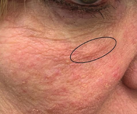
Siperstein7

Table 3. Strategies for the Prevention and Treatment of
Bruising From Injection of HA Filler in the Infraorbital Area

 Prevention of bruising

  Use of a vein finder for entry point

  Use of a cannula

  Use of light force

                                                                                                                                                 Downloaded from https://academic.oup.com/asjopenforum/article/doi/10.1093/asjof/ojac001/6508878 by guest on 02 July 2022
  Immediate postprocedure pressure

  Use of arnica, bromelain, ice

  Discontinue unnecessary medications that can increase the risk of bruising   Video 2. Watch now at http://academic.oup.com/
   such as vitamin E and Ginkgo                                                asjopenforum/article-lookup/doi/10.1093/asjof/ojac001
 Treatment of bruising

  Watchful waiting and reassurance

  Laser treatment (IPL/PDL/Nd:YAG)

HA, hyaluronic acid; IPL, intense pulsed light; Nd:YAG, neodymium-doped yt-
trium aluminum garnet; PDF, pulsed dye laser.

the resolution of swelling dramatically, as shown in Figure
5. Topical steroids along with a strong massage are used
to assist in moving the swelling out of the small confined
under-eye area. Local steroid injections (2.5 mg/mL × 0.05-
0.1 mL of TAC) can be injected with either a needle or a
cannula. However, a cannula is recommended to prevent
                                                                               Video 3. Watch now at http://academic.oup.com/
placing a bolus of the solution in the dermis, which could                     asjopenforum/article-lookup/doi/10.1093/asjof/ojac001
cause temporary atrophy and hypopigmentation (if this
occurs, saline can be used to flush the area for quicker
resolution).18,22,23 In addition, while extremely rare and with                is required. However, for patients who must return to work
higher doses, steroid particles can cause vascular occlu-                      or for whom bruising is mutually exclusive with social ac-
sion, so the same precautions taken with filler should be                      tivity, laser or intense pulsed-light therapy can speed reso-
used with light force using a cannula and injecting small                      lution. In addition, makeup concealers can completely hide
amounts while moving retrograde.24-26 Oral steroids can be                     bruising, especially if these are lightly patted on, so that
used in cases when the swelling is affecting the patient’s                     additional layers can be applied to completely block the
quality of life and other measures are not effective.                          appearance.

Bruising
Bruising is another SE that is far more common and severe
                                                                               Long-Term SEs
around the eye than other areas of the face. Prevention of
bruising is important, as bruising can dramatically lengthen                   Prolonged or Late-Onset Edema
the amount of social downtime associated with the pro-                         Swelling that persists longer than 4 weeks without improve-
cedure. A summary of preventive measures and treatment                         ment is considered prolonged edema. If the swelling is mild
for bruising is presented in Table 3. Like other SEs, preven-                  to moderate and the patient has a more severe baseline
tion is key, and the use of a cannula, vein finder, and gentle                 volumetric deficit and/or is not eligible for surgery, patients
injection technique is all vital. Immediate broad pressure                     may not want treatment as often the swelling is not notice-
following injection is recommended for 5 minutes. Pressure                     able to them. For delayed-onset swelling, it is important
should not be restricted to the insertion point but should                     to take a detailed medical history to identify that the pos-
include the entire treated area. Other measures such as ar-                    sible underlying causes of the swelling, since trauma, in-
nica, bromelain, and ice are also commonly used; however,                      fection, or exposure to an allergen can be a cause that
evidence for these practices is limited.27 Because bruising                    resolves easily. There is some debate that filler migration
is self-limiting, watchful waiting and reassurance are all that                and blockage of lymphatic flow through compression or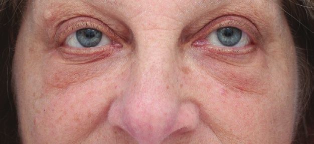
8                                                                                                                     Aesthetic Surgery Journal Open Forum

Table 4. Treatment for Delayed-Onset or Long-Term (>4 weeks) Swelling

 Swelling treatment

  Massage with cortisone cream

  Application of silicate compression cream

  Antihistamines or diuretics

  Triamcinolone and/or hyaluronidase
             Mild long term or delayed onset

                                                                                                                                                                                Downloaded from https://academic.oup.com/asjopenforum/article/doi/10.1093/asjof/ojac001/6508878 by guest on 02 July 2022
             Inject 0.1 mL of 2.5 mg/mLa triamcinoloneb with a cannula in each area.

             Moderate long term or delayed onset
             Inject 0.1-0.2 mL of 2.5 mg/mL triamcinolone with a few units of micro-dose hyaluronidase.c

             Severe long term or delayed onset
             Inject 0.2-0.4 mL of 2.5 mg/mL triamcinolone with 15 units of low-dose hyaluronidased in each location depending on the breadth of swelling.

             No resolution with triamcinolone or low-dose hyaluronidase
             Dilute the hyaluronidase by half (75 units/mL) and slowly increase the dosee with of each treatment from 15 to 75 units until full resolution.

  Cheek filler
             Inject with a cannula under the ZCL to volumize and then with a needle in a vertical retrograde technique along the lateral ZCL to camouflage
                swelling above the ZCL. NEVER inject medial to the lateral limbus deep with a needle.

  Laser resurfacing
             Tightens the skin, preventing the accumulation of fluid.

  Complete reversal with hyaluronidase
            If either no change occurs or swelling improves but then relapses quickly without overall improvement with the above options, inject 150 units per 1
               cc of under-eye filler to completely dissolve and return to baseline. Warn the patient that it may look worse than baseline for 1-2 weeks until the na-
               tive HA rebuilds, and the skin re-adjusts to the new volume (recommend silicate compression cream while waiting).

aAbout  2.5 mg/mL of triamcinolone can be obtained by mixing 0.1 mL of triamcinolone 50 mg/5 mL with 0.3 mL of bacteriostatic saline or a combination of 0.2 mL saline
and 0.1 mL of lidocaine with epinephrinef; bDO NOT REPEAT triamcinolone injections more than once within a month or more than twice within 6 months;
cMicro-dose hyaluronidase with 2.5 mg/mL of triamcinolone can be obtained by mixing 0.05 mL of hyaluronidase with 0.1 mL of triamcinolone (50 mg/5 mL) and 0.25

mL of saline (or 0.15 mL of saline and 0.1 mL of lidocaine with epinephrine)f; dLow-dose hyaluronidase with 2.5 mg/mL of triamcinolone can be obtained by mixing 0.1
cc triamcinolone 50 mg/5 mL, 0.1 mL of hyaluronidase 150 u/mL (15U) with 0.2 mL of saline (or 0.1 mL of saline and 0.1 mL of lidocaine with epinephrine)f; eIf using more
than 0.1 mL of hyaluronidase, an intradermal test for allergy is recommended by placing 0.1 mL in the dermis to create a bleb and waiting 30 minutes to check for a
reaction; fIf using lidocaine with epinephrine, warn the patient that the area will turn white in color for a few hours and will feel numb. The change in color outlining the
treated area assures the injector of proper placement, and the immediate improvement assures the patient that it is swelling and not filler causing the volume change.
The temporary improvement from the epinephrine constricting the vasculature may wane after a few hours. Topical vasoconstrictors such as Mirvaso/Rhofade can
also be used for this purpose but are more expensive and less efficacious than the silicate creams. ZCL, zygomatico-cutaneous ligament.

Table 5. Strategies to Prevent and Treat Bumps and Lumps                                 silicate cream may be helpful. However, most other treat-
From Injection of HA Filler in the Infraorbital Area                                     ments are only effective when they address the underlying
 Prevention of bumps and lumps
                                                                                         cause. For instance, oral antihistamines are recommended
                                                                                         when there is a possible allergic component (eg, new anti-
  Inject lower volumes                                                                   biotic for infection) and this presents with deeper, broader
 Treatment of bumps and lumps                                                            facial swelling. Triamcinolone is recommended when the
                                                                                         swelling is more severe from either trauma, an allergy not
  Watchful waiting and reassurance                                                       responding to antihistamines, or an acute inflammatory
  Silicate compression cream to reduce swelling                                          stimulus like a chalazion. Oral steroids should be reserved
                                                                                         for severe cases.
  Cheek filler to camouflage
                                                                                             If the inciting stimulus remains, patients will only re-
  Laser treatment (Nd:YAG) for veins outside the orbital rim                             spond to local triamcinolone for 2 to 6 weeks before the
                                                                                         swelling returns. In the author’s practice, patients do not
  Injection of 7.5-15 U of micro-dose hyaluronidase to reduce the volume
   of HA filler ± 0.1 mL of 2.5 mg/cc triamcinolone if there is swelling
                                                                                         receive more than 2 injections of triamcinolone alone in
                                                                                         the same area per year to prevent atrophy. Additionally,
HA, hyaluronic acid.                                                                     the injection is subdermal because the intradermal injec-
                                                                                         tion can cause hypopigmentation and dermal atrophy.
exposure to the crosslinker (1,4-butanediol diglycidyl ether)                            Using a cannula can help to ensure that steroids are ad-
as the HA filler is resorbed can also cause this condition.28                            ministered under the skin and decrease the likelihood that
Regardless, in all cases of late-onset swelling, massage and                             it is injected into a vessel, especially with larger-gauge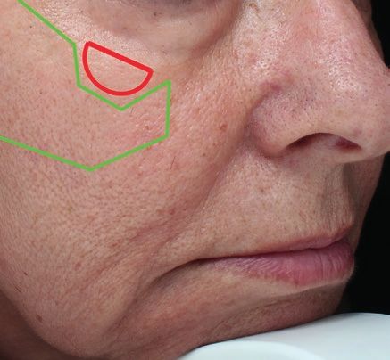
Siperstein9

 A                                                                   B

                                                                                                                                     Downloaded from https://academic.oup.com/asjopenforum/article/doi/10.1093/asjof/ojac001/6508878 by guest on 02 July 2022
Figure 6. A 69-year-old female in 2014, one year after 2 infraorbital hollow treatments (A) presenting with a delayed-onset bump
on her right that was a vein that became more prominent, and (B) after cheek injections to camouflage the protruding vein.

              A                                                      B

Figure 7. A 66-year-old female presenting with (A) a bump 2 weeks after infraorbital filler was diagnosed with a vein finder (B)
as a vein.

cannulae.18,29 An additional option is camouflaging the              a vein finder. Figure 7 illustrates how utilization of a vein
swelling by adding additional volume in the cheek around             finder can aid in the identification of the bump’s origins.
it. If there is skin laxity, laser resurfacing reduces the lax       When managing lumps and bumps, it is critical to identify
area for fluid to accumulate. If swelling cannot be managed          the correct etiology, and baseline images are particularly
by the above methods in Table 4, or continues to return,             helpful to inform a diagnosis. For example, in patients with
the filler must be dissolved using hyaluronidase in part or          some degree of fat pad prolapse, the highest point of the
in full. While studies have demonstrated the successful              fat pad prolapse can often be seen after treatment, espe-
management of swelling using complete removal of the                 cially during eye movement with an upward gaze (Figure 8).
filler with hyaluronidase,30 this is the last resort for patients    In such an instance, not knowing the patient’s baseline fea-
and in many cases is not necessary. If using hyaluronidase,          tures complicates the diagnosis of surface irregularities. In
it is important to warn the patient that the results can get         addition, granulomas are not within the scope of this paper
worse for a few weeks while the skin re-adjusts to the new           but can present as bumps with surrounding erythema and
lower volume.                                                        should be treated with hyaluronidase immediately, either
                                                                     with or without anti-inflammatory antibiotics31 depending
Bumps and Surface Irregularities                                     on symptoms such as pain or warmth.
If a soft HA filler is placed with a cannula and the filler is not       Most often, bumps that arise at the time of filler place-
bolused too superficially, bumps and surface irregularities          ment are due to the patient’s own veins rising to the
are almost never due to the lumpiness of the filler itself.          surface due to the increased volume in the area from
While the filler is the origin of this side effect, it is NOT the    the filler. An analogy of rising tides raising all ships may
direct cause. Most frequently, bumps are distended blood             be used to explain this phenomenon to patients. An
vessels (Figure 6A), which can be shown to the patient with          animation of before and after filler causing increased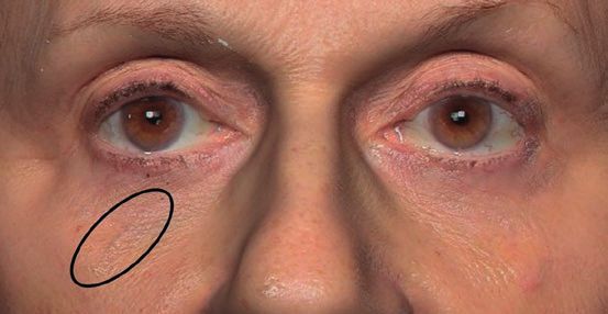
10                                                                                       Aesthetic Surgery Journal Open Forum

              A                                                      B

                                                                                                                                    Downloaded from https://academic.oup.com/asjopenforum/article/doi/10.1093/asjof/ojac001/6508878 by guest on 02 July 2022
Figure 8. A 64-year-old female (A) before treatment and (B) after 2 syringes of infraorbital hyaluronic acid filler to camouflage
prolapsed fat pads with a small surface irregularity from the top of the fat pad.

                                                                   However, if the lump is bothersome, it can often be dis-
                                                                   guised through additional filler injections in the cheeks
                                                                   (Figure 6B) or, alternatively, an Nd:YAG laser and internal
                                                                   metal eye shields may be used to treat the veins out-
                                                                   side the orbital rim. To eliminate some of the filler volume
                                                                   causing compression and to reduce the swelling that may
                                                                   be causing vein protrusion, 2.5 mg/mL triamcinolone and
                                                                   7.5-15 U of hyaluronidase diluted in saline can lower the
                                                                   filler and swelling volume without creating a depression
                                                                   if evenly injected throughout the treated area with a can-
                                                                   nula. Preventive measures and treatment for lumps and
                                                                   bumps are presented in Table 5.

Figure 9. A 72-year-old female presenting 6 months after           Blue-Gray Discoloration
infraorbital hyaluronic acid treatment with bluish-gray
                                                                   The blue discoloration from HA filler in the superfi-
swelling.
                                                                   cial dermis is most often attributed to the Tyndall effect
                                                                   caused by the scattering of blue light by colloid particles.32
                                                                   However, for this effect to occur, the particles should
                                                                   be around the same size as visible light (400-700 nm),
                                                                   which means smaller than 1 micron. As HA filler particles
                                                                   range from approximately 250 to 1000 microns in size,
                                                                   the Tyndall effect is unlikely the explanation. A publica-
                                                                   tion by Rootman and colleagues33 also discusses how the
                                                                   Tyndall effect is not the likely cause for the blue color
                                                                   that HA filler sometimes creates, and they also published
                                                                   other possible theories related to the underlying vessels,
                                                                   though there are no definitive answers to date. The cur-
                                                                   rent author proposes the blue discoloration present after
Video 4. Watch now at http://academic.oup.com/                     IOH filler may be due to the Rayleigh effect from swelling.
asjopenforum/article-lookup/doi/10.1093/asjof/ojac001              The Rayleigh effect is caused by scattering of blue light
                                                                   by nano-colloids which measure 1/10 or less than the size
visualization of a vein is seen in Video 3. Bumps that             of the wavelength of blue light (450-495 nm) which would
surface after some time has passed are often due to a              be 45-50 nm or less. The hypothesis is that superficial
combination of both a vessel and increasing leakage of             edema contains nanoparticles (size ranging from 1 to 50
fluid from vessels causing late-onset swelling, as shown           nm) which causes the Rayleigh effect. In fact, edematous
in Figure 6. In either case, injection with a low volume at        fluid has proteins such as albumin that measures approxi-
initial treatment and reducing injection volume at sub-            mately 5-10 nm.34 It is most important to note that the
sequent injections to reduce the risk of protrusion are            blue color is not necessarily synonymous with visible
preventive measures. In many patients, surface irregular-          superficial filler. In fact, in the infraorbital area, the au-
ities from veins are much less bothersome than the orig-           thor finds that late-onset blue-gray discoloration is nearly
inal infraorbital depression and no treatment is needed.           always synonymous with swelling (Figure 9). Frequent
Siperstein11
resolution of blue-gray discoloration with triamcinolone 8 mg of dexamethasone to treat an eyelid hemangioma.37
(Video 4) in the author’s practice further supports the hy- In another case, an injection of 4 mg/0.05 mL caused fat
pothesis that blue-gray discoloration that does not occur atrophy and hypopigmentation.38 The most serious case
immediately following injection is secondary to late-onset report describes 0.5 mL of methylprednisolone 80 mg/
swelling and not simply significant amounts of HA filler mL (40 mg) injected with a needle into a chalazion on
migrating too superficially. Management is largely the the eyelid immediately after surgery which caused retinal
same as late-onset swelling, as the removal of fluid will occlusion.26
resolve the light-scattering effect. While these SEs need to be taken seriously, our tech-
nique recommends either 1 mg of triamcinolone mixed
Downloaded from https://academic.oup.com/asjopenforum/article/doi/10.1093/asjof/ojac001/6508878 by guest on 02 July 2022
with the filler over a large area for prevention or 0.25 mg
DISCUSSION of triamcinolone placed in an area of swelling, both with
a cannula and in an area distant and inferior to the eyelid.
Though the infraorbital area is prone to more treatment The recommended doses in this paper, therefore, range
SEs, there are many effective prevention and treatment from 1/4th to 1/160th of the amounts used in the previous
measures. With the tools presented above, these common case reports. In addition, while the size of particles in
nonischemic SEs are nearly entirely manageable with triamcinolone varies from 1 to 1000 microns, it was re-
quick, easy, and inexpensive solutions. Many late-onset ported that, in one analysis of triamcinolone 40 mg/mL,
SEs are secondary to swelling, including bumps from the majority (71%) of particles were 1 to 10 microns, with
distended veins and accumulation of edematous fluid 88% of the particles 50 microns or less.39 Since the par-
and resultant Rayleigh effect. Both early- and late-onset ticle size of HA filler (250-1000 microns) is larger than
swelling are so common in the treatment of IOH that the the average particle size of triamcinolone, the addition of
narrative that swelling is due to poor injection technique triamcinolone to HA filler7 should pose no additional risk
is somewhat misleading and may prevent honest edu- for occlusion. Utilizing the recommended triamcinolone
cation of the patient. While poor injection technique can techniques in this paper, the author has treated over 250
certainly worsen the risk of swelling, swelling is not ne- patients with each technique safely without any serious
cessarily indicative of poor technique especially when it or permanent SEs.
occurs years later. In most of the author’s IOH treatments which started
In addition, it makes little sense to automatically use in 2013, a 27-gauge cannula was used. Currently, when
hyaluronidase to dissolve all the filler in these instances. injecting the IOH area, the author continues to use the
Swelling or increased water binding in this area is not in- same blunt-end, flexible (low elastic modulus), 27-gauge
herently a “bad” outcome. In fact, it is likely that water binds cannula. Utilizing the author’s instrument and technique,
around fillers in all areas35,36 but simply is more noticeable as seen in another publication also utilizing a similar
in this tight, small, thin-skinned area. Patients should be en- 27-gauge cannula,19 most patients report decrease pain,
couraged to weigh the impact of the revolumization against bruising, and swelling. In addition, after treating over
the impact of the swelling and resultant irregularities and 1600 IOH in over 800 patients, there have been no re-
participate in collaborative decision making. Before and ports of vascular occlusion. This is likely due to (1) the
after photographs should be referenced, and intermediate specific type of 27-gauge cannula which allows the au-
approaches should be considered. The impact of swelling thor to feel resistance and redirect; (2) the minimal force
on the patient’s impression of their appearance is highly used during advancement of the cannula; (3) injecting
personal, and treatment approaches to swelling should be only retrograde while moving the cannula with minimal
determined with the patient’s wishes in mind. Camouflaging force on the plunger; and (4) withdrawing the cannula
the SE with filler in the cheek, injecting triamcinolone, re- most of the distance before wider re-direction to ensure
ducing laxity with laser resurfacing, and partial reduction of that increased torque is not placed on fixed vessels. It is
filler with evenly applied micro-dose hyaluronidase are all important to mention that not all 27-gauge cannulae are
intermediate options that can achieve long-lasting natural- the same and a more thorough literature review and dis-
appearing results. cussion on this topic will be published separately. While
If considering triamcinolone, it is important to be edu- a more formal analysis in a prospective clinical trial of
cated about several rare case reports in which injection of the percentages of each IOH SE with different cannulae
triamcinolone alone directly into the eyelid or surrounding and needles (differences could include tapering, length,
areas at high doses caused serious SEs.24-26,37,38 However, elastic modulus, gauge, coating, size of opening and
the doses in these case reports were much higher than our bevel) would be informative, this is outside the scope of
recommended dosage and a needle was used. One case this work. Thankfully, more information should be avail-
report described iris depigmentation after an injection with able shortly with the recent FDA approval of the IOH indi-
a needle into the eyelid with 40 mg of triamcinolone and cation for VYC-15L.12 Aesthetic Surgery Journal Open Forum
CONCLUSIONS 2006;22(5):335-341; discussion 341; discussion 341-343.
https://doi.org/10.1097/01.iop.0000235820.00633.61
Interestingly, patients treated with filler in the IOH may not 9. Steinsapir KD, Steinsapir SMG. Deep-fill hyaluronic acid for
need additional filler treatments for several years, if ever, the temporary treatment of the naso-jugal groove: a report
due to the replacement of metabolized filler with increased of 303 consecutive treatments. Ophthalmic Plast Reconstr
amounts of bound water. Filler in this area is more durable Surg. 2006;22(5):344-348.
10. Griepentrog GJ, Lucarelli MJ, Burkat CN, Lemke BN,
in general, but for patients with swelling, the volume re-
Rose JG. Periorbital edema following hyaluronic acid
mains replenished for a very long period, and patients can
gel injection: a retrospective review. Am J Cosm Surg.
be maintained with cheek filler. It matters little to the patient 2011;28(4):251-254.
Downloaded from https://academic.oup.com/asjopenforum/article/doi/10.1093/asjof/ojac001/6508878 by guest on 02 July 2022
whether the volume is from their own fluid or the HA filler 11. Mustak H, Fiaschetti D, Goldberg RA. Filling the periorbital hol-
if the volume is in the correct place and appears natural. lows with hyaluronic acid gel: long-term review of outcomes
Together, the approaches in this paper outline preventive and complications. J Cosmet Dermatol. 2018;17(4):611-616.
measures and treatment for SEs and how to manage them 12. Lederhandler M, Belkin D, Anolik R, Geronemus RG. The
effectively and specifically. Paired with safe injection tech- rise and fall of the pale puffy lower eyelid pillow. J Drugs
nique, the management of these SEs can support good Dermatol. 2021;20(4):475-476.
long-term outcomes for patients and clinicians. 13. Beleznay K, Carruthers JD, Humphrey S, Jones D. Avoiding
and treating blindness from fillers: a review of the world lit-
Supplemental Material erature. Dermatol Surg. 2015;41(10):1097-1117.
14. Beleznay K, Carruthers JDA, Humphrey S, Carruthers A,
This article contains supplemental material located online at Jones D. Update on avoiding and treating blindness from
www.asjopenforum.com. fillers: a recent review of the world literature. Aesthet Surg
J. 2019;39(6):662-674.
Disclosures 15. Cotofana S, Schenck TL, Trevidic P, et al. Midface: clinical
Dr Siperstein is a consultant, clinical trial investigator, speaker, anatomy and regional approaches with injectable fillers.
and trainer for Galderma (Lausanne, Switzerland) and Allergan Plast Reconstr Surg. 2015;136(5 Suppl):219S-234S.
(Irvine, CA, USA). 16. Hirmand H. Anatomy and nonsurgical correction of the tear
trough deformity. Plast Reconstr Surg. 2010;125(2):699-708.
Funding 17. Wollina U, Goldman A. Facial vascular danger zones for
filler injections. Dermatol Ther. 2020;33(6):e14285.
Funding for this research was provided by Allergan, Galderma, 18. Alam M, Kakar R, Dover JS, et al. Rates of vascular occlu-
and Merz (Raleigh, NC, USA). sion associated with using needles vs cannulas for filler
injection. JAMA Dermatol. 2021;157(2):174-180.
REFERENCES 19. Beer KR. Safety and effectiveness of injection of calcium
1. Farkas JP, Pessa JE, Hubbard B, Rohrich RJ. The science hydroxylapatite via blunt cannula compared to injection
and theory behind facial aging. Plast Reconstr Surg Glob by needle for correction of nasolabial folds. J Cosmet
Open. 2013;1(1):e8-e15. Dermatol. 2014;13(4):288-296.
2. Pessa JE, Garza JR. The malar septum: the anatomic basis 20. Hufschmidt K, Bronsard N, Foissac R, et al. The infraorbital
of malar mounds and malar edema. Aesthet Surg J. 1997; artery: clinical relevance in esthetic medicine and identi-
17(1):11-17. fication of danger zones of the midface. J Plast Reconstr
3. Pessa JE, Zadoo VP, Adrian EK, Woodwards R, Garza JR. Aesthet Surg. 2019;72(1):131-136.
Anatomy of a “black eye”: a newly described fascial 21. Alghoul M, Codner MA. Retaining ligaments of the face:
system of the lower eyelid. Clin Anat. 1998;11(3):157-161. review of anatomy and clinical applications. Aesthet Surg
4. Funt DK. Avoiding malar edema during midface/cheek J. 2013;33(6):769-782.
augmentation with dermal fillers. J Clin Aesthet Dermatol. 22. El-Amawy HS, Sarsik SM. Saline in derma-
2011;4(12):32-36. tology: a literature review. J Cosmet Dermatol.
5. Naik MN. Hills and valleys: understanding the under-eye. 2020;20(7):2040-2051.
J Cutan Aesthet Surg. 2016;9(2):61-64. 23. Shiffman MA. Letter: Treatment of local, persistent cuta-
6. Shoukath S, Taylor GI, Mendelson BC, et al. The lymphatic neous atrophy after corticosteroid injection with normal
anatomy of the lower eyelid and conjunctiva and correlation saline infiltration. Dermatol Surg. 2010;36(3):436.
with postoperative chemosis and edema. Plast Reconstr 24. Li G, Xu D, Hu Z, Li H. Embolic retinal and choroidal vas-
Surg. 2017;139(3):628e-637e. cular occlusion after peribulbar triamcinolone injection: a
7. Siperstein R, MJ, Speranza A. A retrospective review of the case report. Medicine (Baltim). 2018;97(17):e0467.
safety and efficacy of low-dose triamcinolone mixed with 25. Edwards AO. Central retinal artery occlusion following
hyaluronic acid fillers to reduce post-injection infraorbital forehead injection with a corticosteroid suspension.
swelling. J Cutan Aesthet Dermatol (In press). Pediatr Dermatol. 2008;25(4):460-461.
8. Goldberg RA, Fiaschetti D. Filling the periorbital hol- 26. Hosal BM, Zilelioglu G. Ocular complication of intralesional
lows with hyaluronic acid gel: initial experience with corticosteroid injection of a chalazion. Eur J Ophthalmol.
244 injections. Ophthalmic Plast Reconstr Surg. 2003;13(9-10):798-799.Siperstein13
27. Ho D, Jagdeo J, Waldorf HA. Is there a role for arnica and 34. Wiwanitkit V. Glomerular pore size corresponding to al-
bromelain in prevention of post-procedure ecchymosis or bumin molecular size, an explanation for underlying struc-
edema? A systematic review of the literature. Dermatol tural pathology leading to albuminuria at nanolevel. Ren
Surg. 2016;42(4):445-463. Fail. 2006;28(1):101.
28. De Boulle K, Glogau R, Kono T, et al. A review of the metab- 35. Becker M, Balague N, Montet X, et al. Hyaluronic acid filler
olism of 1,4-butanediol diglycidyl ether-crosslinked hyaluronic in HIV-associated facial lipoatrophy: evaluation of tissue
acid dermal fillers. Dermatol Surg. 2013;39(12):1758-1766. distribution and morphology with MRI. Dermatology.
29. Ugradar S, Hoenig J. Measurement of the force required 2015;230(4):367-374.
by blunt-tipped microcannulas to perforate the facial ar- 36. Mundada P, Kohler R, Boudabbous S, et al. Injectable
tery. Ophthalmic Plast Reconstr Surg. 2019;35(5):441-446. facial fillers: imaging features, complications, and diag-
Downloaded from https://academic.oup.com/asjopenforum/article/doi/10.1093/asjof/ojac001/6508878 by guest on 02 July 2022
30. Hilton S, Schrumpf H, Buhren BA, Bölke E, Gerber PA. nostic pitfalls at MRI and PET CT. Insights Imaging
Hyaluronidase injection for the treatment of eyelid edema: 2017;8(6):557-572.
a retrospective analysis of 20 patients. Eur J Med Res. 37. Al-Mahdi H. Iris depigmentation: an unusual compli-
2014;19:30. cation of intralesional corticosteroid injection for ca-
31. Pradhan S, Madke B, Kabra P, Singh AL. Anti-inflammatory pillary hemangioma. Middle East Afr J Ophthalmol
and immunomodulatory effects of antibiotics and their use 2010;17(1):100-102.
in dermatology. Indian J Dermatol. 2016;61(5):469-481. 38. Park J, Chang M. Eyelid fat atrophy and depigmentation
32. King M. Management of Tyndall effect. J Clin Aesthet Dermatol. after an intralesional injection of triamcinolone acetonide
2016;9(11):E6-E8. to treat chalazion. J Craniofac Surg. 2017;28(3):e198-e199.
33. Rootman DB, Lin JL, Goldberg R. Does the Tyndall effect 39. Benzon HT, Chew T-L, McCarthy RJ, Benzon HA, Walega DR.
describe the blue hue periodically observed in subdermal Comparison of the particle sizes of different steroids and the
hyaluronic acid gel placement? Ophthalmic Plast Reconstr effect of dilution: a review of the relative neurotoxicities of
Surg. 2014;30(6):524-527. the steroids. Anesthesiology 2007;106(2):331-338.You can also read