Targeting Angiogenesis in Prostate Cancer - MDPI
←
→
Page content transcription
If your browser does not render page correctly, please read the page content below
International Journal of
            Molecular Sciences

Review
Targeting Angiogenesis in Prostate Cancer
Zsombor Melegh 1             and Sebastian Oltean 2, *
 1    Department of Cellular Pathology, Southmead Hospital, Bristol BS10 5NB, UK; zsombor.melegh@nbt.nhs.uk
 2    Institute of Biomedical and Clinical Sciences, Medical School, College of Medicine and Health,
      University of Exeter, Exeter EX12LU, UK
 *    Correspondence: s.oltean@exeter.ac.uk

 Received: 7 May 2019; Accepted: 29 May 2019; Published: 31 May 2019

 Abstract: Prostate cancer is the most commonly diagnosed cancer among men in the Western world.
 Although localized disease can be effectively treated with established surgical and radiopharmaceutical
 treatments options, the prognosis of castration-resistant advanced prostate cancer is still disappointing.
 The objective of this study was to review the role of angiogenesis in prostate cancer and to investigate
 the effectiveness of anti-angiogenic therapies. A literature search of clinical trials testing the efficacy
 of anti-angiogenic therapy in prostate cancer was performed using Pubmed. Surrogate markers of
 angiogenic activity (microvessel density and vascular endothelial growth factor A (VEGF-A) expression)
 were found to be associated with tumor grade, metastasis, and prognosis. Six randomizedstudies
 were included in this review: two phase II trials on localized and hormone-sensitive disease (n = 60
 and 99 patients) and four phase III trials on castration-resistant refractory disease (n = 873 to 1224
 patients). Although the phase II trials showed improved relapse-free survival and stabilisation of
 the disease, the phase III trials found increased toxicity and no significant improvement in overall
 survival. Although angiogenesis appears to have an important role in prostate cancer, the results of
 anti-angiogenic therapy in castration-resistant refractory disease have hitherto been disappointing.
 There are various possible explanations for this lack of efficacy in castration-resistant refractory disease:
 redundancy of angiogenic pathways, molecular heterogeneity of the disease, loss of tumor suppressor
 protein phosphatase and tensin homolog (PTEN) expression as well as various VEGF-A splicing
 isoforms with pro- and anti-angiogenic activity. A better understanding of the molecular mechanisms
 of angiogenesis may help to develop effective anti-angiogenic therapy in prostate cancer.

 Keywords: prostate cancer; angiogenesis; VEGF-A; splicing isoforms

1. Introduction
     Prostate cancer is the most commonly diagnosed cancer in men in the Western world, with a
median age at diagnosis of 66 years [1]. There will be an estimated 160,000 new cases and 30,000 deaths
in 2018 in the USA, representing 19% of all new cancer diagnoses and 9% of all cancer related deaths,
respectively [2]. In the United Kingdom, over 47,000 men are diagnosed with prostate cancer every year,
with over 330,000 men currently living with the disease [3]. The purpose of this literature review is to
assess whether angiogenesis is important in prostate cancer and, if so, whether anti-angiogenic therapies
are effective in the treatment of prostate cancer. To begin with, the current treatment options in prostate
cancer will be discussed, along with a summary of what is already known in relation to angiogenesis in
cancer. This will be followed by the literature review on angiogenesis and anti-angiogenic therapies in
prostate cancer, specifically. Finally, the discussion will consider any treatment difficulties that have
emerged in such studies.

Int. J. Mol. Sci. 2019, 20, 2676; doi:10.3390/ijms20112676                          www.mdpi.com/journal/ijms
Int. J. Mol. Sci. 2019, 20, 2676                                                                           2 of 16

2. Background

2.1. Prostate Cancer
     Prostate cancer is characterized by slow to moderate growth. Consequently, many cases are
indolent and in up to 70% of incidentally diagnosed cases over 60 years death is due to an unrelated
cause [4]. The five-year relative survival rate for men diagnosed in the USA between 2001 and 2007
with local or regional disease was 100%, whilst the rate for distant disease was 28.7% [5]. UK statistics
show similar results: the five-year relative survival for prostate cancer was 100% in localized disease
and 30% in distant disease for patients diagnosed during 2002–2006 in the former Anglia Cancer
Network [6]. Most cases of prostate cancer are diagnosed by prostate specific antigen (PSA) testing
or rarely by rectal examination. Prostate cancer can present with decreased urinary stream, urgency,
hesitancy, nocturia, or incomplete bladder emptying, but these symptoms are non-specific and are
infrequent at diagnosis [7].

2.2. Treatment Options in Prostate Cancer
      Prostate cancer staging is divided into four stages. Stage 1 and 2 cancers are localized to the
prostate whilst stage 3 cancers extend into the periprostatic tissue or the seminal vesicle, without
 involvement of a nearby organ or lymph node and with no distant metastasis [8]. Stage 4 tumors
 represent those that have spread to nearby or distant organs or lymph nodes [8].
      Stage 1 tumors and stage 2 tumors of low and intermediate risk (Table 1) can be followed up by
‘watchful waiting’ or active surveillance and monitoring [9,10]. Watchful waiting has no curative intent,
whilst active surveillance and monitoring defers treatment with curative intent to a time when it is
 needed [9]. Therefore, in active surveillance and monitoring therapy is reserved for tumor progression,
with a 1–10% mortality rate [9].

      Table 1. Risk stratification of localized prostate cancer according to NICE guidance, UK [10]. Gleason
      score: histological pattern of the tumor. Stage T1–T2a: tumor involving
Int. J. Mol. Sci. 2019, 20, 2676 3 of 16
Hormonal manipulation options include surgical castration (orchidectomy) or medical castration
(LH-RH antagonists) [12]. These may be used in stage 3 or 4 cancers and can be enhanced by the addition
of anti-androgenic therapy and adjuvant treatment with bisphosphonates [13]. Recently approved
anti-androgen agents include abiraterone acetate, an inhibitor of cytochrome P450c17, a critical
enzyme in Sci.
Int. J. Mol. androgen
2019, 20, xsynthesis and enzalutamide, a second generation androgen-receptor–signaling
FOR PEER REVIEW 3 of 16
inhibitor [13–15].
Treatment
Treatment optionsfor
options forhigh
highstage
stagemetastatic
metastatic hormone-refractory
hormone-refractory prostate
prostatecancer
cancerinclude
includeactive
active
cellular immunotherapy with sipuleucel-T, which has resulted in increased overall
cellular immunotherapy with sipuleucel-T, which has resulted in increased overall survival in metastatic survival in
metastatic castration-resistant prostate cancer, in a double-blind, placebo-controlled,
castration-resistant prostate cancer, in a double-blind, placebo-controlled, multicenter phase 3 trial [16]. multicenter
phase
This lead 3totrial [16]. This
its approval forlead to its approval
the treatment for the treatment
of asymptomatic of asymptomatic
or minimally symptomatic or patients
minimally
with
symptomatic patients with nonvisceral metastatic castration-resistant prostate cancer in 2010.
nonvisceral metastatic castration-resistant prostate cancer in 2010. Radium-223 dichloride is used
Radium-223 dichloride is used in symptomatic patients with bone metastases and no known visceral
in symptomatic patients with bone metastases and no known visceral metastases [17]. Cabazitaxel,
metastases [17]. Cabazitaxel, a derivative of docetaxel, is approved as a second line chemotherapy
a derivative of docetaxel, is approved as a second line chemotherapy agent [18]. Further possible
agent [18]. Further possible treatment options to prevent bone metastases include denosumab (a
treatment options to prevent bone metastases include denosumab (a monoclonal antibody that inhibits
monoclonal antibody that inhibits osteoclast function) [19] and bone-seeking radionucleotides
osteoclast function) [19] and bone-seeking radionucleotides (strontium chloride Sr 89) [20].
(strontium chloride Sr 89) [20].
Despite
Despite a widening
a wideningarsenal
arsenalofofnew
newtreatment
treatment options,
options, aacure
cureisisrarely
rarelyachieved
achievedinin stage
stage 4 prostate
4 prostate
cancer, although there is astriking difference in treatment response between individual
cancer, although there is astriking difference in treatment response between individual patients [21].patients [21]. Such
outcomes emphasize the need for research into further treatment options in hormone-refractory
Such outcomes emphasize the need for research into further treatment options in hormone-refractory advanced
prostate
advanced cancer. One such
prostate emerging
cancer. One suchtherapeutic
emergingoption is inhibition
therapeutic optionofistumor-related
inhibition of angiogenesis.
tumor-related
angiogenesis.
2.3. Angiogenesis in Cancer
2.3.Angiogenesis
Angiogenesis inisCancer
defined as the development of new vascular vessels from pre-existing blood
vessels.Angiogenesis
It has a critical
is role in wound
defined as the healing and embryonic
development development
of new vascular vesselsand also
from provides collateral
pre-existing blood
formation
vessels. for improved
It has a criticalorgan
role perfusion
in wound in ischaemia
healing and [22]. It is a multi-step
embryonic development process triggered
and also by an
provides
angiogenic
collateral stimulus
formation(Figure 1). The organ
for improved first step of the in
perfusion process is the[22].
ischaemia production of proteases
It is a multi-step which
process
triggered
degrade the by an angiogenic
basement membrane. stimulus
This (Figure 1). The
is followed first step of
by migration andthe process is the
proliferation production
of the of
endothelium,
proteases
resulting which
in the degradeof
formation thea basement membrane.
new vascular channelThis is followed by migration and proliferation of
[23].
the endothelium, resulting in the formation of a new vascular channel [23].
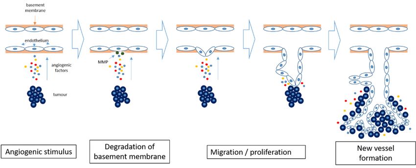
Figure Angiogenesis
1. 1.
Figure Angiogenesisinincancer.
cancer. Hypoxia
Hypoxia within the tumor
within the tumorinduces
inducesthe
therelease
releaseofofpro-angiogenic
pro-angiogenic
factors and results in degradation of the basement membrane by matrix metalloproteinases
factors and results in degradation of the basement membrane by matrix metalloproteinases (MMP). (MMP).
TheThe
endothelial cells
endothelial startstart
cells to differentiate and and
to differentiate proliferate, forming
proliferate, new new
forming bloodblood
vessels. The newly
vessels. formed
The newly
formed
blood blood
vessels vessels
allow allowtumor
further further tumor growth.
growth.
Although
Although angiogenesis
angiogenesis is not entirely
is not necessary
entirely forfor
necessary tumor initialization
tumor (some
initialization (sometumors of the
tumors brain,
of the
brain,
lung, andlung,
liverand
canliver
growcan growpre-existing
along along pre-existing vessels)
vessels) [23],aonce
[23], once tumora tumor reaches
reaches a sizea of
size of more
more than a
fewthan a few millimeters,
millimeters, formation formation of newvessels
of new blood blood vessels is necessary
is necessary to provide
to provide an appropriate
an appropriate bloodblood
supply
to support tumor cell viability and proliferation. Hence, angiogenesis plays an important role in role
supply to support tumor cell viability and proliferation. Hence, angiogenesis plays an important tumor
in tumor progression
progression and is nowand is now recognized
recognized as one
as one of the of the hallmarks
hallmarks of cancerof cancer [24].
[24].
Angiogenesis is controlled by a delicate balance between angiogenesis inducers and
angiogenesis inhibitors. In a growing cancer there is a constant production of angiogenesis inducers,
including vascular endothelial growth factor (VEGF)-A, basic fibroblast growth factor (bFGF, also
known as FGF), angiogenin, tumor necrosis factor (TNF)-α, granulocyte colony-stimulating factor
(G-CSF), platelet-derived endothelial growth factor (PDGF), placental growth factor (PGF),
transforming growth factor (TGF)-α, TGF-β, interleukin-8 (IL-8), hepatocyte growth factor (HGF),Int. J. Mol. Sci. 2019, 20, 2676 4 of 16
Angiogenesis is controlled by a delicate balance between angiogenesis inducers and angiogenesis
inhibitors. In a growing cancer there is a constant production of angiogenesis inducers, including
vascular endothelial growth factor (VEGF)-A, basic fibroblast growth factor (bFGF, also known as
FGF), angiogenin, tumor necrosis factor (TNF)-α, granulocyte colony-stimulating factor (G-CSF),
platelet-derived endothelial growth factor (PDGF), placental growth factor (PGF), transforming
growth factor (TGF)-α, TGF-β, interleukin-8 (IL-8), hepatocyte growth factor (HGF), and epidermal
growth factor (EGF) [22]. This constant production of angiogenesis inducers results in increased
activity of endothelial cells, as long as the production of anti-angiogenic factors is correspondingly
reduced [25]. Among the angiogenesis activators, VEGF-A and bFGF are particularly important in
tumor angiogenesis. The abundance and redundant activities of different angiogenesis inducers may
explain the resistance or suboptimal effectiveness of anti-angiogenic therapies, when inhibitors acting
only on a single angiogenesis activator are being used [25].
Under normal conditions, angiogenesis inducers are balanced by naturally occurring angiogenesis
inhibitors, such as endostatin, angiostatin, IL-1, IL-12, interferons, metalloproteinase inhibitors, and
retinoic acid [25,26]. These inhibitors can either disrupt new vessel formations or can help to remove
already formed vascular channels. Shifting the balance towards angiogenesis inhibition can interfere
with important physiological roles of angiogenesis, such as in embryo development, wound healing,
and renal function. Interference with wound healing is a particularly important concern in cancer
treatment, for example resulting in delayed post-operative healing [27]. Another example involves the
inhibition of VEGF-A, resulting in vasoconstriction by means of elevated NO production, consequently
elevating blood pressure [28], and increasing the risk of thrombogenesis, resulting in stroke or
myocardial infarction. These factors can potentially limit the use of angiogenesis inhibition in cancer,
on account of their potential side effects.
2.4. Angiogenesis Inhibition in Cancer
Although angiogenesis is an essential factor in tumor progression, by means of new vessel formation,
this also means that angiogenesis inhibition may only result in inhibition of further tumor growth and
may not actively eliminate the tumor. This, and the redundancy of the numerous angiogenesis inducers
as listed above, explain why the utilization of angiogenesis inhibitors as a monotherapy has not proved
to be as effective as initially expected [29]. Hence, angiogenesis inhibitor therapeutic regimes may
require a combination of several anti-angiogenic strategies or may need to be complemented by other
non-angiogenesis related chemotherapeutic agents in order to achieve an optimal therapeutic effect [30].
Based on the target of the therapeutic agent, angiogenesis inhibition can be divided into two
main groups: direct and indirect inhibition [31]. Direct inhibitors target growing endothelial cells,
whilst indirect inhibitors target the tumor cells or tumor-associated stromal cells. Small molecular
fragments (e.g., arrestin, tumstatin, canstatin, endostatin, and angiostatin) are the products of proteolytic
degradation of the extracellular matrix, and act as direct inhibitors by means of inhibition of the
endothelial cell proliferation and migration induced by VEGF-A, bFGF, PDGF, and interleukins [32].
The direct anti-angiogenic effect of targeting integrins (cellular adhesion receptors), has also been
demonstrated [32]; an integrin inhibitor—cilentigide—has been shown to inhibit tumor cell invasion [33].
Unfortunately, even though cilentigide acts both on tumor cells and endothelial cells and could be a
prime example of multifactorial treatment, results of clinical trials have proved disappointing so far [34].
The most extensively clinically used direct anti-angiogenic strategy targets VEGF-A or its receptors.
VEGF-A binds to its receptors to stimulate the proliferation of endothelial cells via the RAS–RAF–MAPK
(mitogen-activated protein kinase) signalling pathway [35]. Bevacizumab is a humanised IgG1
monoclonal antibody against VEGF-A. It selectively binds to circulating VEGF-A, preventing its
interaction with its receptor, VEGF-receptor 2, expressed on the surface of endothelial cells. Initial
studies showed clinical improvement when bevacizumab was used in combination with chemotherapy
in a number of cancers, without a marked increase in toxicity [36]. Subsequently it has been approved
as part of a combination therapy in the treatment of various cancers, including metastatic lung,Int. J. Mol. Sci. 2019, 20, 2676 5 of 16
colorectal, and renal cell carcinoma, and as a single agent treatment in adult glioblastoma [37]. However,
subsequent studies have revealed adverse effects, including gastrointestinal perforation, nephrotic
syndrome, thromboembolism, surgical wound healing complications and hypertension [37,38].
In contrast, indirect angiogenesis inhibition involves an interplay between tumor or stromal cells
and angiogenesis. One example involves the inhibition of epidermal growth factor receptor (EGFR), a
tyrosine kinase receptor. Tumor cell expression and activation of EGFR induces interleukin production,
which is demonstrated to promote intratumoral angiogenesis. Thus, blocking the expression and/or
activity of EGFR can result in indirect inhibition of angiogenesis [39].
To summarize, a number of anti-angiogenesis drugs have already been approved and are currently
used in cancer treatment. This prompts the question whether angiogenesis plays any role in prostate
cancer progression and, if so, whether anti-angiogenic therapy would be effective in refractory
castration-resistant prostate cancer, for which the current treatment options are limited.
3. Results
3.1. Angiogenesis in Prostate Cancer
Currently there are no direct markers to assess angiogenic activity in prostate cancer, but it
is reasonable to assume that vascular density is an indicator of intratumoral angiogenic activity.
Microvessel density (MVD) is considered a good surrogate marker of angiogenic activity and has been
demonstrated as a prognostic factor in various tumors, including breast and colon cancers as well
as malignant melanoma [40]. MVD can be assessed by histological examination of the vasculature,
either by assessing the most vascularised area of the tumor (‘hot spot’) or a random representative area.
Preliminary data suggested that MVD is associated with higher tumor grade and stage, and worse
outcome in prostate cancer [41,42]. Moreover, ultrasound imaging studies of haemodynamic indices
have shown a higher peak intensity in high-grade tumors [43]. Later studies, however, have failed to
confirm that MVD is an independent prognostic factor in untreated tumors, and no correlation has yet
been established between MVD and effectiveness of anti-angiogenic treatment in prostate cancer [44].
Reasons for these conflicting results potentially include different counting methods, diferences in
antibodies used, different population sizes, personal experience and pathological background [45].
A further limiting factor is the complex geometrical structure of the newly fromed vascular system,
which is difficult to analyse on a two dimensional histological section [46]. Fractal geometry to estimate
the surface dimension, computer aided automated image analysis, 3D models or magnetic resonance
imaging could potentially be used to overcome these shortcomings, [46,47].
Another possible surrogate marker for tumor angiogenesis is by an assessment of the level of
angiogenic regulators in the tumor. Both physiological and pathological angiogenesis is predominantly
regulated by VEGF, which has various protein isoforms, each acting on their specific tyrosine kinase
receptor at the cell surface [48]. Among the VEGF isoforms, VEGF-A has been extensively studied, and
it has been demonstrated to play an important role in prostate cancer angiogenesis [49]. In addition,
VEGF-A has been found to be overexpressed in prostate cancer and a high level of VEGF-A is associated
with distant metastasis and a poorer prognosis [50–52]. Furthermore, in prostate cancer a high-level
VEGF-A expression has been found not only in endothelial cells, but also in tumor cells [53].
These findings suggest that angiogenesis is important in prostate cancer, prompting subsequent
clinical studies to assess whether anti-angiogenesis therapy is effective in the treatment of prostate cancer.
3.2. Anti-Angiogenesis Clinical Studies in Prostate Cancer
An unfiltered Pubmed search for the keywords “angiogenesis” and “prostate” revealed a steady
increase in published papers between 2000 and 2013 (from 70 per year in 2000 to 213 per year in 2013)
followed by a slow decline (down to 115 in 2018). This appears to reflect the fact that, despite the
promising findings of initial studies, suggesting an important role of angiogenesis in prostate cancer,
phase III clinical trials, mainly conducted after 2010, have proved disappointing so far.Int. J. Mol. Sci. 2019, 20, 2676 6 of 16
Since VEGF-A was demonstrated to be overexpressed in prostate cancer and associated with
poor prognosis and metastasis, most anti-angiogenic clinical studies in prostate cancer have targeted
VEGF-A. A randomizedphase II trial on bevacizumab involving 99 patients with hormone-sensitive
prostate cancer showed improved relapse-free survival when bevacizumab was used alongside
hormone-deprivation therapy (Table 2) [54]. A randomized, double-blind, placebo-controlled phase III
clinical study of 1050 patients with prostate cancer showed some improvement in progression-free
survival, but found no significant improvement in overall survival in metastatic, castration-resistant
prostate cancer, when bevacizumab was used together with docetaxel chemotherapy and prednisone
hormonal therapy [55]. Furthermore, bevacizumab resulted in increased toxicity and a greater incidence
of treatment-related deaths [55]. This suggests that bevacizumab has some positive effect, especially
on hormone-sensitive recurrent prostate cancer, but in hormone-resistant refractory tumors, in which
the conventional treatment options are particularly prone to failure, adding bevacizumab treatment
does not have any clinical benefit (Table 2).
Table 2. Anti-angiogenesis clinical studies in treatment of prostate cancer.
Phase of the Number of
Drug Mechanism of Action Outcome
Clinical Trial Patients
Improved relapse-free
II 99
Bevacizumab Recombinant humanized monoclonal survival [54]
antibody that blocks VEGF-A
No improvement in
III 1050
overall survival [55]
No improvement in
Aflibercept Binds to circulating VEGF-A III 1224
overall survival [56]
No improvement in
Sunitinib Receptor tyrosine kinase inhibitor III 873
overall survival [57]
Multiple mechanisms, including Disease stabilisation,
I/II 60
Lenalidomide inhibition of VEGF-induced decrease in PSA [58]
PI3K-Akt pathway signalling III 1059 Worse overall survival [59]
Aflibercept (a hybrid protein composed of various domains of VEGF-receptors 1 and 2, fused
to human immunoglobulin G1) also targets the VEGF-A pathway, by acting as a decoy receptor for
VEGF-A. Unfortunately, similar to bevacizumab, in a phase III multicentre, randomizeddouble-blind
placebo-controlled parallel group study in 1224 men with castration-resistant refractory tumors,
aflibercept therapy combined with docetaxel chemotherapy and hormonal therapy did not show any
improvement in overall survival [56].
Sunitinib and cediranib are small multireceptor molecule tyrosine kinase inhibitors, with a
demonstrated activity against VEGF-receptors 1 and 2. Sunitininb is approved for the treatment of
gastrointestinal stromal tumor, renal cell carcinoma and pancreatic neuroendocrine tumors. However,
in a randomized, placebo-controlled, phase III trial of sunitinib therapy combined with hormonal
therapy in 873 patients with refractory castration-resistant prostate cancer, there was no improvement
in overall survival compared to placebo [57].
Furthermore, these anti-VEGF-A therapies have been associated with an increased rate of toxicity
and adverse effects, resulting in the discontinuation of treatment (27% vs. 7%) [57]. These toxic and
adverse effects included fatigue, asthenia, hand-foot syndrome, hypertension, bowel perforation,
pulmonary thromboembolism, and gastrointestinal bleeding, seen in both pre-clinical and clinical
studies [60,61]. In addition, treatment-related haematological problems also emerged in up to 20% of
the patients, including lymphopenia, neutropenia, and anaemia [57].
Thalidomide is an immune-modulatory drug, which also has anti-angiogenic effects. Lenalidomide
is a more potent analogue of thalidomide, with less prominent side effects. The mechanism of the
anti-angiogenic effect of lenalidomide is not entirely elucidated, but appears to be through multiple
mechanisms, including inhibition of VEGF-induced phosphatidylinositol-3,4,5-trisphosphate (PI3K)-AktInt. J. Mol. Sci. 2019, 20, 2676 7 of 16
pathway signalling [62]. Lenalidomide therapy in non-metastatic prostate cancer in a phase I/II
double-blinded, randomized study of 60 patients resulted in stabilization of the disease and a decline in
PSA, with minimal toxicity [58]. A randomized, double-blind, placebo-controlled phase III trial in 1059
patients with castration-resistant refractory prostate cancer, however showed worse overall survival
when lenalidomide was added to prednisone, hormonal, and docetaxel chemotherapy, compared to the
placebo group [59]. There was also a 25% increase in adverse events, which included haematological
side effects (34% vs. 20%), diarrhoea (7% vs. 2%), pulmonary embolism (6% vs. 1%), and asthenia
(5% vs. 3%) [59].
To summarize, these findings suggest that anti-angiogenic therapy has no clinical benefit when
added to chemotherapy or hormonal therapy in refractory, castration-resistant prostate cancer.
4. Discussion
Clinical trials that showed an association between high VEGF-A expression and tumor progression
assessed VEGF-A protein levels by immunohistochemistry, ELISA methods, or mRNA levels by
reverse-transcription-polymerase chain reaction (RT-PCR). Despite high VEGF-A expression in advanced
prostate cancer using these methods, anti-angiogenic therapies targeting the VEGF-A pathway have
failed to provide significant treatment benefits [63,64]. There are various possible explanations for
resistance to anti-angiogenic therapy in prostate cancer. Redundancy of angiogenic pathways means
that targeting a single pathway may result in upregulation of alternative pathways. For example, with
long-term bevacizumab treatment, which blocks VEGF-A, there is upregulation of EGF, HGF, and
PDGF [65]. Lindholm et al. demonstrated in breast cancer xenografts that targeting these pathways can
be effective in anti-angiogenic therapy [66]. A combination of different anti-angiogenic therapies in
prostate cancer has also showed some promising results: a phase II study of combined bevacizumab
and lenalidomide therapy, added to docetaxel and prednisone chemotherapy and hormonal therapy in
63 patients with metastatic castration-resistant prostate cancer found that combined anti-angiogenic
therapy can be safely administered, but further randomizedtrials are required to confirm clinical
benefit [67].
Another reason for treatment resistance is due to the fact that prostate cancer is a molecularly
heterogeneous disease,= and there is currently a lack of biomarkers that can help select those patients
who are likely to benefit from anti-angiogenic therapy or that can assess response to anti-angiogenic
treatment [48]. The genetic signature of the VEGF-A pathway or variations in VEGF-A or its receptors
could be possible markers to predict therapy response, but these have as yet not been validated [68,69].
It is hoped that further stage III trials will be able to identify subgroups of patients who could benefit
from anti-angiogenic treatment.
Resistance to sunitinib tyrosine-kinase-inhibitor has been shown to be associated with loss of the
tumor suppressor protein phosphatase and tensin homolog (PTEN). PTEN is a gatekeeper protein that
negatively regulates intracellular levels of PI3K and consequently suppresses the PI3K-Akt pathway,
which normally promotes cell survival and growth [70]. Reinstating PTEN activity, by suppression
of the PI3K-Akt pathway in in vitro studies, has been shown to restore sensitivity to sunitinib in
cancer cells [70]. Loss of PTEN activity is considered a key event in prostate carcinogenesis, and
reinstating PTEN activity in prostate cancer seems to be a promising tool in overcoming sunitinib
resistance. In addition, activation of the PI3K-Akt pathway in tumors with PTEN deletion has been
shown to be associated with repressed androgen signalling in prostate cancer, while suppression
of the PI3K-Akt pathway was demonstrated to activate androgen receptor signalling [71,72]. In a
similar way, suppression of the androgen signaling pathway resulted in activation of the PI3K-Akt
pathway [71]. This suggests that there is a cross-talk between the androgen receptor and PI3K-Akt
pathways, which would at least in part explain the castration-resistant phenotype observed in tumors
with PTEN deletion. Since activation of the PI3-Akt pathway appears to play an important role in
resistance to both sunatininb and anti-androgenic therapy, suppression of the PI3K-Akt pathway could
help overcome difficulties in anti-angiogenic and anti-androgenic therapy. Recent preclinical studiesInt.J.J.Mol.
Int. Mol.Sci.
Sci.2019,
2019,20,
20,2676
x FOR PEER REVIEW 88ofof1616
therapy. Recent preclinical studies on mouse models have shown that targeted inhibition of the PI3K-
on mouse
Akt models
pathway have shown that targeted
in castration-resistant prostateinhibition of the PI3K-Akt
cancer resulted pathwaycancer
in both inhibited in castration-resistant
cell proliferation
prostate cancer resulted in both inhibited cancer cell proliferation
and MVD [73,74]. Suboptimal results with bevacizumab treatment may also relate to the and MVD [73,74]. Suboptimal
interaction
results
between withthe
bevacizumab
androgen treatment
receptor may (AR) also relate toand
signalling the interaction
angiogenicbetween the androgen
pathways. It has beenreceptor
long
(AR) signalling
established thatand angiogenic
androgens pathways.
upregulate It has expression
VEGF-A been long established
[75], although thatthe
androgens
mechanism upregulate
of this is
VEGF-A expression
not entirely [75], although
understood [76]. Most the mechanism
recently, of this is not
an interaction entirely
between understood
epigenetic [76].(Lysine
factors Most recently,
specific
an interaction between
demethylase 1 (LSD1), epigenetic
protein factors (Lysine
arginine specific demethylase
methyltransferase 1 (LSD1),
5 (PRMT5)) proteinzinc-finger
[77,78], arginine
methyltransferase
transcription factors 5 (PRMT5)) [77,78],
(specificity proteinzinc-finger
1 (Sp1), Wilms transcription
tumor gene factors (specificity
1 (WT1), and early protein
growth 1 (Sp1),
factor
Wilms tumor
1 (EGR1)) gene 1different
[76,79], (WT1), and ARearly
splicegrowth
variantsfactor
[80]1and(EGR1)) [76,79],
hypoxia different
mediated byARthesplice variants [80]
hypoxia-inducable
and hypoxia
factor mediated
1 α (HIF-1α) by have
[81] the hypoxia-inducable
emerged as potential factor 1 α (HIF-1α)
mechanisms for[81] have emerged as potential
androgen-dependent VEGF-A
mechanisms
regulation. for androgen-dependent
Furthermore, AR has been VEGF-A
shown regulation.
to regulate Furthermore, AR has in
EGFR expression been showncancer
prostate to regulate
cells.
EGFR expression
[82,83] in prostate
In addition to the cancer
role ofcells.
EGFR[82,83] In addition
in indirect to the role ofpromotion
angiogenesis EGFR in indirect
through angiogenesis
interleukin
promotion
production, through interleukin
[39] it has also beenproduction,
demonstrated [39] to
it has also been
upregulate demonstrated
VEGF-A directlytoand
upregulate VEGF-A
through induction
directly and through induction
of HIF-1α [84,85] (Figure 2). of HIF-1α [84,85] (Figure 2).
Figure 2. Interaction between angiogenic and androgen receptor pathways in prostate cancer cells.
Castration
Figure 2. results in androgen
Interaction betweendepletion which
angiogenic andcauses
androgenhypoxia Hypoxia
receptor enhances
pathways the transcriptional
in prostate cancer cells.
activity of androgen receptor (AR) at low androgen levels, as seen in castration-resistant prostate
Castration results in androgen depletion which causes hypoxia Hypoxia enhances the transcriptional
cancer. The activated androgen receptor promotes the overexpression of vascular endothelial growth
activity of androgen receptor (AR) at low androgen levels, as seen in castration-resistant prostate
factor A (VEGF-A) through hypoxia-inducable factor 1 α (HIF-1α) and (specificity protein 1 (Sp1)
cancer. The activated androgen receptor promotes the overexpression of vascular endothelial growth
related mechanisms and also via regulation of epidermal growth factor receptor (EGFR) expression
factor A (VEGF-A) through hypoxia-inducable factor 1 α (HIF-1α) and (specificity protein 1 (Sp1)
and upregulation of cytokins, mainly interleukin (IL)-6 [86].
related mechanisms and also via regulation of epidermal growth factor receptor (EGFR) expression
and upregulation of cytokins, mainly interleukin (IL)-6. [86].
The interaction and the importance of angiogenesis and hormonal therapy in tumor progression
have initiated a clinical trial implementing dual targeting of angiogenesis and androgen signalling
The interaction and the importance of angiogenesis and hormonal therapy in tumor progression
in hormone-sensitive tumors [54]. As discussed above, this phase II clinical trial, which combined
have initiated a clinical trial implementing dual targeting of angiogenesis and androgen signalling in
short-course androgen deprivation therapy with bevacizumab, improved relapse free survival in
hormone-sensitive tumors [54]. As discussed above, this phase II clinical trial, which combined short-
recurrent, hormone-sensitive tumors. In addition, it has been demonstrated that androgen deprivation
course androgen deprivation therapy with bevacizumab, improved relapse free survival in recurrent,
by castration, causes hypoxia in prostatic tumor cells [87,88]. Hypoxia consequently enhances
hormone-sensitive tumors. In addition, it has been demonstrated that androgen deprivation by
the transcriptional activity of AR in prostatic tumor cells at low androgen levels, such as seen
castration, causes hypoxia in prostatic tumor cells [87,88]. Hypoxia consequently enhances theInt. J. Mol. Sci. 2019, 20, 2676 9 of 16
in castration-resistant prostate cancer [89]. It has been suggested that the activation of AR in hypoxic
conditions is HIF-1α mediated [90], hence targeting HIF-1α could influence the AR stimulatory effect
of hypoxia in castration-resistant prostate cancer. Recently, dual targeting of HIF-1α and AR pathways
by HIF-1α inhibitors and enzalutamide, a second generation AR inhibitor, showed synergistic effect in
castration-resistant prostate cancer cell lines, also resulting in decreased VEGF-A levels [81]. In addition,
suppression of Sp1 binding to VEGF-A promoter resulted in significant reduction of VEGF-A level in
castration-resistant prostate cancer cells [79]. However, a better understanding of the mechanism of
the interaction between VEGF-A and AR is still needed to identify those patients who may benefit
from dual targeting therapy [79,86].
Targeting VEGF-A also raises a further question: does inhibition of VEGF-A result in a pure
anti-angiogenetic effect? Interestingly, it has been shown that VEGF-A has different splice isoforms and
these different isoforms can show pro- or anti-angiogenic functions [91]. In the terminal exon of the
VEGF-A gene, there are two alternative splice sites. Splicing at the proximal splice site results in the
canonical angiogenic VEGF165a isoform. Splicing at the distal splice site results in an alternative splicing
isoform VEGF165b , which has been found to have anti-angiogenic effect by inhibiting vasodilation and
reducing permeability [92,93]. The level of the anti-angiogenic VEGF165b splice variant has also been
found to be decreased in cancer cells, compared to normal tissue cells [93]. This means that, in cancer
cells, there appears to be a shift towards the pro-angiogenic VEGF165a splice variant at the expense of
the anti-angiogenic VEGF165b splice variant. The cause of this shift has not been entirely elucidated,
but nuclear receptor-coregulator complexes have been shown to regulate splicing events, therefore
aberrant recruitment of nuclear receptor-coregulator complexes to the VEGF promoter to promote
VEGF165a splicing has been suggested as a possible explanation [48,94]. Current anti-VEGF-A therapies
lack isoform specificity, as the epitope of bevacizumab binds the N-terminal region of VEGF-A, which
is present in all splice isoforms [95]. Thus, current anti-angiogenic therapies targeting VEGF-A function
may result in both inhibition and promotion of tumor angiogenesis. However, the fact that the two
isoforms appear to have different splice sites and post-translational regulation offers the possibility
of selectively targeting specific isoforms. Serine-arginine protein kinase 1 (SRPK1), a kinase that
phosphorylates SR-protein, appears to stimulate VEGF165a splicing, whilst VEGF165b splicing has
been shown to be stimulated by Clk1/4, a dual specific protein kinase [96–98]. Investigation with
SRPK1 knocked-down cell lines showed a shift towards the anti-angiogenic VEGF165b isoform, while
xenografts showed decreased tumor growth and decreased MVD in tumors [99]. In addition, specific
inhibition of SRPK1 in a mouse tumor model has been shown to be associated with reduced tumor
growth [100] (Figure 3).
Most current mainstream anti-angiogenic treatment therapies focus on direct angiogenesis
inhibition. A further possible treatment option is indirect inhibition of angiogenesis, targeting an
interplay between tumor or stromal cells and angiogenesis. The galectin family of proteins have
emerged as playing an important role in this interplay, facilitating tumor progression. Galectins are
β-galactoside-binding lectin proteins, which are overexpressed in various cancers and have been
associated with poor prognosis and tumor progression in prostate cancer [101]. In addition to their
intracellular function of promoting cell transformation and survival, galectins are also secreted into the
extracellular space. Here they interact with cell surface receptors, resulting in suppression of the immune
response and promotion of angiogenesis, likely by means of interaction with VEGF-receptor2 [102,103].
Rabinovich and colleagues identified that prostate cancer shows a unique galectin expression profile
during cancer progression, and showed that galectin-1 is uniquely expressed at high levels in advanced
prostate cancer [104]. This makes galectin-1 a potential target of angiogenesis therapy in advanced
prostate cancer [105].translational regulation offers the possibility of selectively targeting specific isoforms. Serine-arginine
protein kinase 1 (SRPK1), a kinase that phosphorylates SR-protein, appears to stimulate VEGF165a
splicing, whilst VEGF165b splicing has been shown to be stimulated by Clk1/4, a dual specific protein
kinase [96–98]. Investigation with SRPK1 knocked-down cell lines showed a shift towards the anti-
angiogenic VEGF165b isoform, while xenografts showed decreased tumor growth and decreased MVD
Int. J. Mol. Sci. 2019, 20, 2676 10 of 16
in tumors [99]. In addition, specific inhibition of SRPK1 in a mouse tumor model has been shown to
be associated with reduced tumor growth [100] (Figure 3).
Figure 3. Alternative splicing of VEGF-A. Splicing at the proximal splicing site (PSS) is stimulated
Figure 3. Alternative splicing of VEGF-A. Splicing at the proximal splicing site (PSS) is stimulated by
by serine-arginine protein kinase 1 (SRPK1), and results in the pro-angiogenic VEGF165a splice
serine-arginine protein kinase 1 (SRPK1), and results in the pro-angiogenic VEGF165a splice variant.
variant. Clk1/4 stimulates splicing at the distal splicing site (DSS), which results in the anti-angiogenic
Clk1/4 stimulates splicing at the distal splicing site (DSS), which results in the anti-angiogenic
VEGF165b isoform.
VEGF165b isoform.
5. Materials and Methods
The literature review was conducted by a Pubmed literature search engine using a collection of
keywords with no restriction on publication date. The following word strings were used as keywords:
“angiogenesis”[All Fields]] AND [“prostatic neoplasms”[MeSH Terms] OR [“prostatic”[All Fields]
AND “neoplasms”[All Fields]] OR “prostatic neoplasms”[All Fields] OR [“prostate”[All Fields] AND
“cancer”[All Fields]] OR “prostate cancer”[All Fields]. The search results were subsequently filtered by
article type, specifically clinical trials and review articles. Abstracts were assessed for relevance with
subsequent review of full text versions. Only phase II or III studies were included. Studies cited by
these articles, but not included in the algorithm, were also manually scoped and were also subject of
the review.
6. Conclusions
The association of MVD and overexpression of VEGF-A with tumor prognosis in prostate cancer
suggested that angiogenesis has an important role in prostate cancer progression. Supplementation
of hormonal manipulation and chemotherapy with anti-angiogenesis therapy in hormone-sensitive
prostate cancer showed some positive effect, further supporting the hypothesis that angiogenesis is an
important factor in prostate cancer. Despite this, clinical trials in refractory castration-resistant prostate
cancer hitherto have shown increased toxicity with no clinical benefit. A better understanding of the
mechanism of angiogenesis may help to understand the failure of trials, possibly leading to targeted
anti-angiogenic therapies in prostate cancer. These could include identification of specific subgroups
of patients who might benefit from therapies, targeting tumor-suppressor genes that play a role in
treatment resistance, or by identifying and selectively targeting splice variants of VEGF-A.
Funding: Funding for this study was supported by grants from British Heart Foundation to SO (PG/15/53/31371),
Diabetes UK to SO (17/0005668).
Acknowledgments: We wish to acknowledge Cornelia Szecsei for her critical reading of the manuscript.
Conflicts of Interest: The authors declare no conflict of interest
References
1. National Cancer Institute. SEER Stat Fact Sheets: Prostate; National Cancer Institute: Bethesda, MD, USA.
Available online: https://seer.cancer.gov/statfacts/html/prost.html#prevalence (accessed on 10 March 2018).Int. J. Mol. Sci. 2019, 20, 2676 11 of 16
2. American Cancer Society. Cancer Facts and Figures; American Cancer Society: Atlanta, GA, USA, 2018; Available
online: https://www.cancer.org/content/dam/cancer-org/research/cancer-facts-and-statistics/annual-cancer-
facts-and-figures/2018/cancer-facts-and-figures-2018.pdf (accessed on 10 March 2018).
3. Cancer Research UK. Prostate Cancer Incidence Statistics [Internet]. 2014. Available online: http://www.
cancerresearchuk.org/cancer-info/cancerstats/types/prostate/incidence/#age (accessed on 14 August 2018).
4. Zlotta, A.R.; Egawa, S.; Pushkar, D.; Govorov, A.; Kimura, T.; Kido, M.; Takahashi, H.; Kuk, C.; Kovylina, M.;
Aldaoud, N.; et al. Prevalence of prostate cancer on autopsy: Cross-sectional study on unscreened Caucasian
and Asian men. J. Natl. Cancer Inst. 2013, 105, 1050–1058. [CrossRef] [PubMed]
5. American Cancer Society. Cancer Facts and Figures; American Cancer Society: Atlanta, GA, USA, 2012;
Available online: https://www.cancer.org/content/dam/cancer-org/research/cancer-facts-and-statistics/
annual-cancer-facts-and-figures/2012/estimated-number-of-new-cancer-cases-and-deaths-by-sex-2012.
pdf (accessed on 19 August 2018).
6. The National Cancer Registration Service, Eastern Office [Internet]. Available online: http://www.ncras.nhs.
uk/ncrs-east/ (accessed on 14 August 2018).
7. Zelefsky, M.J.; Eastham, J.A.; Sartor, A.O. Cancer of the prostate. In Cancer: Principles and Practice of Oncology,
9th ed.; De Vita, V.T., Jr., Lawrence, T.S., Rosenberg, S.A., Eds.; Lippincott Williams & Wilkins: Philadelphia,
PA, USA, 2011; pp. 1220–7121.
8. PDQ Adult Treatment Editorial Board. Prostate Cancer Treatment (PDQ® ): Patient Version. 30 April 2018.
In PDQ Cancer Information Summaries [Internet]; National Cancer Institute (US): Bethesda, MD, USA, 2002.
Available online: https://www.ncbi.nlm.nih.gov/books/NBK65915/ (accessed on 19 August 2018).
9. Hamdy, F.C.; Donovan, J.L.; Lane, J.A.; Mason, M.; Metcalfe, C.; Holding, P.; Davis, M.; Peters, T.J.; Turner, E.L.;
Martin, R.M.; et al. 10-Year Outcomes after Monitoring, Surgery, or Radiotherapy for Localized Prostate
Cancer. N. Engl. J. Med. 2016, 375, 1415–1424. [CrossRef] [PubMed]
10. Graham, J.; Kirkbride, P.; Cann, K.; Hasler, E.; Prettyjohns, M. Prostate cancer:summary of updated NICE
guidance. BMJ 2014, 8, 348. [CrossRef]
11. Ragde, H.; Blasko, J.C.; Grimm, P.D.; Kenny, G.M.; Sylvester, J.E.; Hoak, D.C.; Landin, K.; Cavanagh, W.
Interstitial iodine-125 radiation without adjuvant therapy in the treatment of clinically localized prostate
carcinoma. Cancer 1997, 80, 442–453. [CrossRef]
12. The Medical Research Council Prostate Cancer Working Party Investigators Group. Immediate versus
deferred treatment for advanced prostatic cancer: Initial results of the Medical Research Council Trial.
Br. J. Urol. 1997, 79, 235–426.
13. Dearnaley, D.P.; Mason, M.D.; Parmar, M.K.; Sanders, K.; Sydes, M.R. Adjuvant therapy with oral sodium
clodronate in locally advanced and metastatic prostate cancer: Long-term overall survival results from the
MRC PR04 and PR05 randomizedcontrolled trials. Lancet Oncol. 2009, 10, 872–876. [CrossRef]
14. James, N.D.; de Bono, J.S.; Spears, M.R.; Clarke, N.W.; Mason, M.D.; Dearnaley, D.P.; Ritchie, A.W.; Amos, C.L.;
Gilson, C.; Jones, R.J. Abiraterone for Prostate Cancer Not Previously Treated with Hormone Therapy. N. Engl.
J. Med. 2017, 377, 338–351. [CrossRef]
15. Scher, H.I.; Fizazi, K.; Saad, F.; Taplin, M.E.; Sternberg, C.N.; Miller, K.; de Wit, R.; Mulders, P.; Chi, K.N.;
Shore, N.D.; et al. Increased survival with enzalutamide in prostate cancer after chemotherapy. N. Engl.
J. Med. 2012, 367, 1187–1197. [CrossRef]
16. Kantoff, P.W.; Higano, C.S.; Shore, N.D.; Berger, E.R.; Small, E.J.; Penson, D.F.; Redfern, C.H.; Ferrari, A.C.;
Dreicer, R.; Sims, R.B.; et al. Sipuleucel-T immunotherapy for castration-resistant prostate cancer. N. Engl.
J. Med. 2010, 363, 411–422. [CrossRef]
17. Parker, C.; Nilsson, S.; Heinrich, D.; Helle, S.I.; O’sullivan, J.M.; Fosså, S.D.; Chodacki, A.; Wiechno, P.;
Logue, J.; Seke, M.; et al. Alpha emitter radium-223 and survival in metastatic prostate cancer. N. Engl.
J. Med. 2013, 369, 213–223. [CrossRef]
18. De Bono, J.S.; Oudard, S.; Ozguroglu, M.; Hansen, S.; Machiels, J.P.; Kocak, I.; Gravis, G.; Bodrogi, I.;
Mackenzie, M.J.; Shen, L.; et al. Prednisone plus cabazitaxel or mitoxantrone for metastatic castration-resistant
prostate cancer progressing after docetaxel treatment: A randomizedopen-label trial. Lancet 2010, 376,
1147–1154. [CrossRef]
19. Fizazi, K.; Carducci, M.; Smith, M.; Damião, R.; Brown, J.; Karsh, L.; Milecki, P.; Shore, N.; Rader, M.; Wang, H.;
et al. Denosumab versus zoledronic acid for treatment of bone metastases in men with castration-resistant
prostate cancer: A randomized, double-blind study. Lancet 2011, 377, 813–822. [CrossRef]Int. J. Mol. Sci. 2019, 20, 2676 12 of 16
20. Oosterhof, G.O.N.; Roberts, J.T.; de Reijke, T.M.; Engelholm, S.A.; Horenblas, S.; von der Maase, H.;
Neymark, N.; Debois, M. ColletteL. Strontium (89) chloride versus palliative local field radiotherapy in
patients with hormonal escaped prostate cancer: A phase III study of the European Organisation for Research
and Treatment of Cancer, Genitourinary Group. Eur. Urol. 2003, 44, 519–526. [CrossRef]
21. Fizazi, K.; Tran, N.; Fein, L.; Matsubara, N.; Rodriguez-Antolin, A.; Alekseev, B.Y.; Özgüroğlu, M.; Ye, D.;
Feyerabend, S.; Protheroe, A.; et al. Abiraterone plus Prednisone in Metastatic, Castration-Sensitive Prostate
Cancer. N. Engl. J. Med. 2017, 377, 352–360. [CrossRef]
22. Rajabi, M.; Mousa, S.A. The Role of Angiogenesis in Cancer Treatment. Biomedicines 2017, 21, 34. [CrossRef]
23. Winkler, F. Hostile takeover: How tumors hijack pre-existing vascular environments to thrive. J. Pathol. 2017,
242, 267–272. [CrossRef]
24. Hanahan, D.; Weinberg, R.A. Hallmarks of Cancer: The Next Generation. Cell 2011, 144, 646–674. [CrossRef]
25. Pavlakovic, H.; Havers, W.; Schweigerer, L. Multiple angiogenesis stimulators in a single malignancy:
Implications for anti-angiogenic tumor therapy. Angiogenesis 2001, 4, 259–262. [CrossRef]
26. Kerbel, R.S. Tumor angiogenesis. N. Engl. J. Med. 2008, 358, 2039–2049. [CrossRef]
27. Gressett, M.; Shah, S.R. Intricacies of bevacizumab-induced toxicities and their management. Ann. Pharmacother.
2009, 43, 490–501. [CrossRef]
28. Kamba, T.; McDonald, D.M. Mechanisms of adverse effects of anti-VEGF therapy for cancer. Br. J. Cancer
2007, 96, 1788–1795. [CrossRef]
29. Ferrara, N. VEGF as a therapeutic target in cancer. Oncology 2005, 69 (Suppl. 3), 11–16. [CrossRef] [PubMed]
30. Carmeliet, P.; Jain, R.K. Molecular mechanisms and clinical applications of angiogenesis. Nature 2011, 473,
298–307. [CrossRef]
31. El-Kenawi, A.E.; El-Remessy, A.B. Angiogenesis inhibitors in cancer therapy: Mechanistic perspective on
classification and treatment rationales. Br. J. Pharmacol. 2013, 170, 712–729. [CrossRef]
32. Mundel, T.M.; Kalluri, R. Type IV collagen-derived angiogenesis inhibitors. Microvasc. Res. 2007, 74, 85–89.
[CrossRef]
33. Kurozumi, K.; Ichikawa, T.; Onishi, M.; Fujii, K.; Date, I. Cilengitide treatment for malignant glioma: Current
status and future direction. Neurol. Med. Chir. 2012, 52, 539–547. [CrossRef]
34. Su, J.; Cai, M.; Li, W.; Hou, B.; He, H.; Ling, C.; Huang, T.; Liu, H.; Guo, Y. Molecularly Targeted Drugs
Plus Radiotherapy and Temozolomide Treatment for Newly Diagnosed Glioblastoma: A Meta-Analysis and
Systematic Review. Oncol. Res. 2016, 24, 117–128. [CrossRef] [PubMed]
35. Herbert, S.P.; Stainier, D.Y. Molecular control of endothelial cell behaviour during blood vessel morphogenesis.
Nat. Rev. Mol. Cell. Biol. 2011, 12, 551–564. [CrossRef] [PubMed]
36. Margolin, K.; Gordon, M.S.; Holmgren, E.; Gaudreault, J.; Novotny, W.; Fyfe, G.; Adelman, D.; Stalter, S.;
Breed, J. Phase Ib trial of intravenous recombinant humanized monoclonal antibody to vascular endothelial
growth factor in combination with chemotherapy in patients with advanced cancer: Pharmacologic and
long-term safety data. J. Clin. Oncol. 2011, 19, 851–856. [CrossRef]
37. Ferrara, N.; Adamis, A.P. Ten years of anti-vascular endothelial growth factor therapy. Nat. Rev. Drug Discov.
2016, 15, 385–403. [CrossRef]
38. Li, M.; Kroetz, D.L. Bevacizumab-induced hypertension: Clinical presentation and molecular understanding.
Pharmacol. Ther. 2018, 182, 152–160. [CrossRef]
39. Minder, P.; Zajac, E.; Quigley, J.P.; Deryugina, E.I. EGFR Regulates the Development and Microarchitecture
of Intratumoral Angiogenic Vasculature Capable of Sustaining Cancer Cell Intravasation. Neoplasia 2015, 17,
634–649. [CrossRef]
40. Sharma, S.; Sharma, M.C.; Sarkar, C. Morphology of angiogenesis in human cancer: A conceptual overview,
histoprognostic perspective and significance of neoangiogenesis. Histopathology 2005, 46, 481–489. [CrossRef]
41. Bono, A.V.; Celato, N.; Cova, V.; Salvadore, M.; Chinetti, S.; Novario, R. Microvessel density in prostate
carcinoma. Prostate Cancer Prostatic Dis. 2002, 5, 123–127. [CrossRef]
42. Borre, M.; Offersen, B.V.; Nerstrom, B.; Overgaard, J. Microvessel density predicts survival in prostate cancer
patients subjected to watchful waiting. Br. J. Cancer 1998, 78, 940–944. [CrossRef]
43. Jiang, J.; Chen, Y.; Zhu, Y.; Yao, X.; Qi, J. Contrast-enhanced ultrasonography for the detection and
characterization of prostate cancer: Correlation with microvessel density and Gleason score. Clin. Radiol.
2011, 66, 732–737. [CrossRef]Int. J. Mol. Sci. 2019, 20, 2676 13 of 16
44. Tretiakova, M.; Antic, T.; Binder, D.; Kocherginsky, M.; Liao, C.; Taxy, J.B.; Oto, A. Microvessel density is not
increased in prostate cancer: Digital imaging of routine sections and tissue microarrays. Hum. Pathol. 2013,
44, 495–502. [CrossRef] [PubMed]
45. Miyata, Y.; Saka, H. Reconsideration of the clinical and histopathological significance of angiogenesis in
prostate cancer: Usefulness and limitations of microvessel density measurement. Int. J. Urol. 2015, 22,
806–815. [CrossRef] [PubMed]
46. Taverna, G.; Grizzi, F.; Colombo, P.; Seveso, M.; Giusti, G.; Proietti, S.; Fiorini, G.; Lughezzani, G.; Casale, P.;
Buffi, N.; et al. Two-dimensional neovascular complexity is significantly higher in nontumor prostate tissue
than in low-risk prostate cancer. Korean J. Urol. 2015, 56, 435–442. [CrossRef] [PubMed]
47. Taverna, G.; Grizzi, F.; Colombo, P.; Graziotti, P. Is angiogenesis a hallmark of prostate cancer? Front. Oncol.
2013, 3, 15. [CrossRef] [PubMed]
48. De Brot, S.; Ntekim, A.; Cardenas, R.; James, V.; Allegrucci, C.; Heery, D.M.; Bates, D.O.; Ødum, N.; Persson, J.L.;
Mongan, N.P. Regulation of vascular endothelial growth factor in prostate cancer. Endocr. Relat. Cancer 2015,
22, 107–123. [CrossRef]
49. Wong, S.Y.; Haack, H.; Crowley, D.; Barry, M.; Bronson, R.T.; Hynes, R.O. Tumor-secreted vascular endothelial
growth factor-C is necessary for prostate cancer lymphangiogenesis, but lymphangiogenesis is unnecessary
for lymph node metastasis. Cancer Res. 2005, 65, 9789–9798. [CrossRef]
50. Wegiel, B.; Bjartell, A.; Ekberg, J.; Gadaleanu, V.; Brunhoff, C.; Persson, J.L. A role for cyclin A1 in mediating the
autocrine expression of vascular endothelial growth factor in prostate cancer. Oncogene 2005, 24, 6385–6393.
[CrossRef]
51. Green, M.M.; Hiley, C.T.; Shanks, J.H.; Bottomley, I.C.; West, C.M.; Cowan, R.A.; Stratford, I.J. Expression of
vascular endothelial growth factor (VEGF) in locally invasive prostate cancer is prognostic for radiotherapy
outcome. Int. J. Radiat. Oncol. Biol. Phys. 2007, 67, 84–90. [CrossRef]
52. Duque, J.L.; Loughlin, K.R.; Adam, R.M.; Kantoff, P.W.; Zurakowski, D.; Freeman, M.R. Plasma levels of
vascular endothelial growth factor are increased in patients with metastatic prostate cancer. Urology 1999, 54,
523–527. [CrossRef]
53. Hrouda, D.; Nicol, D.L.; Gardiner, R.A. The role of angiogenesis in prostate development and the pathogenesis
of prostate cancer. Urol. Res. 2003, 30, 347–355.
54. McKay, R.R.; Zurita, A.J.; Werner, L.; Bruce, J.Y.; Carducci, M.A.; Stein, M.N.; Heath, E.I.; Hussain, A.;
Tran, H.T.; Sweeney, C.J.; et al. Randomized Phase II Trial of Short-Course Androgen Deprivation Therapy
With or Without Bevacizumab for Patients With Recurrent Prostate Cancer After Definitive Local Therapy.
J. Clin. Oncol. 2016, 34, 1913–1920. [CrossRef]
55. Kelly, W.K.; Halabi, S.; Carducci, M.; George, D.; Mahoney, J.F.; Stadler, W.M.; Morris, M.; Kantoff, P.;
Monk, J.P.; Kaplan, E.; et al. Randomized, double-blind, placebo-controlled phase III trial comparing
docetaxel and prednisone with or without bevacizumab in men with metastatic castration-resistant prostate
cancer: CALGB 90401. J. Clin. Oncol. 2012, 30, 1534–1540. [CrossRef]
56. Tannock, I.F.; Fizazi, K.; Ivanov, S.; Karlsson, C.T.; Fléchon, A.; Skoneczna, I.; Orlandi, F.; Gravis, G.;
Matveev, V.; Bavbek, S.; et al. Aflibercept versus placebo in combination with docetaxel and prednisone for
treatment of men with metastatic castration-resistant prostate cancer (VENICE): A phase 3, double-blind
randomizedtrial. Lancet Oncol. 2013, 14, 760–768. [CrossRef]
57. Michaelson, M.D.; Oudard, S.; Ou, Y.C.; Sengeløv, L.; Saad, F.; Houede, N.; Ostler, P.; Stenzl, A.; Daugaard, G.;
Jones, R.; et al. Randomized, placebo-controlled, phase III trial of sunitinib plus prednisone versus prednisone
alone in progressive, metastatic, castration-resistant prostate cancer. J. Clin. Oncol. 2014, 32, 76–82. [CrossRef]
58. Keizman, D.; Zahurak, M.; Sinibaldi, V.; Carducci, M.; Denmeade, S.; Drake, C.; Pili, R.; Antonarakis, E.S.;
Hudock, S.; Eisenberger, M. Lenalidomide in nonmetastatic biochemically relapsed prostate cancer: Results
of a phase I/II double-blinded, randomized study. Clin. Cancer Res. 2010, 16, 5269–5276. [CrossRef]
59. Petrylak, D.P.; Vogelzang, N.J.; Budnik, N.; Wiechno, P.J.; Sternberg, C.N.; Doner, K.; Bellmunt, J.;
Burke, J.M.; de Olza, M.O.; Choudhury, A.; et al. Docetaxel and prednisone with or without lenalidomide in
chemotherapy-naivepatients with metastatic castration-resistant prostate cancer (MAINSAIL): Arandomized,
double-blind, placebo-controlled phase 3 trial. Lancet Oncol. 2015, 16, 417–425. [CrossRef]
60. Mangoni, M.; Vozenin, M.C.; Biti, G.; Deutsch, E. Normal tissues toxicities triggered by combined
anti-angiogenic and radiation therapies: Hurdles might be ahead. Br. J. Cancer 2012, 107, 308–314. [CrossRef]
[PubMed]You can also read