INDIGO PLANT LEAF EXTRACT INHIBITS THE BINDING OF SARS COV 2 SPIKE PROTEIN TO ANGIOTENSIN CONVERTING ENZYME 2
←
→
Page content transcription
If your browser does not render page correctly, please read the page content below
EXPERIMENTAL AND THERAPEUTIC MEDICINE 23: 274, 2022
Indigo plant leaf extract inhibits the binding of SARS‑CoV‑2
spike protein to angiotensin‑converting enzyme 2
MAN HAGIYAMA1*, FUKA TAKEUCHI1*, AKI SUGANO2, AZUSA YONESHIGE1, TAKAO INOUE1,
AKIHIRO WADA1, HIROSHI KAJIYAMA1, YUTAKA TAKAOKA3,4, KENROH SASAKI5 and AKIHIKO ITO1
1
Department of Pathology, Kindai University Faculty of Medicine, Osaka‑Sayama, Osaka 589‑8511;
2
Center for Clinical Research, Toyama University Hospital; 3Data Science Center for Medicine and
Hospital Management, Toyama University Hospital, Toyama, Toyama 930‑0194; 4Department of Medical Systems,
Kobe University Graduate School of Medicine, Kobe, Hyogo 650‑0017; 5Division of Pharmacognosy,
Tohoku Medical and Pharmaceutical University, Sendai, Miyagi 981‑8558, Japan
Received November 6, 2021; Accepted January 5, 2022
DOI: 10.3892/etm.2022.11200
Abstract. Severe acute respiratory syndrome coronavirus 2 elements as well as tryptanthrin. This extract may be useful
(SARS‑CoV‑2) uses its S1 spike protein to bind to angio‑ for the prevention or treatment of SARS‑CoV‑2 infection.
tensin‑converting enzyme 2 (ACE2) on human cells in the
first step of cell entry. Tryptanthrin, extracted from leaves Introduction
of the indigo plant, Polygonum tinctorium, using d‑limonene
(17.3 µg/ml), is considered to inhibit ACE2‑mediated cell entry Severe acute respirator y syndrome coronavi r us 2
of another type of coronavirus, HCoV‑NL63. The current study (SARS‑CoV‑2) is responsible for coronavirus disease 2019
examined whether this extract could inhibit the binding of the (COVID‑19), and infects human cells by binding to human
SARS‑CoV‑2 spike protein to ACE2. Binding was quantified angiotensin‑converting enzyme 2 (ACE2) with its spike
as cell‑bound fluorescence intensity in live cell cultures in protein (1,2). The SARS‑CoV‑2 spike protein is composed of
which canine kidney MDCK cells overexpressing ACE2 were the S1 receptor‑binding subunit and the S2 fusion subunit (2),
incubated with fluorescein‑labeled S1 spike protein. When and the spike resides on the viral surface as a trimer of this
indigo extract, together with S1 protein, was added at 8,650x protein (2). The spike S1 subunit enables binding to ACE2 and
and 17,300x dilutions, fluorescence intensity decreased in a its portion for the binding is called the receptor‑binding domain
dose‑ and S1 extract‑dependent manner, without affecting cell (RBD) (3). Therefore, compounds with high affinity for RBD,
viability. When 4.0‑nM tryptanthrin was added instead of the if they can interfere with spike protein‑ACE2 binding, are
indigo extract, fluorescence intensity also decreased, but to expected to be promising candidates for prophylactics against
a lesser degree than with indigo extract. Docking simulation SARS‑CoV‑2 infection.
analyses revealed that tryptanthrin readily bound to the Leaves of the indigo plant, Polygonum tinctorium, have
receptor‑binding domain of the S1 protein, and identified long been used for dyeing clothes in Japan. This leaf is not only
2‑ and 7‑amino acid sequences as the preferred binding sites. an excellent source of blue dye, but has also proven to have
The indigo extract appeared to inhibit S1‑ACE2 binding anti‑viral, anti‑inflammatory, and anti‑allergic activities (4‑6).
at high dilutions, and evidently contained other inhibitory In parallel with these studies, remarkable progress has been
made in identifying bioactive compounds in the leaves.
One of the active components is tryptanthrin, indolo[2,1‑b]
quinazolin‑6,12‑dione. Interestingly, Tsai et al reported that
tryptanthrin has antiviral action against human coronavirus
NL63 (HCoV‑NL63) (7). They incubated simian LLC‑MK2
Correspondence to: Professor Akihiko Ito, Department of Pathology, and human Calu‑3 cells with HCoV‑NL63, and found that the
Kindai University Faculty of Medicine, 377‑2 Ohno‑higashi, number of cells containing the virus was reduced by more
Osaka‑Sayama, Osaka 589‑8511, Japan
than 80% in the presence of tryptanthrin, suggesting that
E‑mail: aito@med.kindai.ac.jp
tryptanthrin might kill the virus and/or interfere with viral
*
Contributed equally entry into the cells. Importantly, HCoV‑NL63 also binds to
ACE2, although SARS‑CoV‑2 and HCoV‑NL63 appear to
Key words: natural product, coronavirus disease 2019 prevention, have distinct binding sites on ACE2. We hypothesized that
severe acute respiratory syndrome coronavirus 2 spike protein, indigo plant leaf components might inhibit SARS‑CoV‑2
tryptanthrin, indigo plant, d‑limonene, docking simulation binding to ACE2.
In order to extract active components from indigo plant
leaves, we devised an original extraction method using the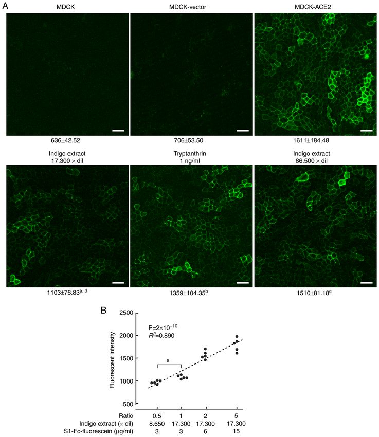
2 HAGIYAMA et al: INDIGO PLANT FOR COVID-19 PREVENTION
solvent, d‑limonene, (+)‑p‑Mentha‑1,8‑diene, an acyclic and the extract was subjected to HPLC. To analyze the compo‑
monoterpene widely used as a fragrance. The resulting indigo sition, the extract was dissolved in ethanol (5.0 mg/ml) and
extract contains tryptanthrin at a fairly high concentration. passed over a COSMOSIL 5PE‑MS column (i.d. 4.6x250 mm;
In the present study, with reference to a deposited preprint Nacalai Tesque) (mobile phase 40% CH 3CN; flow rate,
by Kapczynski et al (unpublished data), we prepared canine 1.0 ml/min; detection 254 nm; temperature 25˚C). Data were
kidney epithelial MDCK cells overexpressing ACE2 and collected with a SIC Chromatocorder12 (System Instruments
established a cell culture system that allowed us to quantify the Co., Ltd.). A tryptanthrin standard was purchased from
degree of fluorescein‑labeled S1 spike protein‑ACE2 binding Sigma‑Aldrich Japan. All other reagents were purchased
by measuring fluorescence intensity. We also conducted from Fujifilm‑Wako Pure Chemicals, Co. In the present study,
computer simulation analyses of docking between tryptanthrin indigo extract was diluted 10‑fold in ethanol (stock solution).
and the spike protein. d‑limonene was also diluted 10‑fold in ethanol as a control
stock solution. For reference, indigo leaves were extracted with
Materials and methods ethanol (99.5%) according to the same procedures.
Cells and reagents. Madin‑Darby canine kidney (MDCK) cells S1 proteins and fluorescein labeling. Recombinant protein
were purchased from the American Type Culture Collection SARS‑CoV‑2 S1 subunit tagged with mouse IgG2a Fc portion
(NBL‑2) and cultured in Eagle's minimal essential medium (S1N‑C5257) and normal mouse IgG (mIgG; 140‑09511) were
with 10% fetal calf serum, as described in our previous purchased from ACROBiosystems and Fujifilm‑Wako Pure
report (8). Human colon adenocarcinoma Caco‑2 cells were Chemicals, Co., respectively. A Fluorescein Labeling Kit‑NH2
previously purchased from the Riken BioResource Center (Dojindo) was used to conjugate fluorescein to the S1 protein
(RCB0988) (9). Tryptanthrin (SML0310; Sigma‑Aldrich; and mouse IgG, according to the manufacturer's instructions.
Merck KGaA) was dissolved in DMSO at a concentration of
1 mg/ml and then diluted 10‑fold in ethanol (final concentra‑ Detection and quantification of fluorescein fluorescence in
tion of the stock solution, 100 µg/ml). MDCK cell cultures. 3x10³ of transfected or untransfected
MDCK cells were suspended in 10 µl of culture medium, and
Plasmid construction and transfection. Total RNA was were transferred separately into the bottom of the micro‑Insert
extracted from Caco‑2 cells using Trizol reagent (Invitrogen; 4‑Well (gasket) on a µ‑Dish (35 mm, high, no. 80406; ibidi
Thermo Fisher Scientific, Inc.), and first strand cDNA was GmbH). After 3 h of incubation, 150 µl of medium were
reverse‑transcribed using total RNA as a template with poured into the insert to fill all wells with identical medium.
Superscript IV (Invitrogen; Thermo Fisher Scientific, Inc.). The next day, fluorescein‑labeled S1 or mIgG was added to
Human ACE2 full‑length cDNA (NM_001371415.1) was the culture medium at a concentration of 3 µg/ml. At the same
obtained by polymerase chain reaction (PCR) where Caco‑2 time, indigo extract, d‑limonene, or tryptanthrin was added
cDNA was used as a template with the primer set: forward, at the indicated concentrations, depending upon the experi‑
5'‑gtggatg tgatct tggctca‑3' and reverse, 5'‑caaa atcacctca ag ment. After cell cultures were incubated 24 h, the micro‑insert
agga aaa a‑3'. The PCR product was inserted into the pTA2 gasket was gently removed, and cells were washed 3x with
TA‑cloning vector (Toyobo). After amplification, the insert culture medium. Then, the µ‑Dish was filled with 1 ml of
was excised at the NotI and HincII sites, and then inserted culture medium, and was placed on the microscope stage
into the pCX4pur vector (10) at the NotI and HpaI sites of a C2+ confocal laser scanning system (Nikon, Tokyo,
(pCX4pur‑hACE2). The absence of mutations was verified by Japan). Fluorescein fluorescent images were captured with a
sequencing. 40x objective lens and analyzed on the Nikon C2+ computer
MDCK cells (8x10⁴) were grown in a 6‑cm dish to 60‑70% system. Fluorescein intensity (arbitrary units per unit area)
confluence and were transfected with either pCX4pur‑hACE2 was measured at five randomly selected high‑power fields for
or the empty pCX4pur vector (5 µg each) using the each well using Analysis Controls tools. Cell cultures in wells
Lipofectamine 3000 (Invitrogen; Thermo Fisher Scientific, on µ‑Dishes were prepared and measured in triplicate for each
Inc.) according to the manufacturer's instructions. Cells were experimental group, and the mean and standard deviation of
selected by resistance to puromycin for two weeks. fluorescein intensities were calculated using ROI Statistics.
Experiments were independently repeated three time with
Plant materials. Leaves of Polygonum tinctorium were similar results.
collected in September 2020 in Aomori Prefecture, Japan. A
voucher specimen was deposited in the herbarium of the medic‑ Immunofluorescence. After having detected the fluores‑
inal herbal garden of Tohoku Medical and Pharmaceutical cence from S1 proteins, we fixed the cells in µ‑Dishes with
University and identified by KS. methanol for 10 min at ‑20˚C, and blocked them with 2%
bovine serum albumin for 30 min at room temperature. At
Preparation of indigo plant leaf extract and high‑performance this time, no fluorescein fluorescence was detectable on cells
liquid chromatography (HPLC). We used our original extract, in any dishes. Then, cells were incubated with an antibody
named ‘AOMORI‑BLUE extract’, which has been patented against ACE2 (E‑11, sc‑390851; Santa Cruz Biotechnology,
in Japan (Japanese Patent no. 6389492). Powdered, air‑dried Inc.) overnight at 4˚C, and were visualized with Alexa Flour
leaves (100 g) were extracted with 1,200 ml of d‑limonene 488‑conjugated secondary antibody (anti‑mouse IgG; Jackson
(Wako Pure Chemical Industries, Ltd.) at room temperature ImmunoResearch). After washing with phosphate‑buffered
for 48 h. After filtration, a pale‑yellow extract was obtained, saline (PBS) three times, nuclei were labeled with DAPI
EXPERIMENTAL AND THERAPEUTIC MEDICINE 23: 274, 2022                                                       3

(Molecular Probes) for 2 h at 4˚C. Fluorescent images were
captured using a C2+ confocal scanning system equipped with
488‑nm argon and 543‑nm helium‑neon lasers (Nikon).

Water‑soluble tetrazolium‑8 assay. Cell viability was
assessed with a water‑soluble tetrazolium‑8 (WST‑8)‑based
colorimetric assay using a Cell Counting Kit 8 (Dojindo,
Kumamoto, Japan). MDCK cells (3x10 4) were seeded in a
96‑well plate in triplicate overnight, and then indigo extract
was added to each well at indicated dilution rates. The next
day, cells were incubated with WST‑8 for 30 min, and the
absorbance was measured at 450 nm using an automated
microplate reader. Measurement of mitochondrial dehy‑
drogenase cleavage of WST‑8 to formazan dye provided an
indication of cell viability.

Western blotting analysis. Cells were washed in PBS and
were lysed in a buffer containing 50 mM Tris‑HCl (pH 8.0),
150 mM NaCl, 1% Triton X‑100 and 1 mM phenylmethylsul‑
fonyl fluoride. After removal of impurities by centrifugation,
lysates were subjected to Western blot analyses, as described
in our previous report (11). Immunoreactive band intensities
were quantified using ImageJ software (National Institutes of
Health), as described previously (12).

Molecular simulation. We obtained the 3D structure of the
of SARS‑CoV‑2 spike protein trimer from the Protein Data
Bank (https://www.rcsb.org/) (PDB ID: 6Z97). We selected
this structure because it satisfies the following conditions:
i) the receptor‑accessible ‘up’ conformation is available in        Figure 1. high performance liquid chromatography analysis of indigo extract.
the trimer, ii) the amino acid sequence is conserved without        (A) Tryptanthrin standard (10.0 µg/ml). (B) Indigo extract (tryptanthrin
deletions, and, iii) the structure is available at a resolution     content, 17.3 µg/ml).
4                                         HAGIYAMA et al: INDIGO PLANT FOR COVID-19 PREVENTION

Figure 2. Expression of ACE2 in MDCK cells. (A) Immunofluorescence of untransfected MDCK, MDCK‑vector and MDCK‑ACE2 cells using an anti‑ACE2
antibody (green; upper). In the lower panels, MDCK‑ACE2 cells were incubated with indigo extract, tryptanthrin or d‑limonene at indicated dilutions or
concentrations, together with S1‑Fc‑fluorescein. Cell nuclei were labeled with DAPI (blue). Merged images of green and blue fluorescent signals are presented
(scale bars=50 µm). (B) Western blot analyses of various types of MDCK cells using an anti‑ACE2 antibody. An arrowhead indicates ACE2‑specific bands.
The blot was re‑probed with an anti‑β‑actin antibody to determine the amount of protein loading per lane. (C) WST‑8 assays of MDCK cellsin the presence
of serially diluted indigo extract. Viability percentages are line‑plotted by dots with bars indicating the mean and standard deviation from triplicate wells,
respectively. P‑values from one‑way ANOVA are shown above the graph. aP= 0.0168 and bP
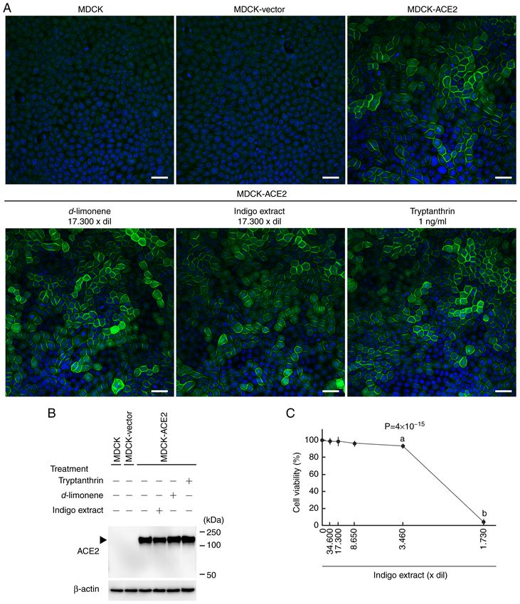
EXPERIMENTAL AND THERAPEUTIC MEDICINE 23: 274, 2022                                                                5

Figure 3. Quantification of S1 proteins bound to ACE2 on MDCK cells. (A) MDCK, MDCK‑ACE2 and MDCK‑vector cells were incubated with
S1‑Fc‑fluorescein (3 µg/ml). In some MDCK‑ACE2 cell cultures, either indigo extract or tryptanthrin was also added at indicated dilution rates or concentra‑
tions. After one day of incubation and wash, fluorescent intensity of fluorescein remaining on the cells was measured using a confocal laser microscopy
system. Representative photomicrographs are presented. Below each image, the mean and standard deviation of the intensity (arbitrary unit) are presented for
the corresponding experimental group. aP=1.20x10 ‑13, bP= 0.0033 and cP>0.999 vs. MDCK‑ACE2 cell intensity; dP= 0.0027 vs. tryptanthrin treatment (scale
bars=50 µm). (B) In MDCK‑ACE2 cell cultures, various ratios of concentrations of indigo extract and S1‑Fc‑fluorescein were used. The ratio was expressed
as 1 under the conditions in A (indigo extract, 17,300‑fold dilution; S1‑Fc‑fluorescein, 3 µg/ml). The ratio (logarithmic in X‑axis) and fluorescent intensity
(linear in Y‑axis) are presented in a scatter plot (n=5 for each ratio group). The dot distribution approximates a linear function (dotted lines). Correlations
and statistical significance were analyzed using Spearman's rank test. R2 and P‑values are presented. aP=3x10 ‑4 as indicated. ACE2, angiotensin‑converting
enzyme 2; MDCK, Madin‑Darby canine kidney.

Inhibition of S1‑ACE2 binding by indigo extract. WST‑8                           (4.39‑7.44). Together with S1‑Fc‑fluorescein, the indigo extract
assays revealed that indigo extract had no substantial effect on                 stock solution was added to confluent MDCK‑ACE2 cell
MDCK cell viability when diluted ≥8,650‑fold (Fig. 2C). The                      cultures in µ‑Dishes at a dilution of 1,730‑fold (17,300‑fold
extract did not change the medium pH in this range of dilution                   for the original extract), and after 24 h, cells were observed
6                                                HAGIYAMA et al: INDIGO PLANT FOR COVID-19 PREVENTION

Table I. Docking results of tryptanthrin‑S1 spike protein                                                Table III. Amino acid residues of SARS‑CoV‑2 S1 spike
docking run per 100 times.                                                                               protein that are involved in binding to ACE2 or tryptanthrin.

                                               Docking score                                                                                          Tryptanthrin (45 poses)
No. of correct     ‑‑‑‑‑‑‑‑‑‑‑‑‑‑‑‑‑‑‑‑‑‑‑‑‑‑‑‑‑‑‑‑‑‑‑‑‑‑‑‑‑‑‑‑‑‑‑‑‑‑‑‑‑‑‑‑‑‑‑‑‑‑‑‑‑‑‑‑‑‑‑‑‑‑‑‑‑‑‑‑‑‑‑   ACE2 amino acids                                  amino acids
binding models     Mean ± SD Most stable Score rankinga
                                                                                                         K417, G446, Y449, Y453, L455a,                 L455a, F456a E484,
58                 ‑5.88±0.10                       ‑6.02                            26                  F456a, A475, F486, N487, Y489a,                G485, C488, Y489a,
Rank of the most stable docking score.
a                                                                                                        Q493a, G496, Q498, T500, N501,                 F490, L492 and
                                                                                                         G502 and Y505                                  Q493a
                                                                                                         a
                                                                                                          Four residues are common to S1‑ACE2 and S1‑tryptanthrin binding.
Table II. Docking results of the same binding mode in 58                                                 For tryptanthrin‑S1 docking, the same binding mode was repeated
correct tryptanthrin‑S1 spike protein bindings.                                                          45 times in 58 correct binding runs (presented in Table I). Binding
                                                                                                         was defined by proximity of 4 Å between S1 and ACE2 or S1 and
No. of the same                 Amino acids                               Docking score                  tryptanthrin. ACE2, angiotensin‑converting enzyme 2.
binding mode                     targeted                                 (mean ± SD)

45                     455‑456 and 488‑494                                   ‑5.88±0.02

alive. Fluorescent signals were weakly detectable (Fig. 3A).
The fluorescence intensity was much lower than that in
MDCK‑ACE2 cell cultures without indigo extract (P
EXPERIMENTAL AND THERAPEUTIC MEDICINE 23: 274, 2022 7
even at 0.6 µl/ml (1,667‑fold v/v dilution) and 0.5 mM, respec‑ open a new avenue for practical use of this natural product as
tively (14,15). We used indigo extract mainly at a 17,300‑fold a prophylactic against SARS‑CoV‑2 infection.
v/v dilution, i.e., 0.35 mM d‑limonene. The WST‑8 assay
showed that this concentration was low enough for MDCK Acknowledgements
cells to remain healthy; therefore, d‑limonene was useful for
indigo leaf extraction. Actually, we did not detect substantial Not applicable.
changes in ACE2 expression in MDCK cells treated with
indigo extract or d‑limonene (Fig. 2A). This also supported Funding
the notion that indigo extract inhibited S1‑ACE2 binding, not
ACE2 expression, at high dilution rates. This study was supported by the Japan Society for the
The present study also showed that tryptanthrin inhibited Promotion of Science KAKENHI (grant nos. 17K08680,
S1‑ACE2 binding, though it did not account for the entire 20K07434, 18K07049 and 21K06978), the Takeda Science
inhibitory effect of indigo extract. Tryptanthrin was reported Foundation (to MH, 2019) and the All‑Kindai University
to reduce the HCoV‑NL63 infectivity with IC50 values of support project against COVID‑19 (to AI, 2020 and 2021). The
0.30 and 1.52 µM in Calu‑3 and LCC‑MK2 cells, respec‑ current study also received funding from Aomori AI Industrial
tively (7). We used tryptanthrin at concentrations of 4.0 nM, Co., Ltd., Aomori, Japan.
suggesting that tryptanthrin may be much more effective
against SARS‑CoV‑2. This speculation, however, may be too Availability of data and materials
simple, because we examined the inhibitory effect only on
S1‑ACE2 binding, not infectivity, in MDCK cells that had been The datasets used and/or analyzed during the current study are
forced to overexpress ACE2. To the best of our knowledge, the available from the corresponding author on request.
minimal 50% cytotoxic concentration (CC50) of tryptanthrin
is 173.2 µM for Calu‑3 cells (7), and tryptanthrin is generally Authors' contributions
thought to have no significant cytotoxicity to human normal
cells (16,17). In terms of cytotoxicity, tryptanthrin can be MH and FT constructed expression vectors and performed
expected to serve as an inhibitor for S1‑ACE2 binding within transfection. MH and FT also conducted cell culture experi‑
its safe concentration range. ments, confocal microscopic studies and western blot analyses.
Since tryptanthrin is much smaller than S1 protein, one may AY, TI, HK and AW helped complete these experiments. KS
wonder how it can inhibit binding. Docking simulation analyses provided plant materials and performed HPLC. AS and YT
revealed that tryptanthrin bound to the RBM of the spike protein conducted simulation analyses. MH conducted the statistical
trimer mainly using nine amino acid residues, four of which are analyses. MH, FT and AI confirmed the authenticity of all the
involved in S1 RBM‑ACE2 binding, and its molecular plane raw data. KS and AI conceived and designed the study, and
was nearly perpendicular to the RBM surface. This binding AI drafted the manuscript. All authors read and approved the
conformation may explain why tryptanthrin inhibits the binding final manuscript.
of S1 protein to ACE2. Even though it is small, tryptanthrin may
bind to residues essential for RBM‑ACE2 binding. Consistent Ethics approval and consent to participate
with this simulation, the competitive nature of indigo extract
was illustrated by cell culture experiments in which we changed Not applicable.
the ratios of concentrations of the extract and S1 protein added
to the cultures (Fig. 3B). However, another possibility remains. Patient consent for publication
Tryptanthrin may have a stronger affinity for ACE2 than
S1 protein. Since ACE2 is crucial to heart function control (18), Not applicable.
this possibility must be examined carefully when clinical appli‑
cations are considered. Competing interests
In addition, the present study suggests that indigo extract
contains other active components beside tryptanthrin. The authors declare no competing interests.
Compared with ethanol, d‑limonene extracts low‑polarity
components, including tryptanthrin, rather than high‑polarity References
components such as glycosides (compare Figs. 1 and S2). With
this feature in mind, we are now trying to isolate and identify the 1. Huang C, Wang Y, Li X, Ren L, Zhao J, Hu Y, Zhang L, Fan G,
active components. Since Polygonum tinctorium is generally Xu J, Gu X, et al: Clinical features of patients infected with 2019
classified as food and its toxicity has not been reported, we novel coronavirus in Wuhan, China. Lancet 395: 497‑506, 2020.
2. Walls AC, Park YJ, Tortorici MA, Wall A, McGuire AT
are also developing in vivo experiments to administer indigo and Veesler D: Structure, function, and antigenicity of the
extract intranasally to mice. Pharmacokinetic analyses are SARS‑CoV‑2 spike glycoprotein. Cell 181: 281‑292.e6, 2020.
planned on the extract and other active components. 3. Wrapp D, Wang N, Corbett KS, Goldsmith JA, Hsieh CL,
Abiona O, Graham BS and McLellan JS: Cryo‑EM structure of
In conclusion, we demonstrated that indigo extract has an the 2019‑nCoV spike in the prefusion conformation. Science 367:
inhibitory effect on binding of S1 to ACE2 at concentrations 1260‑1263, 2020.
low enough not to affect cell viability. One of the active compo‑ 4. Zhong Y, Yoshinaka Y, Takeda T, Shimizu N, Yoshizaki S,
Inagaki Y, Matsuda S, Honda G, Fujii N and Yamamoto N:
nents appears to be tryptanthrin, but the extract likely contains Highly potent anti‑HIV‑1 activity isolated from fermented
other active, unidentified elements. Further investigation may Polygonum tinctorium Aiton. Antiviral Res 66: 119‑128, 2005.8 HAGIYAMA et al: INDIGO PLANT FOR COVID-19 PREVENTION
5. Ishihara T, Okura T, Kohno K, Tanimoto T, Ikegami H and 12. Mimae T, Okada M, Hagiyama M, Miyata Y, Tsutani Y, Inoue T,
Kurimoto M: Polygonum tinctorium extract suppresses nitric Murakami Y and Ito A: Upregulation of notch2 and six1 is asso‑
oxide production by activated macrophages through inhibiting ciated with progression of early‑stage lung adenocarcinoma and
inducible nitric oxide synthase expression. J Ethnopharmacol 72: a more aggressive phenotype at advanced stages. Clin Cancer
141‑150, 2000. Res 18: 945‑955, 2012.
6. Han NR, Kang SW, Moon PD, Jang JB, Kim HM and 13. Lan J, Ge J, Yu J, Shan S, Zhou H, Fan S, Zhang Q, Shi X,
Jeong HJ: Genuine traditional Korean medicine, Naju Jjok Wang Q, Zhang L and Wang X: Structure of the SARS‑CoV‑2
(Chung‑Dae, Polygonum tinctorium) improves 2,4‑dinitro‑ spike receptor‑binding domain bound to the ACE2 receptor.
fluorobenzene‑induced atopic dermatitis‑like lesional skin. Nature 581: 215‑220, 2020.
Phytomedicine 21: 453‑460, 2014. 14. Tang XP, Guo XH, Geng D and Weng LJ: d‑Limonene protects
7. Tsai YC, Lee CL, Yen HR, Chang YS, Lin YP, Huang SH PC12 cells against corticosterone‑induced neurotoxicity by
and Lin CW: Antiviral action of tryptanthrin isolated from activating the AMPK pathway. Environ Toxicol Pharmacol 70:
Strobilanthes cusia leaf against human coronavirus NL63. 103192, 2019.
Biomolecules 10: 366, 2020. 15. Rabi T and Bishayee A: d‑Limonene sensitizes docetaxel‑induced
8. Hosokawa Y, Hagiyama M, Iino T, Murakami Y and Ito A: cytotoxicity in human prostate cancer cells: Generation of reac‑
Noncontact estimation of intercellular breaking force using a tive oxygen species and induction of apoptosis. J Carcinog 8: 9,
femtosecond laser impulse quantified by atomic force micros‑ 2009.
copy. Proc Natl Acad Sci USA 108: 1777‑1782, 2011. 16. Shankar GM, Alex VV, Nisthul AA, Bava SV, Sundaram S,
9. Hagiyama M, Yabuta N, Okuzaki D, Inoue T, Takashima Y, Retnakumari AP, Chittalakkottu S and Anto RJ: Pre‑clinical
Kimura R, Ri A and Ito A: Modest static pressure suppresses evidences for the efficacy of tryptanthrin as a potent suppressor
columnar epithelial cell growth in association with cell shape of skin cancer. Cell Prolif 53: e12710, 2020.
and cytoskeletal modifications. Front Physiol 8: 997, 2017. 17. Han NR, Kim HM and Jeong HJ: Tryptanthrin reduces mast cell
10. Kimura R, Otani T, Shiraishi N, Hagiyama M, Yoneshige A, proliferation promoted by TSLP through modulation of MDM2
Wada A, Kajiyama H, Takeuchi F, Mizuguchi N, Morishita K and and p53. Biomed Pharmacother 79: 71‑77, 2016.
Ito A: Expression of cell adhesion molecule 1 in human and murine 18. Crackower MA, Sarao R, Oudit GY, Yagil C, Kozieradzki I,
endometrial glandular cells and its increase during the prolifera‑ Scanga SE, Oliveira‑dos‑Santos AJ, da Costa J, Zhang L,
tive phase by estrogen and cell density. Life Sci 283: 119854, 2021. Pei Y, et al: Angiotensin‑converting enzyme 2 is an essential
11. Koma Y, Furuno T, Hagiyama M, Hamaguchi K, Nakanishi M, regulator of heart function. Nature 417: 822‑828, 2002.
Masuda M, Hirota S, Yokozaki H and Ito A: Cell adhesion
molecule 1 is a novel pancreatic‑islet cell adhesion molecule This work is licensed under a Creative Commons
that mediates nerve‑islet cell interactions. Gastroenterology 134: Attribution-NonCommercial-NoDerivatives 4.0
1544‑1554, 2008. International (CC BY-NC-ND 4.0) License.You can also read