In situ imaging reveals disparity between prostaglandin localization and abundance of prostaglandin synthases - Nature
←
→
Page content transcription
If your browser does not render page correctly, please read the page content below

ARTICLE
                  https://doi.org/10.1038/s42003-021-02488-1                 OPEN

                  In situ imaging reveals disparity between
                  prostaglandin localization and abundance
                  of prostaglandin synthases
                  Kyle D. Duncan           1,5,   Xiaofei Sun       2,3,5,   Erin S. Baker      4,   Sudhansu K. Dey            2,3   & Ingela Lanekoff      1✉

                  Prostaglandins are important lipids involved in mediating many physiological processes, such
                  as allergic responses, inflammation, and pregnancy. However, technical limitations of in-situ
                  prostaglandin detection in tissue have led researchers to infer prostaglandin tissue dis-
1234567890():,;

                  tributions from localization of regulatory synthases, such as COX1 and COX2. Herein, we
                  apply a novel mass spectrometry imaging method for direct in situ tissue localization of
                  prostaglandins, and combine it with techniques for protein expression and RNA localization.
                  We report that prostaglandin D2, its precursors, and downstream synthases co-localize with
                  the highest expression of COX1, and not COX2. Further, we study tissue with a conditional
                  deletion of transformation-related protein 53 where pregnancy success is low and confirm
                  that PG levels are altered, although localization is conserved. Our studies reveal that the
                  abundance of COX and prostaglandin D2 synthases in cellular regions does not mirror the
                  regional abundance of prostaglandins. Thus, we deduce that prostaglandins tissue localization
                  and abundance may not be inferred by COX or prostaglandin synthases in uterine tissue, and
                  must be resolved by an in situ prostaglandin imaging.

                  1 Department of Chemistry-BMC, Uppsala University, Uppsala, Sweden. 2 Division of Reproductive Sciences, Cincinnati Children’s Hospital Medical Center,

                  Cincinnati, OH 45229, USA. 3 College of Medicine, University of Cincinnati, Cincinnati, OH 45221, USA. 4 Department of Chemistry, North Carolina State
                  University, Raleigh, NC, USA. 5These authors contributed equally: Kyle D. Duncan, Xiaofei Sun. ✉email: Ingela.Lanekoff@kemi.uu.se

                  COMMUNICATIONS BIOLOGY | (2021)4:966 | https://doi.org/10.1038/s42003-021-02488-1 | www.nature.com/commsbio                                          1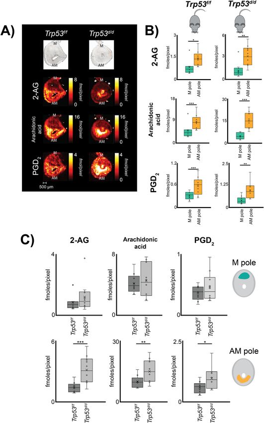
ARTICLE                                                                  COMMUNICATIONS BIOLOGY | https://doi.org/10.1038/s42003-021-02488-1

P
      rostaglandins (PGs) are potent bioactive lipid mediators                  is fully differentiated into primary and secondary decidual zones,
      that play important roles in various biological processes,                and both COX1 and COX2 are expressed. By day 10, COX2 is
      including angiogenesis, inflammation, and reproductive                     expressed around the fetal-maternal interface around the ecto-
events1,2. PG biosynthesis is mediated by several key enzymes,                  placental cone, the presumptive site of placentation15, and by
including phospholipase A2 and diacylglycerol lipase, which                     midgestational stage, COX2 is observed in invasive spiral-artery
cleave acylglycerol chains such as arachidonic acid from phos-                  trophoblast giant cells16. These results imply a role for PGs in
pholipids and diacylglycerols. Subsequently, cyclooxygenases                    placentation, tissue remodeling, and embryonic development.
(COX1 and COX2) mediate the conversion of arachidonic acid to                   PGF2α derived from COX1 is considered indispensable for on-
PGH2, which is considered the rate-limiting step in PG bio-                     time parturition and birth17.
synthesis (Fig. 1A)3–5. Of the two PG synthase enzymes, COX1                       Previous studies have shown that transformation-related pro-
(encoded by Ptgs1) is largely considered as a basal PG regulator,               tein 53 (p53) plays a critical role in many pathophysiological
whereas COX2 (encoded by Ptgs2) is considered an inducible                      processes, including pregnancy and embryonic development18,19.
enzyme6,7. After PGH2 formation, distinct downstream PG syn-                    For example, uterine-specific deletion of p53 has been shown to
thases engage in synthesizing specific PGs, such as PGD2, PGE2,                  promote preterm labor in mice and has been implicated in pre-
and PGF2α (Fig. 1A). Although the molecular structures of PGs                   mature births in humans18. Furthermore, studies have revealed
are similar, individual PGs have different biological functions.                that p53 interacts with AMPK and mTORC1 signaling, which
   In pregnancy, PGs are key regulatory molecules in multiple                   control parturition timing, and we have reported on specific lipid
events, including ovulation, fertilization, embryo implantation,                accumulation and depletion19–21. Further, evidence linking
decidualization, and parturition8. Two major regions in embryo                  deletion of p53 to aberrations in COX2-derived PG signaling
implantation sites are the mesometrial (M) pole and the anti-                   includes elevated levels of COX2 activity on day 16 of
mesometrial (AM) pole (Fig. 1B, C, and D). Embryo homing and                    pregnancy18. The sum of these studies suggests a significant
implantation of the developing embryo are presumed to occur at                  alteration in PGs also at the earlier day 8 of pregnancy. Con-
the AM pole, which decidua is the main provider of nutrition                    sidering the importance of p53 in healthy embryonic develop-
prior to the establishment of a functional placenta at the M pole9.             ment and parturition, relatively little is known of p53’s impact on
The importance of PGs in pregnancy is revealed by defective                     specific PG synthases, PG receptors, PG precursors, or PG spatial
ovulation and fertilization failure in mice with a deficiency in                 distributions. In sum, PGs play a key role throughout the process
Ptgs210,11. To underscore the importance of COX2 in implanta-                   of pregnancy. However, our knowledge of localization of specific
tion, we have previously shown that Ptgs2−/− mice develop                       PGs in pregnancy events remains limited due to the dynamic
implantation failures, and further studies suggested that COX2-                 spatiotemporal pattern of PG synthetic enzymes and the mea-
derived PGI2 plays a key role in embryo implantation12. Mice                    surable accumulation of unstable PGs in uteri at different stages
deficient in Ptgs1 show no apparent implantation defects13, but a                and conditions of pregnancy.
compensatory expression of Ptgs2 is observed for Ptgs1 in a                        Despite the biological importance of PGs, localization of native
similar pattern as Ptgs214, which suggests that PGs derived from                PGs to specific cells in tissues has not yet been possible, although
COX1 play a critical role during implantation. As implantation                  methods using PGs labelled with biotins have helped in this
and decidualization progress, Ptgs2 expression is switched from                 regard22,23. As a result, PG distribution in tissues has been largely
the antimesometrial (AM) pole to the mesometrial (M) pole of                    assumed to correspond to the regional expression of COX2 and/
the implantation chamber11. On day 8 of pregnancy, the decidua                  or downstream PG synthases. Mass spectrometry imaging (MSI)

Fig. 1 Schematic detailing the studied PG biosynthesis pathway and experimental design. A Generation of PG species from phospholipids or 2-AG via
COX and PG synthases, enzymes are in red and metabolites in blue. B Mouse embryo implantation sites from day 8 of pregnancy were used from Trp53f/f
and Trp53d/d mice. C Florescence in situ hybridization was applied to study the in situ localization of synthases. Implantation sites were segmented into the
AM-pole and the M-pole for RT-qPCR analysis of enzyme mRNA expression. D Quantitative nano-DESI MSI was performed on thin sections of
implantation sites to determine in situ small molecule localization.

2                            COMMUNICATIONS BIOLOGY | (2021)4:966 | https://doi.org/10.1038/s42003-021-02488-1 | www.nature.com/commsbio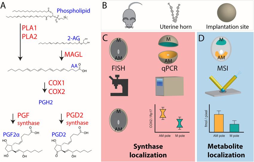
COMMUNICATIONS BIOLOGY | https://doi.org/10.1038/s42003-021-02488-1                                                               ARTICLE

techniques have the ability to localize a wide range of molecules              limit of detection and therefore not included. Quantitative pros-
in thin tissue sections. However, although PG precursors, such as              taglandin imaging of PGD2 and PGF2α was accomplished by
2-arachidonoylglycerol (2-AG), arachidonic acid, and other ara-                introducing heavy isotope-labeled PG standards for direct single-
chidonic acid-containing phospholipids, have been imaged in                    point calibration in each pixel28,30. Isomeric identification
tissues with MSI20,24, localizing PGs to specific cellular regions              determined that the detected isomer was PGD2 and not PGE2,
has been a major technical challenge. This challenge largely arises            which was accomplished by liquid chromatography ion mobility
from low PG ionization efficiencies in combination with very low                spectrometry and mass spectrometry (LC-IMS-MS) (Fig. S2 and
endogenous concentrations, ranging between picomoles and                       Tables S2, S3). The amount of PG in morphological regions of
nanomoles per gram of tissue18,25–27. Recently, we reported a                  the tissue was determined by extracting data from the AM pole
novel approach using silver-doped nanospray desorption elec-                   and the M pole for all biological and technical replicate images to
trospray ionization (nano-DESI) MSI for quantitative imaging of                ensure accurate biological interpretations and correlation to
prostaglandins in thin tissue sections of mouse implantation sites             regional expression of proteins followed by statistical analyses.
of day 4 of pregnancy28. In particular, within one experiment we               Overall, our established workflow-enabled speciation, quantifi-
uncovered the localization and quantity of four different PGs to               cation, and localization of PGs, PG precursors, and expression of
the luminal epithelium and the glandular epithelium of the tissue,             PG synthases.
showing applicability for biological studies.
   Here, we use the unique combination of silver doped nano-
                                                                               Regional distribution of PG and PG precursors. Quantitative
DESI MSI, RNA in situ hybridization, and RT-qPCR to study the
                                                                               nano-DESI MSI enabled direct in situ imaging of PGD2 (Fig. 2A)
correlation between enzyme expression and PG distribution in
                                                                               and PGF2α (Fig. S1) in tissue from day 8 implantation sites.
mouse embryo implantation sites on Day 8 of pregnancy. We
                                                                               Simultaneously, the technique provided in situ imaging of the PG
apply this workflow to study the impact of p53 on the molecular
                                                                               precursor’s 2-AG and arachidonic acid (Fig. 2A and Table S4).
machinery of PG biosynthesis in early embryonic development.
                                                                               The ion images presented in Fig. 2A are quantitative through the
The goal of this study was to: (i) visualize detectable PG species;
                                                                               use of internal standards and the color scale of the pixels are
(ii) detect any perturbations to PG synthesis as a result of p53
                                                                               individually represented for each ion image in fmoles/pixel. The
deletion; and (iii) correlate spatial distributions of PG species and
                                                                               results illustrate that PGD2 and all its precursors localize to the
PG synthases. Overall, this study provides the first evidence of PG
                                                                               AM-pole (Fig. 2A, B), while PGF2α mainly localizes to the myo-
localization in a tissue linked to a spatially defined analysis of key
                                                                               metrium on day 8 of pregnancy (Fig. S1). Relative comparisons of
enzymes involved in PG biosynthetic pathways and demonstrates
                                                                               the spatial distribution for PGD2 and its precursors show that
that the abundance or spatial distribution of PGs in tissue cannot
                                                                               arachidonic acid is ~2.5 times higher in the AM-pole compared to
be determined by monitoring only PG synthases.
                                                                               the M-pole while 2-AG and PGD2 are ~1.5 times higher in the
                                                                               AM-pole (Fig. 2B and Tables S5, S6).
                                                                                  Regional distribution of PG and PG precursors was investigated
Results
                                                                               in mice with a uterine-specific deletion of Trp53. Quantitative
The biosynthetic pathway for PGs includes several key enzymes
                                                                               nano-DESI MSI reveals significant accumulation of 2-AG,
(Fig. 1A), that were monitored in the workflow for spatial cor-
                                                                               arachidonic acid, and PGD2 in the AM-pole of the Trp53d/d mice
relation between PG synthases, PG receptors, PG dehydrogenase,
                                                                               (Fig. 2A, C). Compared to their detected abundances in the AM-
PG precursors, and PGs was established for mouse embryo
                                                                               pole of Trp53f/f mice, 2-AG is increased ~2 times, and arachidonic
implantation sites on day 8 of pregnancy (Fig. 1B−D). Specifi-
                                                                               acid and PGD2 ~1.5 times in Trp53d/d mice (Fig. 2C and Table S5,
cally, the uterine horn was extracted from control mice and mice
                                                                               S6). Negligible differences were observed for PGF2α in Trp53f/f and
with a uterine conditional deletion of p53 on day 8 of pregnancy
                                                                               Trp53d/d mice, which were primarily detected in the myometrium
and individual embryo implantation sites were isolated (Fig. 1B).
                                                                               (Figs. S1 and S3). Overall, changes in PG and PG precursor
Expression of key synthases and dehydrogenase involved in PG
                                                                               abundance as a result of p53 deletion appears to be isolated to the
biosynthesis and signaling pathways were analyzed using RT-
                                                                               AM-pole, as no significant differences were observed for any of
qPCR. Specifically, dissected mouse uterine implantation sites
                                                                               these molecules in the M-pole (Fig. 2A, C and Tables S5, S6).
were partitioned into AM and M-pole segments, and the mRNA
expression was quantified by RT-qPCR for each pole of the
implantation site (Fig. 1C and Table S1). As a result, the levels of           Regional distribution of PG synthases. Fluorescence in situ
mRNA for PG synthases could be statistically compared between                  hybridization (FISH) of COX2 protein shows synthase localization
the two morphologically different poles to explore regional PG                 (Fig. 3A). Segmented RT-qPCR of mouse embryo implantation
biosynthesis on day 8 of pregnancy. To provide additional spatial              sites on day 8 of pregnancy further show that mRNA expression of
information on PG biosynthetic enzyme localization, fluorescence                Ptgs2 encoding COX2 is 15 times higher in the M-pole compared
in situ hybridization and immunostaining were performed on                     to the AM-pole (Fig. 3B). Results from RT-qPCR and FISH of
thin tissue sections of implantation sites (Fig. 1C).                          Ptgs1 both reveal that Ptgs1 expression is primarily found in the
   Quantitative imaging of PGs and PG molecular precursors in                  AM-pole of the uterine implantation site (Fig. 3A, B). Expression
implantation sites was performed by silver doped nano-DESI MSI                 of the two PGD2 synthases Ptgds31 and Hpgds32 with segmented
with subsequent region-of-interest data analysis from molecules                RT-qPCR shows that both are 2 and 4 times higher in the AM-
in the AM and M-pole (Fig. 1D). The analysis was performed                     pole than in the M-pole of the implantation sites on day 8 of
untargeted to enable detection of all PGs and PG precursors in                 pregnancy, respectively (Fig. 3A, B).
one experiment per tissue section, through localized liquid                       Regional distribution of PG synthases was studied in control
extraction of tissue molecules into a liquid bridge between two                mice and mice with a uterine-specific deletion of Trp53. Data
capillaries with subsequent electrospray ionization (nano-DESI,                from segmented RT-qPCR show that Ptgs2 expression is
Fig. 1D)20,28–30. Despite the untargeted approach, only the two                significantly higher in the AM-pole of p53 deficient mice
PG species PGD2 and PGF2α were detected at maximum 4                           compared to control mice (Fig. 3B, C). Further, Ptgs2 expression
fmoles/pixel and 1.2 fmoles/pixel in control mice, respectively                is significantly lower in the M-pole of p53 deficient (Trp53f/f
(Fig. 2A and S1). It is likely that there are additional PG species of         PgrCre/+, Trp53d/d) mice, suggesting that Trp53d/d mice have an
importance present in the tissue; however, these were below the                overall lower level of COX2 compared to Trp53f/f mice (Fig. 3C).

COMMUNICATIONS BIOLOGY | (2021)4:966 | https://doi.org/10.1038/s42003-021-02488-1 | www.nature.com/commsbio                                     3
ARTICLE COMMUNICATIONS BIOLOGY | https://doi.org/10.1038/s42003-021-02488-1 Fig. 2 Distribution of prostaglandins to morphological regions in mouse embryo implantation sites on day 8 of pregnancy using quantitative silver doped nano-DESI MSI. A Optical images and corresponding quantitative ion images of 2-AG, arachidonic acid, and PGD2 in Trp53f/f and Trp53d/d mice on day 8 of pregnancy. Color scaling corresponds to detected fmoles/pixel with the values going from dark to bright. Note that the fixed quantitative color scales for each metabolite facilitates direct visual comparisons. Scale bar 500 µm. M mesometrial pole; AM anti-mesometrial pole. B Regions of interest for the AM-pole and M-pole of Trp53f/f and Trp53d/d mice, respectively, with detected concentrations of 2-AG, arachidonic acid, and PGD2 from three biological and three technical replicates. C Region of interest analysis of the AM-pole and M-pole for 2-AG, AA, and PGD2 of 3 biological and 3 technical replicates comparing the of Trp53f/f and Trp53d/d mice (n = 9 for each genotype). Error bars in represent 1 standard deviation and * p-value < 0.05, ** p-value < 0.01, *** p-value < 0.001. 4 COMMUNICATIONS BIOLOGY | (2021)4:966 | https://doi.org/10.1038/s42003-021-02488-1 | www.nature.com/commsbio
COMMUNICATIONS BIOLOGY | https://doi.org/10.1038/s42003-021-02488-1                                                                     ARTICLE

Fig. 3 Distribution of proteins involved in PG synthesis, hydrolysis, and signaling in implantation sites on day 8 of pregnancy. A Fluorescence in situ
hybridization of Ptgs1 and COX2 in Trp53f/f and Trp53d/d implantation sites on day 8 of pregnancy. The embryonic trophoblasts are outlined by Cytokeratin
8 (CK8) immunostaining. M mesometrial pole; AM anti-mesometrial pole; Em embryo; SDZ secondary decidual zone; Myo myometrium; scale bar,
500 µm. B Segmented RT-qPCR results for Ptgs2, Ptgs1, Ptgds, Hpgds, in the AM and M-poles in Trp53f/f and Trp53d/d implantation sites on day 8 of
pregnancy. C Segmented RT-qPCR results showing the expression of Ptgs2, Ptgs1, Ptgds and Hpgds, Pgdh, Ptgdr, and Ptgdr2 in the M-pole and AM-pole of the
implantation sites comparing the Trp53f/f and Trp53d/d implantation sites. Significance is determined by a two-tailed student’s t-test, where * p-value <
0.05, ** p-value < 0.01, *** p-value < 0.001.

COMMUNICATIONS BIOLOGY | (2021)4:966 | https://doi.org/10.1038/s42003-021-02488-1 | www.nature.com/commsbio                                            5
ARTICLE                                                              COMMUNICATIONS BIOLOGY | https://doi.org/10.1038/s42003-021-02488-1

These results illustrate a clear, region-specific perturbation of            pregnancy13, our data of Ptgs1 and PGD2 suggests that COX1
COX2 expression levels in Trp53d/d mice. FISH imaging                       plays a role in early pregnancy. Further, COX2 is the major
corroborates the results from RT-qPCR, showing that Ptgs1 is                inducible enzyme for the first rate-limiting step in the biosynth-
expressed on decidual cells, mainly in the secondary decidual               esis of PGs, and disruption of its function causes multiple female
zone on the AM-pole, for both Trp53d/d and Trp53f/f mice, while             reproductive failures, including infertility in Ptgs2−/− mice11.
Ptgs2 is mainly expressed in the M-pole (Fig. 3A).                          Therefore, we hypothesize that Ptgs2 is induced to compensate for
   Our RT-qPCR data reveal an opposing impact of p53 on PGD2                COX1 in Ptgs1−/− mice, so that no apparent defects are observed
synthases when compared to Ptgs2 (Fig. 3C). The expression of               in the latter. In fact, our previous study supports this hypothesis,
Ptgds and Hpgds are significantly decreased in the AM-pole of the            in that Ptgs2 is expressed in luminal epithelia in Ptgs1−/− mice on
Trp53d/d mice as compared to control mice; however, the relative            day 4 of pregnancy where Ptgs1 is normally expressed in wild-
levels of Ptgds in the AM-pole are still ~2-fold higher than in the         type mice14.
M-pole (Fig. 3C). In the M-pole, the Ptgds and Hpgds levels of                 The role of COX appears pleiotropic during pregnancy9. Ptgs2
Trp53d/d mice are similar to the Trp53f/f control mice.                     is subject to cell-specific expression in the mouse uterus
   The RT-qPCR data indicates a crucial role of p53 in controlling          depending on the experimental conditions and implantation-
PG synthesis by regulating both Ptgds and Hpgds levels in the               specific gene expression37–39. The excess of available arachidonic
AM-pole (Fig. 3C). Segmented RT-qPCR analysis of Ptgdr and                  acid in the AM-pole corroborates that COX is indeed the rate-
Ptgdr2 expression levels, indicating the abundance of PGD2                  limiting step in PG synthesis, in agreement with the previous
receptors, show that Ptgdr is ~3 times lower in both the AM and             studies3,4. Further, the lower relative abundance of 2-AG to
M-pole of the Trp53d/d mice, compared to that of Trp53f/f mice              arachidonic acid suggests that arachidonic acid does not exclu-
(Fig. 3C). In addition, no significant change in Pgdh expression             sively originate from 2-AG, but rather from a larger diverse pool
was observed between Trp53f/f and Trp53d/d mice suggesting no               of lipids, such as phospholipids and diacylglycerols. This is fur-
alterations in PG dehydrogenase levels resulting from conditional           ther evidenced by our previous study from day 8 implantation
uterine deletion of p53. (Fig. 3C).                                         sites reporting a clear localization of arachidonic acid-containing
                                                                            phospholipids and diacylglycerols to the AM-pole20. In addition
                                                                            to PGD2, PGF2α localizes to the myometrium of the uterus
Discussion                                                                  matching the expression of Ptgs1 encoding COX1, again away
Technical limitations in direct detection of PGs in tissue sections         from COX2 expression and localization. Although Ptgs1 is con-
largely arise from the inability to selectively stain individual PGs        sidered to be a housekeeping gene that maintains basal cell
for in situ localization studies with fluorescent microscopy. Fur-           functions, the disparate expression of COX2 with PG and PG
ther, the low endogenous abundance and poor MS ionization                   precursor localization indicates that COX2 abundance does not
efficiency of PGs have hindered the use of MSI techniques for                directly correlate with in situ PG distributions in tissue.
mapping their distribution in tissue. The inclusion of silver ions             Conditional deletion of Trp53 is known to impact pregnancy
in the nano-DESI solvent increases the ionization efficiency of              progression of mice18. Although the embryo development is
PGs and enables direct visualization of PGs in tissue. However,             compromised in Trp53d/d mice, the overall expression pattern
the identification of specific PGs in selected domains remains                and intensity of Ptgs1 are comparable to those in Trp53f/f mice.
uncharted territory. We combined our novel PG MSI method                    An increase of COX2 in the AM-pole with a concurrent decrease
with traditional RNA in situ hybridization, making it possible to           in the M-pole suggests that the absence of p53 causes a sig-
identify specific PGs in situ, which significantly advances our               nificantly arrested transition of Ptgs2 from the AM-pole to the
current understanding of PG signaling in uterine biology and                M-pole between days 5 and 8 of pregnancy36.
beyond.                                                                        We observed an increase of PGD2 along with 2-AG and ara-
   The main localization of PG and PG precursors is to the AM-              chidonic acid in decidual cells at the anti-mesometrial side is
pole. Thus, at day 8 of pregnancy, it is likely that Ptgds and PGD2         observed in WT mice on day 8 of pregnancy. The physiological
localization to the AM-pole signifies a defined role in AM-pole               roles of PGD2 in decidualization are still unclear. Our earlier
decidualization. Our previous study also suggests PGD2 and PGE2             study revealed that oxidized lipids were found to be decreased in
are major players in endometrial cancer33. Although the role of             the AM-pole of Trp53d/d implantation sites, indicating lower
PGD2 remains unclear in the mouse uterus, cells with positive               levels of reactive oxygen species in p53 deficient uteri20. As a
PGD2 signals undergo tissue remodeling to make room for                     result, the higher PGD2 levels in Trp53d/d mice are not likely to
growing fetuses. There are several known downstream effects of              originate from autoxidation of arachidonic acid through the
PGD2 signaling, which include inhibition of platelet aggregation            isoprostane pathway, but rather from elevated levels of COX2 in
and relaxation of vascular and non-vascular smooth muscle34,35.             the AM-pole. Accumulation of PGD2 and arachidonic acid in the
Therefore, PGD2 localization to the lateral and AM-pole on day              AM-pole of Trp53d/d mice is accompanied with increased poly-
8 suggests its importance to ensure adequate nutrient delivery to           ploid cells and decidual cell senescence18, indicating PGD2’s role
the developing embryo and facilitate tissue reorganization.                 in decidual aging and/or differentiation. It would be interesting to
   Localization of PG precursors and PGD2 to the AM-pole of the             examine whether the elevated PG levels in Trp53d/d decidua
implantation site nicely correlates with the expression of the              extend to parturition, since PGs are closely associated with par-
PGD2 synthases Ptgds and Hpgds in implantation sites on day 8 of            turition timing17 and contribute to spontaneous preterm birth in
pregnancy. This localization further correlates with the expression         Trp53d/d females18.
of Ptgs1 encoding for the housekeeping enzyme COX1 in agree-                   The higher abundance of PGD2 observed in the AM-pole of the
ment with our previous results36. However, PGD2 localization is             Trp53d/d mice might stimulate higher Pgdh activity. However,
in contrast with the expression and localization of COX2, which             since no significant change in Pgdh expression was observed
is primarily observed in the M-pole, suggesting that COX1 plays             between the M-poles and AM-poles of Trp53f/f and Trp53d/d
an important role in decidualization and parturition. Although              mice, the increased levels of PGD2 could signify an increase in
our current results show overlapping localization of PGD2, ara-             PGD2 signaling in the AM-pole of the Trp53d/d mice. PGD2 has
chidonic acid, and 2-AG with Ptgs1, the role of COX1 is obscured            two primary receptors, DP1 and DP2, which are known to have
by the fact that Ptgs2 can compensate for Ptgs1 loss11,13. Speci-           antagonistic roles in regulating cAMP levels and calcium
fically, although Ptgs1−/− mice show no obvious defects in early             mobilization40,41. In combination with the increased PGD2 levels

6                         COMMUNICATIONS BIOLOGY | (2021)4:966 | https://doi.org/10.1038/s42003-021-02488-1 | www.nature.com/commsbio
COMMUNICATIONS BIOLOGY | https://doi.org/10.1038/s42003-021-02488-1 ARTICLE
in the AM-pole of the Trp53d/d mice, the lower levels of DP1 in Institutional Animal Care and Use Committee. All mice were housed in wall-
the AM-pole could impact the balance of cAMP and calcium mount negative airflow polycarbonate cages with corn cob bedding. They were
provided ad libitum with double distilled autoclaved water and a rodent diet
mobilization through PGD2 signaling. (LabDiet 5010). Female mice were mated with WT fertile males to induce preg-
Spatial information of specific PGs greatly advances our nancy (vaginal plug = day 1 of pregnancy). Mice were euthanized by cervical
understanding of COX/PG signaling. For example, whole tissue dislocation right before tissue collection under deep anesthesia. Mice were killed on
digests would have overlooked elevated PGD2 levels in specific day 8 of pregnancy.
cellular regions such as the AM-pole, and the factual increase in
this region would have been diluted throughout the rest of the Fluorescence in situ hybridization (FISH). In situ hybridization was performed
tissue. Further, RT-qPCR of homogenized whole uterine as previously described42. In brief, implantation sites from three individual animals
in each experimental group were collected. Frozen sections (12 μm) from three
implantation sites would have yielded an overall lower level of implantation sites from different females in each group were mounted onto poly-L-
COX2 in the Trp53d/d mice. As a result, the higher abundance of lysine-coated slides and fixed in 4% paraformaldehyde in PBS. Following acetyla-
COX2 in the AM-pole would have been overlooked. The lower tion and permeabilization, slides were hybridized with the DIG-labeled Ptgs1 at
observed abundance of COX2 in combination with lower levels of 55 °C overnight. After hybridization, slides were then washed, quenched in H2O2
(3%), and blocked in blocking buffer (1%). Anti-Dig-peroxidase was applied onto
Ptgds and Hpgds in Trp53d/d mice would have suggested that hybridized slides and color was developed by Tyramide signal amplification (TSA)
PGD2 was decreased in the Trp53d/d mice by RT-qPCR alone. Yet Fluorescein according to the manufacturer’s instructions (PerkinElmer). The slides
the combination of small molecule and synthetic enzyme spatial were then immunostained with antibody for Cytokeratin 8 (Iowa hybridoma bank,
information reveals that PGD2 is indeed higher in abundance in 1:100 dilution). Alexa 594 conjugated secondary antibodies (used in 1:400 dilution)
the Trp53d/d mice, despite the lower Ptgds and Hpgds levels in this were from Jackson Immunoresearch. Nuclear staining was performed using
Hoechst 33342 (H1399, Molecular Probes, 2 µg/ml). Immunofluorescence was
region. These results highlight the importance of both COX1 and visualized under a confocal microscope (Nikon Eclipse TE2000).
COX2 in PG biosynthesis.
In this study, MSI was employed to detect PGs, but only PGD2 Segmented quantitative RT-PCR. Mesometrial and antimesometrial tissues were
and PGF2α were detected. This means that these are the most separated by the top edge of implantation chambers. The whole implantation site
abundant PG species in implantation sites at day 8 of pregnancy, was separated into two halves along the M−AM axis, and the implantation
and suggests that they play an important role in the early stages of chamber with an embryo was exposed. M and AM poles were obtained by cutting a
half of the implantation site following a cutting plane vertical to the M−AM axis at
pregnancy. An earlier study observed PGI2 synthase localizing to the top edge (close to the M pole) of the implantation chamber. The embryonic
the M-pole of day 8 mouse uterus implantation sites, indicating a tissues were removed from the AM pole prior to RNA was collected from Trp53f/f
role also for PGI2 at that stage in early placentation12. We and Trp53d/d implantation sites. RNA was analyzed as previously described43. In
hypothesize that the biosynthesis of PG species that are below the brief, total RNA was extracted with Trizol (Invitrogen, USA) according to the
manufacturer’s protocol. After DNase treatment (Ambion, USA), 1 µg of total RNA
limit of detection in this study would be impacted by the nearly was reverse transcribed with Superscript II (Invitrogen). Quantitative PCR was
two times lower expression of COX2 in the M-pole of the Trp53d/d performed using StepOne™ Real-Time PCR System. All data presented were nor-
mice, thereby contributing to the compromised embryonic malized against levels of rPl7 which served as an internal loading control. For
development in this genotype. These results in combination with statistical analysis between samples, a two-tailed F-test was carried out to deter-
the importance of PGs in cell physiology and signaling will drive mine equal or unequal sample variances. Following the corresponding two-tailed
student’s t-test was used to determine statistical differences between samples.
us to develop additional measures for increasing PG coverage for
in situ localization in tissue.
Nano-DESI MSI of uterine implantation sites. Thin tissue sections from day 8
mouse uterine implantation sites for three Trp53f/f and three Trp53d/d mice were
Conclusion mounted on regular glass slides and subjected to quantitative sliver doped nano-
DESI MSI28. At least three technical replicates from each mouse were conducted,
The most abundant prostaglandin species detected in mouse totaling at least nine replicate images for both Trp53f/f and Trp53d/d mice. Nano-
uterine implantation sites on day 8 of pregnancy was PGD2, DESI images were acquired using 150 µm OD and 50 µm ID primary and sec-
which co-localized with PG precursor metabolites and PGD2 ondary capillaries with an experimental set-up described in an earlier
synthases in the AM-pole. Conversely, Ptgs2 levels were found to manuscript29. The nano-DESI solvent consisted of 9:1 acetonitrile:methanol (LC-
MS grade, Merck, Germany), doped with 10 ppm Ag+(AgNO3, > 99%, Fisher
be 15 times higher in the M-pole, which restricts its use as a Scientific, Gothenburg, Sweden), 0.1% formic acid (100%, Merck), 2.5 µM arachi-
marker for prostaglandin localization. Our data revealed that donic acid-d8 (99% D, Merck), and 0.5 µM PGF2α-d9 (99% D, Merck), and was
COX1 actually co-localized with PGD2 and PGF2α on day 8 of propelled through the nano-DESI capillary interface at 0.5 µL/min. To acquire
pregnancy. Further, upon conditional deletion of p53 in mouse mass spectrometry images, motorized stages propelled the sample under the nano-
DESI capillary interface at 20 µm/s along the x-axis, and stepped along the y-axis in
uterine tissue, PG synthesis was elevated in the AM-pole, which
150 µm increments. Mass spectrometry data were acquired using a QExactive MS
was concomitant with higher expression of Ptgs2. However, (Thermofisher Scientific, Bremen, Germany) in positive ion mode, with an elec-
despite the higher levels of PGD2 and Ptgs2 in the AM-pole of trospray voltage of 3 kV, a heated capillary temperature of 250 °C, an AGC target of
p53 deficient mice, the opposite was true Ptgds and Hgpds2, 1 × 10⁶, and a maximum ion accumulation time of 200 ms. Two scan functions
which showed reduced mRNA expression. Thus, no single syn- were stacked back to back—one narrow window full scan targeted for pros-
taglandin silver adduct detection between m/z 458–473, and a broader range full
thase or combination of synthase enzymes was fully indicative of scan for untargeted investigations between m/z 300 and 700. Overall, the MS duty
in situ PG abundance in tissue. Despite only monitoring PGD2 cycle for each scan and sample motion under the nano-DESI capillary interface
and PG synthases in detail, we anticipate these data can be generated pixels roughly 150 × 20 µm.
interpolated to suggest that similar discrepancies may exist for PG
species not detected in this study due to mRNA for the rate- MSI data processing. All data processing was carried out using custom scripts,
limiting enzymes Ptgs1 and Ptgs2 not mirroring PG abundance. where Thermofisher RAW files were first converted to centroided mzXML files for
direct import into Matlab. To generate ion images, the closest m/z peak of interest
Altogether, our results indicate the importance of COX1 in PG obtained from a targeted list was selected within a 5 ppm mass window, and m/z
biosynthesis, and the importance of in situ detection of each PG peaks were aligned in time along the x-axis to account for different numbers of
species to fully elucidate PG biosynthesis. scans for each line scan on the y-axis (Table S4). For quantitative comparisons of
MSI data, endogenous PGD2 and PGF2α were determined using a 0.5 µM PGF2α-d9
internal standard added to the nano-DESI solvent. PG precursors 2-AG and FA
Materials and methods 20:0 were quantified from a 2.5 µM FA 20:4-d8 internal standard. Regions of
Mice. Trp53loxP/loxPPgrCre/+ (Trp53d/d) mice were generated as described18. All interest were extracted with custom scripts and AM-pole, M-pole, and myome-
genetically modified mice and WT controls were housed in the animal care facility trium anatomical regions were drawn based on the ion image for arachidonic acid.
at the Cincinnati Children’s Hospital Medical Center according to NIH and Internal standard normalized average intensities were extracted for each ROI of the
institutional guidelines for laboratory animals. All protocols of the present study ion image, and the top and bottom 1 percentile of pixels were removed to minimize
were approved by the Cincinnati Children’s Hospital Research Foundation extreme values. Arachidonic acid and 2-AG were normalized to arachidonic acid-
COMMUNICATIONS BIOLOGY | (2021)4:966 | https://doi.org/10.1038/s42003-021-02488-1 | www.nature.com/commsbio 7ARTICLE COMMUNICATIONS BIOLOGY | https://doi.org/10.1038/s42003-021-02488-1
d8, and PGD2 and PGF2α were normalized to PGF2α-d9. To compare values across regulated by mRNA splicing (cyclooxygenase/Rous sarcoma virus/immediate-
replicate mass spectrometry images technical replicates were combined with bio- early gene/pp6OvsIT). Proc. Natl. Acad. Sci. USA 88, 2692−6 (1991).
logical replicates, and an equal variance two-tailed student’s t-test was employed. 7. DeWitt, D. L., Meade, E. A. & Smith, W. L. PGH synthase isoenzyme
selectivity: The potential for safer nonsteroidal antiinflammatory drugs. Am. J.
LC-IMS-MS analyses. Lipids were extracted from the uteri tissue using a Folch Med. 95, S40–S44 (1993).
extraction44, where the tissue was lysed in 400 µl methanol using a tissue lyser with 8. Cha, J., Sun, X. & Dey, S. K. Mechanisms of implantation: strategies for
a 3 mm tungsten carbide bead for 3 min. Samples were then transferred into a vial successful pregnancy. Nat. Med. 18, 1754–1767 (2012).
and 800 µl of chloroform was added. The sample was vortexed for 60 s then shaken 9. Wang, H. & Dey, S. K. Roadmap to embryo implantation: clues from mouse
for 1 h at room temperature at 1000 rpm. The samples were then vortexed again models. Nat. Rev. Genet. 7, 185 (2006).
briefly and 300 µl of water was added to induce a bi-phase separation. The sample 10. Dinchuk, J. E. et al. Renal abnormalities and an altered inflammatory response
was gently mixed, incubated at room temperature for 10 min, and then centrifuged in mice lacking cyclooxygenase II. Nature 378, 406–409 (1995).
for 5 min at 15,000 × g at 4 °C. The lower organic layer was collected and trans- 11. Lim, H. et al. in Cyclooxygenase 2—deficient mice. Cell 91, 197–208 (1997).
ferred into another vial. The remaining sample was washed using a blank lower 12. Lim, H. et al. Cyclo-oxygenase-2-derived prostacyclin mediates embryo
organic layer to collect the remaining lipids. The washed samples were vortexed for implantation in the mouse via PPARδ. Genes Dev. 13, 1561–1574 (1999).
5 s, incubated at room temperature for 10 min, and then centrifuged as stated 13. Langenbach, R. et al. Prostaglandin synthase 1 gene disruption in mice reduces
above. The lower organic layer of the washed sample was added to the first lower arachidonic acid-induced inflammation and indomethacin-induced gastric
organic layer and then dried in vacuo. One hundred fifty microliters of 2:1 ulceration. Cell 83, 483−92 (1995).
chloroform/methanol was added and the samples were stored at −20 °C until 14. Reese, J., Brown, N., Paria, B. C., Morrow, J. & Dey, S. K. COX-2
analysis. compensation in the uterus of COX-1 deficient mice during the pre-
The LC analyses of the uteri tissue were performed using a Waters NanoAquity implantation period. Mol. Cell. Endocrinol. 150, 23−31 (1999).
UPLC system interfaced with the 6560 IMS-QTOF MS instrument (Agilent, Santa 15. Sun, X. et al. Endocannabinoid signaling directs differentiation of trophoblast
Clara, CA)45. Initially, the lipid extracts were dried in vacuo, reconstituted in 200 cell lineages and placentation. Proc. Natl Acad. Sci. USA. 107, 16887–16892
µL of isopropanol, and analyzed with LC-IMS-MS. Here 0.7 µL of the sample was (2010).
injected onto a capillary column (26 cm × 150 µm i.d.) containing HSS T3 reversed- 16. Wang, H. et al. Stage-specific integration of maternal and embryonic
phase material (1.8 µm). Lipids were separated over 90-min using gradient elution peroxisome proliferator-activated receptor δ signaling is critical to pregnancy
(mobile phase A: acetonitrile/water (40:60) containing 10 mM ammonium acetate; success. J. Biol. Chem. 282, 37770–37782 (2007).
mobile phase B: acetonitrile/isopropanol (10:90) containing 10 mM ammonium 17. Gross, G. A. et al. Opposing actions of prostaglandins and oxytocin determine
acetate) at a flow rate of 1 µl/min. The IMS-MS analyses were then performed in the onset of murine labor. Proc. Natl Acad. Sci. USA. 95, 11875–11879 (1998).
both positive and negative ionization and collected from 100 to 3200 m/z at a 18. Hirota, Y. et al. Uterine-specific p53 deficiency confers premature uterine
resolution of 40,000. senescence and promotes preterm birth in mice. J. Clin. Invest. 120, 803–815
(2010).
Statistics and reproducibility. For RT-qPCR analysis, implantation sites were 19. Hirota, Y., Cha, J., Yoshie, M., Daikoku, T. & Dey, S. K. Heightened
dissected and segmented from 3 Trp53f/f and 5 Trp53d/d mice. In total, mRNA was uterine mammalian target of rapamycin complex 1 (mTORC1) signaling
extracted and quantified from the AM and M poles of one implantation site from provokes preterm birth in mice. Proc. Natl Acad. Sci. USA. 108, 18073–18078
each animal. An F-test was employed to determine the equal or unequal variance (2011).
between Trp53f/f and Trp53d/d data, followed by the corresponding two-tailed 20. Lanekoff, I. et al. Trp53 deficient mice predisposed to preterm birth display
student’s t-test. region-specific lipid alterations at the embryo implantation site. Sci. Rep. 6,
For MSI experiments, implantation sites from three Trp53f/f and three Trp53d/d 33023 (2016).
mice were extracted to capture biological variance. At least three technical 21. Deng, W. et al. 2 / AMPK / mTORC1 signaling to govern parturition timing
replicates were performed for each mouse, totaling nine analyzed sections for the find the latest version: p53 coordinates decidual sestrin 2/AMPK/
Trp53f/f implantation sites, and ten analyzed sections for Trp53d/d implantation mTORC1 signaling to govern parturition timing. J. Clin. Invest. 126,
sites. Regions of interest analysis included data from all analyzed sections, where an 2941–2954 (2016).
F-test was used to confirm equal variance between sample sets, followed by a two- 22. Siegel, R. J., Villa, L. C. & Fishbein, M. C. Immunohistochemical localization
tailed student’s t-test. of 6-keto-prostaglandin F1 alpha and prostaglandin E2 in the human
umbilical cord before and after labor. Lab. Invest. 56, 550–553 (1987).
Reporting summary. Further information on research design is available in the Nature 23. Miyauchi, M. et al. Immunohistochemical demonstration of prostaglandins in
Research Reporting Summary linked to this article. various tissues of the rat. Histochem. Cell Biol. 105, 27–31 (1996).
24. Yin, R., Burnum-Johnson, K. E., Sun, X., Dey, S. K. & Laskin, J. High spatial
resolution imaging of biological tissues using nanospray desorption
Data availability electrospray ionization mass spectrometry. Nat. Protoc. 14, 3445–3470 (2019).
All data needed to evaluate the conclusions in the paper are present in the paper and/or
25. Olson, D. M., Shimada, K. & Etches, R. J. Prostaglandin concentrations in
the Supplementary Materials. Source data used to generate the figures are available in the
peripheral plasma and ovarian and uterine plasma and tissue in relation to
Supplementary Data 1 Excel file. Any remaining information can be obtained from the
oviposition in hens. Biol. Reprod. 35, 1140–1146 (1986).
corresponding author upon reasonable request.
26. Reese, J. et al. Coordinated regulation of fetal and maternal prostaglandins
directs successful birth and postnatal adaptation in the mouse. Proc. Natl
Received: 30 January 2021; Accepted: 22 July 2021; Acad. Sci. USA 97, 9759–9764 (2000).
27. Liu, B. et al. Prostaglandin D2 is the major cyclooxygenase-1-derived product
in prepartum mouse uteri where it mediates an enhanced in vitro myometrial
contraction. Eur. J. Pharmacol. 813, 140–146 (2017).
28. Duncan, K. D. et al. Quantitative mass spectrometry imaging of
prostaglandins as silver ion adducts with nanospray desorption electrospray
ionization. Anal. Chem. 90, 7246–7252 (2018).
References 29. Lanekoff, I. et al. Automated platform for high-resolution tissue imaging using
1. Funk, C. D. Prostaglandins and leukotrienes: advances in eicosanoid biology.
nanospray desorption electrospray ionization mass spectrometry. Anal. Chem.
Science 294, 1871–1875 (2001).
84, 8351–8356 (2012).
2. Wang, W. et al. ω-3 Polyunsaturated fatty acids-derived lipid metabolites on
30. Lanekoff, I. et al. Imaging nicotine in rat brain tissue by use of nanospray
angiogenesis, inflammation, and cancer. Prostaglandins Other Lipid Mediat.
desorption electrospray ionization mass spectrometry. Anal. Chem. 85,
113–115, 13–20 (2014).
882–889 (2013).
3. Whiteley, P. J. & Needleman, P. Mechanism of enhanced fibroblast
31. Urade, Y., Fujimoto, N. & Hayaishi, O. Purification and characterization of rat
arachidonic acid metabolism by mononuclear cell factor. J. Clin. Invest. 74,
brain prostaglandin D synthetase. J. Biol. Chem. 260, 12410–12415 (1985).
2249–2253 (1984).
32. Urade, Y., Fujimoto, N., Ujihara, M. & Hayaishi, O. Biochemical and
4. Bailey, J. M., Muza, B., Hla, T. & Salata, K. Restoration of prostacyclin
immunological characterization of rat spleen prostaglandin D synthetase. J.
synthase in vascular smooth muscle cells after aspirin treatment: Regulation by
Biol. Chem. 262, 3820–3825 (1987).
epidermal growth factor. J. Lipid Res. 26, 54–61 (1985).
33. Daikoku, T. et al. Mammalian target of rapamycin complex 1 and
5. Hla, T., Bishop-Bailey, D., Liu, C. H., Schaefers, H. J. & Trifan, O. C.
cyclooxygenase 2 pathways cooperatively exacerbate endometrial cancer. Am.
Cyclooxygenase-1 and -2 isoenzymes. Int. J. Biochem. Cell Biol. 31, 551–557
J. Pathol. 184, 2390–2402 (2014).
(1999).
34. Bushfield, M., McNicol, A. & MacIntyre, D. E. Inhibition of platelet-
6. Xie, W., Chipman, J. G., Robertson, D. L., Eriksont, R. L. & Simmons, D. L.
activating-factor-induced human platelet activation by prostaglandin D2.
Expression of a mitogen-responsive gene encoding prostaglandin synthase is
8 COMMUNICATIONS BIOLOGY | (2021)4:966 | https://doi.org/10.1038/s42003-021-02488-1 | www.nature.com/commsbioCOMMUNICATIONS BIOLOGY | https://doi.org/10.1038/s42003-021-02488-1 ARTICLE
Differential sensitivity of platelet transduction processes and functional Author contributions
responses to inhibition by cyclic AMP. Biochem. J. 232, 267–271 (1985). K.D., X.S., S.D., and I.L. designed the study; K.D. collected the MSI data; X.S. collected
35. Cheng, K. et al. Antagonism of the prostaglandin D2 receptor 1 suppresses the biological material, performed the PCR and FISH experiments; E.S.B. collected the
nicotinic acid-induced vasodilation in mice and humans. Proc. Natl Acad. Sci. LC-IMS-MS data; K.D. and X.S performed the data analysis, all authors discussed the
USA. 103, 6682–6687 (2006). results and prepared the paper.
36. Chakraborty, I., Das, S. K., Wang, J. & Dey, S. K. Developmental expression of
the cyclo-oxygenase-1 and cyclo-oxygenase-2 genes in the peri-implantation
mouse uterus and their differential regulation by the blastocyst and ovarian
Funding
Open access funding provided by Uppsala University.
steroids. J. Mol. Endocrinol. 16, 107–122 (1996).
37. Song, H. et al. Dysregulation of EGF family of growth factors and COX-2 in
the uterus during the preattachment and attachment reactions of the Competing interests
blastocyst with the luminal epithelium correlates with implantation failure in The authors declare no competing interests.
LIF-deficient mice. Mol. Endocrinol. 14, 1147–1161 (2000).
38. Daikoku, T. et al. Conditional deletion of MSX homeobox genes in the uterus
inhibits blastocyst implantation by altering uterine receptivity. Dev. Cell 21, Additional information
1014–1025 (2011). Supplementary information The online version contains supplementary material
39. Sun, X. et al. Kruppel-like factor 5 (KLF5) is critical for conferring uterine available at https://doi.org/10.1038/s42003-021-02488-1.
receptivity to implantation. Proc. Natl Acad. Sci. USA. 109, 1145–1150 (2012).
40. Sawyer, N. et al. Molecular pharmacology of the human prostaglandin D 2 Correspondence and requests for materials should be addressed to I.L.
receptor, CRTH2. Br. J. Pharmacol. 137, 1163–1172 (2002).
Peer review information Communications Biology thanks Wipawee Winuthayanon,
41. Pettipher, R., Hansel, T. T. & Armer, R. Antagonism of the prostaglandin D2
Kang Sun and the other, anonymous, reviewers for their contribution to the peer review
receptors DP1 and CRTH2 as an approach to treat allergic diseases. Nat. Rev.
of this work. Primary Handling Editors: Loredana Quadro and Anam Akhtar. Peer
Drug. Discov. 6, 313–325 (2007).
reviewer reports are available.
42. Yuan, J. et al. Primary decidual zone formation requires Scribble for
pregnancy success in mice. Nat. Commun. 10, 5425 (2019).
Reprints and permission information is available at http://www.nature.com/reprints
43. Das, S. K., Paria, B. C., Chakraborty, I., Dey, S. K. & Sinsheimer, R. L.
Cannabinoid ligand-receptor signaling in the mouse uterus. Pharmacology 92,
4332–4336 (1995). Publisher’s note Springer Nature remains neutral with regard to jurisdictional claims in
44. Folch, J., Lees, M. & Sloane Stanley, G. A simple method for the isolation and published maps and institutional affiliations.
purification of total lipides from animal tissue. J. Biol Chem, 226, 497–509
(1957).
45. May, J. C. et al. Conformational ordering of biomolecules in the gas phase: Open Access This article is licensed under a Creative Commons
Nitrogen collision cross sections measured on a prototype high resolution drift Attribution 4.0 International License, which permits use, sharing,
tube ion mobility-mass spectrometer. Anal. Chem. 86, 2107–2116 (2014). adaptation, distribution and reproduction in any medium or format, as long as you give
appropriate credit to the original author(s) and the source, provide a link to the Creative
Commons license, and indicate if changes were made. The images or other third party
material in this article are included in the article’s Creative Commons license, unless
Acknowledgements indicated otherwise in a credit line to the material. If material is not included in the
This research project was provided by the Swedish Foundation for Strategic Research article’s Creative Commons license and your intended use is not permitted by statutory
(IL), the Swedish Research Council (IL), National Institute on Drug Abuse (DA006668) regulation or exceeds the permitted use, you will need to obtain permission directly from
(SKD), and Eunice Kennedy Shriver National Institute of Child Health and Human the copyright holder. To view a copy of this license, visit http://creativecommons.org/
Development (HD068524 & HD103475) (SKD). The authors acknowledge PNNL ion licenses/by/4.0/.
mobility analyses were supported by the National Institutes of Health (NIH) Eunice
Kennedy Shriver National Institute of Child Health and Human Development grant R21
HD084788. © The Author(s) 2021, corrected publication 2021
COMMUNICATIONS BIOLOGY | (2021)4:966 | https://doi.org/10.1038/s42003-021-02488-1 | www.nature.com/commsbio 9You can also read