Human papillomavirus 16 infection and p16 expression in oral squamous cell carcinoma
←
→
Page content transcription
If your browser does not render page correctly, please read the page content below
ONCOLOGY LETTERS 22: 528, 2021

                       Human papillomavirus‑16 infection and p16
                       expression in oral squamous cell carcinoma
                             NORIHIKO TOKUZEN, KOH‑ICHI NAKASHIRO, SHIN TOJO,
                         HIROYUKI GODA, NOBUYUKI KURIBAYASHI and DAISUKE UCHIDA

             Department of Oral and Maxillofacial Surgery, Ehime University Graduate School of Medicine, Toon,
                                                   Ehime 791‑0295, Japan

                                      Received November 27, 2020; Accepted April 13, 2021

                                                     DOI: 10.3892/ol.2021.12789

Abstract. Human papillomavirus (HPV) is a possible carci‑          Despite the increasing knowledge into the etiology of OSCC
nogenetic factor in oral squamous cell carcinoma (OSCC).           and the advances in chemotherapy, radiation and surgery,
Previous studies have reported the prevalence of HPV in            there has been little improvement in the relative survival
patients with OSCC. However, the association between               time in patients with OSCC in recent decades (2). Smoking
HPV and OSCC remains controversial. The present study              and drinking are major risk factors for OSCC (3). In addition,
aimed to clarify the association between HPV infection, p16        infection with human papillomavirus (HPV) has been identi‑
protein expression and the clinicopathological characteristics     fied as another risk factor for developing carcinoma in the oral
of OSCC. The expression level of HPV‑16E6 mRNA and                 cavity (4).
p16 protein, a known surrogate marker of HPV infection,                HPV is a circular double‑stranded DNA molecule, ~8 kb
was investigated in 100 OSCC cases using TaqMan reverse            and over 100 genotypes have been reported (5). The high‑risk
transcription‑quantitative PCR and immunohistochemistry            types, HPV‑16 and 18 have been associated with 90% of
staining, respectively. HPV‑16E6 mRNA expression level was         uterine cervical cancers (6‑8). The HPV genome is composed
only detected in one case (1%), and positive expression of p16     of early and late genes, which encode the early proteins, E1 to
was found in 10 cases (10%), including an HPV‑positive case.       E7, and the late proteins L1 and L2. Among these genes, E6
Subsequently, the association between p16 expression level         and E7 have critical functions in malignant transformation of
and clinicopathological characteristic factors were analyzed;      squamous cells (9). E6 binds to TP53 and inactivates its func‑
however, no significant association was found. These results       tion by ubiquitin‑dependent degradation (10). E7 manipulates
suggested that HPV‑16 infection was less likely to cause OSCC      and degrades the retinoblastoma tumor suppressor protein
in Japan and p16 expression was not a suitable marker for HPV      (Rb), resulting in the activation of the transcription factor E2F,
infection in OSCC.                                                 which enhances the expression of the cyclin‑dependent kinase
                                                                   inhibitor 2A (CDKN2A; p16) (11). The expression of the p16
Introduction                                                       protein has been used as a surrogate marker for HPV infection
                                                                   in HNSCC (12).
Oral squamous cell carcinoma (OSCC) is the most frequent               The relevance of HPV infection in cervical cancer and
type of head and neck squamous cell carcinoma (HNSCC),             HNSCC is well‑known. Patients with HNSCC and are
with >500,000 new cases annually worldwide (1). OSCC is            HPV‑positive have an improved prognosis compared with those
more likely to invade local tissues and spread to the lymph        who are HPV‑negative (13). According to a systematic review
nodes, and has a mortality rate of ~50% within five years (2).     on HPV detection among 4,852 HNSCC cases worldwide, the
                                                                   overall prevalence rate of HPV was 34.5% (14). HPV‑16 and
                                                                   18 are the most common genotypes in HPV‑positive OSCC,
                                                                   with a frequency rate of 32.4% (204/630) and 11.3% (71/630),
                                                                   respectively (15). Another systematic review on HPV infec‑
Correspondence to: Dr Koh‑ichi Nakashiro, Department of Oral       tion, in patients with OSCC, reported that the prevalence rate
and Maxillofacial Surgery, Ehime University Graduate School of
                                                                   of HPV was 55.5% (76/137) and the most common genotype
Medicine, 454 Shitsukawa, Toon, Ehime 791‑0295, Japan
                                                                   was HPV‑16 (16).
E‑mail: nakako@m.ehime‑u.ac.jp
                                                                       However, the association between HPV infection and
Abbreviations: HPV, human papillomavirus; OSCC, oral squamous      OSCC remains unclear. For example, the prevalence of HPV
cell carcinoma                                                     in patients with OSCC varies in different regions world‑
                                                                   wide (17,18). In addition, a wide range of HPV prevalence was
Key words: head and neck squamous cell carcinoma, human            observed among patients from Japan (0‑78%) (19‑23). The
papillomavirus, infection, oral squamous cell carcinoma, p16       present study aimed to determine the prevalence of HPV and
                                                                   clarify the association between HPV‑16 infection, p16 protein
                                                                   expression and clinicopathological characteristics of OSCC.
2 TOKUZEN et al: HPV16 AND p16 IN OSCC
Materials and methods University Hospital were used as the positive control. Written
informed patient consent was obtained.
Patients and samples. Tissue samples were obtained from The expression of HPV‑18E6 mRNA was detected using
100 patients with OSCC, including basaloid squamous cell RT‑qPCR and the SYBR® system. PCR amplification was
carcinoma (BSCC), who underwent surgical resection at the performed in a 10‑µl final reaction mixture containing 5 µl
Department of Oral and Maxillofacial Surgery at Ehime 2X One Step SYBR® RT‑PCR Buffer 4, 0.4 µl PrimeScript®
University Hospital (Ehime, Japan) between April 2004 and One Step Enzyme Mix 2, 0.2 µl ROX reference Dye II (50X),
March 2013. Tumor staging was assessed according to the 2.6 µl RNase‑free distilled water (all from Takara Bio, Inc.),
Union for International Cancer Control TNM Classification 0.4 µl forward and reverse primers (10 µM each) and 1 µl total
of Malignant Tumors 7th Edition and histological grading RNA (100 ng/µl). The following thermocycling conditions
was performed according to the World Health Organization were used: RT at 45˚C for 5 min and 95˚C for 10 sec, followed
criteria for OSCC (24). The Institutional Review Board of by 40 cycles at 95˚C for 5 sec and 55˚C for 30 sec.
Ehime University Hospital (Ehime, Japan) approved the Amplification and detection were performed using the
present study. ViiA™ 7 real‑time PCR system (Thermo Fisher Scientific,
Inc.). Hydroxymethylbilane synthase (HMBS) was used as
Immunohistochemistry. Surgically resected OSCC specimens an internal control. To confirm the amplicon size, the PCR
were fixed in 10% phosphate‑buffered formalin for 24 h products were electrophoresed on 3% agarose gels (Bio‑Rad
at room temperature and embedded in paraffin. A series Laboratories), stained with ethidium bromide for 5 min at
of 4‑µm thick sections were prepared from each sample. room temperature, and visualized under an ultraviolet transil‑
Immunohistochemical (IHC) staining was performed using luminator (FAS‑III; Toyobo Life Science).
the avidin‑biotin‑peroxidase complex method. Briefly, the The following primers and TaqMan® probe were used:
sections were deparaffinized and heated at 121˚C in an auto‑ HPV‑16E6 forward, 5'‑GAATGTGTGTACAAGCAACAG‑3',
clave for 20 min in 10 mM citrate buffer (pH 6.0) to regenerate reverse, 5'‑TGGATTCCCATCT CTATATACTATG CAT‑3'
epitopes. The sections were incubated with 0.3% hydrogen and TaqMan® probe, 5'‑CGACGTGAGGTATATGACT TT
peroxide in distilled water for 5 min at room temperature GCTTTTCGG‑3' (25); HPV‑18E6 forward, 5'‑CAGAAACCG
to block endogenous peroxidase activity. The sections were TTGA ATCCAG CA‑3' and reverse, 5'‑TTTC TC T GCGTC
then incubated overnight at 4˚C with a specific mouse mono‑ GTTGGAGTC‑3' (25) and HMBS forward, 5'‑CATGCAGGC
clonal antibody to anti‑human p16 (cat. no. 550834; diluted TACCATCCATGTC‑3' and reverse, 5'‑GTTACGAGCAGT
1:50; BD Pharmingen; BD Biosciences). After washing with GATGCCTACCAA‑3'.
TBS+Tween‑20 (TBS‑T; Sigma‑Aldrich; Merck KGaA), the
sections were overlaid with biotinylated anti‑mouse antibody Genomic DNA extraction and exonuclease V‑qPCR. Genomic
(Maravai LifeSciences) at room temperature for 60 min, DNA was extracted from formalin fixed paraffin embedded
washed with TBS‑T, then labeled with streptavidin‑peroxidase (FFPE) OSCC tissues using a GeneRead DNA FFPE kit
complex (Maravai LifeSciences). The sections were subse‑ (Qiagen GmbH). Exonuclease V (ExoV; New England BioLabs,
quently counterstained with hematoxylin for 10 sec at room Inc.) digestion was performed, as previously described (26).
temperature, dehydrated with a series of graded ethanols For detecting HPV‑16E6 DNA, qPCR amplification was
(75, 95, 100 and 100%) for 5 min each at room temperature, conducted in a 10‑µl final reaction mixture containing 5 µl 2X
treated with xylene and enclosed in synthetic resin. IHC PowerTrack™ SYBR™ Green Master Mix (Thermo Fisher
staining was observed at x40 and x100 magnifications under Scientific, Inc.), 0.5 µl each forward and reverse primers (8 µM
a light microscope (Nikon Corporation). Positive expression each), 3 µl nuclease‑free distilled water and 1 µl genomic DNA
of p16 protein was determined as when >70% of tumor cells (10 ng/µl) with or without ExoV digestion. The following ther‑
showed strong and diffuse nuclear and cytoplasmic staining. mocycling conditions were used: Enzyme activation at 95˚C
for 2 min, followed by 40 cycles at 95˚C for 15 sec and 60˚C
RNA extraction and RT‑qPCR. Total RNA was extracted for 1 min. Amplification and detection was performed using
using ISOGEN (Nippon Gene) from lysing the tissues, after a ViiA™ 7 real‑time PCR system (Thermo Fisher Scientific,
homogenization, with a TissueLyser (Qiagen), according to the Inc.). Human ribosomal 18S DNA (rDNA) was used as an
manufacturer's protocol. internal control. To confirm the amplicon size, the PCR prod‑
The presence of HPV‑16E6 mRNA in the tissues was ucts were electrophoresed and visualized using an Agilent 2100
determined using RT‑qPCR and the TaqMan® RNA‑to‑CT™ Bioanalyzer with the DNA 1000 kit (Agilent Technologies).
1‑Step kit (Thermo Fisher Scientific, Inc.). PCR amplification The following primers were used: HPV‑16E6 forward, 5'‑GAG
was performed in a 10‑µl final reaction mixture containing AACTGCAATGTTTCAGGACC‑3' and reverse, 5'‑TGTATA
0.25 µl TaqMan® RT Enzyme Mix (40X), 5 µl TaqMan® GTT TGCAGC TCTGTGC‑3'; rDNA forward, 5'‑GCAATT
RT‑PCR Mix (2X), 0.4 µl each forward and reverse primers CCCCATGAACG‑3' and reverse, 5'‑GGGACTTAATCAACG
(10 µM each), 0.2 µl TaqMan® probe (10 µM) and 1 µl total CAAGC‑3' (26).
RNA (100 ng/µl). The following thermocycling conditions
were used: RT at 48˚C for 15 min, then initial denaturation Statistical analysis. χ2 or Fisher's exact tests was used to
at 95˚C for 10 min, followed by 40 cycles at 95˚C for 15 min determine significant differences between 2 groups. The
and 60˚C for 1 min. Oropharyngeal cancer tissues from biopsy Kaplan‑Meier method was applied for survival analysis after
materials obtained from patients with tonsil cancer treated in follow‑up for 36 months. Differences in patient survival were
the Department of Oral and Maxillofacial Surgery at Ehime determined using the log‑rank test. PONCOLOGY LETTERS 22: 528, 2021 3
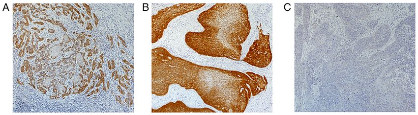
Figure 1. Immunohistochemical staining for p16 protein in oral squamous cell carcinoma tissues. (A) Positive expression in an HPV‑16E6 mRNA negative
case. (B) HPV‑16E6 mRNA positive case showing strong and diffuse staining in the nucleus and cytoplasm of the tumor cells. (C) A representative image
showing negative expression. Magnification, x40
to indicate a statistically significant difference. Statistical of HPV‑16E6 DNA. HPV‑16E6 DNA was ExoV‑resistant,
analyses were performed using GraphPad Prism software suggesting that the circular episome was observed only in the
(v5.04; GraphPad Software, Inc.). HPV‑16E6 DNA and mRNA positive case (Case 1; Fig. S2B).
Since neither HPV‑16E6 DNA nor RNA was detected in 7 of
Results the 10 cases of p16 overexpression (Case 2, 4, 5, 6, 7, 8 and 9),
the false‑positive rate of HPV‑16 infection was 70%. Finally,
Characteristics of the patients. The 100 OSCC cases included only the HPV‑16E6 DNA‑ and mRNA‑positive case (Case 1)
54 men and 46 women, ranging in age from 39 to 93 years was histopathologically determined as BSCC, which is a
(median, 70.3 years). The primary tumor was located in the rare variant of OSCC. The other cases (Case 3 and 10) were
tongue (n=36), mandibular gingiva (n=31), maxillary gingiva determined to be keratinized squamous cell carcinomas.
(n=13), floor of the mouth (n=9), buccal mucosa (n=9), or lower
lip (n=2). All the patients were histopathologically diagnosed Discussion
with squamous cell carcinoma, including BSCC (n=1).
HPV has been recognized as a possible pathogen of oral
Immunohistochemistry staining of p16 protein. Positive cancer (27‑29). HPV has been associated with cancer of the
expression of p16 protein was observed in 10 out of 100 OSCC uterus and cervix and was found in most cases (6‑8). However,
cases (10%). Most p16‑positive cases showed strong and diffuse its role in oral carcinogenesis is still unknown (30‑33). In
staining in the nucleus and the cytoplasm of the tumor cells oropharyngeal HNSCC, HPV‑positive tumors may have
(Fig. 1A and B). The association between p16 immunohisto‑ different clinical and biological functions, with improved
chemistry expression in the tumors from 100 patients with overall survival time and favorable prognosis (34). It has also
OSCC and their clinicopathological parameters was investi‑ been associated with therapeutic response in patients with
gated; however, the differences were not significant (Table I). HNSCC of the oropharynx (35). Another study showed that
Furthermore, following the analysis between p16 expression cases positive for HPV‑16 had lower recurrence rates compared
and survival using the Kaplan‑Meier method, no significant with that for their negative counterparts, indicating an asso‑
association between overall and disease‑free survival times ciation between HPV‑16 infection and a good prognosis in
was found (Fig. S1). OSCC (36). In contrast, HPV‑16 infection reportedly enhanced
the risk of distant metastasis and poor survival in patients with
Prevalence of HPV‑16 in all patients with OSCC. To clarify advanced OSCC (37). In the present study, the prevalence of
HPV‑16 infection, the expression level of HPV‑16E6 mRNA in HPV‑16 in patients with OSCC was only 1%. Therefore, the
100 OSCC cases was determined using RT‑qPCR. HPV‑16E6 association between HPV infection and prognosis could not
mRNA expression was only detected in one case (1%), which be evaluated.
was also positive for p16 expression. PCR products of HPV‑16E6 Several techniques have been used to detect HPV. Examples
and HMBS were visualized using agarose gel electrophoresis include PCR, an HPV genotyping test, morphology, in situ
(Fig. 2). HPV‑18E6 mRNA expression was investigated in hybridization and p16 immunohistochemistry. HPV detection
p16 positive OSCC tissues using RT‑qPCR; however, it was methods, such as morphology, in situ hybridization and p16
not detected (data not shown). Furthermore, the possibility immunohistochemistry lack sensitivity and specificity, as well
of persistent or silent infection with HPV‑16 was investi‑ as the ability to detect high‑risk HPV types. Therefore, PCR
gated using genomic DNA derived from p16 positive OSCC was considered the most sensitive method (38,39). The preva‑
cases using qPCR. HPV‑16E6 DNA was detected in 3 out of lence of HPV infection varied from 0% (19,40) to 100% (41),
10 cases (Cases 1, 3 and 10; Fig. S2A). One of these cases even in oral cavity cases. In the present study, p16 protein
expressed HPV‑16E6 mRNA (Case 1), but the other cases did expression, which was used as a surrogate marker of HPV
not (Case 3 and 10). Subsequently, genomic DNA was digested infection, was detected in 10% of cases, and the rate of HPV‑16
by ExoV, which preserved nicked and supercoiled DNAs but infection with E6 expression was only 1% in 100 OSCC cases.
degraded linear DNAs, followed by qPCR for the detection The one case of HPV‑16E6 mRNA positive expression also4 TOKUZEN et al: HPV16 AND p16 IN OSCC
Table I. Association between p16 status and the characteristics carcinogenic HPV infection, it was not applicable for tongue
of patients with oral squamous cell carcinoma. cancer (19). Furthermore, another study indicated that p16
expression was not a suitable surrogate marker of HPV infec‑
p16‑positive p16‑negative tion in oral lesions and HPV‑16 infection was associated with
Characteristic (n=10) (n=90) P‑value BSCC (42). In fact, only one HPV‑16E6 mRNA‑positive case
was histopathologically determined to be BSCC in the present
Median age, years 71 70 0.383 study. However, both HPV‑16E6 DNA positive cases, without
Sex E6 expression, were keratinized squamous cell carcinomas.
Male 7 47 0.335 Most cervical and oropharyngeal cancers show high expres‑
Female 3 43 sion of E6 and E7; however, OSCC has a lower positive rate of
Primary site 0.185 E6 and E7 mRNA expression compared with that for HPV
Tongue 1 35 DNA positive rate (43). We have hypothesized that there are
Maxillary gingiva 2 11 two types of HPV DNA positive OSCCs. One is HPV‑related
Mandibular gingiva 3 28 OSCC, which is a silent infection with no expression of E6
Floor of mouth 2 7 and E7, but is caused by the integration of HPV DNA into
the genome. The other is non‑keratinized OSCC with E6 and
Buccal mucosa 1 8
E7 expression, in which HPV DNA is actively infected as a
Lip 1 1
nuclear episome and/or genome integration.
Histological grading 0.465 In summary, the results from the present study indicated
G1 7 56 that there were few OSCC cases due to HPV‑16 infection. The
G2 3 22 expression of p16 protein was not an appropriate surrogate
G3 0 12 marker for HPV‑16 infection in OSCC. In addition, HPV‑16
T‑status 0.077 DNA may also be detected in p16 negative OSCC cases. As the
½ 4 63 number of HPV‑16 DNA positive cases was extremely low in
¾ 6 27 the present study, further investigation is required to examine
N‑status 0.515 the presence of episomal and integrated HPV DNA and the
0 5 55 expression of E6 and E7 mRNA, regardless of p16 expression,
1‑3 5 35 using the large number of fresh frozen OSCC tissues.
Clinical stage 0.504
Acknowledgements
I/II 3 42
III/IV 7 48 The authors would like to thank Ms Yumiko Fukuda
Recurrence/metastasis >0.999 (Department of Oral and Maxillofacial Surgery, Ehime
No 6 56 University Graduate School of Medicine, Ehime, Japan) for
Yes 4 34 providing technical assistance.
Funding
This study was supported by a Grant‑in‑Aid for Scientific
Research (B) from the Japan Society for the Promotion of
Science (grant no. 16H05543).
Availability of data and materials
Figure 2. Expression of HPV‑16E6 mRNA in p16 positive OSCC cases. The
expression level of HPV‑16E6 mRNA was analyzed using reverse transcription-
quantitative PCR. Only 1 case was found to be positive. HMBS was used as a
The datasets used and/or analyzed during the current study are
loading control. PC, positive control; HMBS, hydroxymethylbilane synthase; available from the corresponding author on reasonable request.
OSCC, oral squamous cell carcinoma; HPV, human papillomavirus.
Authors' contributions
had expression of the p16 protein. To investigate the association NT and KN confirm the authenticity of all the raw data. KN,
between HPV‑16 infection and p16 expression, the presence of NK and DU advised and supervised the study. KN designed
HPV‑16E6 DNA in genomic DNA samples, which were also the experiments. NT and ST performed the experiments. NT,
p16 positive was determined and HPV‑16E6 DNA was found HG, NK and DU analyzed the data. NT and KN wrote the
not only in the E6 mRNA positive case, but also in additional manuscript. All authors read and approved the final version of
2 cases without E6 expression. The expression of p16 was also manuscript.
70% HPV‑16 false‑positive, indicating the low reliability of the
surrogate marker of HPV infection in OSCC. A previous study Ethics approval and consent to participate
showed 100% HPV false‑positive results in mobile tongue
cancer (19). It suggested that although p16 protein expression The present study was approved by the Institute Research
was a biomarker for cervical or tonsillar cancer arising from Ethics Committee of the Ehime University Hospital (approvalONCOLOGY LETTERS 22: 528, 2021 5
number, 1607005) and written informed consent was provided 20. Shima K, Kobayashi I, Saito I, Kiyoshima T, Matsuo K, Ozeki S,
Ohishi M and Sakai H: Incidence of human papillomavirus 16 and
by all the patients. 18 infection and p53 mutation in patients with oral squamous cell
carcinoma in Japan. Br J Oral Maxillofac Surg 38: 445‑450, 2000.
Patient consent for publication 21. Sugiyama M, Bhawal UK, Kawamura M, Ishioka Y, Shigeishi H,
Higashikawa K and Kamata N: Human papillomavirus‑16 in
oral squamous cell carcinoma: Clinical correlates and 5‑year
Not applicable. survival. Br J Oral Maxillofac Surg 45: 116‑122, 2007.
22. Rushatamukayanunt P, Morita K, Matsukawa S, Harada H,
Shimamoto H, Tomioka H and Omura K: Lack of association
Competing interests between high‑risk human papillomaviruses and oral squamous
cell carcinoma in young japanese patients. Asian Pac J Cancer
The authors declare that they have no competing interests. Prev 15: 4135‑4141, 2014.
23. Ono K, Sugahara K, Nomura T, Takano N, Shibahara T and
Katakura A: Multiple HPV subtypes infection in Japanese
References oral Squamous cell carcinoma. J Oral Maxillofac Surg Med
Pathol 26: 128‑132, 2014.
1. Ferlay J, Shin HR, Bray F, Forman D, Mathers C and Parkin DM: 24. International Union Against Cancer (UICC): TNM classification
Estimates of worldwide burden of cancer in 2008: GLOBOCAN of malignant tumours. 7th edition. Sobin LH, Gospodarowicz MK
2008. Int J Cancer 127: 2893‑2917, 2010. and Wittekind C (eds). Willey‑Blackwell, Hoboken, pp25‑29,
2. Gupta S, Kong W, Peng Y, Miao Q and Mackillop WJ: 2009.
Temporal trends in the incidence and survival of cancers of the 25. Yamakawa‑Kakuta Y, Kawanata H, Doi Y, Fujimori T and
upper aerodigestive tract in Ontario and the United States. Int Imai Y: Does the expression of HPV16/18 E6/E7 in head and
J Cancer 125: 2159‑2165, 2009. neck squamous cell carcinomas relate to their clinicopathological
3. Blot WJ, McLaughlin JK, Winn DM, Austin DF, Greenberg RS, characteristics? Int J Oncol 35: 983‑988, 2009.
Preston‑Martin S, Bernstein L, Schoenberg JB, Stemhagen A 26. Myers JE, Guidry JT, Scott ML, Zwolinska K, Raikhy G,
and Fraumeni JF Jr: Smoking and drinking in relation to oral and Prasai K, Bienkowska‑Haba M, Bodily JM, Sapp MJ and
pharyngeal cancer. Cancer Res 48: 3282‑3287, 1988. Scott RS: Detecting episomal or integrated human papillo‑
4. Leemans CR, Braakhuis BJ and Brakenhoff RH: The molecular mavirus 16 DNA using an exonuclease V‑qPCR‑based assay.
biology of head and neck cancer. Nat Rev Cancer 11: 9‑22, 2011. Virology 537: 149‑156, 2019.
5. Dell G and Gaston K: Human papillomaviruses and their role in 27. Anaya‑Saavedra G, Ramírez‑Amador V, Irigoyen‑Camacho ME,
cervical cancer. Cell Mol Life Sci 58: 1923‑1942, 2001. García‑Cuellar CM, Guido‑Jiménez M, Méndez‑Martínez R and
6. zur Hausen H: Papillomaviruses and cancer: From basic studies García‑Carrancá A: High association of human papillomavirus
to clinical application. Nat Rev Cancer 2: 342‑350, 2002. infection with oral cancer: A case‑control study. Arch Med
7. Clifford GM, Smith JS, Plummer M, Muñoz N and Franceschi S: Res 39: 189‑197, 2008.
Human papillomavirus types in invasive cervical cancer world‑ 28. Iamaroon A, Pattanaporn K, Pongsiriwet S, Wanachantararak S,
wide: A meta‑analysis. Br J Cancer 88: 63‑73, 2003. Prapayasatok S, Jittidecharaks S, Chitapanarux I and
8. Bosch FX and de Sanjosé S: The epidemiology of human papilloma‑ Lorvidhaya V: Analysis of 587 cases of oral squamous cell carci‑
virus infection and cervical cancer. Dis Markers 23: 213‑227, 2007. noma in northern Thailand with a focus on young people. Int
9. Münger K, Baldwin A, Edwards KM, Hayakawa H, Nguyen CL, J Oral Maxillofac Surg 33: 84‑88, 2004.
Owens M, Grace M and Huh K: Mechanisms of human papil‑ 29. Lee SY, Cho NH, Choi EC, Baek SJ, Kim WS, Shin DH and
lomavirus‑induced oncogenesis. J Virol 78: 11451‑11460, 2004. Kim SH: Relevance of human papilloma virus (HPV) infection
10. Narisawa‑Saito M and Kiyono T: Basic mechanisms of high‑risk to carcinogenesis of oral tongue cancer. Int J Oral Maxillofac
human papillomavirus‑induced carcinogenesis: Roles of E6 and Surg 39: 678‑683, 2010.
E7 proteins. Cancer Sci 98: 1505‑1511, 2007. 30. Doorbar J: The papillomavirus life cycle. J Clin Virol 32 Suppl 1:
11. McLaughlin‑Drubin ME and Münger K: The human papilloma‑ S7‑S15, 2005.
virus E7 oncoprotein. Virology 384: 335‑344, 2009. 31. Ibieta BR, Lizano M, Fras‑Mendivil M, Barrera JL, Carrillo A,
12. El‑Naggar Ak and Westra WH: p16 expression as a surrogate marker Ma Ruz‑Godoy L and Mohar A: Human papilloma virus in oral
for HPV‑related oropharyngeal carcinoma: A guide for interpreta‑ squamous cell carcinoma in a Mexican population. Oral Surg
tive relevance and consistency. Head Neck 34: 459‑461, 2012. Oral Med Oral Pathol Oral Radiol Endod 99: 311‑315, 2005.
13. Deng Z, Hasegawa M, Yamashita Y, Matayoshi S, Kiyuna A, 32. Llamas‑Martínez S, Esparza‑Gómez G, Campo‑Trapero J,
Agena S, Uehara T, Maeda H and Suzuki M: Prognostic value Cancela‑Rodríguez P, Bascones‑Martínez A, Moreno‑López LA,
of human papillomavirus and squamous cell carcinoma antigen García‑Núñez JA and Cerero‑Lapiedra R: Genotypic determination
in head and neck squamous cell carcinoma. Cancer Sci 103: by PCR‑RFLP of human papillomavirus in normal oral mucosa,
2127‑2134, 2012. oral leukoplakia and oral squamous cell carcinoma samples in
14. Termine N, Panzarella V, Falaschini S, Russo A, Matranga D, Madrid (Spain). Anticancer Res 28 (6A): 3733‑3741, 2008.
Lo Muzio L and Campisi G: HPV in oral squamous cell carci‑ 33. Uobe K, Masuno K, Fang YR, Li LJ, Wen YM, Ueda Y and
noma vs. head and neck squamous cell carcinoma biopsies: A Tanaka A: Detection of HPV in Japanese and Chinese oral carci‑
meta‑analysis (1988‑2007). Ann Oncol 19: 1681‑1690, 2008. nomas by in situ PCR. Oral Oncol 37: 146‑152, 2001.
15. Sritippho T, Chotjumlong P and Iamaroon A: Roles of Human 34. Marur S, D'Souza G, Westra WH and Forastiere AA:
Papillomaviruses and p16 in Oral Cancer. Asian Pac J Cancer HPV‑associated head and neck cancer: A virus‑related cancer
Prev 16: 6193‑6200, 2015. epidemic. Lancet Oncol 11: 781‑789, 2010.
16. Hobbs CG, Sterne JA, Bailey M, Heyderman RS, Birchall MA and 35. Fakhry C, Westra WH, Li S, Cmelak A, Ridge JA, Pinto H,
Thomas SJ: Human papillomavirus and head and neck cancer: Forastiere A and Gillison ML: Improved survival of patients
A systematic review and meta‑analysis. Clin Otolaryngol 31: with human papillomavirus‑positive head and neck squamous
259‑266, 2006. cell carcinoma in a prospective clinical trial. J Natl Cancer
17. Castillo A, Koriyama C, Higashi M, Anwar M, Bukhari MH, Inst 100: 261‑269, 2008.
Carrascal E, Mancilla L, Okumura H, Matsumoto M, 36. Elango KJ, Suresh A, Erode EM, Subhadradevi L, Ravindran HK,
Sugihara K, et al: Human papillomavirus in upper digestive Iyer SK, Iyer SK and Kuriakose MA: Role of human papil‑
tract tumors from three countries. World J Gastroenterol 17: loma virus in oral tongue squamous cell carcinoma. Asian Pac
5295‑5304, 2011. J Cancer Prev 12: 889‑896, 2011.
18. Krüger M, Pabst AM, Walter C, Sagheb K, Günther C, Blatt S, 37. Lee LA, Huang CG, Liao CT, Lee LY, Hsueh C, Chen TC, Lin CY,
Weise K, Al‑Nawas B and Ziebart T: The prevalence of human Fan KH, Wang HM, Huang SF, et al: Human papillomavirus‑16
papilloma virus (HPV) infections in oral squamous cell carci‑ infection in advanced oral cavity cancer patients is related to
nomas: A retrospective analysis of 88 patients and literature an increased risk of distant metastases and poor survival. PLoS
overview. J Craniomaxillofac Surg 42: 1506‑1514, 2014. One 7: e40767, 2012.
19. Kabeya M, Furuta R, Kawabata K, Takahashi S and Ishikawa Y: 38. Schlecht NF, Brandwein‑Gensler M, Nuovo GJ, Li M, Dunne A,
Prevalence of human papillomavirus in mobile tongue cancer Kawachi N, Smith RV, Burk RD and Prystowsky MB: A compar‑
with particular reference to young patients. Cancer Sci 103: ison of clinically utilized human papillomavirus detection methods
161‑168, 2012. in head and neck cancer. Mod Pathol 24: 1295‑1305, 2011.6 TOKUZEN et al: HPV16 AND p16 IN OSCC
39. Pannone G, Rodolico V, Santoro A, Lo Muzio L, Franco R, 42. Friedrich RE, Sperber C, Jäkel T, Röser K and Löning T: Basaloid
Botti G, Aquino G, Pedicillo MC, Cagiano S, Campisi G, et al: lesions of oral squamous epithelial cells and their association
Evaluation of a combined triple method to detect causative with HPV infection and P16 expression. Anticancer Res 30:
HPV in oral and oropharyngeal squamous cell carcinomas: p16 1605‑1612, 2010.
immunohistochemistry, consensus PCR HPV‑DNA, and in situ 43. Ndiaye C, Mena M, Alemany L, Arbyn M, Castellsagué X,
hybridization. Infect Agent Cancer 7: 4, 2012. Laporte L, Bosch FX, de Sanjose S and Trottier H: HPV
40. de Spíndula‑Filho JV, da Cruz AD, Oton‑Leite AF, Batista AC, DNA, E6/E7 mRNA, and p16INK4a detection in head and
Leles CR, de Cássia Gonçalves Alencar R, Saddi VA and neck cancers: A systematic review and meta‑analysis. Lancet
Mendonça EF: Oral squamous cell carcinoma versus oral Oncol 15: 1319‑1331, 2014.
verrucous carcinoma: An approach to cellular proliferation
and negative relation to human papillomavirus (HPV). Tumor This work is licensed under a Creative Commons
Biol 32: 409‑416, 2011. Attribution-NonCommercial-NoDerivatives 4.0
41. Koyama K, Uobe K and Tanaka A: Highly sensitivity detection International (CC BY-NC-ND 4.0) License.
of HPV‑DNA in paraffin sections of human oral carcinomas.
J Oral Pathol Med 36: 18‑24, 2007.You can also read