Endometrioid Cancer Associated With Endometriosis: From the Seed and Soil Theory to Clinical Practice - Frontiers
←
→
Page content transcription
If your browser does not render page correctly, please read the page content below
ORIGINAL RESEARCH
                                                                                                                                                published: 10 March 2022
                                                                                                                                          doi: 10.3389/fonc.2022.859510

                                              Endometrioid Cancer Associated
                                              With Endometriosis: From the Seed
                                              and Soil Theory to Clinical Practice
                                              Alberto Farolfi 1*, Amelia Altavilla 1, Luca Morandi 2,3, Laura Capelli 4, Elisa Chiadini 4,
                                              Giovanna Prisinzano 4, Giorgia Gurioli 4, Marianna Molari 1, Daniele Calistri 4,
                           Edited by:         Maria Pia Foschini 5 and Ugo De Giorgi 1
                   Claudia Marchetti,
 Agostino Gemelli University Polyclinic
                                              1 Department of Medical Oncology, IRCCS Istituto Romagnolo per lo Studio dei Tumori (IRST) “Dino Amadori”,
                        (IRCCS), Italy        Meldola, Italy, 2 Department of Biomedical and Neuromotor Sciences, University of Bologna, Bologna, Italy, 3 Functional and
                                              Molecular Neuroimaging Unit, IRCCS Istituto delle Scienze Neurologiche di Bologna, Bologna, Italy, 4 Biosciences Laboratory,
                      Reviewed by:
                                              IRCCS Istituto Romagnolo per lo Studio dei Tumori (IRST) “Dino Amadori”, Meldola, Italy, 5 Unit of Anatomic Pathology,
                   Lucia Musacchio,
                                              Department of Biomedical and Neuromotor Sciences, Bellaria Hospital, University of Bologna, Bologna, Italy
 Agostino Gemelli University Polyclinic
                        (IRCCS), Italy
                     Angela Santoro,          Endometriosis is a benign condition characterized by the presence of ectopic endometrial
 Agostino Gemelli University Polyclinic
                        (IRCCS), Italy
                                              tissue. It is still debated whether endometriosis is a disease that can predispose to the
                  *Correspondence:
                                              pathogenesis of endometrial cancer outside the uterus. Deficiencies in mismatch repair
                         Alberto Farolfi       (MMR) genes are a known risk factor for developing endometrioid cancer. Starting from
              alberto.farolfi@irst.emr.it
                                              two cases of patients with abnormal MMR endometrioid carcinoma of the uterus and
                   Specialty section:
                                              synchronous endometrioid carcinoma in non-ovarian and ovarian endometriosis, we
         This article was submitted to        performed a somatic mutation profile and phylogenetic analysis of the lesions in order
             Gynecological Oncology,          to identify if they were metastasis or primary de novo tumors. In the first case, we identified
               a section of the journal
                 Frontiers in Oncology        de novo activating mutations in PIK3CA and KRAS in endometrioid cancer lesions but not
         Received: 21 January 2022            in endometriosis. Although the acquisition of a de novo mutation in ESR1 and a decrease
        Accepted: 14 February 2022            in mutant allele fraction (MAF) from the endometrial tumor to the localizations in the
         Published: 10 March 2022
                                              endometriosis lesions, the clonal relationship was confirmed by the limited number of
                              Citation:
      Farolfi A, Altavilla A, Morandi L,
                                              heteroplasmic mutations in D-loop mitochondrial DNA region. In the other case, the clonal
   Capelli L, Chiadini E, Prisinzano G,       behavior was demonstrated by the overlap of MAF at each site. Our data support the
       Gurioli G, Molari M, Calistri D,       hypothesis of a retrograde dissemination of tumor cells, moving from the primary
 Foschini MP and De Giorgi U (2022)
Endometrioid Cancer Associated With           carcinoma in the endometrium to ectopic sites of endometriosis where localizations of
  Endometriosis: From the Seed and            tumor arise.
      Soil Theory to Clinical Practice.
             Front. Oncol. 12:859510.         Keywords: uterine carcinoma, endometriosis, endometrioid adenocarcinoma of the endometrium, mismatch repair
     doi: 10.3389/fonc.2022.859510            (MMR) deficiency, tumor dissemination

Frontiers in Oncology | www.frontiersin.org                                          1                                         March 2022 | Volume 12 | Article 859510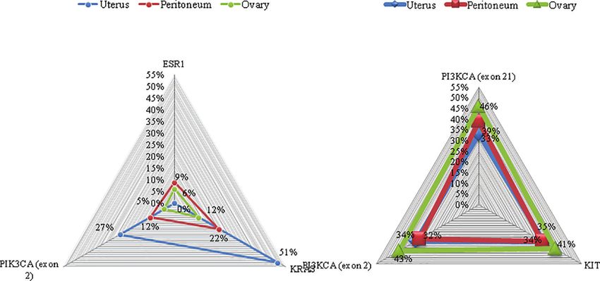
Farolfi et al.                                                                                     Endometrioid Tumor Associated With Endometriosis

INTRODUCTION                                                               MMR (MLH1, MSH2, and MSH6) genes. Moreover, all coding
                                                                           exons and splice junctions of the MLH1, MSH2, and MSH6 genes
Endometriosis is a relatively common disease, characterized by             were analyzed. The analysis for the identification of variants and
the presence of ectopic endometrial tissue outside the uterus (1).         genomic rearrangements [copy number variations (CNVs)] was
Endometriosis affects 10%–15% of all women of reproductive age             performed by next-generation sequencing (NGS) with Miseq-
(2) and approximately 2%–5% of postmenopausal women,                       Illumina sequencer via commercial panel HCS_v1_1
representing a side effect of hormonal replacement therapy or              (Sophia Genetics).
tamoxifen treatment in this population (3). The etiology
underlying endometriosis is controversial, but the processes
proposed in its development closely resemble those involved in             Nucleic Acid Extraction and Quantification
cancer metastasis (4). Ovarian endometriosis has been reported             Dual DNA and RNA isolation was performed from four
to be associated with an increased risk of epithelial ovarian              formalin-fixed paraffin-embedded (FFPE) tissues using the
cancer, representing the direct precursor of clear-cell and                Maxwell® RSC DNA/RNA FFPE Kit with the Maxwell® RSC
endometrioid ovarian carcinomas (5). However, whether                      Instrument. For each sample, areas were characterized by 100%
extraovarian endometriosis may be an endometrioid cancer                   tumor cells. Nucleic acid concentrations were determined by
precursor remains controversial (6, 7).                                    fluorometric quantitation using Qubit 4.0 Fluorimeter with
    Endometrial cancer is a clinically heterogeneous disease.              Qubit dsDNA HS Assay Kit and Qubit RNA HS Assay Kit
Genomic characterization by The Cancer Genome Atlas                        (Thermo Fisher Scientific, Inc.).
(TCGA) classified endometrial cancers into four categories:
POLE ultramutated, microsatellite instability hypermutated,
                                                                           Next-Generation Sequencing
copy-number low, and copy-number high (8). Another
                                                                           To estimate somatic mutation profiling, NGS was performed
classification of endometrial cancer differentiates tumors into
                                                                           with the “Ion Torrent Oncomine Focus Assay” for simultaneous
two subtypes: type I is characterized by a favorable prognosis and         and rapid identification of single-nucleotide variants (SNVs),
represented mostly by endometrioid adenocarcinoma, associated
                                                                           short insertion and deletions (indels; 35 genes), CNVs (19 genes),
with an unopposed estrogen stimulation, often preceded by
                                                                           and gene rearrangements (23 genes) in 52 cancer genes with
endometrial hyperplasia; type II has significantly poorer 5-year
                                                                           therapeutic relevance:
survival predominantly represented by non-endometrioid
                                                                               Hotspot genes (35): AKT1, ALK, AR, BRAF, CDK4,
histology, mostly arising in an atrophic endometrium and
                                                                           CTNNB1, DDR2, EGFR, ERBB2, ERBB3, ERBB4, ESR1,
deriving from intraepithelial carcinoma as a precancerous                  FGFR2, FGFR3, GNA11, GNAQ, HRAS, IDH1, IDH2, JAK1,
lesion (9). Since the evidence of activity of immune checkpoint            JAK2, JAK3, KIT, KRAS, MAP2K1, MAP2K2, MET, MTOR,
inhibitors in patients with advanced mismatch repair (MMR)-
                                                                           NRAS, PDGFRA, PIK3CA, RAF1, RET, ROS1, and SMO.
deficient endometrial cancer (10) and considering that Lynch
                                                                               CNV genes (19): ALK, AR, BRAF, CCND1, CDK4, CDK6,
syndrome may account for about 3% of all endometrial cancers
                                                                           EGFR, ERBB2, FGFR1, FGFR2, FGFR3, FGFR4, KIT, KRAS,
(11), it is recommended to screen all endometrial cancer patients
                                                                           MET, MYC, MYCN, PDGFRA, and PIK3CA.
with the use of immunohistochemical tests for MLH1, MSH2,
                                                                               Fusion driver genes (23): ABL1, ALK, AKT3, AXL, BRAF,
MSH6, and PMS2 (12).                                                       EGFR, ERBB2, ERG, ETV1, ETV4, ETV5, FGFR1, FGFR2,
    Whether patients with endometriosis have increased risks of
                                                                           FGFR3, MET, NTRK1, NTRK2, NTRK3, PDGFRA, PPARG,
development of endometrioid tumors or if endometriosis is the soil
                                                                           RAF1, RET, and ROS1.
                                                                               The Invitrogen SuperScript™ VILO™ cDNA Synthesis Kit
where endometrioid cancer seeds grow remains unclear. In this study,
we report two cases of patients with MMR-deficient endometrioid
                                                                           (Thermo Fisher Scientific) was used for RNA reverse
carcinoma of the uterus and synchronous endometrioid carcinoma in
                                                                           transcription to cDNA before library preparation.
extraovarian and ovarian endometriosis.                                        Libraries were prepared from 10 ng DNA and 10 ng RNA
                                                                           (0.67 ng/µl, 15 µl) with “Oncomine Focus Assay, Chef-Ready
                                                                           Library” reagents on the Ion Chef ™ System (Thermo
METHODS                                                                    Fisher Scientific).
                                                                               Templating and sequencing were performed using “Ion 510™
Tissue Sample and Mismatch Repair                                          & Ion 520™ & Ion 530™ Kit–Chef.”
Evaluation                                                                     For template preparation, we used Ion 520 chip (up to 5
Tissue samples were paraffin-embedded archival specimens.                   million reads per chip, 8 samples) on the Ion Chef™ System,
Immunohistochemistry was performed at the Pathology Unit.                  while sequencing was completed on the Ion Torrent S5 Plus
Antibodies against MLH1, MSH2, MSH6, and PMS2 were                         (Thermo Fisher Scientific).
prediluted according to the manufacturer’s instructions. The                   Sequencing data were analyzed using Ion Reporter ™
expression of MLH1, MSH2, MSH6, and PMS2 was                               Software that helps to identify and prioritize variants.
determined qualitatively to be retained or lost, as is standard.               To define a reliable variant calling, we have considered two
   Molecular analyses on genomic DNA extracted from                        stringent parameters: coverage depth greater than 500× and
peripheral blood were performed to search for variants in                  allele frequency greater than 5%.

Frontiers in Oncology | www.frontiersin.org                            2                                  March 2022 | Volume 12 | Article 859510
Farolfi et al. Endometrioid Tumor Associated With Endometriosis
Phylogenetic Trees lesion was diagnosed [stage pT3aN0M1, Fé dé ration
Phylogenetic analysis starting from different tumor populations Internationale de Gyné cologie et d’Obsté trique (FIGO IVB)].
in the Pathology Unit of Bellaria Hospital (Bologna) was Using immunohistochemistry, tumor cells were shown to have a
performed as previously described (13, 14). In brief, normal pattern of expression of p53, MSH2, and MSH6, whereas
mitochondrial DNA (mtDNA) D-loop region was sequenced MLH1 and PMS2 were lost. For this reason, molecular analysis
in deep onto MiSEQ (Illumina) and processed by Geneious 9.1.8 was performed to search for variants in MMR genes on genomic
(Biomatters Ltd., Auckland, New Zealand) to identify and DNA extracted from peripheral blood. The regions of MLH1,
annotate homoplasmic or heteroplasmic mutations. The 4 MSH2, and MSH6 genes analyzed did not show specific alterations
consensus sequences representative of each of the four tumor nor partial or complete genomic rearrangements (CNVs).
populations were joined and used to construct the phylogenetic Five cycles of chemotherapy with carboplatin and paclitaxel
tree using Multiple Alignment using Fast Fourier Transform were administered. The sixth cycle was omitted for hematologic
(MAFFT; https://mafft.cbrc.jp/alignment/server/) with the toxicity (grade 3 anemia and grade 3 thrombocytopenia). Patient
unweighted pair group method with arithmetic mean and the then received adjuvant external pelvic radiotherapy at a dose of
Jukes-Cantor substitution model. 45 Gray. Until now, patient is disease-free.
Molecular Profiling
RESULTS In order to identify if the endometrioid carcinoma lesions in the
peritoneum and in the ovary were synchronous tumors that
Case Report 1 arose in extraovarian and ovarian endometriosis tissue or
A 58-year-old woman was diagnosed with a grade 2 endometrioid metastasis, a molecular profiling was performed. Targeted
carcinoma in August 2020. After having ruled out other neoplastic sequencing in primary uterine tumor revealed an activating
lesions with thorax and abdomen computed tomography (CT) PIK3CA hotspot exon 2 mutation (c.263G>A) with an allelic
scan and pelvic MRI, the patient underwent a total hysterectomy ratio of 27% and a KRAS exon 2 hotspot mutation (c.34G>A)
with bilateral annexectomy and pelvic lymphoadenectomy. with an allelic ratio of 51%. Comparable mutations were found in
During surgery, two peritoneal lesions in the rectouterine pouch the tumor lesion of the left paracolpium: PIK3CA exon 2
were also removed. The diagnosis was of endometrioid carcinoma mutation (c.263G>A) and a KRAS exon 2 mutation (c.34G>A)
of the uterus grade 3, with more than 50% invasion of the with an allelic ratio of 12% and 22%, respectively. An activating
myometrial wall thickness, 5.2 cm in greatest dimension, with ESR1 hotspot exon 8 mutation (c.1607T>G) with an allelic ratio
infiltration of the cervix stroma, without lymphovascular invasion, of 9% was also identified. The same mutations were detected in
and no pelvic lymph node metastases. The two peritoneal nodules the tumor lesion of the left ovary with less allelic ratio: PIK3CA
were diagnosed as duplex localization of endometrioid carcinoma exon 2 mutation (c.263G>A), KRAS exon 2 mutation (c.34G>A),
grade 3 in endometriosis lesions. In the left ovary, a localization of and ESR1 hotspot exon 8 mutation (c.1607T>G) with an allelic
endometriosis with another endometrioid carcinoma grade 3 ratio of 5%, 12%, and 6%, respectively (Figure 1). No hotspot
A B
FIGURE 1 | Mutations on representative cancer-associated genes of case 1 (A) and case 2 (B). Each color represents a different sample. Triangles represent the
mutant allele frequency (MAF), corresponding to those of the vertical axis.
Frontiers in Oncology | www.frontiersin.org 3 March 2022 | Volume 12 | Article 859510Farolfi et al. Endometrioid Tumor Associated With Endometriosis
mutations were identified (with a sensitivity of detection of 1%) Molecular Profiling
in the endometriosis nodules without tumor lesions. Also in this patient, a molecular profiling was performed to
The phylogenetic relationship of multiple samples from identify if the endometrioid carcinoma lesions in the left ovary
different tumor regions was also evaluated by sequencing D- were synchronous tumors that arose in extraovarian and ovarian
loop mtDNA region. We found 6 heteroplasmic mutations, 4 of endometriosis tissue or metastasis.
which were present only in specimen A1, while 2 were present Targeted sequencing in primary uterine tumor revealed an
only in specimen A12. The phylogenetic tree is shown in activating PIK3CA hotspot exon 2 mutation (c.263G>A) with an
Figure 2. All samples were close to each other, indicating a allelic ratio of 34% and hotspot exon 21 mutation (c c.3129G>T)
possible clonal relationship among them. with an allelic ratio of 33%. Furthermore, a variant of uncertain
significance of cKIT exon 8 mutation (c.1264G>A) with an allelic
ratio of 35% was found. Comparable mutations were found in
Case Report 2 the peritoneum tumor localization: PIK3CA exon 2 mutation
In April 2021, a 60-year-old woman was diagnosed with a grade 2 (c.263G>A) and exon 21 mutation (c c.3129G>T) with an allelic
endometrioid carcinoma. Chest and abdominal contrast-enhanced ratio of 32% and 39%, respectively; cKIT exon 8 mutation
CT scan revealed no metastatic lesions. Patient underwent a (c.1264G>A) with an allelic ratio of 34%. Again, tumor lesion
total hysterectomy with bilateral annexectomy and low anterior of the left ovary presented PIK3CA exon 2 mutation (c.263G>A)
resection of rectum with colostomy. During surgery, a single and exon 21 mutation (c c.3129G>T) with an allelic ratio of 43%
peritoneal lesion in the rectouterine pouch was biopsied. The and 46%, respectively; cKIT exon 8 mutation (c.1264G>A) with
surgical specimen of the uterus showed a 50 × 35 mm polypoid an allelic ratio of 41%.
tumor. Histopathological diagnosis was grade 2 endometrial
carcinoma with more than 50% invasion of the myometrial wall
thickness, infiltration of perirectal fat, and lymphovascular
invasion. In the left ovary, a localization of endometriosis with
another endometrioid carcinoma grade 2 lesion was diagnosed. DISCUSSION
The peritoneal lesion was positive for endometrioid carcinoma
grade 2 (stage pT3bNXM1, FIGO IVB). Tumor cells were shown Although endometriosis is considered to be a benign lesion,
to have a normal pattern of expression of p53 and MLH1 and malignant transformation in endometriosis-related ovarian
PMS2 positives at immunohistochemistry. For this reason, neoplasms is possible (15). In recent years, different studies
molecular analysis was performed to search for variants in MMR demonstrated that endometriosis (ovarian and extraovarian
genes on genomic DNA extracted from peripheral blood. The endometriotic lesions) harbors somatic mutations in cancer
regions of MLH1 and MSH6 genes analyzed did not show any driver genes (6, 16). Since somatic mutations are a widespread
specific alteration nor partial or complete genomic rearrangements event in “normal” tissue (endometrial samples included) and most
(CNVs), while MLH2 gene showed a non-classified variant endometriotic lesions harboring cancer-associated genes do not
(c.728G>A;p). necessarily lead to malignant transformation (17), environmental
Six cycles of chemotherapy with carboplatin and paclitaxel features protective against malignant transformation or that buffer
were administered, and adjuvant external pelvic radiotherapy is the effects of such mutations may prevent the progression from
ongoing. Until now, patient is disease-free. endometriosis to gynecologic cancers (16). Moreover, it was
FIGURE 2 | Phylogenetic tree based on Multiple Alignment using Fast Fourier Transform (MAFFT) with the unweighted pair group method with arithmetic mean and
the Jukes–Cantor substitution model. All the samples of case 1 are very close to each other, as we found a limited number of mutations in heteroplasmy, 4 of which
in the sample of the peritoneum and 2 in endometriosis sample.
Frontiers in Oncology | www.frontiersin.org 4 March 2022 | Volume 12 | Article 859510Farolfi et al. Endometrioid Tumor Associated With Endometriosis
hypothesized that alterations in MMR genes might be involved in Both patients had MMR-deficient tumors. MMR alterations
the malignant transformation of endometriotic lesions (7). may result in genetic tumor heterogeneity (as seen in case 1) but
With these premises, when we faced two similar cases of could also be a risk factor for cancer development in
patients with endometrioid carcinoma of the uterus with MMR endometriotic heterotopic tissue (7, 19). However, our data
deficiency and synchronous endometrioid carcinoma in support the hypothesis of a retrograde dissemination of tumor
extraovarian and ovarian endometriosis, the question was cells, moving from the primary carcinoma in the endometrium
whether the latter was subsequent to malignant transformation to ectopic sites of endometriosis where localizations of tumor
of endometriotic lesions or to secondary localization. To solve may arise, reinforcing literature data on the origin of
this dilemma, we performed a molecular profile of the cancer and endometriosis possibly following retrograde menstruations (20).
endometriotic lesions of the two cases. In the first case, we A brief comment on ESR1 found in one of our cases is needed.
identified de novo activating PIK3CA and KRAS mutations in Our results suggest that the ESR1 mutation was a de novo
endometrioid cancer lesions but not in endometriosis. This was mutation that arose without the selective pressure of a
an unexpected result, since KRAS mutations were associated with hormonal treatment. This observation suggests that ESR1 may
endometriosis sustainability (6, 18). Moreover, case 1 showed a be a biomarker of progression rather than a predictor of endocrine
decrease in mutant allele fraction (MAF) and the acquisition of a therapy resistance (21) in endometrial cancer. Therefore,
de novo mutation (e.g., ESR1) from primary tumor to distant characterization only at the time of progression to hormone
lesions. For this reason, we performed a phylogenetic therapy may be inadequate (22) because ESR1-mutated tumor
relationship of multiple samples from different tumor regions cells may be the result of a clonal expansion of endometrioid
sequencing D-loop mtDNA region. According to this analysis, a cancer cells primarily refractory to hormonal treatment.
limited number of heteroplasmic mutations were found showing In conclusion, endometrioid tumors, especially if an alteration
a close phylogenetic distance among different tumor populations, in the MMR genes occurs, represent a heterogeneous disease, and
demonstrating that peritoneal and ovarian carcinomas are the acquisition of new mutations is an event that needs to be
derived from the same endometrial ancestor clone that considered. Although cancer-associated mutations are frequently
migrated and settled in endometriotic lesions where the observed in endometriosis, our study demonstrated that
endometrioid tumor arose (Figure 3). In case 2., the MAF was endometriosis-associated carcinoma may arise from cancer
almost the same in all the different lesions, supporting the idea of seeds that implant in this permissive soil moving in a
the clonal relationship between the different tumor localizations. retrograde way rather than a direct malignant transformation.
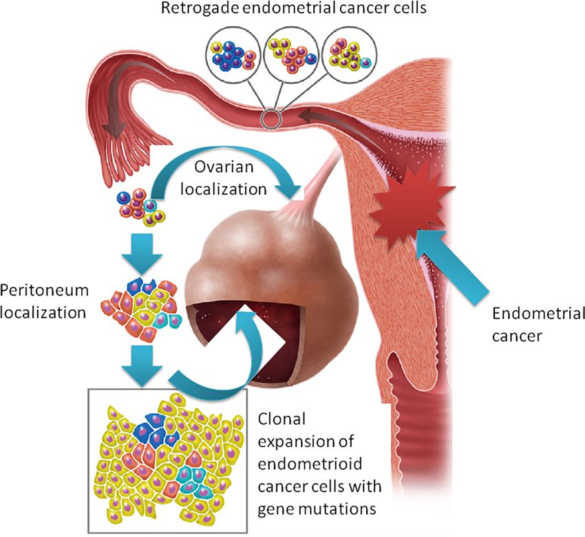
FIGURE 3 | The “seed and soil” hypothesis: retrograde flow of endometrial cells already harboring cancer-associated mutation moving from the primary carcinoma
in the endometrium to ectopic sites of endometriosis where tumor localizations arise.
Frontiers in Oncology | www.frontiersin.org 5 March 2022 | Volume 12 | Article 859510Farolfi et al. Endometrioid Tumor Associated With Endometriosis
How this disease has to be treated and considered (e.g., is really a institutional requirements. The patients/participants provided
stage IV endometrial cancer?) is still a matter of debate. their written informed consent to participate in this study.
DATA AVAILABILITY STATEMENT
The datasets presented in this study can be found in online AUTHOR CONTRIBUTIONS
repositories. The names of the repository/repositories and accession
number(s) can be found in the article/supplementary material. AF, LM, MPF, and UDG: conception and planning of the work.
AF, AA, LC, EC, GP, GG, MM, and DC: acquisition and analysis
of the data. AF, AA, LM, and UDG: interpretation of the data.
ETHICS STATEMENT AF, AA, LM, and GG: drafting and/or critical revision of the
Ethical review and approval was not required for the study on article for important intellectual content. All the authors read
human participants in accordance with the local legislation and and approved the final submitted version of the article.
Carcinoma. Oral Oncol (2017) 67:131–7. doi: 10.1016/j.oraloncology.
REFERENCES 2017.02.017
1. Giudice LC. Clinical Practice. Endometriosis N Engl J Med (2010) 362:2389– 15. Wei JJ, William J, Bulun S. Endometriosis and Ovarian Cancer: A Review of
98. doi: 10.1056/NEJMcp1000274 Clinical, Pathologic, and Molecular Aspects. Int J Gynecol Pathol (2011)
2. Giudice LC, Kao LC. Endometriosis. Lancet (2004) 364:1789–99. 30:553–68. doi: 10.1097/PGP.0b013e31821f4b85
doi: 10.1016/S0140-6736(04)17403-5. 16. Suda K, Nakaoka H, Yoshihara K, Ishiguro T, Tamura R, Mori Y, et al. Clonal
3. Parasar P, Ozcan P, Terry KL. Endometriosis: Epidemiology, Diagnosis and Expansion and Diversification of Cancer-Associated Mutations in
Clinical Management. Curr Obstet Gynecol Rep (2017) 6:34–41. doi: 10.3390/ Endometriosis and Normal Endometrium. Cell Rep (2018) 24:1777–89.
diagnostics10030134 doi: 10.1016/j.celrep.2018.07.037
4. Bulun SE. Endometriosis. N Engl J Med (2009) 360:268–79. doi: 10.1056/ 17. Guo SW. Cancer-Associated Mutations in Endometriosis: Shedding Light on
NEJMra0804690 the Pathogenesis and Pathophysiology. Hum Reprod Update (2020) 26:423–
5. Ogawa S, Kaku T, Amada S, Kobayashi H, Hirakawa T, Ariyoshi K, et al. 49. doi: 10.1093/humupd/dmz047
Ovarian Endometriosis Associated With Ovarian Carcinoma: A 18. Cheng CW, Licence D, Cook E, Luo F, Arends MJ, Smith SK, et al. Activation
Clinicopathological and Immunohistochemical Study. Gynecol Oncol (2000) of Mutated K-Ras in Donor Endometrial Epithelium and Stroma Promotes
77:298–304. doi: 10.1006/gyno.2000.5765 Lesion Growth in an Intact Immunocompetent Murine Model of
6. Anglesio MS, Papadopoulos N, Ayhan A, Nazeran TM, Noë M, Horlings HM, Endometriosis. J Pathol (2011) 224:261–9. doi: 10.1002/path.2852
et al. Cancer-Associated Mutations in Endometriosis Without Cancer. N Engl 19. Bennett JA, Pesci A, Morales-Oyarvide V, Da Silva A, Nardi V, Oliva E.
J Med (2017) 376:1835–48. doi: 10.1056/NEJMoa1614814 Incidence of Mismatch Repair Protein Deficiency and Associated
7. Fuseya C, Horiuchi A, Hayashi A, Suzuki A, Miyamoto T, Hayashi T, et al. Clinicopathologic Features in a Cohort of 104 Ovarian Endometrioid
Involvement of Pelvic Inflammation-Related Mismatch Repair Abnormalities Carcinomas. Am J Surg Pathol (2019) 43(2):235–43. doi: 10.1097/
and Microsatellite Instability in the Malignant Transformation of Ovarian PAS.0000000000001165
Endometriosis. Hum Pathol (2012) 43:1964–72. doi: 10.1016/ 20. Sampson JA. Metastatic or Embolic Endometriosis, Due to the Menstrual
j.humpath.2012.02.005 Dissemination of Endometrial Tissue Into the Venous Circulation. Am J
8. Cancer Genome Atlas Research Network, Kandoth C, Schultz N, Cherniack Pathol (1927) 3:93–110.43.
AD, Akbani R, Liu Y, et al. Integrated Genomic Characterization of 21. Robinson DR, Wu YM, Vats P, Su F, Lonigro RJ, Cao X, et al. Activating ESR1
Endometrial Carcinoma. Nature (2013) 497:67–73. doi: 10.1038/nature12113 Mutations in Hormone-Resistant Metastatic Breast Cancer. Nat Genet (2013)
9. Malik TY, Chishti U, Aziz AB, Sheikh I. Comparison of Risk Factors and 45(12):1446–51. doi: 10.1038/ng.2823
Survival of Type 1 and Type II Endometrial Cancers. Pak J Med Sci (2016) 22. Morel A, Masliah-Planchon J, Bataillon G, Becette V, Morel C, Antonio S,
32:886–90. doi: 10.12669/pjms.324.9265 et al. De Novo ESR1 Hotspot Mutation in a Patient With Endometrial Cancer
10. Oaknin A, Tinker AV, Gilbert L, Samouëlian V, Mathews C, Brown J, et al. Treated With an Aromatase Inhibitor. JCO Precis Oncol (2019) 3:
Clinical Activity and Safety of the Anti-Programmed Death 1 Monoclonal PO.18.00398. doi: 10.1200/PO.18.00398
Antibody Dostarlimab for Patients With Recurrent or Advanced Mismatch
Repair-Deficient Endometrial Cancer: A Nonrandomized Phase 1 Clinical Conflict of Interest: The authors declare that the research was conducted in the
Trial. JAMA Oncol (2020) 6:1766–72. doi: 10.1001/jamaoncol.2020.4515 absence of any commercial or financial relationships that could be construed as a
11. Ryan N, Morris J, Green K, Lalloo F, Woodward ER, Hill J, et al. Association potential conflict of interest.
of Mismatch Repair Mutation With Age at Cancer Onset in Lynch Syndrome:
Implications for Stratified Surveillance Strategies. JAMA Oncol (2017) 3:1702– Publisher’s Note: All claims expressed in this article are solely those of the authors
6. doi: 10.1001/jamaoncol.2017.0619 and do not necessarily represent those of their affiliated organizations, or those of
12. Murali R, Delair DF, Bean SM, Abu-Rustum NR, Soslow RA. Evolving Roles the publisher, the editors and the reviewers. Any product that may be evaluated in
of Histologic Evaluation and Molecular/Genomic Profiling in the this article, or claim that may be made by its manufacturer, is not guaranteed or
Management of Endometrial Cancer. J Natl Compr Canc Netw (2018) endorsed by the publisher.
16:201–9. doi: 10.6004/jnccn.2017.7066
13. Gabusi A, Gissi DB, Tarsitano A, Asioli S, Marchetti C, Montebugnoli L, et al. Copyright © 2022 Farolfi, Altavilla, Morandi, Capelli, Chiadini, Prisinzano, Gurioli,
Intratumoral Heterogeneity in Recurrent Metastatic Squamous Cell Molari, Calistri, Foschini and De Giorgi. This is an open-access article distributed
Carcinoma of the Oral Cavity: New Perspectives Afforded by Multiregion under the terms of the Creative Commons Attribution License (CC BY). The use,
DNA Sequencing and Mtdna Analysis. J Oral Maxillofac Surg (2019) 77:440– distribution or reproduction in other forums is permitted, provided the original
55. doi: 10.1016/j.joms.2018.09.014 author(s) and the copyright owner(s) are credited and that the original publication
14. Gissi DB, Tarsitano A, Leonardi E, Gabusi A, Neri F, Marchetti C, et al. in this journal is cited, in accordance with accepted academic practice. No use,
Clonal Analysis as a Prognostic Factor in Multiple Oral Squamous Cell distribution or reproduction is permitted which does not comply with these terms.
Frontiers in Oncology | www.frontiersin.org 6 March 2022 | Volume 12 | Article 859510You can also read